Anthrax Seroprevalence in Central Java, Indonesia
Dhani Redhono1), Paramasari Dirgahayu2)1)Sub Division Tropical Medicine and Infectious Disease, Internal Medicine,
Faculty of Medicine, Sebelas Maret University, Dr. Moewardi Hospital
2)Biomedic Laboratory, Faculty of Medicine, Sebelas Maret University Surakarta, Indonesia
ABSTRACT
Background: Anthrax is a zoonotic disease that is caused by Bacillus Anthracis is transmitted to humans through infected animal. The transmission to humans occurs when there is a contact to animals or animal products contracting anthrax. Clinical skin manifestations and anthrax serum Ig G antibody can be used to diagnose infected anthrax animals. This study aimed to determine the prevalence of anthrax based on ELISA serum Ig G antibody and clinical skin manifestations occurring in patients with anthrax.
Subjects and Method: This was a descriptive study with cross sectional design conducted in Sragen district, Central, Indonesia, in 2015. A sample of 101 patients infected with anthrax was examined based on clinical skin manifestations and anthrax serum Ig G antibody.
Results: 39.6% of the sample was 21 to 40 years of age. 57.4% of the sample was female. 74% of the sample completed primary school. 21% worked as farmers. 30.5% of the sample who cooked and consumed meat showed positive Ig G. Test results showed serum Ig G antibody negative 50%, 15.8% and 33.7% borderline positive. Clinical manifestations in the skin as much as 11.9%, which is the eschar on all respondents and 92.8% showed positive Ig G. While 88.1% did not show any clinical signs of anthrax.
Conclusion: The increase in serum antibody titer Ig G anthrax is not all respondents were exposed, in an area that otherwise outbreak of anthrax, which is only a third of all respondents, and when it comes up eschar will be followed by an increase in Ig G antibody titer.
Keywords: cutaneous anthrax, Ig G antibody ELISA, eschar
Correspondence:
Dhani Redhon. Sub Division Tropical Medicine and Infectious Disease, Internal Medicine.
BACKGROUND
Anthrax is a zoonotic disease that can be transmitted to humans from animals af-fected of anthrax. A bacillus anthracis bac-terium often affects animals such as cows, sheep, goats, and camels. Transmission to human occurs when there is contact of animals suffered anthrax directly. It can be a skin, blood, and flesh. In addition, trans-mission can also occur when a person in-hales the spores of diseased animal pro-duct, example skin or fur dried (Thunder, 2011)
The prevalence of anthrax in the world, in United States, there were 400
Currently the anthrax incident in In-donesia occurred in eleven provinces spre-ad across islands in the country, namely in Koala, South Sulawesi in 1832, there were 36 patients died after eating meat in 1969. Four years later four patients died after eating meat infected of anthrax in Betung Gulf, Lampung in 1884, Buleleng, Bali and Palembang, East Sumatera in 1885. Bima, NTB in 1976 and Paniai, Irian Jaya in 1985 thousands of dead pigs and 11 patients died from eating pork. A total of 48 cases hap-pened in Semarang, Boyolali, and Demak (Central Java) in 1990 with no death. The last occurrence in 1992, when anthrax attack Kopen, Teras, Boyolali, Central Java on the incident recorded 25 patients tested positive for anthrax and 18 of them dead.
The period of the last ten years has occurs five times the plague that in 1996 of Purwakarta, Subang, Bekasi, and Kara-wang, in 1997 of Purwakarta, Subang and Karawang. In 1999 in Purwakarta, Subng and Bekasi and in 2000 anthrax attack West Java with 32 cases in 2001 in Bogor with 22 patients infected with the death of two patients. This happened when Eid Adha of Coventry Cibinong and Babakan Madang which resulted in two patients died.
On February 2011, in Boyolali obtain a single cow that died of anthrax. The case was attacked nine patients, without death. It because patients do not know when the cow slaughtered and consumed in the vicinity was sick cow. On May 2011 out-break in Sragen, Central Java with patients as 13 cases with no death (Mardianto 2011).
SUBJECTS AND METHOD
This was an analytic observational study with cross sectional design. It performed immunoassay based on patients exposed to anthrax outbreak in 2011. The samples were extraordinary outbreak of anthrax areas in
Boyolali and Sragen, Central Java. This in-cident occurred in February and in No-vember 2011 in Boyolali, and in May in Sra-gen. All persons exposed to animals that died from anthrax, a blood sample for examination Ig G antibody serum by Exam-ination of Enzyme-Linked Immunosorbent Assay (ELISA).
ELISA aimed to determine the serum antibody titer Ig G anthrax exposed to an-thrax analyzed using Calbiotech Anan-thrax Protective Antigen (PA) Ig G ELISA Kit in Biomedical Laboratory of Medical Faculty, Sebelas Maret University of Surakarta.
Definitions
Anthrax is a zoonotic disease that occurs in animals, especially herbivores that can be transmitted to humans, direct or indirectly (Thunder, 2011).
Risk Factors
The risk transmission will occur when there is direct with infected animals or the environment in his life (cage) or from the product of sick animals for example skin, hair that has been dried, it can even happen due to slaughtering, flaying, cooking then eating meat of animal infected with anth-rax. (Redhono, 2011).
Clinical manifestations
lesions in the skin itchy, then raised papuler lesions and develop into vesicles accompanied by edema and pain. These lesions then become local necrosis with eschar formation and soft tissue edema. Germination occurs within 1-3 hours after inoculation, but this germination cannot cause infection of the skin intact. Endospo-res will undergo phagocytosis by macropha-ges and then be taken to the regional lymph nodes, causing lymphadenopathy and lym-phangitis (Scott 2009).
Location is often the case is on the face, extremities or the neck. Endospores enter through skin abrasions or cuts. One to seven days after entry endospores, formed a primary skin lesion that is not painful and itchy papules. Twenty-four to 36 hours later lesions forming vesicles containing clear liquid or serosanguineus containing many gram-positive bacteria. Vesicle then under-goes central necrosis, dry out and cause eschar (necrotic ulcers) distinctive blackish purple with edema and vesicles. Edema usually occurs more severe on the body than the head or neck or leg. Lymphangitis and lymphadenopathy pain can be found following systemic symptoms occur. Al-though anthrax skin can heal itself, but still need to be given antibiotics (to reduce sys-temic symptoms occur). In 80-90% of cases the lesions recover completely without complications or scarring (Scott 2009).
Gastrointestinal anthrax usually oc-curs 2-5 days after eating raw or under-cooked meat that is contaminated with germs. On pathological examination using microscope can be found bacilli in the mucosa and sub-mucosa and lymphadenitis mesenteric lymph tissue. Ulcerations al-most always found. A large number of gram-positive bacteria can be found in the peritoneal fluid. Clinical symptoms can in-clude fever, diffuse abdominal pain,
consti-pation or diarrhea. Ascites can occur with clear liquids until purulent (WHO 2010).
Inhalation anthrax spores began with the entry into the alveolar cavity, then ma-crophages will phagocytic spores and most of the spores will lysis and broken. Spores are still alive will spread to the lymph nodes and mediastina nodes. The process of change in vegetative form occurs approxi-mately 60 days later. The slow process of change in shape is not known with cer-tainty, but well documented in Sverdlovsk that inhalation anthrax cases occurred between day 2 to day 43 after exposure. Once germination has occurred, the disease will arise quickly and replication of the bacteria causing hemorrhage, edema and necrosis (Pile 2005).
Examination Support Anthrax
Diagnosis of anthrax can be confirmed with confirmation of the results of routine blood tests, culture swab on the wound or blood (on the skin), phlegm (on inhalation) chest X-ray (on inhalation), electrolyte (GI) and serology using ELISA (Enzyme Linked Immunosorbent Assay) and PCR (Polyme-rasi Chain Reaction). Samples were taken for laboratory examination of the above is blood serum, rub the injured area, phlegm and land near the cage or a dead animal (Dirgahayu 2011).
ELISA procedure begins with the collection of samples for the ELISA test, namely:
1. The specimens used were obtained from
blood serum suspected anthrax in the field. And serum is obtained by centri-fugation.
2. For the purposes of transport of samples
to be stored for an extended period time for six months.
3. Avoid repeated freezing and thawing
process of the sample. To avoid this, the sample should be made directly aliquots and stored frozen.
Sampling and handling samples for the ELISA test, namely:
1. The specimens were obtained from blood
samples were then centrifuged to obtain the sample.
2. Prior to the analysis, the specimen can
be kept in a refrigerator at a temperature of 2-8˚ C for 6 months. Aliquot samples should be done to prevent damage to the protein.
3. Preparation of reagents required ie
Washing Buffer. Prepared by adding washing buffer (25 ml of 20x washing buffer is added to 475 ml of distilled water. Store at room temperature.
All specimens and Reagent Kit is brought to the room with room tempera-ture (18-26 ° C) and slowly do the mixing, by: (Dirgahayu 2011)
1. Place sticker and make labeling of all
samples correctly
2. The negative control, positive control
and calibrator prepared.
3. Prepare a 1:41 dilution of the sample
solution by adding 200 L diluting the
sample into the sample 5 L. Mix gently.
4. Enter 100 L serum that has been
dilute-ed, the solution calibrator and control solutions in wells ELISA plate. As a
negative control used 100 L diluent
buffer and put into wells at number 1A. Tap-tap the tray slowly to remove the air bubbles and mix gently with the micropipette with its tip replacing each sample change. Leave at room tempera-ture for 30 minutes.
5. Discard the liquid in the tray wells and
wash wells 3 times with 300 L 1x
wash-ing buffer. Attach the surface of the tray to the paper towel so that the liquid can come out entirely.
6. Enter 100 L TMB substrate solution in
wells ELISA tray. Let stand for 10 minutes at room temperature.
7. Add 100 L buffer stop solution to stop
the reaction of the TMB working solute-ion.
8. Later in the ELISA tray insert into the
machine ELISA (micro well reader)to read the OD value of absorbance at 450 nm
Interpretation of ELISA test results are: <0.9: Negative, is not detected IgG anti-bodies against the protein PA on ELISA. 0.9-1.1: Borderline positive. Recommended for reexamination.
> 1.1: Positive, is detected IgG anti-bodies against proteins PA, pa-tients indicated be-ing infected or have been infected with a Bacillus Anthracis.
RESULTS
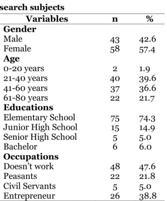
The basic characteristics of research sub-jects.
Table 1. Basic characteristics of re-search subjects
Variables n %
Gender
Male 43 42.6
Female 58 57.4
Age
0-20 years 2 1.9
21-40 years 40 39.6
41-60 years 37 36.6
61-80 years 22 21.7
Educations
Elementary School 75 74.3
Junior High School 15 14.9
Senior High School 5 5.0
Bachelor 6 6.0
Occupations
Doesn’t work 48 47.6
Peasants 22 21.8
Civil Servants 5 5.0
In this study, 101 patients with a his-tory of contact with animals infected with anthrax. The youngest age is 6 years (1%) and the oldest 80 years (1%). The dis-tribution of age is the highest in the 21 to 40 years as much as 39.6%, and most were female gender, i.e. 57.4%. The education level of most respondents is 74.3% finished primary school, farmers as much as 21.8%.
The results obtained Ig G serum antibody showed a negative 50.5%, 15.8% and 33.7% borderline positive. ELISA sero-logy results are shown in Table 2.
Table 2. ELISA Inspection Results
Variabels n %
Positive 34 33.7
Borderline 16 15.8
Negative 51 50.5
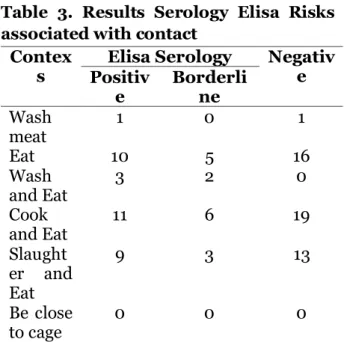
In cross-table analysis results between the risk factors contact with ELISA serology results obtained respondents who cook and eat at the same time is the highest risk on the results of serological positive 20.8%. Serology results Elisa Risks associated with the contact listed in Table 3.
Table 3. Results Serology Elisa Risks associated with contact
Contex
s PositivElisa Serology Negative
e Borderline
Wash
meat 1 0 1
Eat 10 5 16
Wash
and Eat 3 2 0
Cook
and Eat 11 6 19
Slaught er and Eat
9 3 13
Be close
to cage 0 0 0
Overall there are 11.9% of respondents who showed clinical signs of the emergence in the skin in the form of vesicles, accompa-nied by fever and ulcers that ended with the formation of eschar. The skin manifestat-ions can be seen in Table 4.
Table 4. Skin Manifestations in The Form of Eschar
Skin Manifestations n %
Eschar 12 11.9
No eschar 89 88.1
At respondents with positive serology results showed 10.9% of skin manifestation of the emergence of eschar, while only 1.0% with borderline serology that shows cutane-ous manifestations.
Twenty two percent of respondents with positive ELISA results, but does not cause any manifestation in the skin of eschar or other clinical signs (fever, myal-gia, cough, tightness, nausea and vomiting). Elisa serology associated with skin manifes-tations in the form of eschar can be seen in Table 5.
Table 5. Results Serology Elisa Associa-ted with Cutaneous Manifestations in The Form of Eschar
Eschar Serology Elisa Negative Positive Borderline
Exist 11 (10.9%) 1 (1%) 0 (0%) Doesn’t
ExisT 23 (22.8%) 15 (14.8%)
51 (50.5%)
con-tacts with the manifestation of the emer-gence of eschar can be seen in Table 6.
Table 6. Relationship between contacts with the manifestation of the emer-gence of eschar.
Variables
Eschar
Available Not
Availabe
Washing meat
1 (1%) 1 (1%)
Eating 2 (2%) 29 (28.7%)
Washing and Eating
3 (3%) 2 (2%)
Cooking and Eating
0 (0%) 36 (35.6%)
Slaughtering and Eating
6 (6%) 19 (18.9%)
Be Close to The Cage
0 (0%) 2 (2%)
DISCUSSION
In January 2011 obtained a cow belonging to one of the people who suddenly fall and accompanied by seizures. The owners de-cided to slaughter cattle meat and sold to citizens as much as 40 packs. Samples of meat and cow's blood is checked in Lab-kesda Central Java Province and tested positive for anthrax. Seven days later seven residents who complained there was little bumps and itching, accompanied by swell-ling and wet lesions in the area under the eyes, hands, legs or feet, then taken to a health center and declared suspected anth-rax. Furthermore, the derah declared out-breaks of anthrax. Then in May 2011 in Sragen also occurred the same thing and some people who show symptoms of ant-hrax skin contact.
Clinical manifestations such as eschar present in 11.9% of the respondents and all bermanifetasi as Antrak skin (cutaneous anthrax). Respondents were drawn from these two locations, got 101 serological
samples are then examined serum Ig G Antibody anthrax. Of these 50.5% negative and 33.7% positive, while 15.8% borderline. Clinical manifesttations in the form of a skin disorder that begins their benjoalan or injury which later lead to edema and end with eschar present in 10.9% of the res-pondents were Ig G antibody positive and 1.0% of respondents with Ig G borderline results. This is due to the emergence of antibodies against the anthrax bacteria on respondents who had clinical manifestat-ions in the skin, but that cannot be explain-ed is the result of antibodies obtainexplain-ed also borderline clinical manifesttations (see Picture 1).
Picture 1. Anthrax manifestations in the skin with eschar appearance.
Theriskof direct contact,namely cook-ing and eatcook-ing the flesh of infected animals showed 30.5% IgG positive results, but did not cause clinical manifestations with the advent of eschar (0%). This may be due to immune factors from the patient and the virulence of B antrhacis that enter the body. Risk factors for eating just 32% of respon-dents showed positive results. While the risk factors slaughter and eat 24% of the skin manifests with the appearance of the eschar. The increase in serum antibody titer Ig G anthrax is not all respondents were ex-posed, in an area that otherwise outbreak of anthrax, which is only a third of all res-pondents, and when it comes up eschar will be followed by an increase in Ig G antibody titer.
REFERENCE
Braunwald E, Isselbacher KJ, Wilson JD, Martin JB, Kasper DL. Eds. Harris-on's Principles of Internal Medicine. 16th ed. McGraw-Hill; New York: 892-899.
Centers for Disease Control and Prevention
(2011).Guidelines anthrax. www.CDC.
Cieslak TJ, Eitzen E (2005). Clinical and epidemiologic principles of anthrax. Emerging infectious diseases (5): 552-555.
Dirgahayu P (2011). Laboratory tests im-munoassay based anthrax detection. Herman G. Anthrax: 18- 26.
Dixon TC, Meselson BSM, Guillemin J, Hanna PC (2005). Anthrax. N Engl J Med 341: 815-826.
Stern EJ, Uhde KB, Shadomy SV, Messon-nier N (2008) CDC Case Definition. Public Health and Clinical Guidelines for Anthrax Affiliation 14.
Friedlander AM (2008). Anthrax. In: Me-dical aspects of chemical and
biologic-alwarfare.www.nbcmed.org/Site
Con-tent/HomePage/WhatsNew/MedAsp ects/Ch-22/electrv699.pdf.
Geoffrey Scott (2009). Anthrax. In: Man-sons's Tropical Diseases 21st ed. El-sevier: China: 1109 – 1111.
Holmes RK (2009). Diphtheria, other cory-nebacterial infection and anthrax. In: Fauci AS.
Inglesby TV, Henderson DA, Bartlett JG (2005). Anthrax as a biological wea-pon of medical and public health management. JAMA 281: 1735-1745. Inglesby TV, O'Toole T, Henderson DA,
Bartlett JG, Ascher MS, Eitzen (2002). Anthrax as a biological wea-pon: Recommendations for manage-ment updated. JAMA 287 (17): 2236-2252.
Mardiatmo (2011).Anthrax disease
prevent-ion policies. Herman G. Anthrax: 32-36.
Pile JC, JD Malone, Eitzen EM, Friedlander AM (2005). Anthrax as a potential biological warfare agent. Arch Intern Med 158: 429-34.
Redhono D, Sumandjar T, Hermawan G (2011). Mapping anthrax in Central Java. Herman G. Anthrax: 11- 17. Shafazand S, Doyle R, Ruoss S, Weinacker
A, Raffin TA (2005). Inhalation
anth-rax,Epidemiology, diagnosisand
ma-nagement. Chest 116: 1369-1376. Sutarti E (2011). Anticipation of anthrax in
animals. Herman G. Anthrax: 40-46. Swartz MN (2001). Recognition and
mana-gement of anthrax-an update. NEJM 345: 1621-1626.
WHO (2010). Guidelines for the survey-lance and control of anthrax in hu-mans and animals.www/who.int/emc document/zoonoses/docs/