ARTICLE
Genome-wide Trans-ethnic Meta-analysis Identifies
Seven Genetic Loci Influencing Erythrocyte Traits
and a Role for
RBPMS
in Erythropoiesis
Frank J.A. van Rooij,
1Rehan Qayyum,
2Albert V. Smith,
3,4Yi Zhou,
5,6Stella Trompet,
7,8Toshiko Tanaka,
9Margaux F. Keller,
10Li-Ching Chang,
11Helena Schmidt,
12Min-Lee Yang,
13Ming-Huei Chen,
14,15James Hayes,
16Andrew D. Johnson,
15Lisa R. Yanek,
2Christian Mueller,
17,46Leslie Lange,
18James S. Floyd,
19Mohsen Ghanbari,
1,20Alan B. Zonderman,
21J. Wouter Jukema,
7Albert Hofman,
1,22Cornelia M. van Duijn,
1Karl C. Desch,
23Yasaman Saba,
12Ayse B. Ozel,
23Beverly M. Snively,
24Jer-Yuarn Wu,
11,25Reinhold Schmidt,
26Myriam Fornage,
27Robert J. Klein,
16Caroline S. Fox,
15Koichi Matsuda,
28Naoyuki Kamatani,
29Philipp S. Wild,
30,31,32David J. Stott,
33Ian Ford,
34P. Eline Slagboom,
35Jaden Yang,
36Audrey Y. Chu,
37Amy J. Lambert,
38Andre´ G. Uitterlinden,
1,39Oscar H. Franco,
1Edith Hofer,
26,40David Ginsburg,
23Bella Hu,
5,6Brendan Keating,
41,42Ursula M. Schick,
43,44Jennifer A. Brody,
19Jun Z. Li,
23(Author list continued on next page)
Genome-wide association studies (GWASs) have identified loci for erythrocyte traits in primarily European ancestry populations. We conducted GWAS meta-analyses of six erythrocyte traits in 71,638 individuals from European, East Asian, and African ancestries using a Bayesian approach to account for heterogeneity in allelic effects and variation in the structure of linkage disequilibrium between eth-nicities. We identified seven loci for erythrocyte traits including a locus (RBPMS/GTF2E2) associated with mean corpuscular hemoglobin and mean corpuscular volume. Statistical fine-mapping at this locus pointed toRBPMSat this locus and excluded nearbyGTF2E2. Using zebrafish morpholino to evaluate loss of function, we observed a strong in vivo erythropoietic effect forRBPMSbut not forGTF2E2, sup-porting the statistical fine-mapping at this locus and demonstrating thatRBPMSis a regulator of erythropoiesis. Our findings show the utility of trans-ethnic GWASs for discovery and characterization of genetic loci influencing hematologic traits.
Introduction
Erythrocyte disorders are common worldwide,
contrib-uting to substantial morbidity and mortality.
1Erythrocyte
counts and indices are heritable (estimated
h
2¼
0.40–
0.90
2–4), exhibit different patterns across ethnic groups,
and have been influenced by selection in various ethnic
groups, most notably for protection against infection by
parasites such as those that cause malaria.
5–7Erythrocyte
traits have been studied most extensively in European
1Department of Epidemiology, Erasmus MC, 3000 CA Rotterdam, the Netherlands;2GeneSTAR Research Program, Johns Hopkins University School of
Med-icine, Baltimore, MD 21287, USA;3Faculty of Medicine, University of Iceland, 101 Reykjavik, Iceland;4Icelandic Heart Association, 210 Kopavogur, Iceland; 5Harvard Department of Stem Cell and Regenerative Biology, Harvard University, Cambridge, MA 02138, USA;6Stem Cell Program and Division of
Hema-tology/Oncology, Children’s Hospital Boston, Pediatric Hematology/Oncology at DFCI, Harvard Stem Cell Institute, Harvard Medical School and Howard Hughes Medical Institute, Boston, MA 02115, USA;7Department of Cardiology, Leiden University Medical Center, 2300 AC Leiden, the Netherlands; 8Department of Gerontology and Geriatrics, Leiden University Medical Center, 2300 AC Leiden, the Netherlands;9National Institute on Aging, NIH,
Bal-timore, MD 21224, USA;10Laboratory of Neurogenetics, National Institute on Aging, NIH, Bethesda, MD 20892, USA;11Institute of Biomedical Sciences,
Academia Sinica, Taipei 115, Taiwan;12Institute of Molecular Biology and Biochemistry, Centre for Molecular Medicine, Medical University of Graz, 8010
Graz, Austria;13Division of Cardiovascular Medicine, Department of Internal Medicine, Department of Human Genetics, University of Michigan, 1500 E.
Medical Center Drive, Ann Arbor, MI 48109, USA;14Department of Neurology, Boston University School of Medicine, Boston, MA 02118, USA;15
Framing-ham Heart Study, Population Sciences Branch, Division of Intramural Research, National Heart, Lung, and Blood Institute, NIH, FramingFraming-ham, MA 01702, USA;16Icahn Institute for Multiscale Biology, Department of Genetics and Genomic Sciences, Icahn School of Medicine at Mount Sinai, New York, NY
10029, USA; 17Department of General and Interventional Cardiology, University Heart Centre Hamburg-Eppendorf, 20246 Hamburg, Germany; 18Department of Genetics, University of North Carolina, Chapel Hill, NC 27599, USA;19Department of Medicine, University of Washington, Seattle,
WA 98195-6420, USA;20Department of Genetics, School of Medicine, Mashhad University of Medical Sciences, 91375-345 Mashhad, Iran;21National
Insti-tute on Aging, NIH, Bethesda, MD 20892-9205, USA;22Department of Epidemiology, Harvard T.H. Chan School of Public Health, Boston, MA 02115, USA;
23University of Michigan Medical School, Ann Arbor, MI 48109, USA; 24Department of Biostatistical Sciences, Wake Forest School of Medicine,
Winston-Salem, NC 27101, USA;25School of Chinese Medicine, China Medical University, Taichung 40402, Taiwan;26Clinical Division of Neurogeriatrics,
Department of Neurology, Medical University Graz, 8010 Graz, Austria;27Human Genetics Center, School of Public Health, University of Texas Health
Science Center at Houston, Houston, TX 77030, USA;28Laboratory of Molecular Medicine, Human Genome Center, Institute of Medical Science, The
University of Tokyo, Tokyo 108-8639, Japan;29Laboratory for Statistical Analysis, RIKEN Center for Integrative Medical Sciences, Yokohama 230-0045,
Japan;30Center for Thrombosis and Hemostasis (CTH), University Medical Center Mainz, 55131 Mainz, Germany;31German Center for Cardiovascular
Research (DZHK), Partner Site RhineMain, Mainz, Germany;32Preventive Cardiology and Preventive Medicine, Center for Cardiology, University Medical
Center of the Johannes Gutenberg-University Mainz, 55131 Mainz, Germany;33Institute of Cardiovascular and Medical Sciences, Faculty of Medicine,
Uni-versity of Glasgow, Glasgow G12 8QQ, UK;34Robertson Center for Biostatistics, University of Glasgow, Glasgow G12 8QQ, UK;35Department of Medical
ancestry populations,
8–10with smaller studies in
non-European populations, and have shown both shared and
distinct genetic loci influencing erythrocyte traits.
11,12Trans-ethnic meta-analysis of genome-wide association
studies (GWASs) offers improved signal detection in a
com-bined meta-analysis when heterogeneity of allelic effects,
allele frequencies, and differences in linkage
disequilib-rium (LD) between ethnicities are accounted for.
Trans-ethnic meta-analysis can also enable fine-mapping of
association intervals by evaluating differences in LD
struc-ture between diverse populations, thereby enhancing the
detection of causal variants.
13We conducted trans-ethnic GWAS meta-analyses with
the goal of elucidating the genetic architecture of
erythro-cyte traits and to evaluate (1) whether combining data
across populations of diverse ancestry may improve power
to detect associations for erythrocyte traits and (2) whether
differences in LD structure can be exploited to identify
causal variants driving the observed associations with
common SNPs. In this study, we analyzed GWAS summary
statistics from 71,638 individuals from three diverse
popu-lations of European (EUR), East Asian (EAS), and African
(AFR) ancestry. We conducted replication analyses in
inde-pendent samples and performed functional testing to
sup-port our approach to fine-mapping.
Subjects and Methods
Study Samples
We aggregated HapMap-imputed GWAS results from 71,638 indi-viduals represented in 23 cohorts embedded in the CHARGE Con-sortium (40,258 individuals of EUR ancestry), the RIKEN/BioBank Japan Project and AGEN cohorts (15,252 individuals of EAS ancestry), and the COGENT Consortium (16,128 individuals of AFR ancestry). Phenotypic information on all participating
Statistics and Bioinformatics, Section of Molecular Epidemiology, Leiden University Medical Center, 2300 AC Leiden, the Netherlands;36Quantitative Sci-ences Unit, School of Medicine, Stanford University, Stanford, CA 94304, USA;37Division of Preventive Medicine, Brigham and Women’s Hospital and
Harvard Medical School, Boston, MA 02215, USA;38The Jackson Laboratory, Bar Harbor, ME 04609, USA;39Department of Internal Medicine, Erasmus
MC, 3000 CA Rotterdam, the Netherlands;40Institute of Medical Informatics, Statistics and Documentation, Medical University Graz, 8010 Graz, Austria; 41Center for Applied Genomics, Children’s Hospital of Philadelphia, Philadelphia, PA 19104, USA;42Department of Pediatrics, University of Pennsylvania,
Philadelphia, PA 19104, USA;43Public Health Sciences Division, Fred Hutchinson Cancer Research Center, Seattle, WA 98109, USA;44The Charles
Bronf-man Institute for Personalized Medicine, Icahn School of Medicine at Mount Sinai, New York, NY 10029, USA;45Department of Epidemiology and
Biosta-tistics, Mel and Enid Zuckerman College of Public Health, University of Arizona, Tucson, AZ 85724, USA;46German Center for Cardiovascular Research
(DZHK), Partner Site Hamburg, Lu¨beck, Kiel, Hamburg 20246, Germany;47Department of Epidemiology and Public Health, University of Maryland School
of Medicine, Baltimore, MD 21201, USA;48Division of Genetics, Brigham and Women’s Hospital and Harvard Medical School, Boston, MA 02115, USA;
49Laboratory for Genotyping Development, RIKEN Center for Integrative Medical Sciences, Yokohama 230-0045, Japan;50Cardiovascular Medicine
Divi-sion, Department of Medicine, Stanford University School of Medicine, Stanford, CA 94304, USA;51Institute for Translational Genomics and Population
Sciences, Departments of Pediatrics and Medicine, LABioMed at Harbor-UCLA Medical Center, Torrance, CA 90502, USA;52Division of Cardiovascular
Med-icine, Brigham and Women’s Hospital and Harvard Medical School, Boston, MA 02115, USA;53Department of Genetics, Stanford University School of
Med-icine, Stanford, CA 94305, USA;54Laboratory of Epidemiology, Demography, and Biometry, National Institute on Aging, Intramural Research Program,
NIH, Bethesda, MD 20892-9205, USA;55Department of Biostatistics, University of Washington, Seattle, WA 98195, USA;56Department of Clinical
Epide-miology, Leiden University Medical Center, Leiden 2300 AC, the Netherlands;57Health Disparities Research Section, Clinical Research Branch, National
Institute on Aging, NIH, Baltimore, MD 20892, USA;58Max Planck Institute of Immunobiology and Epigenetics, Freiburg 79108, Germany;59Whitehead
Institute for Biomedical Research, Cambridge, MA 02142, USA;60Biostatistics and Biomathematics, Fred Hutchinson Cancer Research Center, Seattle, WA
98109, USA;61Institute for Health Promotion, Graduate School of Public Health, Yonsei University, Seoul 03722, Korea;62Johns Hopkins Bloomberg School
of Public Health, George W. Comstock Center for Public Health Research and Prevention, Comstock Center & Cardiovascular Epidemiology, Welch Center for Prevention, Epidemiology and Clinical Research, Baltimore, MD 21205, USA;63Laboratory of Genetics and Genomics, National Institute on Aging, NIH,
Baltimore, MD 21225, USA;64Department of Biostatistics, Boston University of Public Health, Boston, MA 02118, USA;65Departments of Epidemiology,
Health Services, and Medicine, University of Washington, Seattle, WA 98195, USA;66Group Health Research Institute, Group Health Cooperative, Seattle,
WA 98101, USA;67Department of BESC, Epidemiology Section, King Faisal Specialist Hospital and Research Centre, Riyadh, Saudi Arabia;68Department of
Public Health and Primary Care, Leiden University Medical Center, 2300 AC Leiden, the Netherlands;69Department of Physiology and Biophysics,
Uni-versity of Mississippi Medical Center, Jackson, MS 39216, USA;70Department of Biostatistics and Epidemiology, MRC-PHE Centre for Environment and
Health, School of Public Health, Imperial College, W2 1PG London, UK;71Department of Biostatistics, University of Liverpool, Block F, Waterhouse
Building, 1-5 Brownlow Street, Liverpool L69 3GL, UK;72Wellcome Trust Centre for Human Genetics, University of Oxford, Roosevelt Drive, Oxford
OX3 7BN, UK;73Department of Statistical Genetics, Osaka University Graduate School of Medicine, Osaka 565-0871, Japan;74Department of Epidemi-ology, University of Washington, Seattle, WA 98195, USA
*Correspondence:[email protected] http://dx.doi.org/10.1016/j.ajhg.2016.11.016.
Zhao Chen,
45Tanja Zeller,
17,46Jack M. Guralnik,
47Daniel I. Chasman,
37,48Luanne L. Peters,
38Michiaki Kubo,
49Diane M. Becker,
2Jin Li,
50Gudny Eiriksdottir,
4Jerome I. Rotter,
51Daniel Levy,
15Vera Grossmann,
30Kushang V. Patel,
21Chien-Hsiun Chen,
11,25The BioBank Japan Project,
Paul M. Ridker,
37,52Hua Tang,
53Lenore J. Launer,
54Kenneth M. Rice,
55Ruifang Li-Gao,
56Luigi Ferrucci,
9Michelle K. Evans,
57Avik Choudhuri,
5,6Eirini Trompouki,
6,58Brian J. Abraham,
59Song Yang,
5,6Atsushi Takahashi,
29Yoichiro Kamatani,
29Charles Kooperberg,
60Tamara B. Harris,
54Sun Ha Jee,
61Josef Coresh,
62Fuu-Jen Tsai,
25Dan L. Longo,
63Yuan-Tsong Chen,
11Janine F. Felix,
1Qiong Yang,
15,64Bruce M. Psaty,
65,66Eric Boerwinkle,
27Lewis C. Becker,
2Dennis O. Mook-Kanamori,
56,67,68James G. Wilson,
69Vilmundur Gudnason,
3,4Christopher J. O’Donnell,
15Abbas Dehghan,
1,70L. Adrienne Cupples,
15,64Michael A. Nalls,
10Andrew P. Morris,
71,72Yukinori Okada,
29,73cohorts is provided in Table S1 and has been reported previ-ously.8,11,12,14,15 We conducted replication analyses of the identified trait-loci associations in six independent studies: the Gutenberg Health Study (GHS cohorts 1 and 2, both EUR ancestry), the Genes and Blood-Clotting Study (GBC, EUR ancestry), the NEO study (EUR ancestry), the JUPITER trial (EUR ancestry), and the HANDLS study (AFR ancestry)16–21(total replication size N¼16,389).
Erythrocyte Phenotype Modeling
We analyzed six erythrocyte traits: hemoglobin concentration (Hb, g/dL), hematocrit (Hct, percentage), mean corpuscular he-moglobin (MCH, picograms), mean corpuscular hehe-moglobin concentration (MCHC, g/dL), mean corpuscular volume (MCV, femtoliters), and red blood cell count (RBC, 1M cells/cm3). Trait units were harmonized across all studies. MCH, MCHC, MCV, and RBC were transformed to obtain normal distributions. We excluded samples deviating more than 3 SD from the ethnic- and trait-specific mean within each contributing study, because we focused on determinants of variation in the general population rather than on specific hematological diseases that are overrepresented at the extremes of the trait distribution (Table S2).
Genotyping
In brief, the cohorts comprise unrelated individuals, except for the Framingham Heart Study (related individuals of European ancestry) and GeneSTAR (related individuals of European or Afri-can ancestry). SNPs with a minor allele frequency<1%, missing-ness > 5, or HWE p < 107 were excluded. Genotypes were
imputed to approximately 2.5 million SNPs using HapMap Phase II CEU. The RIKEN and the BioBank Japan Project and AGEN cohorts comprise unrelated individuals of East Asian ancestry (EAS). SNPs with a minor allele frequency<0.01, miss-ingness>1%, or HWE p<107were excluded. Individuals with
a call rate<98% were excluded as well. Genotypes were imputed to approximately 2.5 million SNPs using HapMap Phase II JPT and CHB. The COGENT consortium cohorts comprise individuals of African American ancestry (AFR). SNPs with a minor allele fre-quency<1% or missingness>10% were excluded. Genotypes were imputed to approximately 2.5 million SNPs using HapMap Phase II CEU and YRI.
Cohort-Specific GWASs
For the initial GWA analyses, each cohort used linear regression to assess the association of all SNPs meeting the quality control criteria with each of the six traits separately. An additive genetic model was used and the regressions were adjusted for age, sex, and study site (if applicable). The Framingham Heart Study and the GeneSTAR study used linear mixed effects models to account for relatedness, and these models included adjustment for prin-cipal components.
Ethnic-Specific GWAS Meta-analyses
GWAS results of SNPs with a minor allele frequency (MAF)R1% and an imputation quality>30% were analyzed in a fixed-effect meta-analysis (METAL software22) within each ancestry group, with genomic control (GC) correction of the individual GWAS results of each contributing cohort and the final meta-analysis results.23
Trans-ethnic Meta-analyses
For the trans-ethnic meta-analyses, the three sets of the ethnic-specific meta-analysis summary statistics were then combined with three approaches. First, we performed for each trait a trans-ethnic fixed-effect inverse variance-weighted meta-analysis of the EUR, EAS, and AFR GWAS summary statistics using METAL. Second, the ethnic-specific GWAS summary statistics were also combined using the MANTRA (Meta-Analysis of Trans-ethnic Association Studies) package, a meta-analysis software tool allow-ing for heterogeneity in allelic effects due to differences in LD structure in different ancestry clusters.24MANTRA results are re-ported as log10 Bayes’s factors (log10BF). Finally, the three sets of
ethnic-specific results were analyzed by means of the Han and Eskin RE2 model, a meta-analysis method developed for higher statistical power under heterogeneity.25We used the METASOFT 3.0c tool as developed by the Buhm Han laboratories (Web
Resources). For the fixed-effects and the RE2 models, we applied
a genome-wide significance threshold adjusted for multiple testing, as we analyzed six traits in our study. Given that the traits under investigation are correlated (Table S10), we used eigenvalues to assess the effective number of independent traits according to Ji and Li,26and we estimated this number at 4.0549 using the Matrix Spectral Decomposition tool (Web Resources). We therefore considered p values smaller than 1.253108(i.e., 53108/
4.0549) as genome-wide significant. For the MANTRA discovery analyses, a log10BF>6.1 was considered as a genome-wide
signif-icant threshold value.27
Replication in Human Cohorts
The six independent replication studies—the Gutenberg Health Study (GHS cohorts 1 and 2, both EUR ancestry), the Genes and Blood-Clotting Study (GBC, EUR ancestry), the NEO study (EUR ancestry), the JUPITER trial (EUR ancestry), and the HANDLS study (AFR ancestry)16–21(total replication size N¼ 16,389)—pro-vided linear regression results for the nine trait-locus combina-tions. Their results were meta-analyzed with a fixed effects inverse variance weighted method (METAL) and the RE2 methodology. Additionally, we meta-analyzed replication results with the discov-ery data using fixed-effects, MANTRA, and RE2 methods. For the replication analyses of the nine individual trait-locus combina-tions, we applied a threshold of p<0.05/9. Additional human replication findings are provided inSupplemental Data.
Fine-Mapping
previously identified regions that may not have showed up in the more stringent MANTRA discovery analyses.
Heterogeneity Analysis
Heterogeneity of the associations across the different ethnicities was assessed by the I2and Cochran’s Q statistics as reported by
METAL22 and the posterior probability of heterogeneity as re-ported by MANTRA.24
ENCODE Annotation
We evaluated the SNPs identified in the discovery analyses against the ENCODE Project Consortium’s database of functional ele-ments in the K562 erythroleukemic line.29
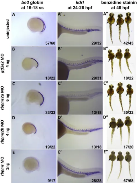
Experiments in Zebrafish
To substantiate the fine mapping of theRBPMS/GTF2E2region biologically, we tested the effect of morpholino knockdown in zebrafish for both RBPMS and GTF2E2 orthologous genes, fol-lowed by assays of erythrocyte development.
Zebrafish rbpms, rbpms2, and gtf2e2 were identified and confirmed by peptide sequence homology study and gene synteny analysis. Forrbpms, we relied solely on peptide homology compar-ison and domain structure since no syntenic region was previously annotated and found by this study.
For each morpholino (MO), its design incorporated information about gene structure and translational initiation sites (Gene-Tool Inc.). MOs targeting each transcript were injected into single-cell embryos at 1, 3, and 5 ng/embryo to find an optimal dose at which there was minimal non-specific toxicity. The stepwise doses also give a range of phenotypes from a hypomorph to a near complete knockdown for most transcripts, which were used to assess the ad-ditive model of genetic association. After injection, embryos were collected at specified time points, 16–18 ss, 22–26 hpf, and 48 hpf using both standard morphological features of the whole embryo and hours post-fertilization (hpf) to minimize differences in em-bryonic development staging caused by the MO injection.30,31 The embryos were then assayed for hematopoietic development by whole-mount in situ hybridization and benzidine staining. We conducted two assays simultaneously for globin transcription and hemoglobin formation. For the globin transcription, devel-oping erythrocytes in the intermediate cell mass of the embryos were assayed by embryonicb-globin 3 expression at the 16 somite stage, or 16–18 hpf.31Benzidine staining phenotype was catego-rized from subtle decrease to complete absence of staining, which was categorized as mild, intermediate, or strong effect. Morpho-logically normal morphants with decreased blood formation were scored for hematopoietic effect.
In zebrafish,rbpmswas not annotated in the known EST and cDNA databases, although a genomic sequence in the telomeric re-gion on chromosome 7 predicting a coding sequence (80% pep-tide sequence similarity) was identified. In addition, the synteny between humanRBPMSandGTF2E2is not conserved in zebrafish whererbpmsandgtf2e2are located on two separate chromosomes, chromosomes 7 and 1, respectively.rbpms2was annotated with two paralogs on chromosome 7 (26 Mb away from and centro-meric to the truerbpms) and chromosome 25 of the zebrafish genome. This orthology mapping was confirmed again by this research based on gene synteny and 88% and 91% sequence sim-ilarity, respectively, forrbpms2bandrbpms2ato humanRBPMS2. These two zebrafish RBPMS2 orthologs have a higher overall sequence similarity to human RBPMS than the true zebrafish
rbpms, but both have a RBPMS2-signature stretch of alanine in the C terminus of the protein. Therefore, to confirm ourrbpms
orthology study and to confirm functional conservation ofrbpms
in zebrafish, MO individual knockdown of both rbpms2a and
rbpms2b was also performed in independent experiments, showing much less or no effect byrbpms2aknock-down and mod-erate effect byrbpms2bimpact on erythropoiesis, suggesting func-tional compensation of the genes in therbpmsfamily in zebrafish during embryonic erythropoiesis.
Chromatin Immunoprecipitation and Assay for
Transposase Accessible Chromatin in Human CD34
þCell Lines
For ChIP-seq experiments, the following antibodies were used: Gata1 (Santa Cruz cat# sc265X), Gata2 (Santa Cruz cat# sc9008X), and H3K27ac (Abcam cat# ab4729; RRID: AB_ 2118291). ChIP experiments were performed as previously described with slight modifications.32,33 In brief, 20–30 million cells for each ChIP were crosslinked by the addition of 1/10 vol-ume 11% fresh formaldehyde for 10 min at room temperature. The crosslinking was quenched by the addition of 1/20 volume 2.5 M glycine. Cells were washed twice with ice-cold PBS and the pellet was flash-frozen in liquid nitrogen. Cells were kept at80C until the experiments were performed. Cells were lysed in 10 mL of lysis buffer 1 (50 mM HEPES-KOH [pH 7.5], 140 mM NaCl, 1 mM EDTA, 10% glycerol, 0.5% NP-40, 0.25% Triton X-100, and protease inhibitors) for 10 min at 4C. After centrifugation, cells were resuspended in 10 mL of lysis buffer 2 (10 mM Tris-HCl [pH 8.0], 200 mM NaCl, 1 mM EDTA, 0.5 mM EGTA, and protease inhibitors) for 10 min at room temperature. Cells were pelleted and resuspended in 3 mL of sonication buffer for K562 and U937 and 1 mL for other cells used (10 mM Tris-HCl [pH 8.0], 100 mM NaCl, 1 mM EDTA, 0.5 mM EGTA, 0.1% Na-Deoxycholate, 0.05% Nlauroylsarcosine, and protease inhibi-tors) and sonicated in a Bioruptor sonicator for 24–40 cycles of 30 s followed by 1 min resting intervals. Samples were centrifuged for 10 min at 18,0003gand 1% of TritonX was added to the su-pernatant. Prior to the immunoprecipitation, 50 mL of protein G beads (Invitrogen 100-04D) for each reaction were washed twice with PBS, 0.5% BSA. Finally, the beads were resuspended in 250 mL of PBS, 0.5% BSA, and 5 mg of each antibody. Beads were rotated for at least 6 hr at 40C and then washed twice with PBS, 0.5% BSA. Cell lysates were added to the beads and incu-bated at 40C overnight. Beads were washed 13with 20 mM Tris-HCl (pH 8), 150 mM NaCl, 2 mM EDTA, 0.1% SDS, 1% Triton X-100, 13with 20 mM Tris-HCl (pH 8), 500 mM NaCl, 2 mM EDTA, 0.1% SDS, 1% Triton X-100, 13 with 10 mM Tris-HCl (pH 8), 250 nM LiCl, 2 mM EDTA, 1% NP40, and 13with TE and finally resuspended in 200 mL elution buffer (50 mM Tris-HCl [pH 8.0], 10 mM EDTA, and 0.5%–1% SDS). 50 mL of cell lysates prior to addition to the beads was kept as input. Crosslink-ing was reversed by incubatCrosslink-ing samples at 65C for at least 6 hr. Afterward the cells were treated with RNase and proteinase K and the DNA was extracted by phenol/chloroform extraction.
(Life Technologies, 12027). A tail was added to the end-repaired DNA using NEB Klenow Fragment Enzyme (30-50 exo, M0212L), 13NEB buffer 2, and 0.2 mM dATP (Invitrogen, 18252-015) and incubating the reaction mix at 37C for 30 min. A-tailed DNA was cleaned up using AMPure beads (1.83reaction volume). Sub-sequently, cleaned-up dA-tailed DNA went through Adaptor liga-tion reacliga-tion using Quick Ligaliga-tion Kit (NEB, M2200L) according to the manufacturer’s protocol. Adaptor-ligated DNA was first cleaned up using AMPure beads (1.83of reaction volume), eluted in 100mL and then size-selected using AMPure beads (0.93of the final supernatant volume, 90mL). Adaptor ligated DNA fragments of proper size were enriched with PCR reaction using Fusion High-Fidelity PCR Master Mix kit (NEB, M0531S) and specific index primers supplied in NEBNext Multiplex Oligo Kit for Illumina (Index Primer Set 1, NEB, E7335L). Conditions for PCR used are as follows: 98C, 30 s; (98C, 10 s; 65C, 30 s; 72C, 30 s)315 to 18 cycles; 72C, 5 min; hold at 4C. PCR-enriched fragments were further size selected by running the PCR reaction mix in 2% low-molecular-weight agarose gel (Bio-Rad, 161-3107) and subsequently purifying them using QIAquick Gel Extraction Kit (28704). Libraries were eluted in 25 mL elution buffer. After measuring concentration in Qubit, all the libraries went through quality-control analysis using an Agilent Bioanalyzer. Samples with proper size (250–300 bp) were selected for next generation sequencing using Illumina Hiseq 2000 or 2500 platform.
Alignment and visualization ChIP-seq reads were aligned to the human reference genome (hg19) using bowtie with parameters -k 2 -m 2 -S.34WIG files for display were created using MACS35with parameters -w -S–space¼50–nomodel–shiftsize¼200 and were displayed in IGV.36,37
High-confidence peaks of ChIP-seq signal were identified using MACS with parameters–keepdup¼auto -p 1e-9 and correspond-ing input control. Bound genes are RefSeq genes that contact a MACS-defined peak between 10,000 bp from the TSS andþ5,000 bp from the TES.
For the assay for transposase accessible chromatin (ATAC-seq), CD34þcells were expanded and differentiated using the protocol mentioned above. Before collection, cells were treated with 25 ng/mL hrBMP4 for 2 hr. 53104cells per differentiation stage were harvested by spinning at 5003gfor 5 min, 4C. Cells were washed once with 50 mL of cold 13 PBS and spun down at 5003gfor 5 min, 4C. After discarding supernatant, cells were lysed using 50mL cold lysis buffer (10 mM Tris-HCl [pH 7.4], 10 mM NaCl, 3 mM MgCl2, 0.1% IGEPAL CA-360) and spun
down immediately at 5003g for 10 min, 4C. The cells were then precipitated and kept on ice and subsequently resuspended in 25mL 2X TD Buffer (Illumina Nextera kit), 2.5mL transposase enzyme (Illumina Nextera kit, 15028252), and 22.5mL nuclease-free water in a total of 50mL reaction for 1 hr at 37C. DNA was then purified using QIAGEN MinElute PCR purification kit (28004) in a final volume of 10mL. Libraries were constructed according to Illumina protocol using the DNA treated with trans-posase, NEB PCR master mix, Sybr green, and universal and li-brary-specific Nextera index primers. The first round of PCR was performed under the following conditions: 72C, 5 min; 98C, 30 s; (98C, 10 s; 63C, 30 s; 72C, 1 min)35 cycles; hold at 4C. Reactions were kept on ice and, using a 5mL reaction aliquot, the appropriate number of additional cycles required for further amplification was determined in a side qPCR reaction: 98C, 30 s; (98C, 10 s; 63C, 30 s; 72C, 1 min)320 cycles; hold at 4C. Upon determining the additional number of PCR cycles required further for each sample, library amplification was
con-ducted using the following conditions: 98C, 30 s; (98C,10 s; 63C, 30 s; 72C, 1 min)3appropriate number of cycles; hold at 4C. Libraries prepared went through quality-control analysis using an Agilent Bioanalyzer. Samples with appropriate nucleo-somal laddering profiles were selected for next generation sequencing using Illumina Hiseq 2500 platform.
All human ChIP-seq datasets were aligned to build version NCBI37/HG19 of the human genome using Bowtie2 (v.2.2.1)34 with the following parameters:–end-to-end, -N0, -L20. We used the MACS2 v.2.1.035peak finding algorithm to identify regions of ATAC-seq peaks, with the following parameter: –nomodel– shift 100–extsize 200. A q-value threshold of enrichment of 0.05 was used for all datasets.
Evaluation in Mouse Crosses
To further affirm the trait loci we identified, and in an attempt to further fine-map the intervals identified in our discovery analyses through cross-species comparisons, we evaluated the new loci in syntenic regions in 12 inter-strain mouse QTL crosses.38
In brief, mice from 12 different strains were inter-crossed38and the same erythrocyte traits we have studied by GWAS were measured in peripheral blood. The Jackson Laboratory Animal Care and Use Committee approved all protocols. The number of markers genotyped per cross varied by the platform used, and the total number per cross is provided inTable S9. QTL analysis was per-formed for each erythrocyte trait using R/qtl v1.07-12 (Web
Re-sources).39Genetic map positions of all markers used were updated
to the new mouse genetic map using online mouse map converter tool (Web Resources).40All phenotypic data were ranked-Z trans-formed to approximate the normal distribution prior to analysis. The QTL analysis was performed as a genome-wide scan with sex as an additive covariate. Permutation testing (1,000 permutations) was used to determine significance, and LOD scores greater than the 95thpercentile (p<0.05) were considered significant. QTL
con-fidence intervals were determined by the posterior probability.41,42 For each candidate region in the mouse, the coordinates were ob-tained from the Mouse Genome Database, which is part of Mouse Genome Informatics (MGI), using the ‘‘Genes and Markers’’ query
(Web Resources). Protein coding genes, non-coding RNA genes,
and unclassified genes were queried.
Results
In this study we analyzed the association of genetic
varia-tion in 71,638 individuals and 6 clinically relevant
eryth-rocyte traits which are commonly measured, accounting
for the diverse ethnic background of the participants.
We identified 44 previously reported loci
7–12,43–47(Table
S3) and 9 other significant trait-locus associations at 7 loci
(p
<
5
3
10
8or log
10BF
>
6.1,
Table 1).
SHROOM3
was
simultaneously identified in an exome chip analysis by
our group in overlapping samples.
48Ethnic-specific results
are presented in
Table S4. Regional association plots are
shown for each region in
Figure S1, showing
ethnic-spe-cific results, the trans-ethnic meta-analysis, and plots of
pairwise LD across the regions for EUR, EAS, and AFR
ancestry.
the Bayesian MANTRA analyses, and in the RE2 analyses;
these were
TMEM163
/
ACMSD
for Hct,
PLCL2:rs2060597
for MCH, and
ID2
,
PLCL2:rs9821630
, and
RBPMS
for
MCV. Two loci (
MET
and
FOXS1
) showed a borderline
sig-nificant effect in METAL and RE2 and a strong sigsig-nificant
effect in MANTRA for HB and MCV, respectively. The
asso-ciation of rs2979489 (
RBPMS
) further showed a strong
association with MCH in the multi-ethnic Bayesian
meta-analysis and in the RE2 model but was not detected
in the multi-ethnic fixed-effects meta-analysis, nor in any
of the ethnic-specific meta-analyses for this trait.
Interest-ingly, MCH and MCV are correlated traits, yet strong
het-erogeneity of effect was observed for this SNP’s association
with MCH only, as indicated by both METAL (I
2statistic
94%, p value Cochran’s Q statistic of heterogeneity
6.48
3
10
8) and MANTRA (posterior probability of
het-erogeneity
¼
1) (Table 1). Inspection of the discovery
data-sets showed that one of the African American cohorts
supplied data for MCV but not for MCH, which resulted
in a stronger positive association of rs2979489 with MCH
than with MCV in the AFR meta-analyses. This
phenome-non was accompanied by greater evidence of
heterogene-ity for MCH in the trans-ethnic meta-analyses because
the EUR and EAS associations were in the opposite
direc-tion to that observed in the AFR meta-analysis. The
MANTRA and RE2 analyses were able to account for this
heterogeneity and thus yield a stronger result as compared
to METAL for this trait locus.
Replication Analyses
In the meta-analyses of the replication cohorts, the
trait-SNP combinations HT-
TMEM163
/
ACMSD
and
MCH-RBPMS
achieved
a
Bonferroni-corrected
significance
threshold with both fixed effects and RE2 methods
(p
<
0.05/9).
ID2
was Bonferroni-significant in the
fixed-effects model and nominally significant in the RE2 model.
Furthermore, we found nominal significance for
MCV-RBPMS
(fixed-effects analyses) and
FOXS1
(fixed-effects
and RE2) (Table S5).
When we compared the discovery and replication
com-bined meta-analyses with the discovery analyses alone, we
observed stronger associations for Hct-
TMEM163/ACMSD
,
MCH-
PLCL2
, MCV-
ID2
, and MCV-
RBPMS
in all three
models (fixed-effects, MANTRA, and RE2). For
MCH-RBPMS
, we found a stronger association in the fixed-effects
analysis (Table S6).
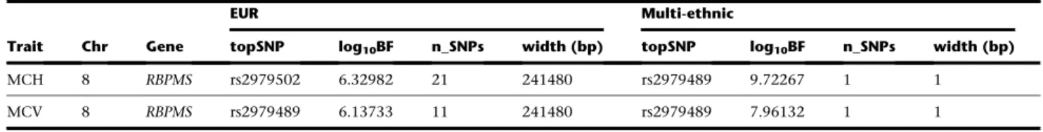
Statistical Fine-Mapping
We found that 31 trait-specific trans-ethnic 99% CSs
showed a decrease in length of at least 50% as compared
to their EUR-only CS counterparts (26 unique loci across
the 6 erythrocyte traits) (Table S7).
Among the loci identified in this study, the chromosome
8
RBPMS
locus showed fine-mapping according to this
cri-terion (Table 2,
Figure 1). For MCH, the EUR credible set
spanned 204,200 bp, encompassing
RBPMS
and
GTF2E2
.
The multi-ethnic credible set comprised just one SNP,
rs2979489, within the first intron of
RBPMS
(Figure 1).
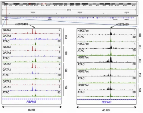
Remarkably, this associated SNP rs2979489 is located
adja-cent to a GATA-motif where a gradual switch of binding
from GATA2 to GATA1 takes place during commitment
of human CD34 progenitors toward erythroid lineage
(Figure 2, bottom left). Moreover, an assay for chromatin
accessibility sites (ATAC-seq) and H3K27a ChIP-seq clearly
identify that the genomic region proximal to this SNP is
actively regulated during human erythroid differentiation
(Figure 2, bottom right).
Among the known loci, fine mapping narrowed signals
as shown in
Table S7.
Interestingly, trans-ethnic fine-mapping of the
XRN1
lo-cus (MCH) led us to the rs6791816 polymorphism. Van der
Harst et al. identified the same SNP in their exploration of
nucleosome-depleted regions (NDRs, representing active
regulatory elements for erythropoeisis) in a follow-up
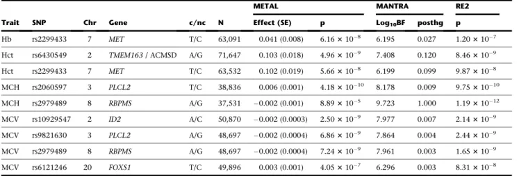
Table 1. Findings from the METAL and MANTRA Trans-ethnic Analyses
Trait SNP Chr Gene c/nc N
METAL MANTRA RE2
Effect (SE) p Log10BF posthg p
Hb rs2299433 7 MET T/C 63,091 0.041 (0.008) 6.163108 6.195 0.027 1.203107
Hct rs6430549 2 TMEM163/ ACMSD A/G 71,647 0.103 (0.018) 4.963109 7.408 0.120 8.463109
Hct rs2299433 7 MET T/C 63,532 0.102 (0.019) 5.663108 6.199 0.099 9.873108
MCH rs2060597 3 PLCL2 T/C 38,836 0.006 (0.001) 4.1831010 8.178 0.009 9.7531010
MCH rs2979489 8 RBPMS A/G 37,531 0.002 (0.001) 8.893105 9.723 1.000 1.1931012
MCV rs10929547 2 ID2 A/C 50,870 0.002 (0.0003) 2.503109 7.977 0.007 2.143109
MCV rs9821630 3 PLCL2 A/G 48,697 0.002 (0.0004) 6.863109 7.864 0.004 2.443109
MCV rs2979489 8 RBPMS A/G 48,697 0.002 (0.0004) 7.243109 7.961 0.003 1.653109
MCV rs6121246 20 FOXS1 T/C 49,896 0.003 (0.001) 4.053107 6.296 0.003 8.313108
Abbreviations are as follows: chr, chromosome number; c/nc, coding/non-coding allele; n, number of participants; SE, standard error; p, p value; log10BF,
analysis of their GWAS results.
10By means of subsequent
formaldehyde-assisted isolation of regulatory elements
fol-lowed by next-generation sequencing (FAIRE-seq), they
pinpointed rs6791816 as an NDR SNP in LD with their
initial index SNP for MCH and MCV.
Furthermore, fine-mapping of both the
MPND
locus
(MCH) and
SH3GL1
locus (MCV) pointed to the rs8887
SNP within the 3
0UTR of
PLIN4
. The rs8887 SNP minor
allele has been shown experimentally to create a novel
seed site for miR-522, resulting in decreased
PLIN4
expres-sion.
49miR-522 is expressed in circulating blood,
50and
these data suggest that an allele-specific miR-522
regula-tion of
PLIN4
by rs8887 could serve as a functional
mech-anism underlying the identified association.
We additionally showed fine mapping in several other
intervals (Table S7) with fine-mapped genes about which
less is known about their potential biologic role in
erythro-poeisis or red blood cell function. These regions are of
in-terest for further hypothesis generation based upon the
GWAS findings.
ENCODE Analyses
We further evaluated the SNPs from the chromosome 8
RBPMS
region against the ENCODE Project Consortium’s
database of numerous functional elements in the K562
erythroleukemic line.
29The lone SNP that was fine
map-ped at the locus, rs2979489, was found in a strong
enhancer element as defined by Segway, supporting a
func-tional role for this SNP and
RBPMS
. The other SNPs in the
RBPMS
region, excluded by the statistical fine-mapping
ex-ercise, were not annotated as regulatory in the ENCODE
data (Table S8).
Table 2. Fine Mapping of a Chromosome 8 Locus Identified in European Ancestry Meta-analysis by MANTRA Trans-ethnic Analysis
Trait Chr Gene
EUR Multi-ethnic
topSNP log10BF n_SNPs width (bp) topSNP log10BF n_SNPs width (bp)
MCH 8 RBPMS rs2979502 6.32982 21 241480 rs2979489 9.72267 1 1
MCV 8 RBPMS rs2979489 6.13733 11 241480 rs2979489 7.96132 1 1
Abbreviations are as follows: chr, chromosome number; log10BF, logarithm of Bayes Factor; n_SNPs, number of SNPs in the region.
Figure 1. Fine Mapping of the Chromosome 8RBPMS/GTF2E2Locus
Experiments in Zebrafish
We identified a erythropoietic effect for the zebrafish
rbpms
. Both embryonic globin expression at 16 ss and
o-di-anisidine/benzidine staining at 48 hpf significantly
decreased in morphants, indicating a decrease in both
globin transcription and Hb levels (Figure 3). This
loss-of-function finding is consistent with a decreased mean
erythrocyte Hb content observed in our human
associa-tion results. In zebrafish, the
rbpms
orthology mapping
included
rbpms2a
,
rbpms2b
, and
rbpms
, and
loss-of-func-tion phenotypes of all orthologs were tested
experimen-tally. The results suggested a clear erythropoietic effect
with limited functional compensation of the genes in the
rbpms family in zebrafish during embryonic
erythropoi-esis. On the other hand, morpholino knockdown
experi-ments with the zebrafish ortholog of
GTF2E2
did not
show an apparent erythropoietic effect.
Review of the human association results showed no
evi-dence of pleiotropy across the RBPMS family of genes and
denote that the human association is specific to
RBPMS
(Supplemental Data). This review was conducted because
the orthology in the fish led to inclusion of
rbpms2
in
the zebrafish analyses as well. These findings indicate
that the statistical fine-mapping was useful to home in
on
RPBMS
as a causal gene influencing erythropoiesis.
Evaluation in Mouse Crosses
In the eight regions from our discovery analysis, six had
evidence of cross-species validation by evidence of
syn-tenic gene within the linkage peak in the mouse QTL
re-sults (Table 3). However, the human GWAS intervals were
not narrowed by the mouse QTL results for any of these
loci (Table S9).
Figure 2. rs2979489 Is Localized to a Po-tential Regulatory Site that Involves Tran-sition Binding of GATA2 to GATA1 during Erythrocyte Differentiation
Top shows gene-track view of rs2979489 location in the RBPMS/GTF2E2 gene re-gion. Bottom left: gene track of RBPMS gene showing overlap of GATA2, GATA1, and ATAC-seq peaks (red, blue, and green, respectively) during human erythroid differentiation. Bottom right: overlap of ATAC-seq (green) and H3K27ac ChIP-seq (black) during differentiation at the region proximal to the SNP rs2979489. The gray horizontal line indicates the position of SNP rs 2979489. D0, day 0; H6, hour 6; D3, day 3; D4, day 4; and D5, day 5 of erythroid differentiation time-course post-induction of differentiation.
Discussion
We conducted GWASs and
meta-ana-lyses of six erythrocyte traits (Hb,
Hct, MCH, MCHC, MCV, and RBC)
in 71,638 individuals from European,
Asian, and African American ancestry. While prior
genome-wide association studies have identified loci
asso-ciated with erythrocyte traits through the analysis of
ancestrally homogeneous cohorts and consortia, largely
biased toward European ancestry studies, trans-ethnic
analysis has not previously been performed while
account-ing for differences in genetic architecture in ethnically
diverse groups.
We identified seven loci for erythrocyte traits (nine
locus-trait combinations) and replicated 44 previously
identified loci. We fine-mapped several known and new
loci. One fine-mapped locus led us to a region on
chromo-some 8 associated with MCH and MCV.
In the chromosome 8
RBPMS/GTF2E2
locus, the index
variant rs2979489, which was associated with MCV and
MCH and highlighted in the trans-ethnic fine-mapping
analyses, is located within the first intron of
RBPMS
(RNA
binding protein with multiple splicing), notably at an
open chromatin site at which a switch of GATA1/2
bind-ing occurs durbind-ing erythroid differentiation. The
RBPMS
protein product regulates a variety of RNA processes,
including pre-mRNA splicing, RNA transport, localization,
translation, and stability.
51,52RBPMS
is expressed at
rela-tively low levels in mammalian erythroblasts and the
pro-tein product has not been detected in mature human
erythrocytes.
53,54populations may lead to a marginal effect of the SNP in
opposing directions by different selection pressures. If,
however, rs2979489 is not causal, but rather a marker in
LD with the causal variant, then the opposing direction
of effects could reflect very different LD structures in the
different populations, also indicating selection, or
theoret-ically it could even reflect different causal variants in AFR
and EUR/EAS—and rs2979489 being just in strong LD
with both causal variants.
The SNP rs2979489 is located adjacent to a GATA-motif
where a gradual switch of binding from GATA2 to GATA1
takes place during commitment of human CD34
progeni-tors toward erythroid lineage. These observations suggest
that rs2979489 localizes at a potential regulatory site
where a modulation of erythroid cell differentiation occurs
and the presence of rs2979489 may lead to observed red
cell trait alterations in human populations, possibly
through regulation of
RBPMS
expression timing, level,
and/or splicing variation. Although
RBPMS
previously
Figure 3. Loss-of-Function Analysis of
theRBPMS, RBPMS2, and GTF2E2
Ortho-logs in Zebrafish
After injection of 0–3 ng ATG and splicing morpholinos (MOs) against the RBPMS zebrafish ortholog (row E), both the o-dia-nisidine/benzidine staining (arrows) in embryos at 48 hpf (right) and the embry-onicbe3 globin expression in embryos at 16–18 ss (left) are obviously decreased, indicating a dose-dependent disruption in erythropoiesis in the experimentally treated embryos as compared to uninjected andgtf2e2-,rbpms2a-, andrbpms2b -MO-in-jected controls (rows A–D). Representative results are shown for the embryos injected with MOs against the RBPMS ortholog in (E) as well as for the embryos injected with MOs againstrbpms2a(C) andrbpms2b
(D) at higher doses. Injections of MO against the zebrafishGTF2E2ortholog (B) also at a higher dose show no obvious ef-fect onbe3 globin expression at 16–18 ss and o-dianisidine/benzidine staining at 48 hpf. Expression pattern of vascular marker genekdrl(A–E, middle) is relatively normal in all MO-injected embryos at 24– 26 hpf, suggesting grossly normal develop-ment of cells in other organs. The numbers on the lower right corner of each image indicate the number of embryos with phe-notypes similar to the ones shown on each of the images over the total number of em-bryos examined in each of the experi-mental groups.
have previously been associated with blood metabolite
levels, obesity, and Parkinson disease.
60–62A genetic
variant in the first intron of
MET
was significantly
associ-ated with both Hb and Hct; however, association was not
observed in replication samples, possibly due to lower
po-wer in the replication experiment. Three additional loci
were intergenic but close to a coding gene (rs10929547
near
ID2
[inhibitor of DNA binding 2, dominant-negative
helix-loop-helix protein], rs6121246 near
FOXS1
[forkhead
box S1], and rs2060597 approximately 40 kbp upstream
of
PLCL2
[phospholipase C-like 2]). The roles of variants
in these regions in determining erythrocyte traits are
unknown.
53,63In the statistical fine-mapping analyses, the trans-ethnic
meta-analysis approach resulted in smaller 99% credible
intervals in all of the loci identified in this study. Since
these loci were identified in analyses that accounted for
heterogeneity in allelic effects between ethnic groups, in
which the heterogeneity may be due to variation in LD
patterns, we examined the LD patterns in these loci. Not
surprisingly, we noted that the consistent decrease in the
size of 99% credible interval across all loci is likely due to
the inclusion of cohorts of African ancestry, an ethnic
group with generally smaller LD blocks throughout the
genome. The loss-of-function screens in zebrafish for the
chromosome 8 signal suggested that these analyses
suc-cessfully identified a single gene (
RBPMS
) with
erythropoi-etic effect within one of the fine-mapped intervals. We also
fine-mapped previously known regions such as the
chro-mosome 6p21.1 region associated with RBC count and
highlighted
CCND3
, which has been experimentally
shown to regulate RBC count experimentally in a
knock-out mouse model.
64These examples suggest that attempts
to refine association signals using these types of
ap-proaches in existing samples may yield functional
candi-dates for further mechanistic hypothesis testing, which is
a major goal of GWASs.
Trans-ethnic genome-wide meta-analyses of common
variants have aided in the characterization of genetic
loci for various complex traits.
13,65–67Our data
demon-strate the benefits of trans-ethnic genome-wide
meta-anal-ysis in identifying and fine-mapping genetic loci of
eryth-rocyte traits. By exploiting the differences in genetic
architecture of the associations within these loci in various
ethnic groups, we may identify causal genes influencing
clinically relevant hematologic traits. Use of a similar
approach for other complex traits is likely to provide
deeper insights into the biological mechanisms underlying
human traits.
Accession Numbers
Summary data have been deposited in the database of Genotypes and Phenotypes (dbGaP) under CHARGE (Cohorts for Heart and Aging Research in Genomic Epidemiology) Consortium Summary Results from Genomic Studies. The dbGaP study accession number is phs000930.
Supplemental Data
Supplemental data include Supplemental Acknowledgments, indi-vidual study methods and cohort descriptions, pleiotropy anal-ysis, 10 tables, and a figure with 123 panels.
Acknowledgments
B.M.P. serves on the DSMB of a clinical trial funded by the manu-facturer (Zoll LifeCor) and on the Steering Committee of the Yale Open Data Access project funded by Johnson & Johnson.
Received: February 15, 2016 Accepted: November 16, 2016 Published: December 22, 2016
Web Resources
Center for Genome Dynamics,http://cgd.jax.org
dbGaP,http://www.ncbi.nlm.nih.gov/gap
Matrix Spectral Decomposition,http://neurogenetics.qimrberghofer. edu.au/matSpD/
METASOFT 3.0c,http://www.buhmhan.com/software
MGI Genes and Markers Query,http://www.informatics.jax.org/ marker
R/qtl v1.07-12,http://www.rqtl.org
Table 3. Mouse QTL Validation of the Findings from MANTRA Trans-ethnic Analyses
Trait Chr Gene
Human (hg18/Build 36) Mouse (37 mm9)
Significant and Suggestive Mouse QTLa
LOD (Chromosome:Position) (Chromosome:Position) Peak (95% CI) (Mb)
Hct 2 TMEM163/ACMSD chr2: 135,196,450–135,438,613 chr1: 129,581,372–129,711,586b 141.0 (54.8–158.9)* 3.72*
Hct 4 SHROOM3 chr4: 77,586,311–77,629,342 chr5: 93,112,461–93,394,344 46.0 (19.6–106.5) 2.34 Hct 7 MET chr7: 116,118,114–116,131,947 chr6: 17,432,318–17,447,418b 37.6 (6.6–127.9) 2.75
MCH 8 RBPMS chr8: 30,400,375–30,400,375 chr8: 34,893,115–35,040,335 78.9 (28.0–96.1)* 3.98* MCV 3 PLCL2 chr3: 16,860,239–16,945,942 chr17: 50,604,848–50,698,773b 46.0 (28.6–55.3)* 5.46*
MCV 20 FOXS1 chr20: 29,684,484–29,897,013 chr2: 152,576,419–152,758,874b 170.1 (147.6–179.3)* 4.69* aGene found in a significant (indicated with asterisk) or suggestive 95% CI mouse QTL, not corresponding to the human interval.
References
1. Koury, M.J. (2014). Abnormal erythropoiesis and the
patho-physiology of chronic anemia. Blood Rev.28, 49–66.
2. Whitfield, J.B., and Martin, N.G. (1985). Genetic and
environ-mental influences on the size and number of cells in the
blood. Genet. Epidemiol.2, 133–144.
3. Evans, D.M., Frazer, I.H., and Martin, N.G. (1999). Genetic and
environmental causes of variation in basal levels of blood
cells. Twin Res.2, 250–257.
4. Lin, J.-P., O’Donnell, C.J., Jin, L., Fox, C., Yang, Q., and
Cup-ples, L.A. (2007). Evidence for linkage of red blood cell size and count: genome-wide scans in the Framingham Heart
Study. Am. J. Hematol.82, 605–610.
5. Guindo, A., Fairhurst, R.M., Doumbo, O.K., Wellems, T.E., and
Diallo, D.A. (2007). X-linked G6PD deficiency protects hemi-zygous males but not heterohemi-zygous females against severe
malaria. PLoS Med.4, e66.
6. Tishkoff, S.A., Varkonyi, R., Cahinhinan, N., Abbes, S.,
Argyr-opoulos, G., Destro-Bisol, G., Drousiotou, A., Dangerfield, B., Lefranc, G., Loiselet, J., et al. (2001). Haplotype diversity and linkage disequilibrium at human G6PD: recent origin of alleles
that confer malarial resistance. Science293, 455–462.
7. Lo, K.S., Wilson, J.G., Lange, L.A., Folsom, A.R., Galarneau, G.,
Ganesh, S.K., Grant, S.F.A., Keating, B.J., McCarroll, S.A., Moh-ler, E.R., 3rd., et al. (2011). Genetic association analysis high-lights new loci that modulate hematological trait variation
in Caucasians and African Americans. Hum. Genet. 129,
307–317.
8. Ganesh, S.K., Zakai, N.A., van Rooij, F.J.A., Soranzo, N., Smith,
A.V., Nalls, M.A., Chen, M.-H., Kottgen, A., Glazer, N.L., Deh-ghan, A., et al. (2009). Multiple loci influence erythrocyte
phenotypes in the CHARGE Consortium. Nat. Genet. 41,
1191–1198.
9. Soranzo, N., Spector, T.D., Mangino, M., Ku¨hnel, B., Rendon,
A., Teumer, A., Willenborg, C., Wright, B., Chen, L., Li, M., et al. (2009). A genome-wide meta-analysis identifies 22 loci associated with eight hematological parameters in the
HaemGen consortium. Nat. Genet.41, 1182–1190.
10. van der Harst, P., Zhang, W., Mateo Leach, I., Rendon, A.,
Ver-weij, N., Sehmi, J., Paul, D.S., Elling, U., Allayee, H., Li, X., et al. (2012). Seventy-five genetic loci influencing the human
red blood cell. Nature492, 369–375.
11. Kamatani, Y., Matsuda, K., Okada, Y., Kubo, M., Hosono, N.,
Daigo, Y., Nakamura, Y., and Kamatani, N. (2010). Genome-wide association study of hematological and biochemical
traits in a Japanese population. Nat. Genet.42, 210–215.
12. Chen, Z., Tang, H., Qayyum, R., Schick, U.M., Nalls, M.A.,
Handsaker, R., Li, J., Lu, Y., Yanek, L.R., Keating, B., et al.; BioBank Japan Project; and CHARGE Consortium (2013). Genome-wide association analysis of red blood cell traits in African Americans: the COGENT Network. Hum. Mol. Genet.
22, 2529–2538.
13. Franceschini, N., van Rooij, F.J.A., Prins, B.P., Feitosa, M.F.,
Karakas, M., Eckfeldt, J.H., Folsom, A.R., Kopp, J., Vaez, A., An-drews, J.S., et al.; LifeLines Cohort Study (2012). Discovery and fine mapping of serum protein loci through transethnic
meta-analysis. Am. J. Hum. Genet.91, 744–753.
14. Nalls, M.A., Couper, D.J., Tanaka, T., van Rooij, F.J.A., Chen,
M.-H., Smith, A.V., Toniolo, D., Zakai, N.A., Yang, Q., Grei-nacher, A., et al. (2011). Multiple loci are associated with white
blood cell phenotypes. PLoS Genet.7, e1002113.
15. Chen, P., Takeuchi, F., Lee, J.-Y., Li, H., Wu, J.-Y., Liang, J.,
Long, J., Tabara, Y., Goodarzi, M.O., Pereira, M.A., et al.; CHARGE Hematology Working Group (2014). Multiple non-glycemic genomic loci are newly associated with blood level
of glycated hemoglobin in East Asians. Diabetes63, 2551–
2562.
16. Wild, P.S., Zeller, T., Beutel, M., Blettner, M., Dugi, K.A.,
Lack-ner, K.J., Pfeiffer, N., Mu¨nzel, T., and Blankenberg, S. (2012).
Die Gutenberg Gesundheitsstudie. Bundesgesundheitsblatt
Gesundheitsforschung Gesundheitsschutz55, 824–829.
17. Desch, K.C., Ozel, A.B., Siemieniak, D., Kalish, Y., Shavit, J.A.,
Thornburg, C.D., Sharathkumar, A.A., McHugh, C.P., Laurie, C.C., Crenshaw, A., et al. (2013). Linkage analysis identifies a locus for plasma von Willebrand factor undetected by
genome-wide association. Proc. Natl. Acad. Sci. USA 110,
588–593.
18. de Mutsert, R., den Heijer, M., Rabelink, T.J., Smit, J.W.A.,
Romijn, J.A., Jukema, J.W., de Roos, A., Cobbaert, C.M., Klop-penburg, M., le Cessie, S., et al. (2013). The Netherlands Epidemiology of Obesity (NEO) study: study design and data
collection. Eur. J. Epidemiol.28, 513–523.
19. Ridker, P.M.; and JUPITER Study Group (2003). Rosuvastatin
in the primary prevention of cardiovascular disease among patients with low levels of low-density lipoprotein choles-terol and elevated high-sensitivity C-reactive protein:
ratio-nale and design of the JUPITER trial. Circulation 108,
2292–2297.
20. Qayyum, R., Snively, B.M., Ziv, E., Nalls, M.A., Liu, Y., Tang,
W., Yanek, L.R., Lange, L., Evans, M.K., Ganesh, S., et al. (2012). A meta-analysis and genome-wide association study of platelet count and mean platelet volume in african
ameri-cans. PLoS Genet.8, e1002491.
21. Reiner, A.P., Lettre, G., Nalls, M.A., Ganesh, S.K., Mathias, R.,
Austin, M.A., Dean, E., Arepalli, S., Britton, A., Chen, Z., et al. (2011). Genome-wide association study of white blood cell count in 16,388 African Americans: the continental ori-gins and genetic epidemiology network (COGENT). PLoS
Genet.7, e1002108.
22. Willer, C.J., Li, Y., and Abecasis, G.R. (2010). METAL: fast and
efficient meta-analysis of genomewide association scans.
Bio-informatics26, 2190–2191.
23. Devlin, B., Roeder, K., and Wasserman, L. (2001). Genomic
control, a new approach to genetic-based association studies.
Theor. Popul. Biol.60, 155–166.
24. Morris, A.P. (2011). Transethnic meta-analysis of genomewide
association studies. Genet. Epidemiol.35, 809–822.
25. Han, B., and Eskin, E. (2011). Random-effects model aimed
at discovering associations in meta-analysis of genome-wide
association studies. Am. J. Hum. Genet.88, 586–598.
26. Li, J., and Ji, L. (2005). Adjusting multiple testing in
multilo-cus analyses using the eigenvalues of a correlation matrix.
Heredity (Edinb)95, 221–227.
27. Wang, X., Chua, H.-X., Chen, P., Ong, R.T.-H., Sim, X., Zhang,
W., Takeuchi, F., Liu, X., Khor, C.-C., Tay, W.-T., et al. (2013). Comparing methods for performing trans-ethnic meta-anal-ysis of genome-wide association studies. Hum. Mol. Genet.
22, 2303–2311.
28. Maller, J.B., McVean, G., Byrnes, J., Vukcevic, D., Palin, K., Su,
Z., Howson, J.M., Auton, A., Myers, S., Morris, A., et al.; Well-come Trust Case Control Consortium (2012). Bayesian refine-ment of association signals for 14 loci in 3 common diseases.
29. The ENCODE Project Consortium (2011). A user’s guide to the Encyclopedia of DNA Elements (ENCODE). PLoS Biol. Published online April 19, 2011.http://dx.doi.org/10.1371/
journal.pbio.1001046.
30. Kimmel, C.B., Ballard, W.W., Kimmel, S.R., Ullmann, B., and
Schilling, T.F. (1995). Stages of embryonic development of
the zebrafish. Dev. Dyn.203, 253–310.
31. Huang, H.-T., Kathrein, K.L., Barton, A., Gitlin, Z., Huang,
Y.-H., Ward, T.P., Hofmann, O., Dibiase, A., Song, A., Tyeku-cheva, S., et al. (2013). A network of epigenetic regulators guides developmental haematopoiesis in vivo. Nat. Cell Biol.
15, 1516–1525.
32. Lee, T.I., Johnstone, S.E., and Young, R.A. (2006). Chromatin
immunoprecipitation and microarray-based analysis of
pro-tein location. Nat. Protoc.1, 729–748.
33. Trompouki, E., Bowman, T.V., Lawton, L.N., Fan, Z.P., Wu,
D.-C., DiBiase, A., Martin, C.S., Cech, J.N., Sessa, A.K., Leb-lanc, J.L., et al. (2011). Lineage regulators direct BMP and Wnt pathways to cell-specific programs during differentiation
and regeneration. Cell147, 577–589.
34. Langmead, B., Trapnell, C., Pop, M., and Salzberg, S.L. (2009).
Ultrafast and memory-efficient alignment of short DNA
se-quences to the human genome. Genome Biol.10, R25.
35. Zhang, Y., Liu, T., Meyer, C.A., Eeckhoute, J., Johnson, D.S.,
Bernstein, B.E., Nusbaum, C., Myers, R.M., Brown, M., Li, W., and Liu, X.S. (2008). Model-based analysis of ChIP-Seq
(MACS). Genome Biol.9, R137.
36. Robinson, J.T., Thorvaldsdo´ttir, H., Winckler, W., Guttman,
M., Lander, E.S., Getz, G., and Mesirov, J.P. (2011). Integrative
genomics viewer. Nat. Biotechnol.29, 24–26.
37. Thorvaldsdo´ttir, H., Robinson, J.T., and Mesirov, J.P. (2013).
Integrative Genomics Viewer (IGV): high-performance geno-mics data visualization and exploration. Brief. Bioinform.
14, 178–192.
38. Peters, L.L., Shavit, J.A., Lambert, A.J., Tsaih, S.-W., Li, Q., Su,
Z., Leduc, M.S., Paigen, B., Churchill, G.A., Ginsburg, D., and Brugnara, C. (2010). Sequence variation at multiple loci
influences red cell hemoglobin concentration. Blood 116,
e139–e149.
39. Broman, K.W., Wu, H., Sen, S., and Churchill, G.A. (2003). R/
qtl: QTL mapping in experimental crosses. Bioinformatics19,
889–890.
40. Cox, A., Ackert-Bicknell, C.L., Dumont, B.L., Ding, Y., Bell,
J.T., Brockmann, G.A., Wergedal, J.E., Bult, C., Paigen, B., Flint, J., et al. (2009). A new standard genetic map for the laboratory
mouse. Genetics182, 1335–1344.
41. Churchill, G.A., and Doerge, R.W. (1994). Empirical threshold
values for quantitative trait mapping. Genetics138, 963–971.
42. Sen, S., and Churchill, G.A. (2001). A statistical framework for
quantitative trait mapping. Genetics159, 371–387.
43. Chambers, J.C., Zhang, W., Li, Y., Sehmi, J., Wass, M.N.,
Zaba-neh, D., Hoggart, C., Bayele, H., McCarthy, M.I., Peltonen, L., et al. (2009). Genome-wide association study identifies vari-ants in TMPRSS6 associated with hemoglobin levels. Nat.
Genet.41, 1170–1172.
44. Ding, K., de Andrade, M., Manolio, T.A., Crawford, D.C.,
Ras-mussen-Torvik, L.J., Ritchie, M.D., Denny, J.C., Masys, D.R., Jouni, H., Pachecho, J.A., et al. (2013). Genetic variants that confer resistance to malaria are associated with red blood cell traits in African-Americans: an electronic medical
re-cord-based genome-wide association study. G3 (Bethesda)3,
1061–1068.
45. Kullo, I.J., Ding, K., Jouni, H., Smith, C.Y., and Chute, C.G.
(2010). A genome-wide association study of red blood cell
traits using the electronic medical record. PLoS ONE 5,
e13011.
46. Li, J., Glessner, J.T., Zhang, H., Hou, C., Wei, Z., Bradfield, J.P.,
Mentch, F.D., Guo, Y., Kim, C., Xia, Q., et al. (2013). GWAS of blood cell traits identifies novel associated loci and epistatic interactions in Caucasian and African-American children.
Hum. Mol. Genet.22, 1457–1464.
47. Pistis, G., Okonkwo, S.U., Traglia, M., Sala, C., Shin, S.-Y.,
Mas-ciullo, C., Buetti, I., Massacane, R., Mangino, M., Thein, S.-L., et al.; CHARGE Consortium Hematology Working (2013). Genome wide association analysis of a founder population identified TAF3 as a gene for MCHC in humans. PLoS ONE
8, e69206.
48. CHARGE Consortium Hematology Working Group (2016).
Meta-analysis of rare and common exome chip variants iden-tifies S1PR4 and other loci influencing blood cell traits. Nat.
Genet.48, 867–876.
49. Richardson, K., Louie-Gao, Q., Arnett, D.K., Parnell, L.D., Lai,
C.-Q., Davalos, A., Fox, C.S., Demissie, S., Cupples, L.A., Fer-nandez-Hernando, C., and Ordovas, J.M. (2011). The PLIN4 variant rs8887 modulates obesity related phenotypes in hu-mans through creation of a novel miR-522 seed site. PLoS
ONE6, e17944.
50. Williams, Z., Ben-Dov, I.Z., Elias, R., Mihailovic, A., Brown, M.,
Rosenwaks, Z., and Tuschl, T. (2013). Comprehensive profiling of circulating microRNA via small RNA sequencing of cDNA libraries reveals biomarker potential and limitations.
Proc. Natl. Acad. Sci. USA110, 4255–4260.
51. Shimamoto, A., Kitao, S., Ichikawa, K., Suzuki, N., Yamabe, Y.,
Imamura, O., Tokutake, Y., Satoh, M., Matsumoto, T., Kuro-mitsu, J., et al. (1996). A unique human gene that spans over 230 kb in the human chromosome 8p11-12 and codes multiple family proteins sharing RNA-binding motifs. Proc.
Natl. Acad. Sci. USA93, 10913–10917.
52. Ascano, M., Hafner, M., Cekan, P., Gerstberger, S., and
Tuschl, T. (2012). Identification of RNA-protein interaction
networks using PAR-CLIP. Wiley Interdiscip. Rev. RNA 3,
159–177.
53. Trakarnsanga, K., Wilson, M.C., Griffiths, R.E., Toye, A.M.,
Carpenter, L., Heesom, K.J., Parsons, S.F., Anstee, D.J., and Frayne, J. (2014). Qualitative and quantitative comparison of the proteome of erythroid cells differentiated from human iPSCs and adult erythroid cells by multiplex TMT labelling
and nanoLC-MS/MS. PLoS ONE9, e100874.
54. Kingsley, P.D., Greenfest-Allen, E., Frame, J.M., Bushnell, T.P.,
Malik, J., McGrath, K.E., Stoeckert, C.J., and Palis, J. (2013).
Ontogeny of erythroid gene expression. Blood121, e5–e13.
55. Ramalho-Santos, M., Yoon, S., Matsuzaki, Y., Mulligan, R.C.,
and Melton, D.A. (2002). ‘‘Stemness’’: transcriptional profiling
of embryonic and adult stem cells. Science298, 597–600.
56. Georgantas, R.W., 3rd, Tanadve, V., Malehorn, M., Heimfeld,
S., Chen, C., Carr, L., Martinez-Murillo, F., Riggins, G., Kowal-ski, J., and Civin, C.I. (2004). Microarray and serial analysis of gene expression analyses identify known and novel tran-scripts overexpressed in hematopoietic stem cells. Cancer
Res.64, 4434–4441.
57. Wagner, W., Ansorge, A., Wirkner, U., Eckstein, V., Schwager,
hematopoietic progenitor cells by genome-wide analysis.
Blood104, 675–686.
58. Sun, Y., Ding, L., Zhang, H., Han, J., Yang, X., Yan, J., Zhu, Y.,
Li, J., Song, H., and Ye, Q. (2006). Potentiation of Smad-medi-ated transcriptional activation by the RNA-binding protein
RBPMS. Nucleic Acids Res.34, 6314–6326.
59. He, W., Dorn, D.C., Erdjument-Bromage, H., Tempst, P.,
Moore, M.A.S., and Massague´, J. (2006). Hematopoiesis
controlled by distinct TIF1g and Smad4 branches of the
TGFbeta pathway. Cell125, 929–941.
60. Shin, S.-Y., Fauman, E.B., Petersen, A.-K., Krumsiek, J., Santos,
R., Huang, J., Arnold, M., Erte, I., Forgetta, V., Yang, T.-P., et al.; Multiple Tissue Human Expression Resource (MuTHER) Con-sortium (2014). An atlas of genetic influences on human
blood metabolites. Nat. Genet.46, 543–550.
61. Comuzzie, A.G., Cole, S.A., Laston, S.L., Voruganti, V.S.,
Haack, K., Gibbs, R.A., and Butte, N.F. (2012). Novel genetic loci identified for the pathophysiology of childhood obesity
in the Hispanic population. PLoS ONE7, e51954.
62. Nalls, M.A., Plagnol, V., Hernandez, D.G., Sharma, M.,
Sheerin, U.M., Saad, M., Simo´n-Sa´nchez, J., Schulte, C.,
Les-age, S., Sveinbjo¨rnsdo´ttir, S., et al.; International Parkinson
Disease Genomics Consortium (2011). Imputation of
sequence variants for identification of genetic risks for Parkin-son’s disease: a meta-analysis of genome-wide association
studies. Lancet377, 641–649.
63. Otsuki, M., Fukami, K., Kohno, T., Yokota, J., and Takenawa, T.
(1999). Identification and characterization of a new
phospho-lipase C-like protein, PLC-L(2). Biochem. Biophys. Res.
Com-mun.266, 97–103.
64. Sankaran, V.G., Ludwig, L.S., Sicinska, E., Xu, J., Bauer, D.E.,
Eng, J.C., Patterson, H.C., Metcalf, R.A., Natkunam, Y., Orkin, S.H., et al. (2012). Cyclin D3 coordinates the cell cycle during differentiation to regulate erythrocyte size and number. Genes
Dev.26, 2075–2087.
65. Keller, M.F., Reiner, A.P., Okada, Y., van Rooij, F.J.A., Johnson,
A.D., Chen, M.-H., Smith, A.V., Morris, A.P., Tanaka, T., Fer-rucci, L., et al.; CHARGE Hematology; COGENT; and BioBank Japan Project (RIKEN) Working Groups (2014). Trans-ethnic meta-analysis of white blood cell phenotypes. Hum. Mol.
Genet.23, 6944–6960.
66. Dastani, Z., Hivert, M.-F., Timpson, N., Perry, J.R.B., Yuan, X.,
Scott, R.A., Henneman, P., Heid, I.M., Kizer, J.R., Lyytika¨inen,
L.-P., et al.; DIAGRAMþ Consortium; MAGIC Consortium;
GLGC Investigators; MuTHER Consortium; DIAGRAM Con-sortium; GIANT ConCon-sortium; Global B Pgen ConCon-sortium; Procardis Consortium; MAGIC investigators; and GLGC Con-sortium (2012). Novel loci for adiponectin levels and their in-fluence on type 2 diabetes and metabolic traits: a multi-ethnic
meta-analysis of 45,891 individuals. PLoS Genet.8, e1002607.
67. Liu, C.-T., Buchkovich, M.L., Winkler, T.W., Heid, I.M.,
Bor-ecki, I.B., Fox, C.S., Mohlke, K.L., North, K.E., Adrienne Cupples, L.; African Ancestry Anthropometry Genetics Con-sortium; and GIANT Consortium (2014). Multi-ethnic
fine-mapping of 14 central adiposity loci. Hum. Mol. Genet.23,