Updated Clinical Guidelines
for Diagnosing Fetal Alcohol
Spectrum Disorders
H. Eugene Hoyme, MD, a, b Wendy O. Kalberg, MA, LED, c Amy J. Elliott, PhD, a Jason Blankenship, PhD, c, †
David Buckley, MA, c Anna-Susan Marais, B Cur Nursing, d Melanie A. Manning, MD, e Luther K. Robinson, MD, f Margaret P. Adam, MD, g Omar Abdul-Rahman, MD, h Tamison Jewett, MD, i Claire D. Coles, PhD, j
Christina Chambers, PhD, MPH, k Kenneth L. Jones, MD, k Colleen M. Adnams, MBChB, l Prachi E. Shah, MD, m Edward P. Riley, PhD, n Michael E. Charness, MD, o Kenneth R. Warren, PhD, p Philip A. May, PhDa, c, q
The adverse effects of prenatal alcohol exposure constitute a continuum of disabilities (fetal alcohol spectrum disorders [FASD]). In 1996, the Institute of Medicine established diagnostic categories delineating the spectrum but not specifying clinical criteria by which diagnoses could be assigned. In 2005, the authors published practical guidelines operationalizing the Institute of Medicine categories, allowing for standardization of FASD diagnoses in clinical settings. The purpose of the current report is to present updated diagnostic guidelines based on a thorough review of the literature and the authors’ combined expertise based on the evaluation of >10 000 children for potential FASD in clinical settings and in epidemiologic studies in conjunction with National Institute on Alcohol Abuse and Alcoholism– funded studies, the Collaborative Initiative on Fetal Alcohol Spectrum Disorders, and the Collaboration on FASD Prevalence. The guidelines were formulated through conference calls and meetings held at National Institute on Alcohol Abuse and Alcoholism offices in Rockville, MD. Specific areas addressed include the following: precise definition of documented prenatal alcohol exposure; neurobehavioral criteria for diagnosis of fetal alcohol syndrome, partial fetal alcohol syndrome, and alcohol-related neurodevelopmental disorder; revised diagnostic criteria for alcohol-related birth defects; an updated comprehensive research dysmorphology scoring system; and a new lip/philtrum guide for the white population, incorporating a 45-degree view. The guidelines reflect consensus among a large and experienced cadre of FASD investigators in the fields of dysmorphology, epidemiology, neurology, psychology, developmental/ behavioral pediatrics, and educational diagnostics. Their improved clarity and specificity will guide clinicians in accurate diagnosis of infants and children prenatally exposed to alcohol.
abstract
aSanford Research and Department of Pediatrics, Sanford
School of Medicine, University of South Dakota, Sioux Falls, South Dakota; bCenter for Applied Genetics and Genomic
Medicine and Department of Pediatrics, University of Arizona College of Medicine, Tucson, Arizona; cCenter on Alcoholism,
Substance Abuse and Addictions, University of New Mexico, Albuquerque, New Mexico; dStellenbosch University
Faculty of Medicine and Health Sciences, Stellenbosch, South Africa; Departments of ePathology and Pediatrics,
Stanford University School of Medicine, Stanford, California;
fDepartment of Pediatrics, State University of New York
at Buffalo School of Medicine and Biomedical Sciences, Buffalo, New York; gDepartment of Pediatrics, University
of Washington School of Medicine, Seattle, Washington;
hDepartment of Pediatrics, University of Mississippi School
of Medicine, Jackson, Mississippi; iDepartment of Pediatrics,
Wake Forest University School of Medicine, Winston-Salem, North Carolina; jDepartment of Psychiatry and
Behavioral Sciences, Emory University School of Medicine, Atlanta, Georgia; kDepartment of Pediatrics, University of
California, San Diego School of Medicine, La Jolla, California;
lDepartment of Psychiatry and Mental Health, University of
Cape Town Faculty of Health Sciences, Cape Town, South Africa; mDepartment of Pediatrics and Communicable
Diseases, University of Michigan Medical School, Ann Arbor, Michigan; nDepartment of Psychology, San Diego State
University, San Diego, California; oVA Boston Healthcare
System, Department of Neurology, Harvard Medical School, and Department of Neurology, Boston University School of Medicine, Boston, Massachusetts; pNational Institute on
Alcohol Abuse and Alcoholism, Bethesda, Maryland; and
qDepartment of Nutrition, Gillings School of Global Public
Health, Nutrition Research Institute, University of North Carolina at Chapel Hill, Chapel Hill, North Carolina
The adverse effects of alcohol on the developing fetus were described independently by Lemoine et al in 1968 1 and by Jones et al in 1973. 2 As
with most malformation syndromes, the most severely affected children were described first, with the associated pattern of malformation termed
fetal alcohol syndrome (FAS). 2 As
pediatricians became more familiar with the clinical presentation of children prenatally exposed to alcohol, it became clear that the associated disabilities represent a spectrum, from mild to severe (fetal alcohol spectrum disorders or FASD). In 1996, the Institute of
NIH
To cite: Hoyme HE, Kalberg WO, Elliott AJ, et al. Updated Clinical Guidelines for Diagnosing Fetal Alcohol Spectrum Disorders. Pediatrics. 2016; 138(2):e20154256
Medicine (IOM) described 4 distinct diagnostic categories within FASD: FAS, partial fetal alcohol syndrome (PFAS), alcohol-related neurodevelopmental disorder (ARND), and alcohol-related birth defects (ARBD). 3 However, the
task force did not specify the clinical process by which individual children could be assigned to the groups. Since that time, a number of diagnostic systems have been proposed.4 –10 In
2005, Hoyme et al 4 described specific
clinical guidelines that allowed for assigning diagnoses within the 1996 IOM classification.
Subsequently, the authors have evaluated >10 000 children for potential FASD in clinical settings and epidemiologic studies as part of National Institute on Alcohol Abuse and Alcoholism (NIAAA) supported studies, the Collaborative Initiative on Fetal Alcohol Spectrum Disorders (CIFASD), and the Collaboration on FASD Prevalence (CoFASP). CIFASD was established by NIAAA in 2003 to investigate data-driven methods for complete diagnosis of the continuum of FASD, prevention of the adverse effects of prenatal alcohol exposure, and effective interventions for affected individuals. 11, 12 CoFASP
seeks to establish the prevalence of FASD among school-age children in US communities by using active case ascertainment methodology. 12
Based on this broad multidisciplinary experience, the purpose of this special article is to propose updated clinical guidelines for diagnosing FASD that clarify and expand on the original 2005 guidelines. These updated diagnostic criteria have been formulated, reviewed, and accepted by the investigators and collaborating sites of CoFASP and the administrative core of CIFASD. They do not
necessarily represent the policy of the American Academy of Pediatrics.
BACKGROUND AND SCOPE OF THE PROBLEM
FASDs are the leading cause of preventable developmental
disabilities in the world. Recent school-based studies in the United States estimate the prevalence of FASD to be much higher than previously thought. May et al 13
recently recorded combined rates of FAS and PFAS of 10.9 to 25.2 per 1000 (1.1%–2.5%) in a Rocky Mountain community, whereas the complete continuum of FASD (including ARND) was observed to be 24 to 48 per 1000 (2.4%–4.8%) in a community in the Northern Plains. 14
In the mixed race population of the Western Cape Province in South Africa, the highest prevalence rates of FASD in the world have been documented, 135.1 to 207.5 per 1000 (13.5%–20.8%). 15 The World Health
Organization (WHO) is currently planning prevalence studies in several countries in Europe, Asia, Africa, and North America, which should lead to global data about the frequency of this continuum of disabilities. 16
The high prevalence of FASD produces an immense burden to society in financial terms, unrealized productivity, and human suffering. In the United States, annual cost estimates have ranged from $74.6 million in 1984 17 to $4.0 billion in
1998. 18 In 2007, the estimated annual
cost of FASD in Canada was CAD $5.3 billion. 19
The soaring prevalence and burden of FASD in children recently led the American Academy of Pediatrics to stress the following: no amount of alcohol intake during pregnancy can be considered safe; there is no safe trimester to drink alcohol; all forms of alcohol pose a similar risk; and binge drinking poses a dose-related risk to the fetus. 20
PREPARATION OF UPDATED DIAGNOSTIC GUIDELINES
These guidelines were formulated by the authors over a 12-month period, through a series of conference calls and face-to-face
meetings at the offices of NIAAA in Rockville, MD. The following working subgroups of investigators were organized to revisit diagnostic criteria: dysmorphology evaluation, neurobehavioral assessment, and definition of significant documented prenatal alcohol exposure.
Recommendations from the working committees were brought back to the larger group for discussion and revision. The guidelines presented herein are the result of a thorough review of the literature and the longstanding collective expertise of the authors. The updated clinical guidelines for diagnosis of FASD are set forth in Table 1.
APPLICATION OF THE GUIDELINES IN THE DIAGNOSIS OF FASD
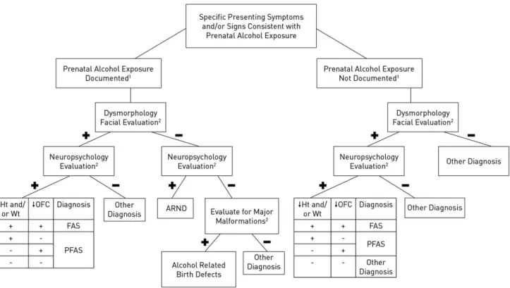
An FASD diagnostic algorithm incorporating the updated diagnostic guidelines is depicted in Fig 1.
Optimal Diagnostic Setting and the Role of the Pediatrician
TABLE 1 Updated Criteria for the Diagnosis of FASD
Diagnostic Categories (See Table 2 for defi nition of documented prenatal alcohol exposure)
I. FAS
(With or without documented prenatal alcohol exposure) A diagnosis of FAS requires all features, A–D:
A. A characteristic pattern of minor facial anomalies, including ≥2 of the following: 1. Short palpebral fi ssures (≤10th centile)
2. Thin vermilion border of the upper lip (rank 4 or 5 on a racially normed lip/philtrum guide, if available) 3. Smooth philtrum (rank 4 or 5 on a racially normed lip/philtrum guide, if available)
B. Prenatal and/or postnatal growth defi ciency
1. Height and/or weight ≤10th centile (plotted on a racially or ethnically appropriate growth curve, if available) C. Defi cient brain growth, abnormal morphogenesis, or abnormal neurophysiology, including ≥1 of the following: 1. Head circumference ≤10th percentile
2. Structural brain anomalies
3. Recurrent nonfebrile seizures (other causes of seizures having been ruled out) D. Neurobehavioral impairmenta
1. For children ≥3 y of age (a or b): a. WITH COGNITIVE IMPAIRMENT:
−Evidence of global impairment (general conceptual ability ≥1.5 SD below the mean, or performance IQ or verbal IQ or spatial IQ ≥1.5 SD below the mean) OR
−Cognitive defi cit in at least 1 neurobehavioral domain ≥1.5 SD below the mean (executive functioning, specifi c learning impairment, memory impairment or visual-spatial impairment)
b. WITH BEHAVIORAL IMPAIRMENT WITHOUT COGNITIVE IMPAIRMENT:
−Evidence of behavioral defi cit in at least 1 domain ≥1.5 SD below the mean in impairments of self-regulation (mood or behavioral regulation impairment, attention defi cit, or impulse control)
2. For children <3 y of age:
−Evidence of developmental delay ≥1.5 SD below the mean II. PFAS
-For children with documented prenatal alcohol exposure, a diagnosis of PFAS requires features A and B: A. A characteristic pattern of minor facial anomalies, including ≥2 of the following:
1. Short palpebral fi ssures (≤10th centile)
2. Thin vermilion border of the upper lip (rank 4 or 5 on a racially normed lip/philtrum guide, if available) 3. Smooth philtrum (rank 4 or 5 on a racially normed lip/philtrum guide, if available)
B. Neurobehavioral impairmenta 1. For children ≥3 y of age (a or b): a. WITH COGNITIVE IMPAIRMENT:
−Evidence of global impairment (general conceptual ability ≥1.5 SD below the mean, or performance IQ or verbal IQ or spatial IQ ≥1.5 SD below the mean) OR
−Cognitive defi cit in at least 1 neurobehavioral domain ≥1.5 SD below the mean (executive functioning, specifi c learning impairment, memory impairment or visual-spatial impairment)
b. WITH BEHAVIORAL IMPAIRMENT WITHOUT COGNITIVE IMPAIRMENT:
−Evidence of behavioral defi cit in at least 1 domain ≥1.5 SD below the mean in impairments of self-regulation (mood or behavioral regulation impairment, attention defi cit, or impulse control)
2. For children <3 y of age:
−Evidence of developmental delay ≥1.5 SD below the mean
-For children without documented prenatal alcohol exposure, a diagnosis of PFAS requires all features, A–C: A. A characteristic pattern of minor facial anomalies, including ≥2 of the following:
1. Short palpebral fi ssures (≤10th centile)
2. Thin vermilion border of the upper lip (rank 4 or 5 on a racially normed lip/philtrum guide, if available) 3. Smooth philtrum (rank 4 or 5 on a racially normed lip/philtrum guide, if available)
B. Growth defi ciency or defi cient brain growth, abnormal morphogenesis, or abnormal neurophysiology
1. Height and/or weight ≤10th centile (plotted on a racially or ethnically appropriate growth curve, if available), or: 2. Defi cient brain growth, abnormal morphogenesis or neurophysiology, including ≥1 of the following:
a. Head circumference ≤10th percentile b. Structural brain anomalies
c. Recurrent nonfebrile seizures (other causes of seizures having been ruled out) C. Neurobehavioral impairmenta
1. For children ≥3 y of age (a or b): a. WITH COGNITIVE IMPAIRMENT:
−Evidence of global impairment (general conceptual ability ≥1.5 SD below the mean, or performance IQ or verbal IQ or spatial IQ ≥1.5 SD below the mean) OR
Diagnostic Categories b. WITH BEHAVIORAL IMPAIRMENT WITHOUT COGNITIVE IMPAIRMENT:
−Evidence of behavioral defi cit in at least 1 domain ≥1.5 SD below the mean in impairments of self-regulation (mood or behavioral regulation impairment, attention defi cit, or impulse control)
2. For children <3 y of age:
−Evidence of developmental delay ≥1.5 SD below the mean III. ARND
Requires features A and B (this diagnosis cannot be made defi nitively in children <3 y of age): A. Documented prenatal alcohol exposure
B. Neurobehavioral impairmenta For children ≥3 y of age (a or b): a. WITH COGNITIVE IMPAIRMENT:
−Evidence of global impairment (general conceptual ability ≥1.5 SD below the mean, or performance IQ or verbal IQ or spatial IQ ≥1.5 SD) OR
−Cognitive defi cit in at least 2 neurobehavioral domains ≥1.5 SD below the mean (executive functioning, specifi c learning impairment, memory impairment or visual-spatial impairment)
b. WITH BEHAVIORAL IMPAIRMENT WITHOUT COGNITIVE IMPAIRMENT:
−Evidence of behavioral defi cit in at least 2 domains ≥1.5 SD below the mean in impairments of self-regulation (mood or behavioral regulation impairment, attention defi cit, or impulse control)
IV. ARBD
Requires features A and B:
A. Documented prenatal alcohol exposure
B. One or more specifi c major malformations demonstrated in animal models and human studies to be the result of prenatal alcohol exposure: cardiac: atrial septal defects, aberrant great vessels, ventricular septal defects, conotruncal heart defects; skeletal: radioulnar synostosis, vertebral segmentation defects, large joint contractures, scoliosis; renal: aplastic/hypoplastic/dysplastic kidneys, “horseshoe” kidneys/ureteral duplications; eyes: strabismus, ptosis, retinal vascular anomalies, optic nerve hypoplasia; ears: conductive hearing loss, neurosensory hearing loss
Diagnostic Caveats: The assignment of an FASD is a complex medical diagnostic process best accomplished through a multidisciplinary approach. As is the case with many medical conditions, sound clinical judgment must be used. Differential diagnoses should always include genetic disorders or conditions arising from other teratogens. Additionally, because head circumference, growth, and many cognitive and behavioral characteristics have moderate to high degrees of heritability, when information is available about the biological parents, these data should be considered in the fi nal diagnostic decision.
a Adaptive skills should be assessed, but such defi cits cannot stand alone for diagnosis.
TABLE 1 Continued
FIGURE 1
educators, audiologists, and/or ophthalmologists. 10, 21–23
The essential role of the pediatrician in the identification and care of children with FASD cannot be overstated. Pediatricians are among the most likely practitioners to first encounter children with prenatal alcohol exposure who are potentially at risk for FASD. Jones et al 24 demonstrated the accuracy
of pediatricians in recognizing FAS on the basis of physical and other common associated features after a relatively short training session. In addition, once a diagnosis is assigned, pediatricians are called on to provide a medical home for affected children, coordinate mental health services, and manage other comorbid mental health disorders. Pediatricians also play an important role in the prevention of future alcohol-exposed pregnancies through counseling women with affected children. 25
Documentation of Signifi cant Prenatal Alcohol Exposure
Assessment of maternal prenatal alcohol intake is an essential part of the diagnostic process and is the first step in the diagnostic algorithm outlined in Fig 1. It is best measured by quantity of alcohol consumed per occasion (standard drinks per drinking day), frequency that it is consumed (eg, daily, times per week), and timing
during gestation, because timing of significant exposure (even in the early weeks of pregnancy) can produce different physical and neurobehavioral phenotypes. 26 –30
Binge drinking (3–5 drinks or more per occasion) has been shown in animal and human studies to be the most detrimental to fetal development. 26, 31 Asking about
use of other potential teratogens during pregnancy is also important because, in addition to their own potential teratogenicity, women who abuse drugs are more likely to use alcohol during pregnancy. 13, 14, 32
Because in many populations it is likely that prenatal alcohol use will be denied completely or be significantly underreported, 13, 14, 33–35 biomarkers
can assist in documenting prenatal alcohol exposure. Most frequently, alcohol exposure information is collected retrospectively. It is well documented that accurate information on a particular pregnancy can be obtained from a willing respondent years after the birth of a child 36–38 or from the
medical or social service records or a collateral informant (eg, spouse, close relative, or friend) who had regular contact with the mother during pregnancy.15, 26
In maternal interviews, because of potential stigmatization associated with prenatal alcohol use, and for accuracy, questions should
be asked in a timeline followback manner, 39, 40 progressing from the
broader context of health history (childbearing, general illness, nutrition, and dietary intake 26, 41, 42)
to the more sensitive alcohol use questions. It is important to also consider the overall drinking pattern immediately before pregnancy recognition, as it is common for the drinking pattern of 3 months before pregnancy to persist into early pregnancy. 13, 14, 43 –49
A consensus definition of significant prenatal alcohol exposure is set forth in Table 2. Note that although certain circumstances permit the diagnosis of FAS or PFAS without firm documentation of gestational alcohol use ( Table 1), positive confirmation of alcohol exposure must be available for the
diagnosis of ARND or ARBD to be assigned.
Dysmorphology Evaluation
After assessing prenatal alcohol exposure, the presence or absence of the characteristic structural features of FASD must be
evaluated. For the dysmorphology examination, height, weight, and head circumference should first be measured and plotted by using population-specific growth curves. In the United States, the authors advise following the Centers for Disease Control and Prevention (CDC) recommendations: use the
TABLE 2 Defi nition of Documented Prenatal Alcohol Exposure (as Applied to the Diagnostic Categories Set Forth in Table 1)
One or more of the following conditions must be met to constitute documented prenatal alcohol exposure during pregnancy (including drinking levels reported by the mother 3 mo before her report of pregnancy recognition or a positive pregnancy test documented in the medical record). The information must be obtained from the biological mother or a reliable collateral source (eg, family member, social service agency, or medical record):
−≥6 drinks/wk for ≥2 wk during pregnancya
−≥3 drinks per occasion on ≥2 occasions during pregnancya
− Documentation of alcohol-related social or legal problems in proximity to (before or during) the index pregnancy (eg, history of citation[s] for driving while intoxicated or history of treatment of an alcohol-related condition)
− Documentation of intoxication during pregnancy by blood, breath, or urine alcohol content testing
− Positive testing with established alcohol-exposure biomarker(s) during pregnancy or at birth (eg, analysis of fatty acid ethyl esters, phosphatidylethanol, and/ or ethyl glucuronide in maternal hair, fi ngernails, urine, or blood, or placenta, or meconium) 50 –55
− Increased prenatal risk associated with drinking during pregnancy as assessed by a validated screening tool of, for example, T-ACE (tolerance, annoyance, cut down, eye-opener) or AUDIT (alcohol use disorders identifi cation test) 56
Assignment of documented prenatal alcohol exposure to any individual case requires the sound judgment of an experienced clinician.
WHO growth charts for children from birth to 2 years to assess height and weight. (The WHO growth standards for children younger than 2 years have been adapted for use in the United States.) Use the CDC growth charts for children and teenagers aged 2 to 19 years. 61 In other
countries, we recommend using more-specific population-normed charts, if available. If growth curves specific to the population studied are not available, we endorse the recommendations of the CDC for US children. 61 Prenatal growth
restriction can be determined from reference data published by Oken et al 62 by gestational age.
In these diagnostic guidelines, we define growth deficiency as ≤10th centile. 4, 8 Prenatal growth
restriction should be exhibited, or a pattern of postnatal growth deficiency should be documented if possible (decreased height and/ or weight on >1 occasion over 12 months, and unrelated to postnatal environmental deprivation). With respect to determination of head circumference centiles, we have used the head circumference growth charts from Nellhaus 63 in all
populations, in lieu of more-specific
population-based norms. For the purposes of these guidelines, a small head circumference is defined as ≤10th centile. 4, 8
The presence or absence of the 3 cardinal facial characteristics of FASD must next be objectively assessed: short palpebral fissures, smooth philtrum, and thin vermilion border of the upper lip ( Fig 2). Although other investigators have advocated for measurement of facial anthropometry from 2-dimensional photography, we feel that direct examinations are more practical in an office setting. Here we define short palpebral fissures as ≤10th centile. 4, 8 Palpebral fissure length
centiles can be estimated from a number of published norms; we have used the curves derived from direct examination of children published by Thomas et al. 64 If facial
anthropometry is measured live, palpebral fissure norms derived from live examinations must be used. (If palpebral fissure lengths are measured from photographs, published norms obtained from 2-dimensional photography are available.65) Similar to the experience
of the authors, Avner et al 66 found
palpebral fissure lengths measured from photographs to be consistently
smaller than those measured live. Similarly, Astley 67 found the norm
for palpebral fissures measured from 2-dimensional photographic software to fall 1.6 SDs below the mean on a palpebral fissure chart derived from live examinations. Figure 3 A and B depicts the technique for direct measurement of palpebral fissure length, and Fig 3C demonstrates why, in our experience, 2-dimensional photographic assessment of palpebral fissure length is prone to inaccuracy because of individual variation in the zygomatic angle that cannot be corrected for by a single mathematical adjustment. However, it should be noted that investigators disagree on the method that results in the most accurate measurement of palpebral fissure length.67–69 The
morphology of the philtrum and the vermilion border of the upper lip are objectively scored by comparison with a racially normed lip/philtrum guide ( Fig 4). 70, 71 Scores of 4 or 5
are consistent with the effects of prenatal alcohol exposure. If 2 of the 3 cardinal facial characteristics are present (short palpebral fissures, smooth philtrum, and/or thin vermilion border of the upper lip) the child is classified as having a positive dysmorphology facial evaluation for FASD.
Neurodevelopmental Assessment and Neuropsychology Evaluation
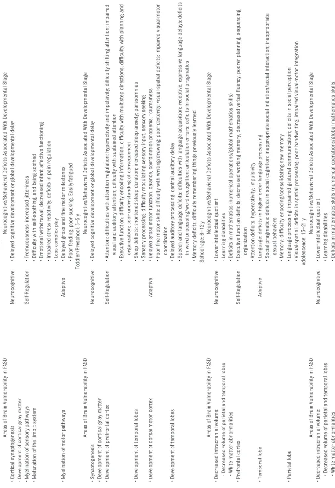
Because the primary manifestations of the teratogenic effects of alcohol are demonstrated by changes in brain structure and/or function, comprehensive neurodevelopmental assessment is essential. Although the dysmorphology assessment of infants and small children for the growth and facial characteristics of FASD is feasible, a comprehensive cognitive/ developmental evaluation may not be possible by using conventional assessment tools until after age 3 years. 72 However, the cognitive
and neurobehavioral phenotype of affected children evolves predictably over time 73 –76 and can be correlated FIGURE 2
with areas of brain vulnerability ( Table 3).
The authors promote the use of standardized tests that were developed by using normative groups that are representative of the population being tested. Therefore, in the updated guidelines, ≥1.5 SD below the mean refers to the mean of the normative group on which the tests were standardized. Therefore, both groups (alcohol-exposed children as well as unexposed children) are tested by using the same well-normed testing battery, thereby making the comparisons appropriate.
Multidisciplinary Case Conference
Once the prenatal exposure history, dysmorphology assessment, and neuropsychological testing have been obtained, a multidisciplinary case conference offers the best opportunity for full discussion of the case before assignment of an FASD or other diagnosis ( Fig 1).
Phenocopies of FASD
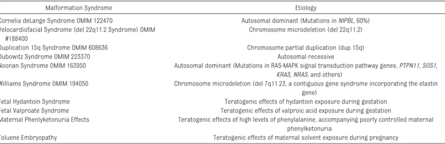
Clinicians should be aware that the facial phenotype of FAS, although most commonly associated with prenatal alcohol exposure, is also observed in a variety of genetic and teratogenic conditions ( Table 4).
Therefore, physicians should use a low threshold for ordering additional genetic testing of children with potential FASD. A chromosome microarray has been shown to be the highest-yield diagnostic test when a genetic phenocopy of FASD is being considered. 77, 78
DISCUSSION
In the decade since the original operationalized IOM diagnostic criteria 4 were published, extensive
international research on the teratogenic effects of alcohol and the authors’ broad clinical experience have allowed for the development of further clarity and specificity in the diagnostic guidelines presented in this article. However, it should be noted that agreement on a universal diagnostic system for FASD is lacking among investigators in the field of FASD, especially concerning some of the features of the diagnostic guidelines set forth in Table 1. A discussion of the debated elements follows.
Diagnostic Categories Within the Continuum of FASD
It is the authors’ assertion that the 4 original IOM diagnostic categories 3
within the continuum of FASD remain the most apt descriptors of the range of disabilities observed. These longstanding categories have heretofore been accepted by many of the diagnostic systems, 4, 8, 9
and we see no need to introduce additional confusion into a field in which diagnostic consensus is critical. In addition, classification of individuals into 1 of the existing specific IOM categories allows for determination of prognosis and treatment planning. We also assert that the category of ARBDs, although uncommon, remains necessary, especially in epidemiologic
studies.82, 83 Our extensive database
of alcohol-exposed children reveals many examples of affected children not fitting into 1 of the other categories who display 1 of the major malformations set forth in Table 1 and whose mothers binged during the embryonic stage critical to the developmental pathology of the malformation.
It should be noted that the Canadian diagnostic guideline for FASD recently was updated, collapsing the diagnostic categories under the diagnosis of “fetal alcohol spectrum disorder” to 2: FASD with sentinel facial features and FASD without
FIGURE 3
sentinel facial features. 10 Whether
this simplified diagnostic scheme will result in practical improvements in the clinical care of affected individuals and more accurate epidemiologic studies estimating the prevalence of FASD remains to be demonstrated.
Sensitivity Versus Specifi city in Clinical Diagnosis
Similar to others, our goals in the formulation of FASD diagnostic guidelines have been improved sensitivity and greater inclusion of children in the complete continuum of FASD 4, 8; thus, we have set cutoff
levels for growth deficiency, head circumference, and palpebral fissure length at ≤10th centile and required 2, rather than 3, cardinal facial features for a diagnosis of FAS and PFAS. Because we advocate for a structured expert-led multidisciplinary diagnostic approach to the diagnosis of FASD, casting a broad net early in the diagnostic process and later using the case conference to carefully assign diagnoses has been our standard. Other diagnostic systems advocate for more stringent cutoffs: growth deficiency, head circumference, and palpebral fissure length less than or equal to the third centile and requiring all 3 of the cardinal facial features for alcohol-related diagnoses. 5, 9, 10 Sensitivity and
specificity are 2 sides of a diagnostic coin. Theoretically, the guidelines presented here demonstrating increased sensitivity could lead to overdiagnosis; thus, our advocacy for a structured expert multidisciplinary approach. On the other hand, strict diagnostic cutoffs associated with increased specificity could lead to underdiagnosis of affected children. Children with FASD are subject to a host of societal, educational, health, and judicial problems, all of which are affected by the time of diagnosis. 84, 85 Because
importance, the authors assert that improved, sensitive, and inclusive diagnostic criteria for FASD should continue to be imperatives in the diagnostic process.
Defi cient Brain Growth, Abnormal Morphogenesis, or Abnormal Neurophysiology
In the updated criteria, we have added documentation of recurrent nonfebrile seizures to the potential assignment of children to the diagnostic categories of FAS or PFAS. A child with FAS must now exhibit deficient brain growth, structure, or neurophysiology. This modification was prompted by a growing body of research that indicates that epilepsy is a frequent accompaniment of FASD. 86, 87 More commonly observed
in children with FASD, a small head circumference is a reliable, easily obtained proxy for decreased brain volume. 88, 89 Finally, a number of
structural brain anomalies have been observed in imaging studies in animals and human subjects with FASD. Although no specific anatomic region of the brain is preferentially affected, malformations resulting from migration abnormalities, changes in size and shape of the corpus callosum, cerebellar vermis hypoplasia, and hypoplasia of the basal ganglia and hippocampus have been documented.90, 91
The 4-digit diagnostic code 5 assesses
these features as “structural evidence of central nervous system damage, ” and the updated Canadian guideline for diagnosis of FASD 10 includes
a similar category (abnormal neuroanatomy/neurophysiology) as 1 of the 10 central nervous system domains that may be impaired, although this category is not a
universal part of other diagnostic systems. 6–8
Other Minor Anomalies in Children With FASD
In dysmorphology, clinical diagnoses are based on recognizable patterns of major and minor anomalies. Although the dysmorphology contribution to FASD diagnoses is derived from objective evaluation of the face, a number of other minor anomalies have been observed consistently and more commonly in children prenatally exposed to alcohol than in nonexposed controls. 4, 13, 14, 92, 93 The
clinical assessment of the presence or absence of these features should be part of the dysmorphology evaluation of children with potential FASD. The overall dysmorphic variation in any individual child can be quantified by calculation of a dysmorphology score (an updated dysmorphology scoring system based on objective observations of growth and minor anomalies in 370 children with FAS is presented in Table 5). The dysmorphology score allows for objective comparison among groups of children with FASD and has proven to be a valuable research tool. It is also a useful instrument to review as part of the differential diagnostic process when assessing features of genetic or other teratogenic disorders that may mimic FASD ( Table 4). The score has been observed to correlate significantly with prenatal maternal alcohol intake, as well as with the cognitive and neurobehavioral characteristics of the affected child. 26, 94
Specifi city of Neurobehavioral Impairment
The updated guidelines now require that all children assigned FASD
diagnoses (with the exception of those with ARBD) must display neurobehavioral impairment (cognitive impairment or behavioral impairment without cognitive impairment). The original guidelines allowed for children with the requisite facial features, growth restriction, and/or microcephaly to be assigned an FASD diagnosis in the absence of significant neurobehavioral impairment. However, because neurocognitive impairment and abnormal behavior are the principal sources of disability in FASD, assignment of children with prenatal alcohol exposure into an FASD category without neurobehavioral impairment has no practical utility for either the child or the child’s family.
The definition of neurobehavioral impairment in FASD has become more specific over the past decade. 36, 72 –76
The original 1996 IOM criteria and the 2005 guidelines defined neurobehavioral impairment as “evidence of a complex pattern of behavioral or cognitive abnormalities inconsistent with developmental level that cannot be explained by genetic predisposition, family background, or environment alone.” 3, 4
Although the 2005 criteria outlined areas of marked neurobehavioral impairment, levels of deficit and affected functional domains were not clearly articulated. The guidelines set forth in Table 1 clearly delineate domains of functioning to be assessed and levels of deficit to be reached to meet the diagnostic criteria for FAS, PFAS, and ARND.
The domains of function outlined in the updated criteria encompass the following: (1) global intellectual ability (full-scale, verbal,
performance, or spatial IQ), (2) cognition (executive functioning, learning, memory, and visual-spatial skills), (3) behavior and self-regulation (mood, behavioral regulation, attention, and impulse control), and (4) adaptive skills.
FIGURE 4
Lip/philtrum guide for the white population, incorporating a 45-degree view. This guide was produced by analysis of photographs of >800 white children from school-based studies in the United States. 13, 14 Scores are assessed separately for the philtrum and vermilion border; scores of 4 or 5 are compatible with FAS or PFAS.
TABLE 3
Developmental Emer
gence of Neurocognitive and Behavioral Defi
cits Associated With FASD
Infancy: 0–2 y
Areas of Brain Vulnerability in FASD
Neurocognitive/Behavioral Defi
cits Associated With Developmental Stage
• Cor
tical synaptogenesis
Neurocognitive
• Delayed cognitive development or global developmental delay
• Development of cor
tical gray matter
• Myelination of sensory pathways
Self-Regulation
• T
remulousness, increased jitteriness
• Maturation of the limbic system
• Diffi
culty with self-soothing, and being soothed
• Emotional withdrawal, decreased infant affective functioning • Impaired stress reactivity; defi
cits in pain regulation
• Less complex play
• Myelination of motor pathways
Adaptive
• Delayed gross and fi
ne motor milestones
• Poor feeding: poor sucking. Easily fatigued
Toddler/Preschool: 3–5 y
Areas of Brain Vulnerability in FASD
Neurocognitive/Behavioral Defi
cits Associated With Developmental Stage
• Synaptogenesis
Neurocognitive
• Delayed cognitive development or global developmental delay
• Development of cor
tical gray matter
• Development of prefrontal cor
tex
Self-Regulation
• Attention: diffi
culties with attention regulation; hyperactivity and impulsi
vity; diffi
culty shifting attention; impaired
visual and auditory attention; diffi
culty with sustained attention
• Executive function: diffi
culty encoding information; diffi
culty with multistep directions; diffi
culty with planning and
or
ganization; poor understanding of consequences
• Development of temporal lobes
• Sleep defi
cits: shor
tened sleep duration; increased sleep anxiety; parasomnias
• Sensory processing: diffi
culty modulating sensory input; sensory seeking
• Development of dorsal motor cor
tex
Adaptive
• Delayed gross motor function: balance, coordination problems; “clumsiness” • Poor fi
ne motor skills: diffi
culty with writing/drawing; poor dexterity; visual-spatial defi
cits; impaired visual-motor
coordination
• Development of temporal lobes
• Delayed auditory processing: central auditory delay • Speech and language defi
cits: diffi
culties with language acquisition; receptive, expressive language delays; defi
cits
in word processing/word recognition; ar
ticulation errors; defi
cits in social pragmatics
• Memory defi
cits: diffi
culty remembering things previously learned
School-age: 6–12 y
Areas of Brain Vulnerability in FASD
Neurocognitive/Behavioral Defi
cits Associated With Developmental Stage
• Decreased intracranial volume:
Neurocognitive
• Lower intellectual quotient
• Decreased volume of parietal and temporal lobes
• Learning disabilities
• White matter abnormalities
• Defi
cits in mathematics (numerical operations/global mathematics skills)
• Prefrontal cor
tex
Self-Regulation
• Executive function defi
cits: decreased working memory, decreased verbal fl
uency, poorer planning, sequencing,
or
ganization
• Attention defi
cits: hyperactivity; impulsivity
• Temporal lobe
Adaptive
• Language: defi
cits in higher order language processing
• Social pragmatics: defi
cits in social cognition: inappropriate social initiation/social interaction; inappropriate
sexual behaviors
• Memory: diffi
culty encoding/consolidating new memory
• Parietal lobe
• Language processing: impaired gestural communication; defi
cits in social perception
• Visual-spatial: defi
cits in spatial processing; poor handwriting; impaired visual-motor integration
Adolescence: 13–21 y
Areas of Brain Vulnerability in FASD
Neurocognitive/Behavioral Defi
cits Associated With Developmental Stage
• Decreased intracranial volume:
Neurocognitive
• Lower intellectual quotient
• Decreased volume of parietal and temporal lobes
• Learning disabilities
• White matter abnormalities
• Defi
These functional domains were selected based on the empirical evidence of deficits in children prenatally exposed to alcohol and/or who have been given a diagnosis of FASD. 32, 95 –107
For children >3 years of age, diagnoses of FAS or PFAS require evidence of global cognitive impairment (reflected in a deficit of ≥1.5 SDs below the mean on a measure of global intelligence [full-scale IQ score] or performance, verbal, or visual/spatial IQ) or evidence of behavioral deficit ≥1.5 SDs below the mean in ≥1 domain in impairments of self-regulation (mood or behavioral regulation impairment, attention deficit, or impulse control).
A diagnosis of ARND can be made only if there is confirmed prenatal alcohol exposure and global cognitive impairment, reflected in a deficit of ≥1.5 SDs below the mean on a measure of global intelligence (full-scale IQ score) or performance, verbal, or visual/ spatial IQ. If cognitive impairment is not present (often the case with individuals prenatally exposed to alcohol), cognitive deficits in at least 2 additional neurobehavioral domains (executive functioning, specific learning, memory, or visual-spatial) are required at ≥1.5 SDs below the mean. Additionally, the new guidelines provide for an ARND diagnosis based on behavioral impairment without cognitive impairment, as evidenced by deficits at ≥1.5 SDs below the mean in at least 2 behavioral domains: mood or behavioral regulation, attention deficit, or impulse control. Adaptive skills also should be assessed. 108–110
The adaptive scores can be used to assist with the diagnosis; however, specific cutoffs and adaptive behavior requirements are not included in the diagnostic criteria.
For children who are ≤3 years of age, a diagnosis of FAS and PFAS can be made if there is evidence of developmental delay ≥1.5 SDs below
the mean on a standardized measure of developmental trajectory. However, for ARND, a definitive diagnosis cannot be made before 3 years of age.
The neurobehavioral criteria for diagnoses within the FASD continuum differ from those proposed by other investigators 5, 9, 10
(our guidelines require: cutoffs of –1.5 SDs rather than –2 SDs, for neurobehavioral assessment and less stringent neurobehavioral criteria for those affected children who demonstrate the requisite dysmorphology allowing classification into the categories of FAS and PFAS). Our previously published data confirm that because the dysmorphology score has the highest correlation with confirmed diagnoses in the FASD continuum, confidence in an FAS or PFAS diagnosis can be ensured with impairment in fewer neurobehavioral domains. 26, 94
Differentiation Between ARND and Neurobehavioral Disorder With Prenatal Alcohol Exposure
These updated criteria continue to include ARND as a necessary diagnostic category. With the introduction of Neurobehavioral Disorder with Prenatal Alcohol Exposure (ND-PAE) into the Diagnostic and Statistical Manual of Mental Disorders, Fifth Edition as a “condition in need of further study, ” 7 there has been significant
confusion about the necessity of retaining both ARND and ND-PAE as diagnostic entities. To be clear, ARND is a complex medical diagnosis, best assigned as part of a multidisciplinary team evaluation for FASD. It has been widely applied in epidemiologic studies 14, 93 and in clinical settings and
has been found to accurately describe the end of the continuum of FASD without dysmorphology. 111, 112 In
contrast, ND-PAE is an experimental mental health diagnostic code that is intended to be used in clinical settings by clinicians from a variety
• Myelination of prefrontal cor
tex (PFC)
Self-Regulation
• Executive function defi
cits: decreased verbal fl
uency, poorer planning, sequencing, or
ganization; slow processing;
defi
cits in judgment and metacognition
• Development of connections between PFC and basal ganglia
• Attention defi
cits: inattention
• Temporal lobe
Adaptive
• Language: defi
cits in higher order language processing
• Social pragmatics: defi
cits in social cognition: inappropriate social initiation/social interaction; inappropriate
sexual behaviors
• Working memory: diffi
culty encoding new memories; diffi
culty with memory recall
• Parietal lobe
• Language processing: defi
cits in social perception
• Visual-spatial: defi
cits in spatial processing; poor handwriting; impaired visual-motor integration
TABLE 3
of theoretical orientations, including psychiatrists (and other physicians), psychologists, social workers, nurses, occupational and rehabilitation therapists, and counselors. This code triggers payment for services related to the condition as well as helps individuals access needed interventions and treatments. 113
According to the Diagnostic and Statistical Manual of Mental Disorders, Fifth Edition, ND-PAE requires ≥1 deficits in neurocognition and in self-regulation plus ≥2 deficits in adaptive functioning (with at least 1 in communication or social communication and interaction). 99, 114
An ARND diagnosis can be made based on global cognitive deficits alone without the behavioral issues that fall into the psychiatric realm. ARND also can be diagnosed if there is evidence of behavioral deficits in at least 2 behavioral domains in the absence of cognitive deficits. Whether in the long run they will merge into a single entity will depend on further study and refinement of both ARND and ND-PAE as they are applied in practice.
Future Directions
The guidelines presented here are based on the most recent FASD research and clinical data. However, their accuracy will need to be
reevaluated over time as their validity is more extensively assessed. Among
TABLE 4 Genetic and Teratogenic Conditions to Be Considered in the Differential Diagnosis of FASD 79–81
Malformation Syndrome Etiology
Cornelia deLange Syndrome OMIM 122470 Autosomal dominant (Mutations in NIPBL, 60%) Velocardiofacial Syndrome (del 22q11.2 Syndrome) OMIM
#188400
Chromosome microdeletion (del 22q11.2)
Duplication 15q Syndrome OMIM 608636 Chromosome partial duplication (dup 15q)
Dubowitz Syndrome OMIM 223370 Autosomal recessive
Noonan Syndrome OMIM 163950 Autosomal dominant (Mutations in RAS-MAPK signal transduction pathway genes, PTPN11, SOS1,
KRAS, NRAS, and others)
Williams Syndrome OMIM 194050 Chromosome microdeletion (del 7q11.23, a contiguous gene syndrome incorporating the elastin gene)
Fetal Hydantoin Syndrome Teratogenic effects of hydantoin exposure during gestation
Fetal Valproate Syndrome Teratogenic effects of valproic acid exposure during gestation
Maternal Phenlyketonuria Effects Teratogenic effects of high levels of phenylalanine, accompanying poorly controlled maternal phenylketonuria
Toluene Embryopathy Teratogenic effects of maternal solvent exposure during pregnancy
This list is not comprehensive. OMIM, Online Mendelian Inheritance in Man. 56
TABLE 5 Revised Dysmorphology Scoring System (Based on Quantitative Analysis of Growth
Restriction and Minor Anomalies in 370 Children With FAS)
Feature No. Affected Score
OFC ≤10% 354 3
Growth defi ciency
Height ≤10% 327 2
Weight ≤10% 322 1
Short PFL (≤10%) 313 3
Smooth philtrum 307 3
Thin vermilion 293 3
Hypoplastic midface 216 2
Epicanthal folds 204 2
Decreased IPD/ICD (≤25%) 202/104 2
Flat nasal bridge 179 2
Altered palmar crease 173 2
5th fi nger clinodactyly 149 2
Long philtrum (≥90%) 122 2
Anteverted nares 118 2
Camptodactyly 114 2
Ptosis 64 1
“Railroad track” ears 57 1
Heart murmur/confi rmed CHD 50/6 1
Strabismus 35 1
Limited elbow supination 31 1
Hypoplastic nails 23 1
Prognathism 21 1
Hypertrichosis 19 1
Total possible score 41
CHD, congenital heart disease; ICD, intercanthal distance; IPD, interpupillary distance; OFC, occipitofrontal (head) circumference; PFL, palpebral fi ssure length.
The Revised Dysmorphology Score was derived from analysis of growth and structural data from 370 children with full-blown FAS. The subjects were among the international cohort of children examined by the dysmorphology experts (HEH, MAM, LKR, MPA, OAR, TJ, KLJ) involved in NIAAA-supported CIFASD and CoFASP studies. The children were examined blindly by the investigators as part of school-based epidemiology studies of children in grade 1 (ages 5–8). Interexaminer agreement on anthropometric measures was high (Cronbach’s α scores ranged from 0.975 to 0.855 for craniofacial assessment items).
The cardinal diagnostic features (small head circumference, growth restriction [height and weight combined], short palpebral fi ssures, smooth philtrum, and thin vermilion border of the upper lip) were assigned a score of 3. Other features observed in ≥100 children were assigned a score of 2. Features observed in <100 children received a score of 1. The score provides an objective method of quantifying dysmorphic features and comparing the structural phenotype of FASD among affected children; it is not used in assigning FASD diagnoses. However, compilation of the minor anomalies cataloged in the score is useful in differentiating children with FASD from genetic and teratogenic phenocopies ( Table 5).
areas in need of further study are the following: potential use of improved and more practical 3-dimensional photographic imaging as an accurate proxy for live facial anthropometric measurements 115; improved
noninvasive biomarkers for alcohol exposure throughout pregnancy and postnatally 50 –55; postnatal
epigenetic markers as a proxy for documentation of prenatal maternal alcohol intake 116–118; improved
definition of which fetal and postnatal growth patterns are most consistent with the teratogenic effects of alcohol; and a more precise definition of what constitutes minimal criteria for adverse fetal alcohol exposure during gestation. Finally, other diagnostic approaches to FASD that can be readily applied in resource-poor settings should be explored.
CONCLUSIONS
FASD continues to represent a pressing global public health
challenge. The first step in addressing this dilemma is to recognize the magnitude of the problem through careful case definition. Since the authors’
diagnostic guidelines were published in 2005, considerable progress has been made in further specifying the anatomic and neurobehavioral characteristics of FASD. These updated guidelines reflect consensus among a large and experienced cadre of FASD investigators in the fields of dysmorphology, epidemiology, neurology, psychology, developmental/ behavioral pediatrics, and educational diagnostics. They do not necessarily represent the policy of the American Academy of Pediatrics. The improved specificity of these guidelines will aid clinicians in assignment of more accurate diagnoses of alcohol-exposed infants and children, thereby leading to more widespread early intervention and improved prevention efforts.
ABBREVIATIONS
ARBD: alcohol-related birth defects
ARND: alcohol-related neurode-velopmental disorder CDC: Centers for Disease Control
and Prevention
CIFASD: Collaborative Initiative on Fetal Alcohol Spectrum Disorders CoFASP: Collaboration on Fetal
Alcohol Spectrum Disorders Prevalence FAS: fetal alcohol syndrome FASD: fetal alcohol spectrum
disorders
IOM: Institute of Medicine ND-PAE: Neurobehavioral
Disorder with Prenatal Alcohol Exposure NIAAA: National Institute on
Alcohol Abuse and Alcoholism PFAS: partial fetal alcohol
syndrome
WHO: World Health Organization
Dr Hoyme was the fi rst author of the original diagnostic guidelines for fetal alcohol spectrum disorders (FASD) published in Pediatrics in 2005, conceptualized and designed the study, and drafted the initial document; Ms Kalberg with Dr Coles formulated the psychological testing battery used for the subjects and assisted in assignment of FASD diagnoses, along with Drs Elliott and Coles she was charged with clarifying the defi nition of alcohol-related neurodevelopmental disorder, and she revised the document; Dr Elliott assisted with assignment of FASD diagnoses, along with Ms Kalberg and Dr Coles she was charged with clarifying the defi nition of alcohol-related neurodevelopmental disorder, and she revised the document; Dr Blankenship and Mr Buckley comprised the data analysis group for the FASD team collaboration, they oversaw the gathering, storing, and analysis of sensitive subject data, and produced tables and fi gures for the manuscript; Dr Blankenship died before submission of the paper; Mr Buckley revised the document; Ms Marais coordinated the multidisciplinary diagnostic case conferences on the children from whom the data for this report were collected and revised the document; Drs Manning, Robinson, Adam, Abdul-Rahman, Jewett, and Jones examined all of the children in the studies on which the updated criteria are based, assigned diagnostic categories to the subjects, aided in crafting the defi nitions of the updated diagnostic criteria, and revised the document; Dr Coles contributed signifi cantly to the clarifi cation of alcohol-related neurodevelopmental disorder and revised the manuscript; Dr Chambers contributed signifi cantly to the development of the overall diagnostic scheme for FASD and revised the manuscript; Dr Adnams developed the psychological and neuropsychological testing battery and oversaw the testing of the large South African cohort of children examined over the past decade, participated in developing the neuropsychological criteria set forth in the article, and revised the document; Dr Shah authored the sections on the natural history of the neurobehavioral phenotype of FASD over the life span, and revised the document; Drs Riley and Charness added signifi cantly to the formulation of the specifi c revised diagnostic guidelines and revised the document; Dr Warren is the driving force behind the epidemiological investigations that form the basis of the revised guidelines, participated in formulating the specifi c guidelines, and edited the document; Dr May is principal investigator of the school-based population studies in South Africa, Italy, and the United States on which this manuscript is based, and revised the document; and all authors agree to be accountable for all aspects of the work and approved the fi nal manuscript as submitted.
†Deceased.
DOI: 10.1542/peds.2015-4256 Accepted for publication Apr 27, 2016
Address correspondence to H. Eugene Hoyme, MD, Chief, Genetics and Genomic Medicine, Sanford Health, PO Box 5039, Sioux Falls, SD 57117-5039. E-mail: gene. [email protected]
PEDIATRICS (ISSN Numbers: Print, 0031-4005; Online, 1098-4275). Copyright © 2016 by the American Academy of Pediatrics
REFERENCES
1. Lemoine P, Harousseau H, Borteyru JP, Menuet JC. Les enfants des parents alcoholiques: anomolies observees a propos de 127 cas [The children of alcoholic parents: anomalies observed in 127 cases]. Quest Medical. 1968;25:476–482
2. Jones KL, Smith DW, Ulleland CN, Streissguth P. Pattern of malformation in offspring of chronic alcoholic mothers. Lancet. 1973;1(7815):1267–1271
3. Stratton K, Howe C, Battaglia F, eds.
Institute of Medicine. Fetal Alcohol Syndrome: Diagnosis, Epidemiology, Prevention, and Treatment. Washington, DC: National Academies Press; 1996
4. Hoyme HE, May PA, Kalberg WO, et al. A practical clinical approach to diagnosis of fetal alcohol spectrum disorders: clarifi cation of the 1996 Institute of Medicine criteria.
Pediatrics. 2005;115(1):39–47
5. Astley SJ, Clarren SK. Diagnosing the full spectrum of fetal alcohol-exposed individuals: introducing the 4-digit diagnostic code. Alcohol. 2000;35(4):400–410
6. World Health Organization. The ICD-10 Classifi cation of Mental and Behavioral Disorders: Clinical Descriptions and Diagnostic Guidelines. Geneva, Switzerland: World Health Organization; 1992
7. American Psychiatric Association.
Diagnostic and Statistical Manual of Mental Disorders, Fifth Edition. Arlington, VA: American Psychiatric Publishing; 2013:798–801
8. Centers for Disease Control and Prevention. Fetal Alcohol Spectrum Disorders: Guidelines for Referral and Diagnosis. Atlanta, GA: Centers for Disease Control and Prevention; 2004
9. Chudley AE, Conry J, Cook JL, Loock C, Rosales T, LeBlanc N; Public Health Agency of Canada’s National Advisory
Committee on Fetal Alcohol Spectrum Disorder. Fetal alcohol spectrum disorder: Canadian guidelines for diagnosis. CMAJ. 2005;172(suppl 5):S1–S21
10. Cook JL, Green CR, Lilley CM, et al. Fetal alcohol spectrum disorder: a guideline for diagnosis across the lifespan.
CMAJ. 2015;188(3):191–197
11. Mattson SN, Foroud T, Sowell ER, et al; CIFASD. Collaborative initiative on fetal alcohol spectrum disorders: methodology of clinical projects.
Alcohol. 2010;44(7-8):635–641
12. National Institute on Alcohol Abuse and Alcoholism. Major initiatives. Fetal alcohol spectrum disorders. Available at: www. niaaa. nih. gov/ research/ major- initiatives/ fetal- alcohol- spectrum- disorders. Accessed March 29, 2016
13. May PA, Keaster C, Bozeman R, et al. Prevalence and characteristics of fetal alcohol syndrome and partial fetal alcohol syndrome in a Rocky Mountain Region City. Drug Alcohol Depend. 2015;155:118–127
14. May PA, Baete A, Russo J, et al. Prevalence and characteristics of fetal alcohol spectrum disorders.
Pediatrics. 2014;134(5):855–866
15. May PA, Blankenship J, Marais AS, et al. Approaching the prevalence of the full spectrum of fetal alcohol spectrum disorders in a South African population-based study. Alcohol Clin Exp Res. 2013;37(5):818–830
16. Centre for Addiction and Mental Health. Fetal alcohol spectrum disorders: How widespread are they in Canada? Available at: www. camh. ca/ en/ hospital/ about_ camh/ newsroom/ news_ releases_ media_ advisories_ and_ backgrounders/ current_ year/ Pages/ Fetal- Alcohol- Spectrum- Disorders- How- Widespread- Are- They- in- Canada. aspx. Accessed March 29, 2016
17. Abel EL, Sokol RJ. A revised conservative estimate of the
incidence of FAS and its economic impact. Alcohol Clin Exp Res. 1991;15(3):514–524
18. Harwood H. Updating Estimates of the Economic Costs of Alcohol Abuse in the United States: Estimates, Updated Methods and Data. Report Prepared by the Lewin Group. Bethesda, MD: National Institute on Alcohol Abuse and Alcoholism; 2000
19. Stade B, Ali A, Bennett D, et al. The burden of prenatal exposure to alcohol: revised measurement of cost. Can J Clin Pharmacol. 2009;16(1):e91–e102
20. Williams JF, Smith VC; Committee on Substance Abuse. Fetal alcohol spectrum disorders. Pediatrics. 2015;136(5). Available at: www. pediatrics. org/ cgi/ content/ full/ 136/ 5/ e1395
21. Clarren SK, Lutke J, Sherbuck M. The Canadian guidelines and the interdisciplinary clinical capacity of Canada to diagnose fetal alcohol spectrum disorder. J Popul Ther Clin Pharmacol. 2011;18(3):e494–e499
22. Grundfast KM. The role of the audiologist and otologist in the identifi cation of the dysmorphic child.
Ear Hear. 1983;4(1):24–30
23. Strömland K. Visual impairment and ocular abnormalities in children with fetal alcohol syndrome. Addict Biol. 2004;9(2):153–157, discussion 159–160
24. Jones KL, Robinson LK, Bakhireva LN, et al. Accuracy of the diagnosis of physical features of fetal alcohol syndrome by pediatricians after specialized training. Pediatrics. 2006;118(6). Available at: www. pediatrics. org/ cgi/ content/ full/ 118/ 6/ e1734
25. Gahagan S, Sharpe TT, Brimacombe M, et al. Pediatricians’ knowledge, training, and experience in the care of children with fetal alcohol syndrome.
Pediatrics. 2006;118(3). Available at:
FUNDING: This project was funded by the National Institutes of Health (NIH): National Institute on Alcohol Abuse and Alcoholism grants R01 AA11685, RO1/UO1 AA01115134, and U01 AA019879-01/NIH-NIAAA (Collaboration on Fetal Alcohol Spectrum Disorders Prevalence); and by the Oxnard Foundation, Newport Beach, CA. Funded by the National Institutes of Health (NIH).
www. pediatrics. org/ cgi/ content/ full/ 118/ 3/ e657
26. May PA, Blankenship J, Marais AS, et al. Maternal alcohol consumption producing fetal alcohol spectrum disorders (FASD): quantity, frequency, and timing of drinking. Drug Alcohol Depend. 2013;133(2):502–512
27. Sulik KK. Fetal alcohol spectrum disorder: pathogenesis and mechanisms. Handb Clin Neurol. 2014;125:463–475
28. Lipinski RJ, Hammond P, O’Leary-Moore SK, et al. Ethanol-induced face-brain dysmorphology patterns are correlative and exposure-stage dependent. PLoS One. 2012;7(8):e43067
29. Parnell SE, Holloway HE, Baker LK, Styner MA, Sulik KK. Dysmorphogenic effects of fi rst trimester-equivalent ethanol exposure in mice: a magnetic resonance microscopy-based study. Alcohol Clin Exp Res. 2014;38(7):2008–2014
30. Schambra UB, Goldsmith J, Nunley K, Liu Y, Harirforoosh S, Schambra HM. Low and moderate prenatal ethanol exposures of mice during gastrulation or neurulation delays neurobehavioral development. Neurotoxicol Teratol. 2015;51:1–11
31. Maier SE, West JR. Drinking patterns and alcohol-related birth defects.
Alcohol Res Health. 2001;25(3):168–174
32. Ceccanti M, Fiorentino D, Coriale G, et al. Maternal risk factors for fetal alcohol spectrum disorders in a province in Italy. Drug Alcohol Depend. 2014;145:201–208
33. Manich A, Velasco M, Joya X, et al. Validity of a maternal alcohol consumption questionnaire in detecting prenatal exposure [in Spanish]. An Pediatr (Barc). 2012;76(6):324–328
34. Wurst FM, Kelso E, Weinmann W, Pragst F, Yegles M, Sundström Poromaa I. Measurement of direct ethanol metabolites suggests higher rate of alcohol use among pregnant women than found with the AUDIT—a pilot study in a population-based sample of Swedish women. Am J Obstet Gynecol. 2008;198(4):407.e1–407.e5
35. Bryanton J, Gareri J, Boswall D, et al. Incidence of prenatal alcohol exposure
in Prince Edward Island: a population-based descriptive study. CMAJ Open. 2014;2(2):E121–E126
36. Hannigan JH, Chiodo LM, Sokol RJ, et al. A 14-year retrospective maternal report of alcohol consumption in pregnancy predicts pregnancy and teen outcomes. Alcohol. 2010;44(7-8):583–594
37. Czarnecki DM, Russell M, Cooper ML, Salter D. Five-year reliability of self-reported alcohol consumption. J Stud Alcohol. 1990;51(1):68–76
38. Alvik A, Haldorsen T, Groholt B, Lindemann R. Alcohol consumption before and during pregnancy comparing concurrent and
retrospective reports. Alcohol Clin Exp Res. 2006;30(3):510–515
39. Sobell LC, Agrawal S, Annis H, et al. Cross-cultural evaluation of two drinking assessment instruments: alcohol timeline followback and inventory of drinking situations. Subst Use Misuse. 2001;36(3):313–331
40. Sobell LC, Sobell MB, Leo GI, Cancilla A. Reliability of a timeline method: assessing normal drinkers’ reports of recent drinking and a comparative evaluation across several populations.
Br J Addict. 1988;83(4):393–402
41. King AC. Enhancing the self-report of alcohol consumption in the community: two questionnaire formats. Am J Public Health. 1994;84(2):294–296
42. May PA, Gossage JP, Brooke LE, et al. Maternal risk factors for fetal alcohol syndrome in the Western cape province of South Africa: a population-based study. Am J Public Health. 2005;95(7):1190–1199
43. Floyd RL, Decoufl é P, Hungerford DW. Alcohol use prior to pregnancy recognition. Am J Prev Med. 1999;17(2):101–107
44. Tan CH, Denny CH, Cheal NE, Sniezek JE, Kanny D. Alcohol use and binge drinking among women of childbearing age - United States, 2011-2013. MMWR Morb Mortal Wkly Rep. 2015;64(37):1042–1046
45. Mallard SR, Connor JL, Houghton LA. Maternal factors associated with heavy periconceptional alcohol intake and drinking following pregnancy recognition: a post-partum survey of
New Zealand women. Drug Alcohol Rev. 2013;32(4):389–397
46. Parackal SM, Parackal MK, Harraway JA. Prevalence and correlates of drinking in early pregnancy among women who stopped drinking on pregnancy recognition. Matern Child Health J. 2013;17(3):520–529
47. Russell M, Czarnecki DM, Cowan R, McPherson E, Mudar PJ. Measures of maternal alcohol use as predictors of development in early childhood. Alcohol Clin Exp Res. 1991;15(6):991–1000
48. Skagerström J, Alehagen S,
Häggström-Nordin E, Årestedt K, Nilsen P. Prevalence of alcohol use before and during pregnancy and predictors of drinking during pregnancy: a cross sectional study in Sweden. BMC Public Health. 2013;13:780
49. Alvik A, Haldorsen T, Lindemann R. Consistency of reported alcohol use by pregnant women: anonymous versus confi dential questionnaires with item nonresponse differences. Alcohol Clin Exp Res. 2005;29(8):1444–1449
50. Ostrea EM Jr. Testing for exposure to illicit drugs and other agents in the neonate: a review of laboratory methods and the role of meconium analysis. Curr Probl Pediatr. 1999;29(2):37–56
51. Bearer CF, Jacobson JL, Jacobson SW, et al. Validation of a new biomarker of fetal exposure to alcohol. J Pediatr. 2003;143(4):463–469
52. Soderberg BL, Salem RO, Best CA, Cluette-Brown JE, Laposata M. Fatty acid ethyl esters. Ethanol metabolites that refl ect ethanol intake. Am J Clin Pathol. 2003;119(suppl):S94–S99
53. Kulaga V, Pragst F, Fulga N, Koren G. Hair analysis of fatty acid ethyl esters in the detection of excessive drinking in the context of fetal alcohol spectrum disorders. Ther Drug Monit. 2009;31(2):261–266
54. Pichini S, Marchei E, Vagnarelli F, et al. Assessment of prenatal exposure to ethanol by meconium analysis: results of an Italian
multicenter study. Alcohol Clin Exp Res. 2012;36(3):417–424