DYNAMIC COMBINATORIAL CHEMISTRY AS A TOOL IN THE IDENTIFICATION OF NOVEL RECEPTORS FOR BIOMOLECULES
Lindsey Ann Ingerman
A dissertation submitted to the faculty of the University of North Carolina at Chapel Hill in partial fulfillment of the requirements for the degree of Doctor of Philosophy in the Department of Chemistry.
Chapel Hill 2010
© 2010
Abstract
Lindsey Ann Ingerman: Dynamic Combinatorial Chemistry as a Tool in the Identification of Novel Receptors for Biomolecules
(Under the direction of Marcey L. Waters)
The work presented in this thesis highlights various advances in the field of dynamic combinatorial chemistry (DCC), both in the development of new types of dynamic libraries and in the investigation of molecular receptors for biomolecules of interest. DCC has emerged in recent years as a new strategy for the discovery of host-guest systems based on the generation of libraries via reversible chemistry. The true utility of DCC lies in the fact that recognition of a guest molecule causes the equilibrium to shift, allowing for amplification and detection of novel receptors.
Acknowledgements
First and foremost I would like to thank my advisor, Professor Marcey Waters, for her tireless support, encouragement, inspiration, and friendship. I admire her enthusiasm for her work and am extremely thankful for her mentorship over the past five years. She has undoubtedly shaped the type of scientist that I have become and for that I will always be grateful.
I would also like to thank my entire committee, Professor Michel Gagné, Professor Maurice Brookhart, Professor David Lawrence, and Professor James Jorgenson, for their guidance and assistance along the way, both in and out of the classroom. My learning experience was enhanced considerably by my interactions with my committee, and I owe them all a great deal of gratitude. I owe a special thank you to Dr. Gagné for his
collaborative efforts and continual advice.
There are various other members of the UNC scientific community that I could not have succeeded without, including Dr. Ashutosh Tripathy, Dr. Marc ter Horst, Dr. David Harris, Dr. Sohrab Habibi, and Dr. Matthew Crowe. I am extremely thankful for their scientific expertise, as well as the time they each spent teaching and assisting me with a range of scientific problems. I also thank our collaborators, Professor Brian Strahl and Dr. Stephen Fuchs from the Department of Biochemistry and Biophysics.
importantly laughter. I can’t imagine having spent these five years working with a better group of people.
Table of Contents
List of Tables………xiv
List of Figures………xv
List of Schemes………...xxiv
Abbreviations………...xxv
Chapter I. Dynamic Combinatorial Chemistry……….1
A. Introduction………1
B. Reversible exchange reactions………...3
C. Building block design………7
D. Experimental considerations………..9
E. Possible selection methods………..10
F. Analytical methodology………...12
G. Purpose of this work………14
II. Small Molecule Receptors for Protein Post-Translational Modifications……….19
A. Background and significance………...19
i. Histone PTMs and their role in gene transcription………19
ii. The histone code………23
iv. Current methods for identifying post-translational modifications……….31
B. Library design and precedence for using dynamic combinatorial chemistry for the recognition of ammonium cations………35
C. Selective recognition of trimethyllysine: receptors rac-A2B and meso-A2B...40
i. Results and discussion………...40
a. Design and synthesis of guests and monomers………....40
b. Unbiased library templation studies……….42
c. Biased library templation studies……….48
d. Investigation of a histone 3 K9Me3 peptide template……….50
e. Preparative scale biased libraries and isolation of rac-A2B and meso-A2B……….52
f. NMR analysis of trimethyllysine binding to rac-A2B……….53
g. Binding studies by fluorescence anisotropy………54
h. Binding studies by isothermal titration calorimetry……….58
i. Structure-function studies………60
ii. Conclusions………67
iii. Experimental section………..68
a. Monomer synthesis………..68
b. Synthesis of methylated lysine and arginine peptides and fluorescent labeling………..71
c. Dynamic combinatorial library screens………...72
d. Analytical LC-MS………73
f. NMR spectroscopy………...76
g. A2B extinction coefficient determination………78
h. Fluorescent anisotropy binding experiments………...80
i. Isothermal titration calorimetry binding experiments………..86
D. Selective recognition of trimethyllysine and dimethyllysine: receptor BD2...88
i. Results and discussion………...88
a. Naphthalene based monomers……….88
b. Unbiased library templation studies……….89
c. Biased library templation studies……….93
d. Investigation of a histone 3 K9Me3 peptide template……….94
e. Preparative scale biased libraries and isolation of BD2………...95
f. Characterization of BD2………...96
g. NMR analysis of trimethyllysine binding to BD2………98
h. BD2 aggregation by NMR and fluorescence………..100
i. Structure-function studies………..103
ii. Conclusions………..106
iii. Experimental section………107
a. Dynamic combinatorial library screens……….107
b. Analytical LC-MS………..107
c. Synthesis and isolation of BD2………..109
d. NMR spectroscopy……….110
E. Selective recognition of asymmetric dimethylarginine and trimethyllysine:
receptor A2D………..118
i. Results and discussion……….118
a. Unbiased library templation studies………..118
b. Biased library templation studies………...123
c. Preparative scale biased libraries and isolation of A2D……….127
d. Characterization of A2D……….128
e. NMR analysis of asymmetric dimethyl arginine binding to A2D…..131
f. Binding studies by fluorescence quenching………...132
ii. Conclusions………..136
iii. Experimental section………137
a. Dynamic combinatorial library screens……….137
b. Analytical LC-MS………..138
c. Synthesis and isolation of A2D………..138
d. NMR spectroscopy……….140
e. Fluorescence quenching binding experiments………...142
F. Use of small molecule post-translational modification receptors in peptide microarrays………147
i. Background and significance………...147
ii. Results, discussion, and ongoing work………150
a. Generation 1 affinity labeled A2B receptor: A2(B-flagtag)………...150
b. Microarray and binding studies with A2(B-flagtag)………..152
iii. Experimental Section………...156
a. Synthesis of A2(B-flagtag)……….156
b. Fluorescence anisotropy binding experiments………...158
c. Synthesis of (A-flagtag2)2B………161
III. Photoswitchable Dynamic Combinatorial Libraries………167
A. Background and significance……….167
i. Doubly dynamic libraries……….167
ii. Azobenzene as an optical trigger……….169
iii. Hydrazone exchange in dynamic combinatorial libraries………170
iv. Goal of this work……….171
B. Results and discussion………...171
i. Azobenzene monomer design and synthesis………171
ii. Library formation under thermodynamic conditions………...173
iii. Library diversification with proline based hydrazine monomers………175
iv. Effect of photoisomerization on library distribution………...177
v. Templation studies with a polyproline peptide………181
vi. Other templates investigated and limitations of azobenzene hydrazone DCLs………186
C. Conclusions………193
D. Experimental section………..194
i. Synthesis of azobenzene monomer………..194
ii. Synthesis of proline monomers………196
iv. Synthesis of polyproline peptide (Ac-Pro-Pro-Pro-Pro-Pro-NH2)……..201
v. Synthesis of N-methylisoquinoline triflate………..201
vi. Dynamic Combinatorial Chemistry and LC-MS analysis………...202
vii. Photoisomerization………..202
IV. Dynamic Cyclic Thiodepsipeptide Libraries From Thiol-Thioester Exchange...206
A. Background and significance……….206
B. Results and discussion………...208
i. Design and synthesis………208
ii. Preliminary DCLs: optimization and initial observations………...210
iii. Systematic investigation of monomer reactivity………..215
iv. Generation of diverse libraries……….219
C. Conclusions………221
D. Experimental section………..222
i. Peptide synthesis………..222
ii. Thioester libraries………223
iii. Analytical LC-MS………223
iv. Monomer rate studies………...224
V. Selective Recognition of 7-Methyl GMP by Macrocyclic Peptide Receptors Identified by Dynamic Combinatorial Chemistry………229
A. Background and significance……….229
i. β-Hairpin model systems for the recognition of GTP……….229
ii. Significance of 7-methyl GTP……….232
B. Results and discussion………...235
i. General library design………..235
ii. Hairpin monomer generation 2: DPro-Gly turn………238
iii. Hairpin monomer generation 3: WCWC……….243
iv. Nucleotide templation studies………..244
v. Stability of 7-methyl GMP………..249
vi. Isolation of hairpin receptors………...251
vii. NMR characterization of hairpin receptors………..251
C. Conclusions………253
D. Experimental section………..254
i. Peptide synthesis………..254
ii. 7-methyl GMP synthesis………..255
iii. Dynamic combinatorial library screens………...255
iv. Analytical HPLC and LC-MS………..256
v. Synthesis and isolation of hairpin receptors………257
vi. NMR spectroscopy………...258
VI. Binding Induced Folding of a Photocontrolled β-Hairpin Peptide………..265
A. Background and significance……….265
i. β-Hairpin model systems for the recognition of nucleotides…………...265
ii. Photoswitchable β-hairpins………..266
iii. Goal of this work……….268
B. Results and discussion………...269
ii. Biophysical characterization of a photoswitchable WKWK peptide…..271
iii. ATP recognition by a photoswitchable WKWK peptide……….274
iv. Peptide mutations……….278
C. Conclusions………281
D. Experimental section………..282
i. Peptide synthesis………..282
ii. Peptide photoisomerization………..283
iii. UV-Vis measurements……….283
iv. Analytical LC-MS………283
v. NMR spectroscopy………...285
List of Tables
Table Page
2.1 Dissociation constants of rac-A2B and meso-A2B for H3 K9Mex peptides determined by fluorescence anisotropy………... 57 2.2 Dissociation constants of rac-A2B for H3 R8Mex peptides determined by
fluorescence anisotropy………... 58 2.3 Analytical LC methods used to analyze ABC, A2B, and A2B’ DCLs……. 74 2.4 Proton chemical shift assignments for peptide H-KMe3-G-NH2…………. 77 2.5 Analytical LC methods used to analyze BCDH and BD2 DCLs…………. 108 2.6 Proton chemical shift assignments for peptide H-KMe3-G-NH2 with 10%
acetonitrile-d3 at 25 °C………. 114 2.7 Proton chemical shift assignments for H-KMe3-G-NH2 bound to BD2
with 10% acetonitrile-d3 at 25 °C……… 115 2.8 Proton chemical shift assignments for peptide H-KMe3-G-NH2 with 10%
acetonitrile-d3 at 5 °C………... 116 2.9 Proton chemical shift assignments for H-KMe3-G-NH2 bound to BD2
with 10% acetonitrile-d3 at 5 °C……….. 117 2.10 Dissociation constants of A2D for H3 K9Mex and H3 R8Mex peptides
determined by fluorescence quenching……… 135 2.11 Analytical LC methods used to analyze ABD and A2D DCLs……… 138 2.12 Proton chemical shift assignments for peptide Ac-aRMe2-G-NH2………. 141 2.13 Proton chemical shift assignments for Ac-aRMe2-G-NH2 bound to A2D... 142
4.1 Thioester monomer sequences, half-lives, and products formed upon
cyclization……… 216 5.1 Expected and observed masses for macrocycles formed in DCLs
List of Figures
Figure Page
1.1 Generation and templation of a DCL………. 2
1.2 Unsymmetrical covalent exchange reactions involving carbonyl groups.. 5
1.3 Self-sorting of rigid building blocks……….. 8
1.4 Analysis of DCLs by LC-MS………. 12
2.1 Basic structure of a histone-DNA complex……… 20
2.2 Post-translational modifications observed on histone tails……… 21
2.3 Methylated lysine and arginine PTMs………... 22
2.4 Phosphorylation as a PTM switch……….. 24
2.5 Binding pockets for lysine PTMs………... 26
2.6 Crystal structure of the HP1 chromodomain in complex with H3 Lys9Me3………. 29
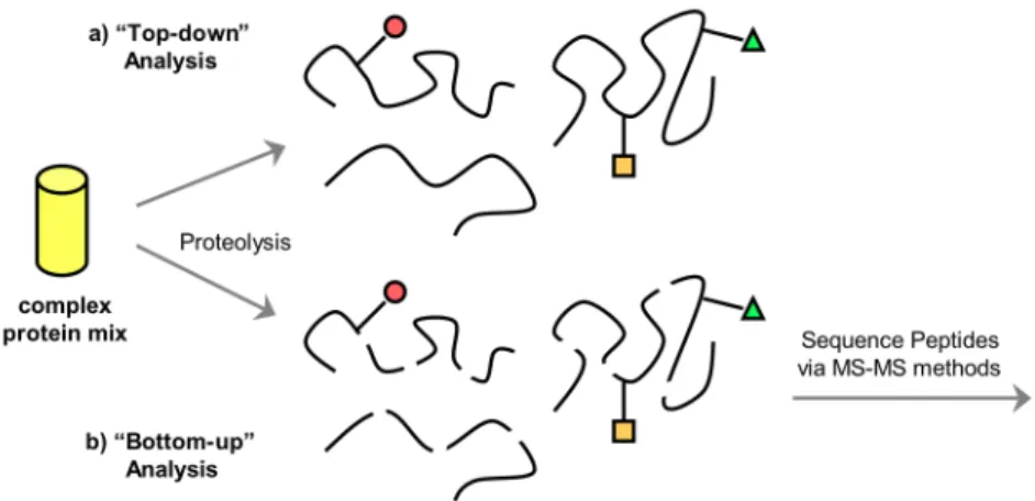
2.7 Approaches to identifying PTMs via mass spectrometry………... 33
2.8 Reversible disulfide exchange……… 36
2.9 Design of dithiol building blocks A and B………. 37
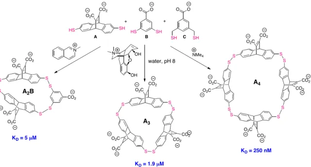
2.10 Selective amplification and binding of three different quaternary ammonium guests………... 38
2.11 Receptors amplified by acetylcholine……… 39
2.12 Monomers used in DCLs……… 40
2.13 Methylated lysine and arginine dipeptide guests………... 42
2.14 Amplified receptors rac-A2B and meso-A2B in an ABC DCL …………. 43
2.15 HPLC trace of an ABC DCL templated with a KMe3 dipeptide………... 44
2.17 HPLC traces of ABC DCLs templated with RMex dipeptides…………... 46
2.18 Extent of amplification of rac-A2B and meso-A2B in an ABC library….. 47
2.19 HPLC traces of A2B DCLs templated with KMex dipeptides……… 49
2.20 Extent of amplification of rac-A2B and meso-A2B in an A2B biased library……….. 50
2.21 Methylated histone tail peptide guests……… 51
2.22 Extent of amplification of rac-A2B and meso-A2B in an A2B biased library templated with methylated histone peptides………... 52
2.23 Fluorescently labeled H3 K9Mex peptide………... 55
2.24 H3 K9Me3 peptide used in ITC experiments………. 60
2.25 Possible variations of monomer B……….. 60
2.26 HPLC traces of A2B’ DCLs templated with KMex & RMex dipeptides… 63 2.27 HPLC trace of an AB’3 DCL templated with a KMe3 dipeptide………… 64
2.28 Extent of amplification of A2B’ in an A2B’ templated library…………... 66
2.29 HPLC traces of AFH DCLs templated with KMe3 & aRMe2 dipeptides……… 67
2.30 Isolation of A2B by HPLC………. 75
2.31 Analytical LC traces of purified rac-A2B and meso-A2B……….. 76
2.32 Mass spectra of purified A2B……….. 76
2.33 1H NMR of peptide H-KMe3-G-NH2………. 77
2.34 1H NMR of peptide H-KMe3-G-NH2 bound to rac-A2B……… 78
2.35 Determination of the extinction coefficient of monomer B………... 79
2.36 Determination of the extinction coefficient of monomer A………... 80
2.38 Fluorescence anisotropy of rac-A2B with H3 K9Me2……… 82
2.39 Fluorescence anisotropy of rac-A2B with H3 K9Me………. 82
2.40 Fluorescence anisotropy of rac-A2B with H3 K9……….. 83
2.41 Fluorescence anisotropy of meso-A2B with H3 K9Me3………. 83
2.42 Fluorescence anisotropy of meso-A2B with H3 K9Me2………. 84
2.43 Fluorescence anisotropy of rac-A2B with H3 G8K9Me3………... 84
2.44 Fluorescence anisotropy of rac-A2B with H3 aRMe2……… 85
2.45 Fluorescence anisotropy of rac-A2B with H3 sRMe2……… 85
2.46 Fluorescence anisotropy of rac-A2B with H3 RMe………... 86
2.47 ITC titration of rac-A2B with KMe3………... 87
2.48 ITC titration of meso-A2B with KMe3……… 88
2.49 Amplified receptor BD2 in a BCDH DCL………. 90
2.50 HPLC trace of a BCDH DCL templated with a KMe3 dipeptide……….. 91
2.51 Extent of amplification of BD2 in a BCDH library……… 92
2.52 HPLC trace of a BD2 biased library templated with a KMe3 dipeptide…. 94 2.53 Extent of amplification of BD2 in a BD2 biased library………. 94
2.54 HPLC trace of a BD2 biased library templated with a H3 K9Me3 peptide……… 95
2.55 Structural isomers of BD2………... 97
2.56 Upfield shifts of H-KMe3-G-NH2 bound to BD2……… 99
2.57 Evidence of aggregation of BD2 by NMR……….. 101
2.59 Fluorescence spectra of BD2 with a KMe3 dipeptide………. 103
2.60 HPLC traces of B’D2 biased DCLs templated with KMex and RMex dipeptides……….... 106
2.61 Isolation of BD2 by HPLC……….. 110
2.62 Mass spectra of purified BD2……….. 110
2.63 1H NMR of BD2 at 40 °C………... 111
2.64 1H NMR of BD2 at 25 °C………... 112
2.65 1H NMR of BD2 at 5 °C………. 112
2.66 BD2 TOSCY and NOESY cross peaks………... 113
2.67 1H NMR of H-KMe3-G-NH2 with 10% acetonitrile-d3 at 25 °C………… 114
2.68 1H NMR of H-KMe3-G-NH2 bound to BD2 with 10% acetonitrile-d3 at 25 °C………... 115
2.69 1H NMR of H-KMe3-G-NH2 with 10% acetonitrile-d3 at 5 °C………….. 116
2.70 1H NMR of H-KMe3-G-NH2 bound to BD2 with 10% acetonitrile-d3 at 5 °C………. 117
2.71 Receptors amplified in an ABD DCL………. 120
2.72 HPLC traces of ABD DCLs templated with KMe3 and aRMe2 dipeptides……… 122
2.73 Extent of amplification of A2D in an ABD library………. 123
2.74 HPLC traces of A2D biased DCLs templated with KMex and RMex dipeptides……… 125
2.75 Extent of amplification of A2D in an A2D biased library………... 125
2.76 Isomers of A2D………... 128
2.77 1H NMR of A2D………. 130
2.79 Upfield shifts of Ac-aRMe3-G-NH2 bound to A2D……… 132
2.80 Isolation of A2D by HPLC………. 139
2.81 Mass spectra of purified A2D………. 139
2.82 1H NMR of peptide Ac-aRMe2-G-NH2……….. 141
2.83 1H NMR of peptide Ac-aRMe2-G-NH2 bound to A2D………... 142
2.84 Uncorrected fluorescence intensity data of H3 K9Mex and H3 R8Mex peptides with A2D………... 143
2.85 Fluorescence titration of H3 asymmetric R8Me2 with A2D………... 144
2.86 Fluorescence titration of H3 symmetric R8Me2 with A2D………. 144
2.87 Fluorescence titration of H3 R8Me with A2D……… 145
2.88 Fluorescence titration of H3 K9Me3 with A2D……….. 145
2.89 Fluorescence titration of H3 K9Me2 with A2D……….. 146
2.90 Fluorescence titration of H3 K9Me with A2D……… 146
2.91 Fluorescence titration of unmethylated H3 R8K9 with A2D……….. 147
2.92 Peptide microarray assays……….. 149
2.93 Structure of A2(B-flagtag) receptor………... 152
2.94 Structure of monomer A-flagtag2………... 155
2.95 Structure of (A-flagtag2)2B receptor……….. 156
2.96 Isolation of A2(B-flagtag) by HPLC……….. 158
2.97 Fluorescence anisotropy of rac-A2(B-flagtag) with H3 K9Me3………… 160
2.98 Fluorescence anisotropy of rac-A2(B-flagtag) with H3 K9Me3 in the presence of a FLAG-tag antibody……….. 160
3.1 Cis-trans isomerization of azobenzene derivatives……… 169 3.2 Reversible hydrazone exchange………. 170 3.3 Azobenzene monomer 1 for photoswitchable DCLs……….. 172 3.4 HPLC trace of a photoswitchable DCL of 1 at thermal equilibrium…….. 174 3.5 UV-Vis spectra of cis and trans azobenzene monomer 1………... 175 3.6 Proline hydrazide building blocks 2 and 3………. 176 3.7 HPLC trace of a photoswitchable DCL containing 1 and 2 at thermal
equilibrium……….. 176 3.8 HPLC trace of a photoswitchable DCL containing 1 and 3 at thermal
equilibrium……….. 177 3.9 HPLC trace of a DCL of 1 in the photostationary state……….. 178 3.10 HPLC trace of a photoswitchable DCL containing 1 and 2 in the
photostationary state………... 179 3.11 HPLC trace of a photoswitchable DCL containing 1 and 3 in the
photostationary state………... 179 3.12 HPLC traces of a DCL of 1 and 3 immediately after photolysis, after
thermal relaxation, and equilibrated in the photostationary state………... 181 3.13 Controlling molecular recognition with a photoswitchable receptor……. 182 3.14 Templation of a photoswitchable DCL with a polyproline peptide……... 183 3.15 HPLC traces of a DCL of 1 and 2 templated with polyproline………….. 184 3.16 Extent of amplification of a cis-azobenzene macrocycle by polyproline... 185 3.17 Other templates investigated in photoswitchable DCLs………. 186 3.18 HPLC traces of a DCL of 1 and 2 templated with benzyltrimethyl
ammonium chloride at thermal equilibrium………... 188 3.19 HPLC traces of a DCL of 1 and 2 templated with benzyltrimethyl
3.20 HPLC traces of a DCL of 1 and 2 templated with methylisoquinoline
iodide at thermal equilibrium and after photolysis………. 190 3.21 Control experiments with azobenzene amino acid 8……….. 191 3.22 Extent of thermal relaxation of amino acid 8 with and without
benzyltrimethyl ammonium……… 193 3.23 1H NMR of azobenzene monomer precursor 5……….. 197 3.24 1H NMR of azobenzene monomer precursor 6……….. 198 3.25 1H NMR of azobenzene monomer precursor 7……….. 198 3.26 1H NMR of trans-azobenzene hydrazide monomer 1……… 199 3.27 1H NMR of cis-azobenzene hydrazide monomer 1……… 199 3.28 1H NMR of meta-substituted proline hydrazide monomer 2……….. 200 3.29 1H NMR of para-substituted proline hydrazide monomer 3………... 200 4.1 Peptidic monomers used to generate DCLs via hydrazone exchange…… 207 4.2 Reversible thioester exchange……… 208 4.3 Reversible formation of dimeric cyclic thiodepsipeptides………. 211 4.4 Preliminary HPLC trace of a DCL containing thioester monomers 3, 6,
and 7………... 213
4.5 HPLC traces of an equilibrating DCL containing thioester 1 over time.... 214 4.6 HPLC traces of cyclic macrocycles of thioester 5 forming over time…… 215 4.7 Rate of consumption of thioester monomers 1 – 3 and 5 – 7…………... 217 4.8 HPLC traces of cyclic macrocycles of thioester 6 forming over time…… 218 4.9 Rate of consumption of thioester monomers 7 and 15……….. 219 4.10 HPLC trace of a DCL containing thioesters 1, 3, and 5 – 7 after
4.12 Rate of consumption of thioester monomers 6 and 8………. 225 4.13 Rate of consumption of thioester monomers 7, 10, 14, and 15………….. 225 4.14 Rate of consumption of thioester monomers 3 and 4………. 226 4.15 Rate of consumption of thioester monomers 5 and 9………. 226 4.16 Molecular models of thioester monomers 5 and 9………. 227 5.1 Structure of hairpin peptide WKWK……….. 231 5.2 NMR structure of ATP bound to WKWK……….. 231 5.3 Structures of nucleotide substrates ATP and GTP………. 232 5.4 Structure of 7-methyl GTP………. 233 5.5 Structures of hairpin dithiol monomers 1 and 2………. 237 5.6 Structures of small molecules monomers B and C………. 238 5.7 Deamidation of hairpin monomer 2b………. 240 5.8 Structure of SKWS hairpin 3 used to investigate peptide deamidation…. 242 5.9 Structures of hairpin monomers 4 and 5 containing a DPro-Gly turn……. 242 5.10 Structure of WCWC hairpin monomer 6……… 244 5.11 DCL screens containing monomers 6, B, and C……… 245 5.12 HPLC traces of DCLs containing monomers 6, B, and C templated with
GMP and 7-Me GMP………. 246 5.13 Macrocycles 6B and 6B2 amplified by 7-Me GMP……… 247 5.14 HPLC traces of DCLs containing monomers 6 and B templated with
GMP and 7-Me GMP………. 248
5.18 Isolation of 6B and 6B2……….. 258 5.19 Mass spectra of purified 6B……… 258 5.20 1H NMR of peptide SKWS 3……….. 259 5.21 1H NMR of peptide SKWS 3 after exposure to basic solution…………... 260 5.22 Asn and Gln 1H NMR signals of 3 before and after exposure to basic
solution………... 261 5.23 1H NMR of peptide WCWC 6……… 262 6.1 NMR structure of a photoswitchable TrpZip hairpin………. 267 6.2 Structure of photoinducible WKWK hairpin 9………... 271 6.3 UV-Vis spectra of hairpin 9 before and after photolysis……… 272 6.4 CD spectra of hairpin 9 at thermodynamic equilibrium and in the
photostationary state………... 274 6.5 HPLC traces of hairpin 9 in the presence and absence of ATP at
thermodynamic equilibrium and in the photostationary state……… 275 6.6 CD spectra of hairpin 9 at thermodynamic equilibrium with ATP……… 276 6.7 CD spectra of hairpin 9 in the photostationary state with ATP………….. 278 6.8 CD spectra of control peptide WKWK with a Gly-Gly turn in the
presence and absence of ATP……….
279
List of Schemes
Scheme Page
Abbreviations
ACN Acetonitrile
AF Amplification factor Arg, R Arginine
ArgMe, RMe Monomethylarginine ArgMe2, RMe2 Dimethylarginine
aRMe2 Asymmetric dimethylarginine Asn, N Asparagine
Asp, D Aspartic acid
ATP Adenosine 5’-triphosphate CD Circular dichroism
CHCl3 Chloroform Cys, C Cysteine
DCC Dynamic combinatorial chemistry DCL Dynamic combinatorial library DCM Dichloromethane
DIPEA Diisopropylethyl amine DMF Dimethylformamide DMSO Dimethyl sulfoxide DNA Deoxyribonucleic acid
DSS 3-(Trimethylsilyl)-1-propanesulfonic acid sodium salt EDT 1,2-Ethanedithiol
eIF4E Eukaryotic initiation factor 4E ESI Electrospray ionization
FAM Carboxyfluorescein
Fmoc N-9-Fluorenylmethyloxycarbonyl Gln, Q Glutamine
GTP Guanosine 5’-triphosphate
H3 Histone 3
HBTU 2-(1H-Benzotriazole-1-yl)-1,1,3,3-tetramethyluronium hexafluorophosphate
His, H Histidine
HOBT N-hydroxybenzotriazole HP1 Heterochromatin protein 1
HPLC High pressure liquid chromatography Hyp Hydroxyproline
IsoAsp Isoaspartic acid
ITC Isothermal titration calorimetry
LC-MS Liquid chromatography – mass spectrometry Lys, K Lysine
LysMe, KMe Monomethyllysine LysMe2, KMe2 Dimethyllysine LysMe3, KMe3 Trimethyllysine MS Mass spectrometry
NMR Nuclear magnetic resonance
Pbf 2,2,4,6,7-Pentamethyl-dihydrobenzofurance-5-sulfonyl PEG Polyethylene glycol
Phe, F Phenylalanine Pro, P Proline
PTM Post-translational modification sRMe2 Symmetric dimethylarginine TCEP Tris(2-carboxyethyl)phosphine TFA Trifluoroacetic acid
TFE Trifluoroethanol TIPS Triisopropyl silane Trp, W Tryptophan
Trt Trityl
CHAPTER I
DYNAMIC COMBINATORIAL CHEMISTRY
A. Introduction
In attempt to mimic the thriving process of natural selection where those deemed most “fit” are rewarded with the ability to successfully reproduce, since the mid-1990s chemists have been developing their own natural selection process involving both selection and amplification, known as dynamic combinatorial chemistry (DCC).
Dynamic combinatorial chemistry is an attractive alternative to traditional rational design in the development of novel receptors, in which molecular recognition guides the
synthesis of complex host systems from simple building blocks using reversible linkages under thermodynamic control (Figure 1.1).1 It takes advantage of the ability of a
Figure 1.1. Generation and templation of a dynamic combinatorial library (DCL).
There are several key components of DCC that together allow for a system that can facilitate Darwinian like evolution, each of which will be introduced to some degree to provide adequate background for the work in this thesis. First, a set of designed monomers which serve as building blocks in the generation of a larger library of constituents are needed. Second, the implementation of reversible chemistry between monomers is required to generate a group of library members which are under
B. Reversible exchange reactions
While the principal requirement of a reaction used to facilitate exchange between building blocks in a dynamic combinatorial library is that it be reversible, there are multiple other criteria that must be met in order for the system to serve as an effective selection process.1a To begin, the rate of reversible exchange is important in that it should occur on a reasonable time scale. While this requirement is often a matter of convenience, it can also be important when degradation of the target molecule over time is an issue. Next, due to the simultaneous nature of the equilibration and selection processes, the reversible reaction must proceed under conditions that are compatible with the target of interest, including all functional groups on the molecule. This becomes particularly relevant, and often challenging, in the screening of biomolecules which generally require physiological conditions (neutral pH in buffered aqueous solution). While the reaction conditions must be compatible with the experimental conditions for selection, they must also be fairly mild (for example, ambient temperature and pressure) as to not interfere with the non-covalent interactions involved in molecular recognition. The reversible reaction must also ensure the solubility of all library members formed at equilibrium, as any insoluble species can potentially act as a thermodynamic trap. Lastly, the ability to turn off the exchange reaction and kinetically “freeze” the library
distribution is essential for analysis of the library distribution and to fully assess the degree of amplification. The isolation and further investigation of identified receptors is also only feasible if exchange has been halted.
covalent bonds often exhibit slower kinetics, they are more widespread due to the fact that enhanced compounds can be easily isolated and handled further. In contrast, libraries formed based on noncovalent interactions achieve equilibration rapidly, yet the often weak and labile bonds generate products that are difficult to analyze in solution and even more problematic to isolate. Despite this, both hydrogen bonds4 and metal-ligand coordination bonds5 have been successfully applied to DCC experiments in numerous cases. While the full scope of potential reversible reactions is exhaustive and beyond the scope of this thesis, it is worth addressing those that have received significant attention thus far in this field.
Nearly all reversible unsymmetrical covalent bonds used in DCC involve carbonyl compounds or some derivative thereof (Figure 1.2). Acyl exchange reactions such as trans-esterification, allyl ester exchange, amide exchange, aldol exchange, and thioester exchange are all facilitated by the relatively fragile nature of the C(O)-X bonds, which can consequently be reversibly broken. One of the earliest examples where reversible transesterification was utilized in DCC studies was reported by Sanders and coworkers, where a cholic acid methyl ester derivative was used to form an equilibrating mixture of linear and cyclic oligomers in the presence of a potassium methoxide-crown ether complex.6 Amide bond exchange, while slightly more challenging due to the stability of amide bonds, has also been demonstrated based on approaches which employ
has been employed in the context of libraries targeting hydrolases9 and in the evaluation of the higher order structural stability in polypeptides.10
O X X
O X
N X
Acyl transfer
Acetyl-type exchange C=N exchange
Figure 1.2. Different types of unsymmetrical covalent exchange reactions involving carbonyl groups: acyl transfer (X = NHR, OR, SR, CH2R), C=N exchange (X = R, NHR, OR), and acetal-type exchange (X = OR, SR, NHR).
Several studies have also explored exchange reactions that benefit from the labile C=N double bond, such as imine, oxime, and hydrazone exchange. The first
demonstration of imine exchange in DCC was described by Huc and Lehn in the generation of a library targeting the production of carbonic anhydrase inhibitors,11 and imine exchange has since remained prevalent in the DCC literature. Structurally related hydrazones and oximes are often viewed as advantageous due to their great hydrolytic stability in comparison to imines. While the application of reversible oxime chemistry in the generation of libraries has been somewhat scarce since its first demonstration by Eliseev and coworkers in the late 1990s,12 hydrazone exchange has proven to be a quite successful and powerful technique in the generation of diverse DCLs. Numerous
examples have been presented based on pseudo-mono-and dipeptides in the generation of acyl hydrazone libraries,13 many of which have resulted in receptors for cationic
Transacetalization chemistry based on the well-known formation of cyclic acetals from diols and aldehydes has been demonstrated in relatively few studies in comparison to acyl and C=N exchange reactions. An initial example revealed the transacetalation between a D-threitol derivative and a diacetal to generate a very complex mixture of cyclic and linear compounds.17 Acetal exchange between formaldehyde acetals to explore cyclophane formation via DCC has also been presented,18 while more recently the production of a cyclic polyether DCL was reported resulting in the amplification of small macrocyclic library members by ammonium ions.19
DCLs. Nicolaou and coworkers used reversible olefin metathesis to prepare a library of vancomycin dimers in water, although not initially intended as a DCC experiment per se. A significant catalyst dependence has also been demonstrated in the templated synthesis of porphyrin boxes from alkene functionalized porphyrin building blocks.22
C. Building block design
For the successful use of DCC, particularly in the event of a selection process, there are various elements that must be considered in the design and choice of building blocks. Specific functionality is required to facilitate an identified reversible reaction, and the reactive functionality can be incorporated either once or twice in each monomer,
depending on whether a library of linear dimeric species or cyclic oligomers are desired. The presence of two reactive groups is generally advantageous in that it results in an increase in the structural diversity of the library, which is particularly crucial in the search for molecular receptors. The generation of species of varying size is feasible, albeit often at an entropic cost for the larger oligomers, however the placement of the reactive groups within each monomer is quite important in dictating the formation of linear versus cyclic oligomers.
The structural diversity desired is often tied to the extent of flexibility incorporated into each designed monomer. When monomers are quite rigid they have a tendency to self-sort, particularly when they differ from each other significantly in terms of size and “bite angle” (Figure 1.3).1a In this case, heterodimers tend to be destabilized through ring strain and therefore represent only minor species within the mixture. A degree of
investigation of a single building block library. Furthermore, diversity can be increased by the incorporation of a small, flexible unit, which can bridge monomers that would otherwise not cyclize or be quite strained.
Figure 1.3. The tendency of rigid building blocks to self-sort.
A consideration of solubility and molecular weight are also worth nothing in the design of monomers. While often difficult to predict, all species formed in a particular library must maintain complete solubility throughout library equilibration to ensure a complete analysis. This becomes particularly problematic upon the generation of larger library members, as the appropriate solubility of each monomer does not ensure that each macrocycle formed will remain soluble. The solubility must also be maintained in a solvent system that is appropriate for each application, be it templation or otherwise. In the case of templation studies, the hosts and guests must sustain complete solubility as individual components and upon binding. In regards to molecular weight, a unique mass is desired for each monomer, as most libraries are analyzed by mass spectrometry which can not differentiate between structural differences. However, in some cases this can be circumvented as a result of different UV absorption spectra of mass degenerate
D. Experimental considerations
As in most reactions, upon choosing reagents and conditions under which the reaction is performed, the main consideration that remains is the concentration of the reagents. In the case of DCC, the building block concentrations tend to be quite influential on the composition of the resulting library. This is a key parameter because it influences the rates of the exchange reactions, as well as the oligomer to polymer ratio. It has been proposed previously that there exists a critical concentration below which the equilibrium composition of the system consists entirely of small macrocycles.23 This concentration dependence should be evaluated in the context of each library in order to generate macrocycles of appropriate size and diversity.
template concentration ensures amplification of those best receptors without a serious loss in templating efficiency. This behavior reflects the truly complex nature of such DCL molecular networks.
E. Possible selection methods
While initial investigations in this field were focused on library development and the underlying reversible chemistry, attention has more recently moved toward evaluating shifts in product distribution resulting from some type of favorable noncovalent
interaction. It is this unique ability of reversible DCLs to efficiently select and amplify a species that is most “fit” in the context of the library that highlights the true utility of this methodology. The majority of the selection methods that have been presented to date involve the use of templating to select for molecules that act as hosts or receptors for particular guests of interest. While synthetic chemists have proven to be quite good at designing elegant syntheses for complex molecular receptors, developing new and
modified versions of such receptors often involves a lengthy process of design, synthesis, evaluation, and often redesign. By simply designing potential receptor fragments and leaving the rest up to the recognition properties of the guest, DCC has allowed for the identification and synthesis of quite complex and unpredictable synthetic receptors, with often impressive affinity. This type of selection has been applied to the identification of receptors for a wide range of molecules, from biomolecules to metal ions, using a variety of reversible chemistries and reaction conditions.
the use of an enzyme’s active site, for example, can lead to amplification of potential inhibitors. The subtle mechanisms of protein folding, the difficulty in predicting the strength of ligand-protein interactions, and the fact that the structure of many proteins is not precisely known often hampers the design and discovery of new inhibitors by
traditional methods. DCC is particularly attractive in this context in that the exact nature of the binding site need not be well understood. Although the association between the ligand and biomolecule is generally entirely noncovalent, a tethering approach has also been developed in which a reversible covalent linkage is used to tether the ligand to the biomolecule.26 This concept is advantageous in that it facilitates the binding of ligands so that low-affinity binders that would otherwise not bind strongly enough can be detected. It should be noted that such systems can be somewhat limiting due to the physiological conditions and high protein concentrations required.
In addition, the selection of the most stable structure within a mixture of structures with different conformational properties can also be facilitated via DCC. Through the use of reversible chemistry, folding into secondary and tertiary structures by proteins or nucleic acids, as well as polymers or oligomers, can be monitored. Molecular recognition takes place intramolecularly, and there is generally no need for a formal template
molecule. Somewhat related is the use of DCC to investigate the stabilization of specific library members through intermolecular noncovalent interactions. In this case the library composition is biased toward those species that form stable assemblies or aggregates. While this selection method has yet to gain a significant amount of attention, it has much potential in the investigation of self-assembling molecules, such as interlocked or
F. Analytical methodology
One primary advantage to DCC as a combinatorial technique is the simultaneous nature of the synthesis and selection processes. Library analysis via liquid
chromatography coupled with mass spectrometry has by far evolved as the primary method of library analysis, allowing for a full identification of all species in solution, in both templated and untemplated reactions. Often more important than characterizing all members of the library, LC-MS allows for the accurate identification of all species enhanced due to favorable molecular recognition events (Figure 1.4). The untemplated reaction ideally serves as an internal control for self-selection processes, and allows for a direct comparison to a library containing a molecular guest of interest. Those species enhanced become visually apparent via inspection of the chromatography traces, and subsequent MS analysis facilitates their immediate identification. Further MS-MS can also be utilized in the analysis of libraries involving sequence isomers and
regioisomers.27
Figure 1.4. Representative chromatography traces of an untemplated library distribution (left), and a library templated with an external template (right). Molecular recognition events guide the amplification of those most favorable receptors (shown in green).
also presents many challenges, as library mixtures can only become so complex before accurate analysis is no longer possible. When the chromatography becomes quite involved, not only is the library composition difficult to ascertain, but the ability to identify amplified, active compounds is often lost. With increasing complexity the probability of generating strong binders increases, yet this results in concentrations of individual library members that are quite low, often below a reasonable concentration for either detection or amplification. The question of how many compounds a DCL can contain while still allowing the amplification of strong binders to useful concentrations is still under debate, and theoretical studies have aided in this regard.28 This again
highlights the importance of careful monomer design, as maximum diversity is desired within the limits of the analytical method. The use of larger solution phase libraries (> 9000 compounds) have been reported in the successful identification of molecular receptors, however most studies continue to make use of relatively simple libraries.29
immobilized, and the detection of selected hits is facilitated by the use of fluorescently labeled targets.33 Beyond liquid-solid phase segregation, the use of liquid-liquid phase segregation has also be reported using two immiscible solvents to increase the diversity and scope of libraries that can be generated and explored via DCC.34 Lastly, as an extension to traditional DCL selection methods, Rayner and coworkers have described a process that combines DCC and SELEX (for Systematic Evolution of Ligands by Exponential enrichment) for the in vitro selection of small molecule conjugated RNA aptamers that bind tightly to the TAR (transactivation-responsive) element of HIV-1.35
G. Purpose of this work
In recent years, many contributions have been made to the field of DCC and it has found application among a variety of disciplines, ranging from drug discovery to material science.36 Although the potential utility of DCC is unquestionable, it remains a relatively limited and young field, allowing for further contributions in a range of areas, while a number of challenges have yet to be addressed. Although the groundwork has been laid in the development of reversible reactions for library generation, new types of reactions will continually be of interest, while the use of known reactions under more
physiological-like conditions is undoubtedly desired for biological applications. The potential to generate new monomers and libraries with novel functionality is unlimited, while there are also numerous targets, biological and otherwise, for which DCC may provide an effective method in the identification of host molecules.
functionality, generate new types of libraries for future applications, and to investigate the recognition of biologically interesting targets. The identification of synthetic receptors for post-translationally modified amino acids such as methylated lysine and arginine, as well as methylated nucleotides, has been investigated due to the critical role of each in controlling gene expression. Our interests in both photochemistry and the development of libraries containing more than one reversible reaction prompted an investigation of azobenzene containing monomers. An array of thioester peptide monomers were also evaluated for their ability to readily generate libraries of cyclic macyrocycles for future DCC assays, while the application of larger peptide monomers with known secondary structures have also been investigated in the context of dynamic combinatorial libraries. It is our hope that these studies ultimately play a role in
1
For reviews, see: (a) Corbett, P. T.; Leclaire, J.; Vial, L.; West, K. R.; Wietor, J-L.; Sanders, J. K. M.; Otto, S. Chem. Rev.2006, 106, 3652-3711. (b) Lehn, J-M. Chem. Eur. J.1999, 5, 2455-2463. (c) Ladame, S. Org. Biomol. Chem. 2008, 6, 219-226. (d)
Ludlow, R. F.; Otto, S. Chem. Soc. Rev.2008, 37, 101-108. (e) Otto, S. Curr. Opin. Drug Disc. Devel.2003, 6, 509-520.
2
For example, see: (a) Otto, S.; Furlan, R. L. E.; Sanders, J. K. M. Science2002, 297, 590-593. (b) Baolu, S.; Stevenson, R.; Campopiano, D. J.; Greaney, M. F. J. Am. Chem. Soc.2006, 128, 8459-8467.
3
(a) Lam, R. T. S.; Belenguer, A.; Roberts, S. L.; Naumann, C.; Jarrosson, T.; Otto, S.; Sanders, J. K. M. Science2005, 308, 667-669. (b) Hamilton, D. G.; Feeder, N.; Teat, S. J.; Sanders, J. K. M. New J. Chem.1998, 1019-1021.
4
For example, see: (a) Crego Calama, M.; Hulst, R.; Fokkens, R.; Nibbering, N. M. M.; Timmerman, P.; Reinhoudt, D. N. Chem. Commun.1998, 1021-1022. (b) Hof, F.; Nuckolls, C.; Rebek, J. Jr. J. Am. Chem. Soc. 2000, 122, 4251-4252.
5
For example, see: (a) Baxter, P. N. W.; Khoury, R. G.; Lehn, J.-M.; Baum, G.; Fenske, D. Chem. Eur. J. 2000, 6, 4140-4148. (b) Mal, P.; Schultz, D.; Beyeh, K.; Rissanen, K.; Nitschke, J. R. Angew. Chem. Int. Ed.2008, 47, 8297-8301. (c) Albrecht, M.; Blau, O.; Frohlich, R. Chem. Eur. J.1999, 5, 48-56.
6
Brady, P. A.; Sanders, J. K. M. J. Chem. Soc. Perkin I1997, 3237-3253.
7
Swann, P. G.; Casanova, R. A.; Desai, A.; Frauenhoff, M. M.; Urbancic, M.;
Slomczynska, U.; Hopfinger, A. J.; LeBreton, G. C.; Venton, D. L. Biopolymers1996, 40, 617.
8
(a) Eldred, S. E.; Stone, D. A.; Gellman, S. H.; Stahl, S. S. J. Am. Chem. Soc.2003, 125, 3422-3423. (b) Hoerter, J. M.; Otte, K. M.; Gellman, S. H.; Cui, Q.; Stahl, S. S. J. Am. Chem. Soc.2008, 130, 647-654.
9
Larsson, R.; Ramstrom, O. Eur. J. Org. Chem.2006, 2006, 285-291.
10
Woll, M. G.; Gellman, S. H. J. Am. Chem. Soc. 2004, 126, 11172-11174.
11
Huc, I.; Lehn, J. M. Proc. Natl. Acad. Sci. USA1997, 94, 2106-2110.
12
(a) Polyakov, V. A.; Nelen, M. I.; Nazarpack-Kandlousy, N.; Ryabov, A. D.; Eliseev, A. V. J. Phys. Org. Chem. 1999, 12, 357-363. (b) Nazarpack-Kandlousy, N.;
Zweigenbaum, J.; Henion, J.; Eliseev, A. V. J. Comb. Chem. 1999, 1, 199-206.
13
14
For example, see: (a) Lam, R. T. S.; Belenguer, A.; Roberts, S. L.; Naumann, C.; Jarrosson, T.; Otto, S.; Sanders, J. K. M. Science2005, 308, 667-669. (b) Furlan, R. L. E.; Yiu-Fai, N.; Cousins, G. R. L.; Redman, J. E.; Sanders, J. K. M. Tetrahedron2002, 58, 771-778.
15
Furlan, R. L. E.; Ng, Y.-F.; Otto, S.; Sanders, J. K. M. J. Am. Chem. Soc.2001, 123, 8876.
16
Voshell, S. M.; Gagné, M. R. J. Am. Chem. Soc.2006, 128, 12422-12423.
17
Fuchs, B.: Nelson, A.; Star, A.; Stoddart, J. F.; Viidal, S. Angew. Chem. Int. Ed. 2003, 42, 4220-4224.
18
Cacciapaglia, R.; Di Stefano, S.; Mandolini, L. J. Am. Chem. Soc.2005,127, 13666-13671.
19
Berkovich-Berger, D.; Lemcoff, N. G. Chem. Commun.2008, 1686-1688.
20
Otto, S.; Furlan, R. L. E.; Sanders, J. K. M. J. Am. Chem. Soc.2000, 122, 12063-12064.
21
Boul, P. J.; Reutenauer, P.; Lehn, J.-M. Org. Lett.2005, 7, 15-18.
22
Van Gerven, P. C. M.; Elemans, J. A. A. W.; Gerritsen, J. W.; Speller, S.; Nolte, R. J. M.; Rowan, A. E. Chem. Commun. 2005, 3535-35537.
23
Jacobsen, H.; Stockmayer, W. H. J. Chem. Phys.1950, 18, 1600-1607.
24
(a) Grote, Z.; Scopelliti, R.; Severin, K. Angew. Chem. Int. Ed.2003, 42, 3821-3825. (b) Severin, K. Chem. Eur. J.2004, 10, 2565-2580. (c) Corbett, P. T.; Otto, S.; Sanders, J. K. M. Chem. Eur. J.2004, 10, 3139.
25
Corbett, P. T.; Sanders, J. K. M.; Otto, S. J. Am. Chem. Soc. 2005, 127, 9390-9392.
26
Erlanson, D. A.; Hansen, S. K. Curr. Opin. Chem. Biol.2004, 8, 399.
27
Nazarpack-Kandlousy N.; Nelen, M. I.; Goral, V.; Eliseev, A. V. J. Org. Chem.2002, 67, 59-65.
28
Corbett, P. T.; Otto, S.; Sanders, J. K. M. Org. Lett.2004, 6, 1825-1827.
29
Ludlow, R. F.; Otto, S. J. Am. Chem. Soc.2008, 130, 12218-12219.
30
Ramstrom, O.; Lehn, J.-M. ChemBioChem2000, 1, 41-48.
31
Roberts, S. L.; Furlan, R. L. E.; Cousins, G. R. L.; Sanders, J. K. M. Chem. Commun.
2002, 938-939.
32
33
McNaughton, B. R.; Miller, B. L. Org. Lett.2006, 8, 1803-1806.
34
(a) Pérez-Fernández, R.; Pittelkow, M.; Belenguer, A. M.; Sanders, J. K. M. Chem. Commun.2008, 1738-1740. (b) Choudhary, S.; Morrow, J. R. Angew. Chem. Int. Ed.
2002, 41, 4096-4098.
35
Bugaut, A.; Toulmé, J.-J.; Rayner, B. Org. Biomol. Chem.2006, 4, 4082-4088.
CHAPTER II
SMALL MOLECULE RECEPTORS FOR PROTEIN POST-TRANSLATIONAL
MODIFICATIONS
(Reproduced, in part, with permission from The Royal Society of Chemistry and
Ingerman, L. A.; Cuellar, M. E.; Waters, M. L. Chem. Commun. 2010, 46, 1839-1841.)
A. Background and Significance
i. Histone post-translational modifications and their role in gene transcription.
Developing a better understanding of the factors that control gene expression is crucial to
the study of both development and disease, and consequently, epigenetics has emerged as
a major challenge and field of study.1 In studying the control of genetic information, it
has become clear that post-translational modifications (PTMs), particularly those of
histone proteins, are intimately involved in gene regulation. These modifications
encompass a range of structural changes to natural amino acids, including methylation,
acylation, phosphorylation, ubiquitination, and glycosylation, among others, and they
have equally diverse functions. Post-translational modifications have been seen to play a
critical role in the regulation of signaling pathways and in tagging proteins for
degradation, while they can also act as chemical switches to induce or repress
Histone proteins are largely considered to be the framework for packaging genetic
material. Within the nucleus, DNA is wrapped around histone proteins to form
nucleosomes, which are then further condensed to give chromatin (Figure 2.1).
Chromatin can exist as tightly packed inactive genetic material, known as
heterochromatin, or switch to actively transcribed genetic material in a lightly packed
state, or euchromatin.
a) b)
Figure 2.1. a) Packaging of DNA within the nucleus (Adapted from a National Human Genome Research Institute Image). b) The atomic structure of the nucleosome core (pdb 1eqz), with each strand of DNA shown in a different shade of blue, surrounding the histone octamer.
There are eight histone proteins which assemble to form an octamer, composed of
two H2A-H2B heterodimers and two H3-H4 heterodimers.3 While DNA is wrapped
around the core of this octamer, each histone protein contains disordered termini which
protrude from the nucleosome. By extending beyond the nucleosome, these unstructured
peptide tails are readily available for enzymatic modification (Figure 2.2). Lysine and
serine residues in particular are abundant throughout the histone tail sequences, resulting
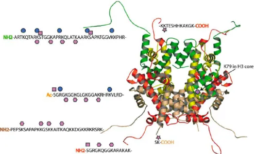
Figure 2.2. The types of post-translational modifications observed on the core histones, comprised of a tetramer of H3 (green) and H4 (yellow) and two dimers of H2A (red) and H2B (beige). The modifications are shown, including methylated lysine and arginine (blue circle), acetylated lysine (pink hexagon), phosphorylated serine (pink square), and ubiquitinated lysine (pink star).4
While it was initially proposed that these histone modifications exerted their
biological function by significantly altering the nucleosome structure, it has since been
realized that they instead act as markers for the initiation or repression of specific
interactions or for the precise recruitment of non-histone proteins, dictating the
higher-order chromatin structure in which DNA is packaged and resulting in gene expression or
gene silencing depending on the type and location of the PTM.5 While lysine acylation is
commonly associated with transcriptional activation, as it is thought to negate the
favorable electrostatic interaction between lysine and the phosphate backbone of DNA
resulting in the loosening of chromatin, lysine methylation often initiates the recruitment
of other proteins through specific binding interactions with the modified tail, resulting in
expression. Although various PTMs have been documented for over forty years, it has
only been within the past decade or so that the impact of certain modifications on gene
transcription have emerged.
Solving the puzzle of histone PTMs is complicated by the fact that the same chemical
modification occurring at different positions within a single protein can have different
effects on gene expression. This is particularly apparent with methylation. For example,
within the histone 3 tail, methylation of lysine 4, 36, or 79 results in activation of
transcription, whereas methylation of lysine 9 or 27 results in transcriptional repression.
This situation is further complicated by the fact that both arginine and lysine can be
variably methylated. Histone methyltransferases can add up to three methyl groups to a
single lysine side chain, while arginine can exist as either monomethyl or dimethyl
arginine (Figure 2.3). Some enzymes specifically produce symmetric dimethylarginine
(sRMe2), while others produce asymmetric dimethylarginine (aRMe2). It has become
clear that both the position of the residue and the degree of methylation together dictate
the transcriptional outcome of the modification.7
H N NH N H N H N H O O H N N N H O O H N NH N H O O H N
H2N N H O O H N NH NH2 N N H O O H N NH NH2 N H N H O O
KMe KMe2 KMe3
RMe sRMe2 aRMe2
ii. The histone code. With the investigation of these modifications and their specific
functions in isolation, it soon became apparent that there is a form of communication
between PTMs which allows them to work together in a very elaborate signaling
pathway. This idea that distinct histone modifications, on one or more tails, act
sequentially or in combination to form a code that is read by other proteins to bring about
distinct downstream events has since been deemed the “histone code.”8 Thus, it is
considered to be the effect of multiple modifications that together control gene
transcription.
Mechanistically it can be envision that this communication between modifications
may occur at several different levels. First, the presence of multiple modifications
(particularly those resulting in an increase or decrease of charge such as phosphorylation
and acetylation, respectively) are thought to amplify the readout of signaling pathways,
causing greater changes in the overall charge density of histone tails, and leading to a
greater change in the extent of chromatin condensation (or decondensation). Second, it is
possible that the covalent modification of a histone tail by one enzyme influences the rate
or efficiency with which a second enzyme follows. Many sites of modification are close
enough within the histone tail to influence, either positively or negatively, the ability of
enzymes to further modify the protein. An enzyme may recognize its substrate more
effectively in the context of other modifications, while the catalytic activity of an enzyme
may also be compromised by prior modification of its substrate. Thirdly, the recruitment
of PTM binding proteins may be affected by the presence of multiple modifications, in
some cases creating a stronger association, and in others working in tandem to recruit
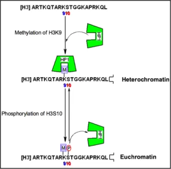
switches to control gene transcription, whereby adjacent PTMs establish a switch
mechanism to regulate protein-protein interactions. One such example is seen with
phosphorylation of serine 10 of the H3 protein, which disrupts the binding interaction
between the neighboring methylated lysine 9 and heterochromatin protein 1 (HP1)
(Figure 2.4).9 By ejecting the HP1 protein, this PTM switch serves to reverse the effects
of histone methylation and in turn reactive transcription. Furthermore, the reversibility of
these modifications allows for an extremely intricate control of gene transcription.
Figure 2.4. Phosphorylation as a PTM switch in the H3 tail, serving to reverse the transcriptional outcome of lysine methylation.
iii. Post-translational modification binding domains. Although the mechanisms
by which cells decipher a PTM-mediated histone code are far from understood, the
emerging school of thought is that histone PTMs are read by protein receptors, or effector
proteins, which facilitate downstream events via the recruitment and/or stabilization of
chromatin-templated machinery.10 Many epigenetic readers have been identified that
understanding of how histone PTMs regulate biological functions.11 These conserved
protein domains bind histone PTMs in a way that is dependent on both the type of
modification and the position within the histone sequence. The identification of binding
modules for methylated lysines has been largely successful, and are known to include
chromodomains, Tudor domains, MBT-repeats, WD40-repeats, and PHD fingers,
whereas protein receptors for methylated arginines in histone proteins have yet to be
identified.12 The identification of such methylated arginine reader proteins remains a
challenge, as does the broader goal of uncovering the relationship between PTM binding
proteins and human disease.
Unlike the elimination of charge upon lysine acetylation, all methylated forms of
lysine are cationic at physiological pH, with trimethyllysine containing a fixed positive
charge. As the number of lysine methyl groups increase, the hydrophobicity and the
distribution of positive charge of the methylammonium group increases, while
concomitantly it’s ability to serve as a hydrogen bond donor decreases. As a result, each
PTM requires a protein partner that can adapt to these inherent physical properties, in
turn leading to a great deal of specificity.
The recognition of these modifications results largely from contacts made between
the methylammonium group and aromatic residues in the protein receptor, forming an
aromatic “cage” about the PTM (Figure 2.5). These aromatic cages are generally highly
specific for a certain methylation state, discriminating between PTMs based on only very
slight differences in size and shape. The cages are thought to be preorganized and static
in nature, showing little or no appreciable structural perturbation upon binding, and
methylammonium and the aromatic cage is mediated largely by cation-π interactions,
while hydrophobic desolvation effects also have an appreciable role. The cation-π
interaction is generally thought of as a charge-quadrupole interaction between a
positively charged species and an aromatic ring, primarily electrostatic in nature. More
specifically, as the quadrupole moment places partial negative charge above each face of
the aromatic ring, favorable interactions with cations occur perpendicular to the plane of
the ring. The role of this interaction in proteins is a topic of great interest, particularly in
the investigation of protein structures, protein-protein interactions, and protein-ligand
interactions.13
a) b)
Figure 2.5. Binding pockets for lysine PTMs. a) Histone 3 K9Me3 (green) bound to the HP1 chromodomain (gray) via cation-π interactions with three aromatic side chains (pdb 1KNE). B) Histone 3 K4Me2 (green) bound to the 3-MBT domain (gray) via cation-π interactions with three aromatic residues and a hydrogen bond to glutamic acid (blue) (pdb 2RHI).
The magnitude of the cation-π interaction in proteins is dependent on numerous
factors, including the electron density of the aromatic ring (for example, phenylalanine
versus tryptophan), the distribution of positive charge across the cation, the degree of
solvent exposure of the interaction, as well as the contribution of other forces such as van
importance of the charge-quadrupole interaction to KMe3 binding and specificity in the
context of a β-hairpin model system, as well as in a histone peptide, indicating that the
driving force for binding is indeed the cation-π interaction as opposed to the hydrophobic
effect.14 Specifically, replacement of KMe3 in a histone 3 peptide with its neutral
analogue, tert-butylnorleucine, resulted in binding to the HP1 chromodomain nearly as
weak as unmethylated lysine, demonstrating the essential nature of the cation-π
interaction to the recognition of the H3 tail by the HP1 chromodomain with good affinity
and selectivity.
In the recognition of the lower methylation states, hydrogen bonding and steric
exclusion also become increasingly important. Nearby acidic residues in the protein are
also known to form salt bridges with the protonated amines of KMe2 and KMe. This has
been demonstrated in the context of a PHD domain by engineering dimethyllysine
recognition specificity. While the wild-type PHD finger’s binding preference is for
histone 3 K4Me3 over K4Me2, the binding preference was reversed for that of KMe2
over KMe3 through the mutation of a key tyrosine residue to glutamic acid.15 The
change in selectivity is associated with hydrogen bonding between the KMe2 proton and
the carboxylate group of the glutamic acid side chain.
Furthermore, complexes with a lower methylation state PTM are bound via the
cavity-insertion recognition mode, where the methylammonium group is inserted into and
buried deep within a protein cleft. This allows the protein pocket to bind only those
PTMs of the appropriate size.12 In contrast, the higher methylation states are known to
use a surface-groove recognition mode whereby the binding pockets are both wider and
consequently the effector proteins have slightly less stringent preferences for specific
methylation states.
Beyond selectivity for different methylation states, effector proteins display marked
selectivity for the sequence context within which a PTM is presented. The PTM
containing peptides are generally unstructured in the unbound state, but undergo an
induced-fit conformational change, adopting secondary structure when bound to their
recognition proteins. The histone tail peptides tend to adopt a β-sheet conformation,
pairing through an antiparallel alignment with an exposed face of an existing β-sheet of
the effector protein, as seen in the case of H3 K4Me3 recognition by the PHD finger of
NURF.16 Similarly, six residues of the H3 peptide containing K9Me3 insert as a β-strand
on the surface of the protein receptor, HP1 chromodomain, and complete a β-sandwich
overall fold.17 While backbone hydrogen bonds between residues adjacent to the PTM
and the effector protein are formed, strengthening the interaction, the sequence selectivity
is largely due to complementary side chain interactions between the two domains. The
steric compatibility, intermolecular hydrogen-bonding, and electrostatic interactions of
the surrounding residues all play a role in determining the binding affinity and specificity.
A significant protein-protein interaction induced by lysine methylation most relevant
to the work presented in this thesis is the binding of histone 3 K9Me3 to the HP1
chromodomain, which results in gene silencing (Figure 2.6). It is thought that the
function of the chromatin binding is to passively stabilize a dense conformation of the
chromatin fiber associated with gene repression.18 The binding affinities have been
reported to be in the micromolar range for both H3 K9Me3 and H3 K9Me2, ranging from
binding for the corresponding unmethylated peptide.14,17 The chromodomain adopts an
incomplete β-barrrel secondary structure, with the methyllysine binding pocket
positioned at one end. The aromatic binding cage is made up of three aromatic residues,
two tyrosines and a tryptophan, to form a conserved aromatic pocket into which the
methylammonium group inserts itself. Mutation of any of these aromatic residues
drastically reduces affinity for the methylated histone tail. Furthermore, residues 5-10 of
the histone tail (QTARK9S) interact with the chromodomain by an induced-fit
sandwiching between terminal β-strands, completing a five-stranded antiparallel β-sheet.
Mutation studies of residues in both the peptide and protein have confirmed the
contribution of intermolecular contacts along the extended surface groove to both binding
affinity and selectivity.19
Figure 2.6. Crystal Structure of the HP1 chromodomain (yellow surface) in complex with Lys9Me3 H3 tail residues 5 through 10 (gray stick).
To discuss all of the remaining lysine methyl binding domains would be exhaustive
and beyond the scope of this thesis, however there are two general classes of protein folds