USE OF T CELL RECEPTOR-LIKE ANTIBODY FRAGMENTS FOR IMAGING AND IMMUNOTHERAPY
Keith Russell Miller
A dissertation submitted to the faculty of the University of North Carolina at Chapel Hill in partial fulfillment of the requirements for the degree of Doctorate of Philosophy in the Department of Biochemistry and Biophysics.
Chapel Hill 2013
Approved by:
ii Abstract
KEITH RUSSELL MILLER: Use of T Cell Receptor-like Antibody Fragments for Imaging and Immunotherapy
(Under the direction of Edward J. Collins)
The cellular proteome, in both healthy and diseased cells, is presented on the cell membrane surface as peptides bound to the major histocompatibility complex (pMHC). During disease, the interaction of specific disease associated pMHC with T cell receptors (TCR) expressed on CD8+ cytotoxic T cells allows the priming and activation of the immune system. Thus, the immune system can actively identify and kill diseased cells by recognition of the pMHC on the diseased cells’ surface.
Sometimes non-disease associated pMHC are misidentified on healthy cells, which are attacked leading to autoimmune disease. The association of the pMHC
molecule with infection, cancer, and autoimmunity has made the pMHC a valuable target for immunotherapeutic development. In order to identify disease-associated pMHC molecules, high affinity antibodies endowed with TCR-like specificity have been developed as a novel means to target tumor and virus-infected cells and for studying autoimmune disease.
Our goal is to improve disease treatment by using TCR-like antibody fragments (Fabs) that mimic the specificity of a TCR to study cancer and
peptide, E75, bound to the MHC called Human Leukocyte Antigen-A2 expressed on many human cancer cells. fE75 binding improved in vivo imaging of human tumors in a tumor mouse model. Translation of such technology into the clinic may
iv Dedication
To my loving parents, Carl and Mary Miller, and to my amazing girlfriend, Samantha Greenlee, your patience, advice, continual love, and unwavering support have made
Acknowledgements
It is difficult to place the significance of a person’s contribution to another’s life. Often, lives become busy with the tasks at hand and time is never quite
available to reflect on the numerous positive impacts people have on one’s life. In a day, there are 86,400 seconds and I intend to spend a few of these to express my gratitude and thanks.
First, I would like to thank my many collaborators that have assisted in making my research and this dissertation possible. These include Shoihe Koide at the University of Chicago, the Macromolecular Interactions Facility, and the Tisch, Jay, Cairns, and Bourett labs. Special thanks for their generosity in reagents and experiment design advice goes to Mark Johnson, Nick Spidale, Ashutosh Tripathy, Bob Immormino, Jonathan Fitzsimmons, Peter Thompson, and Rob Maile.
I truly appreciate the opportunities provided by the biological and biomedical sciences program (BBSP), the training initiative in biomedical and biological
sciences (TIBBS), and the biophysics training program especially the ability to mentor and advise undergraduate research for several of the summer
undergraduate research opportunities. Individuals that made this possible and provided this opportunity for me include Barry Lentz, Lisa Phillippie, Patrick Brandt, Brenda Brock, and Jeff Steinbach.
vi
pleasure and memory filled experience. In particular, I would like to thank Bob Immormino for his brain teasers, editing expertise, great listener, and being an encouraging running partner, Peter Thompson for his friendship and assisting in my transition to NC, Mike Henderson for his insistence that we play all the boardgames in my closet and organization of camping/social activities, and Jose Roques for his cooking prowess and advice during the trying times of graduate school.
I thank the undergraduates that I have had the privilege to mentor and advise. It allowed me to practice my teaching skills and to improve my own scientific
knowledge. I have had the joy of working with Meredith, Taylor, Almin, Brittany, James, Youseff, Blake, Carlie, Joe, Rachel, Brandon, and Aver. Your hard work is greatly appreciated. Good luck with your future endeavors.
The experiences and education I received during my graduate education will serve me well in the future. I have much thanks and gratitude for the support, advice, encouragement, and mentoring I received from Ed in his laboratory over these past five years. His breadth of scientific knowledge, love of research, and ability to convey scientific concepts easily are all characteristics I will continue to emulate in my own teaching career.
Finally, I thank my loving, supporting, and ever-wise family members. Their support and encouragement have helped me overcome the stressful times of graduate school. I thank my wonderful parents for their skype conversations and making the nine-hour drive to NC throughout the years. We have been on
viii
Table of Contents
Chapter 1: A review of T cell receptor-like antibodies ... 1
1.1 Introduction ... 1
1.2 Identification of disease associated peptide/MHC class I molecules ... 3
1.3 Generation of TCR-like antibodies ... 4
1.4 TCR-like antibody applications ... 8
1.5 Conclusion ... 12
1.6 References ... 14
Chapter 2: T cell receptor-like recognition of tumor in vivo by synthetic ________ _antibody fragment ... 20
2.1 Introduction ... 20
2.2 Materials and methods ... 22
2.3 Results... 31
2.4 Discussion ... 49
2.5 References ... 54
Chapter 3: T cell receptor-like antibody fragment binds to insulin secreting _________ beta cells in vivo ... 64
3.1 Introduction ... 64
3.2 Materials and methods ... 67
3.3 Results... 75
3.4 Discussion ... 89
Chapter 4: The future applications of T cell receptor-like molecules ... 100
4.1 Introduction ... 100
4.2 Probing antigen-presentation ... 101
4.3 Improving cancer therapeutics with TCR-like molecules ... 102
4.4 TCR-like proteins for elucidating type 1 diabetes autoimmune __ _mechanisms ... 104
4.5 Improving production of TCR-like proteins ... 107
4.6 Conclusion ... 108
x
Table of Tables
Table of Figures
2.1 Figure 1: Phage-Display isolation of Fabs specific for pMHC molecules ... 33 2.2 Figure 2: TCR-like Fabs bind cognate pMHC with nanomolar affinity ... 35
2.3 Figure 3: Fabs bind specifically to endogenously processed and
___________ presented levels of pMHC molecules ... 38
2.4 Figure 4: HLA-A2 and HER2/neu expression is highly variable on ___________ each tumor cell line ... 39
2.5 Figure 5: TCR-like Fab binds specifically to human tumor cells in ___________ SCID mice ... 44
2.6 Figure 6: TCR-like Fab binds specifically to human tumor cells in ___________ HLA-A2 transgenic SCID mice ... 45
2.7 Figure S1: Saturation binding curves of 64Cu-DOTA-fE75 and
____________ SKOV3 HLA-A2 (E75/HLA-A2 pMHC positive) cells ... 46
2.8 Figure S2: Radiography images of excised human tumors from _____________64Cu-DOTA-fE75 injected SCID and HLA-A2
_____________transgenic SCID mice ... 47
2.9 Figure S3: Additional PET/CT images of SCID and HLA-A2
____________ transgenic SCID mice ... 48
3.1 Figure 1: Isolation of TCR-like fabs by phage display ... 77 3.2 Figure 2: TCR-like Fabs bind cognate pMHC with nanomolar affinity ... 79 3.3 Figure 3: TCR-like Fab binds to insulin producing cells in pancreas
___________ cryosections ... 80
3.4 Figure 4: TCR-like Fabs accumulate on beta cells when injected in vivo ... 82
3.5 Figure 5: TCR-like Fabs block recognition by autoreactive T cells ... 85
3.6 Supplemental Figure 1: TCR-like Fabs accumulate on beta cells _______________________when injected in vivo single staining controls ... 86
xii
3.8 Supplemental Figure 3: TCR-like Fabs block intracellular _______________________ interferon-gamma production of autoreactive
List of Abbreviations
Ab Antibody
APC Antigen-presenting cell
β2M Beta-2-microglobulin
CAR Chimeric antibody T cell receptors
CD Cluster of differentiation
CDR Complementarity determining regions
CHO Chinese hamster ovary
Ci Curie
CT Computed tomography
CTL Cytotoxic T lymphocyte
64Cu Copper-64
DAPI 4’-6-Diamidino-2-phenylindole
DARPin Designed ankyrin repeat proteins
DIC Differential interface contrast
DNA Deoxyribonucleic acid
DOTA-NCS S-2-(4-Isothiocyanatobenzyl)-1,4,7,10-tetraazacyclo-dodecane-tetraacetic acid
E. coli Escherichia coli
EDTA Ethylenediaminetetraacetic acid EGFR Epidermal growth factor receptor
eIF4G Eukaryotic translation initiation factor 4 gamma
xiv
ENV Envelope
EPR Enhanced retention and permeability
Fab Antibody fragment
Fc Fragment crystallizable region
GAD Glutamic acid decarboxylase
GP100 Glycoprotein 100
HA Hemaglutinin
HBV Hepatitis B virus
HEL Hen egg lysozyme
HER2/neu Human epidermal growth factor receptor-2
HIV Human immunodeficiency virus
HLA Human leukocyte antigen
HRP Horseradish peroxidase
HTLV-1 Human T cell lymphotropic virus type I
IGRP Islet-specific glucose-6-phosphatase catalytic subunit related protein
IFN-γ Interferon-gamma
KD Dissociation constant
LN Lymph node
LNCAP Left supraclavicular lymph node prostate adenocarcinoma
M Molar
M1 Matrix protein 1
Mart-1 Melanoma antigen recognized by T cells
MCF7 Michigan Cancer Foundation-7
MDA-MB-231 Mammary derived adenocarcinoma – mammary breast-231
MHC Major histocompatibility complex
MFI Mean fluorescence intensity
MOG Myelin oligodendrocyte glycoprotein
MRI Magnetic resonance imaging
MUC Mucin
Nef Negative regulatory factor
nM Nanomolar
Ni-NTA Nickel-nitrilotriacetic acid
NiSO4 Nickel sulfate
NOD Non-obese diabetic
NY-ESO New York esophageal squamous cell carcinoma OSEM Ordered subsets-expectation maximization
PBS Phosphate buffered saline
PCR Polymerase chain reaction
PE38 Pseudomonas exotoxin
PE R-phycoerythrin
PET Positron emission tomography
PhoA Alkaline phosphatase
xvi
PMSF Phenylmethanesulfonylfluoride
pMHC Peptide bound major histocompatibility complex
PP65 polypeptide-65
PR1 Proteinase
ROI Region of interest
RU Response units
S Seconds
SARS severe acute respiratory syndrome
SCID Severe combined immunodeficiency
SKOV3 Human ovarian adenocarcinoma
SOD1 Superoxide dismutase-1
SSX Synovial sarcoma
SUV Standardized uptake values
T1D Type 1 diabetes
TAP Transporter associated with antigen processing TARP TCR-γ Alternative Reading frame Protein
TAX Trans-activator X
TCR T cell receptor
Chapter 1
A review of T cell receptor-like antibodies
1.1 Introduction
Over 100 years ago, Paul Ehrlich, the German physician and scientist, first proposed the idea of antibodies as “magic bullets” that not only activate effective immune system responses, but also can be used to deliver drugs or toxins to disease sites. Now, antibodies are commonly used to treat a variety of diseases including cancer (1). One of the most successful examples is the use of monoclonal antibodies directed against the Human Epidermal Growth Factor Receptor-2
2
The immune system differentiates infected or cancerous abnormal cells from endogenous normal healthy cells via the Major Histocompatibility Complex (MHC) class I molecules. These MHC molecules are expressed constitutively on the surface of all nucleated cells and function to present peptides from proteins
synthesized within the cell. Eight to twelve amino acid long peptides derived from proteasome-degraded proteins are bound to the MHC in the endoplasmic reticulum and presented on the surface of the cell. Thus, the peptide-bound MHC (pMHC) provides a view of the intracellular protein content of the cell; if the cell becomes abnormal, the proteins that are translated are abnormal, the peptides produced by proteasomal degradation are abnormal, and the composition of peptide then presented by the MHC are abnormal. Similarly, the distribution of peptides presented by MHC on a heart cell is different from the population of peptides presented by a beta cell. This process results in a display of cell-specific antigens on the cell surface that could be detected. Instead of antibodies, the immune
1.2 Identification of disease associated peptide/MHC class I molecules Before antibodies against pMHC are generated, disease associated pMHC molecules must be identified. There are two main approaches, indirect and direct discovery, for identifying peptides associated with MHC molecules. Indirect
discovery relies on genomic, proteomic, or immunologic data to predict the peptides that are bound to particular MHC molecules in a variety of diseases (5). Often, it is assumed that proteins that are over-expressed as a result of a disease state will be over-represented as pMHC molecules on the surface of diseased cells. Following identification of disease-associated genes and proteins, algorithms and/or
computational modeling peptide binding assays are used to identify theoretically high binding affinity peptides for the MHC. These identified peptides are then
synthesized and tested in vitro for MHC binding and/or activation of a CTL response (6). Direct discovery methods for identifying disease-associated pMHC molecules rely on peptides elution from pMHC complexes purified from diseased cell lysate. The primary sequence of the peptides is determined by mass spectrometry (7).
Both indirect and direct peptide binding MHC identification approaches have limitations. The indirect methods suffer from relying on computational algorithms and selection criteria based on assumptions that may be wrong. Moreover, even though it is logical that highly over-expressed proteins should be over-represented on pMHC molecules, this is not necessarily always the case. The peptide
4
limitations, indirect discovery methods may not uncover main disease associated pMHC or give false positive hits for irrelevant pMHC molecules. In direct discovery methods, the peptides eluted are often from cell lines, which can provide their own bias to peptide discovery. Moreover, this technique of direct purification of peptides relies on the solubility and ionization potential of the given peptides. It is assumed that peptides with high hydrophobicity and/or poor ionization are not detected by direct discovery. Thus, both indirect and direct discovery methods have their own technical issues, but these methods provide starting points for identifying disease-associated pMHC molecules upon careful validation (6).
1.3 Generation of TCR-like antibodies
The use of CTL TCR for targeting disease-associated pMHC has been attempted by numerous approaches, but has ultimately failed as a reliable research tool. Genetically engineered T cells expressing only one TCR have been used to indirectly assess pMHC molecules via cell lysis and cytokine assays and CTL proliferation assays with varied success. The maintenance of such cell lines is costly and labor intensive with the added difficulty of quality control. Therefore, interest in using soluble TCRs as reagents grew, but recombinant TCRs had issues including: low affinities (high micromolar) and limited stability (8-14). Both of these issues were addressed by multimerization of the TCRs and protein engineering for improved affinity and stability (15, 16).
TCR-like antibodies can be used under a variety of different assay conditions including: immunoprecipitations and immunohistochemistry. Unfortunately, TCR-like
antibodies have been difficult to make due to unknown reasons, but several have been produced by either classical hybridoma fusion technology after immunization or by phage display (3, 6). A list of isolated and tested TCR-like antibodies/antibody fragments is given in Table 1.
In the immunization and classical hybridoma technology approach for
isolating TCR-like antibodies, B cells from antigen-immunized animals are fused with myeloma cells to create an antibody-producing hybridoma. Most attempts at using this technology have failed (17-19). Although there are a few groups that have been successful (20, 21). The success of these few instances is hypothesized to be due to the efficiency in inducing a specific B cell response during the immunization. This relies heavily on the pMHC immunogen formulation, which should be stable,
homogeneous, and induce antibody responses to both peptide and the MHC
combined instead of either alone (6). Initially, cells expressing the pMHC of interest were used as immunogens resulting in approximately one to three out of
approximately 1000 growth-positive clones able to produce TCR-like antibodies specific for the target pMHC (22, 23). The use of recombinant purified pMHC class I molecules as immunogens showed successful production of monoclonal antibodies with affinities that were ten-fold higher than TCRs (24). The efficiency of this
6
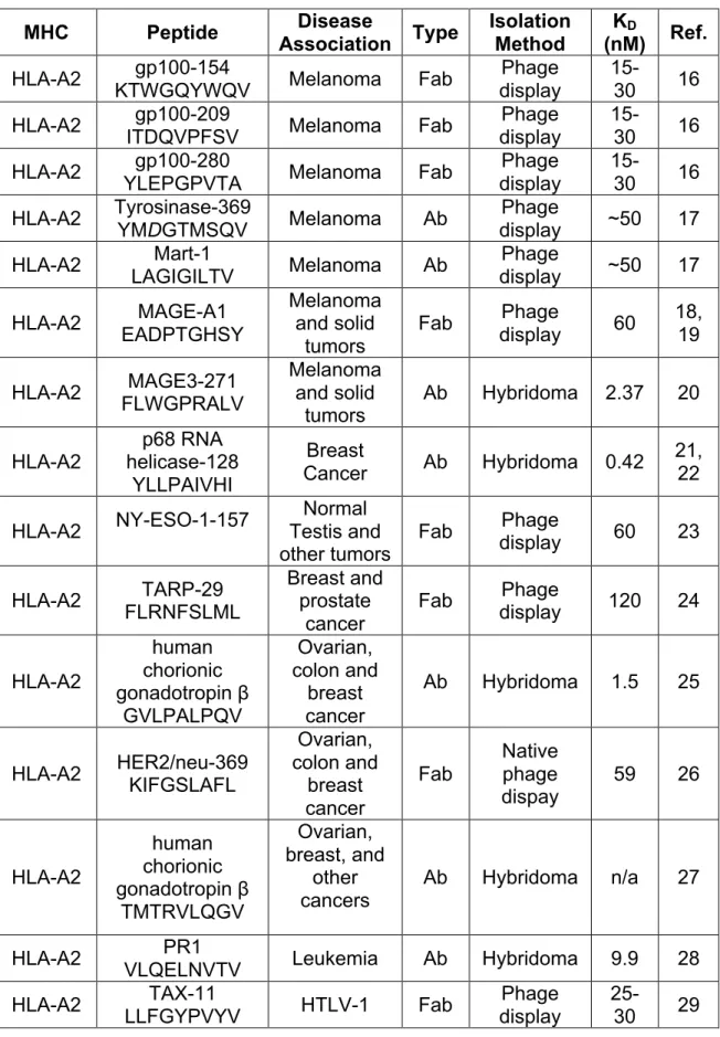
Table 1: List of TCR-like Molecules and Their Associated Specificities
MHC Peptide Association Disease Type Isolation Method (nM) KD Ref. HLA-A2 KTWGQYWQV gp100-154 Melanoma Fab display Phage 15-30 16 HLA-A2 gp100-209
ITDQVPFSV Melanoma Fab
Phage display
15-30 16 HLA-A2 gp100-280
YLEPGPVTA Melanoma Fab
Phage display
15-30 16 HLA-A2 Tyrosinase-369
YMDGTMSQV Melanoma Ab
Phage
display ~50 17 HLA-A2 Mart-1
LAGIGILTV Melanoma Ab
Phage
display ~50 17 HLA-A2 EADPTGHSY MAGE-A1
Melanoma and solid
tumors
Fab display Phage 60 18, 19
HLA-A2 FLWGPRALV MAGE3-271
Melanoma and solid
tumors
Ab Hybridoma 2.37 20
HLA-A2
p68 RNA helicase-128
YLLPAIVHI
Breast
Cancer Ab Hybridoma 0.42
21, 22
HLA-A2 NY-ESO-1-157
Normal Testis and
other tumors Fab
Phage
display 60 23
HLA-A2 TARP-29 FLRNFSLML
Breast and prostate
cancer
Fab Phage
display 120 24
HLA-A2
human chorionic gonadotropin β
GVLPALPQV
Ovarian, colon and
breast cancer
Ab Hybridoma 1.5 25
HLA-A2 HER2/neu-369 KIFGSLAFL
Ovarian, colon and breast cancer Fab Native phage dispay
59 26
HLA-A2
human chorionic gonadotropin β
TMTRVLQGV
Ovarian, breast, and
other cancers
Ab Hybridoma n/a 27
HLA-A2 PR1
VLQELNVTV Leukemia Ab Hybridoma 9.9 28 HLA-A2 TAX-11
LLFGYPVYV HTLV-1 Fab
Phage display
MHC Peptide Disease
Association Type
Isolation Method
KD
(nM) Ref. HLA-A2 MUC-1-D6-13 LLLTVLTVV Glandular cancer Fab display Phage 15-25 29 HLA-A2 Telomerase-540 ILAKFLHWL Most human cancers Fab display Phage ~5 30 HLA-A2 Telomerase-865 RLVDDFLLV Most human cancers Fab display Phage 10-15 30
HLA-A2 SSX2-103 RLQGISPKI
Normal Testis and melanoma
Fab Phage
display 270 31 HLA-A2 GILGFVFTL M1-58 Influenza Fab display Phage n/a 32 HLA-A2 KRQDILDLWVY Nef-105 HIV-1 Ab display Phage 4 33 HLA-A2 ENV-183
FLLTRILTI EBV Ab Hybridoma n/a 34
HLA-A2 eIF4G-720
VLMTEDIKL HIV-1 Ab Hybridoma n/a 35
HLA-A2 Nef138-10/A24
RYPLTFGWCF HIV-1 Ab
Phage
display 2700 36 HLA-A2 NLVPMVATV pp65-495 CMV Ab display Phage 300 37
I-Ak
HEL46 NTDGSTDYGIL
QINSR
n/a Ab Hybridoma n/a 38, 39
I-Ak
HEL116 KGTDVQAWIRG
CRL
n/a Ab Hybridoma n/a 40
HLA-DR2 MOG-35-55 MEVGWYRPPF SRVVHLYRNGK Multiple
Sclerosis Ab
Phage display
30-60 41 H-2Kb Ovalbumin
SIINFEKL n/a Ab Hybridoma 256 41
H-2Kk HA255
FESTGNLI Influenza Ab
Phage
display 56
8
The major advancement for isolation of TCR-like antibodies was the development of antibody phage-display approaches as an alternative to classical hybridoma technologies. This technology utilizes phage particle libraries expressing antibodies as fusion proteins on their surface. One unique antibody fragment or single-chain variable fragment is displayed on each phage particle and is encoded by the phage genes. Repeated rounds of selection using magnetic beads displaying the pMHC molecules followed by bacterial amplification are used to isolate phage expressing antibody-derived molecules specific for pMHC (25-28). Using this methodology, high affinity TCR-like antibody-derived molecules have been produced. These molecules generally have low affinity, but can be genetically-engineered for higher affinity for their target pMHC molecule. Random and rational affinity maturation strategies have been reported to increase binding affinities of phage-display isolated TCR-like antibody-derived molecules into the nanomolar range (~50-300 nM) (29, 30). Therefore, large phage-display libraries are now the method of choice and the most common approach for isolating TCR-like antibody-derived molecules.
1.4 TCR-like antibody applications
immune responses, targeted delivery of toxins to sites of disease or infection, and activation of effective immune responses against tumors (3, 6).
TCR-like antibodies are novel reagents that may expand the tool set
immunologists use to monitor and quantify antigen presentation during disease. The small set of available antibodies have been used to determine the presence of
particular pMHC on cancer cells (27, 31-34). Moreover, methods for quantifying levels of specific pMHC on cell surfaces by flow cytometry are being developed (35). Using these and similar methods, TCR-like antibodies have quantified that there are several hundreds of specific pMHC complexes on non-peptide pulsed cancer cells (32). This number is in agreement with estimates on the level of pMHC density required for CTL activation and lysis (36, 37). In other studies, an expression hierarchy of T cell epitopes from melanoma antigens was analyzed. Analysis of pMHC expression levels of human gp100, Melan-A/Mart-1, and tyrosinase antigen showed that there was high level of tyrosinase derived pMHC molecules on the surface of melanoma compared to the other antigens. It was determined that the quantity of the melanoma pMHC antigens did not correlate with the gene expression profiles. Moreover, melanoma cells given a drug that stabilizes tyrosinase showed a decrease in the tyrosinase antigen derived pMHC levels. Thus, it was hypothesized that protein stability was the dominating factor resulting in the differences between the antigens in their antigen presentation (34). In addition, TCR-like antibodies have been shown to aid in developing cancer vaccines. In one example, TCR-like
10
differentiate between the CTL reactivity to immunological eliciting NY-ESO-1 vaccination peptides compared to cryptic, nonimmune responsive peptides (33).
The specificity of some TCR-like antibodies for cancer pMHC molecules makes them promising agents to deliver toxins to the cancer cells or as antitumor agents in themselves. TCR-like antibodies fused to Pseudomonas exotoxin (38-40) and saporin (41) are effective at killing cancer cells in vitro and in vivo. These TCR-like antibodies fused to toxins specifically identified and killed a variety of cancer cells including: prostate (40), breast (41), and melanoma (38, 39). In addition, TCR-like antibodies can induce effective immune responses against tumors without use of toxins. Full TCR-like antibodies with Fc domains can elicit complement-dependent cytotoxicity (CDC) and activate antibody-dependent cellular cytotoxicity (ADCC) of cancer cells. Activation of any of these immune responses leads to inhibition of tumor growth in mouse models injected with the TCR-like antibodies (21, 42-45).
Alternatively, it has become possible to engineer T cells to attack cancer by transfection of T cells with viruses encoding chimeric antibody T cell receptors (CARs) consisting of a tumor specific antibody fused to an intracellular signaling domain able to activate T cells. Often, these CARs consist of an extracellular single chain variable fragment of a monoclonal antibody fused to a co-stimulatory signaling domain such as CD28 and an intracellular CD3 zeta chain (46). By using TCR-like antibodies, the specificity of T cells can be engineered against certain disease-associated pMHC molecules. In one example, T cells were designed to display chimeric receptors of the TCR-like Fabs fused to the FcεRI γ-chain signaling
killing capacity were observed for these engrafted T cells (29). All together, these results demonstrate that TCR-like antibodies specific for tumor antigens can be isolated and effectively deliver drugs, kill tumors directly and may be used to engineer autologous T cells for immunotherapy.
TCR-like antibodies have been used to understand antigen presentation during viral infection and to specifically target virally infected cells for therapy. TCR-like antibodies have been isolated against viral pMHC presented on cells infected with human immunodeficiency virus (HIV) (47, 48), influenza (49, 50),
cytomegalovirus (51), human T cell lymphotropic virus type I (HTLV-1) (52), and Hepatitis B virus (HBV) (20, 53). One TCR-like antibody, specific for the HTLV-1-derived Tax11-19 peptide bound to HLA-A2, detected pMHC complexes as low as 100 complexes per cell (52). This is near the threshold number of pMHC molecules required for cytokine secretion by T cells (54, 55). Thus, TCR-like antibodies can be isolated that have similar if not greater sensitivity for pMHC than T cells. The
12
display TCR-like antibodies and Fas ligand specifically killed HIV-1 Nef protein expressing cells. In this study, the TCR-like antibody endowed the lentivirus with the specificity and the Fas ligand expression provided the lentivirus with the ability to kill based on the interaction between the Fas ligand and the Fas receptor on the
targeted cells inducing apoptosis of the virally infected cells (47). Also, similar to the cancer therapies, TCR-like antibodies fused to toxins can kill influenza infected target cells in a pMHC specific manner. In one example, a TCR-like antibody specific for the MHC class I H-2Kk bound to the virus-derived hemagglutinin peptide HA was fused with the Pseudomonas exotoxin, PE38. This TCR-like antibody fused toxin specifically killed influenza virus-infec
ted cells presenting the HA peptide bound H-2Kk molecule on their surface (49). As a whole, TCR-like antibodies provide necessary tools to understand antigen presentation during infection and are versatile targeting probes for therapeutics.
1.5 Conclusion
important aspects of antigen processing and pMHC presentation mechanisms in cancer and virally infected cells. Furthermore, these antibodies provide a new realm of targets for delivery of therapeutics to the sites of disease. Our work discussed in this dissertation focuses on generating and utilizing TCR-like antibody fragments for in vivo xeno-transplanted tumor imaging for cancer treatment and
imaging/immunotherapy of beta cells in type 1 diabetes. We show that TCR-like antibody fragments with high affinity and specificity for their disease-specific pMHC molecule can be isolated and used as imaging agents and potential
immunotherapeutics. Therefore, TCR-like molecules may just be a “magic bullet” to assist in understanding MHC-based immunity and improving our use of
14 1.6 References
1. Rahbarizadeh, F., D. Ahmadvand, and Z. Sharifzadeh. 2011. Nanobody; an old concept and new vehicle for immunotargeting. Immunol. Invest. 40: 299–338. 2. Leyland-Jones, B. 2002. Trastuzumab: hopes and realities. Lancet Oncol 3: 137– 144.
3. Dahan, R., and Y. Reiter. 2012. T-cell-receptor-like antibodies - generation, function and applications. Expert Rev Mol Med 14: e6.
4. Abbas, A. K., and A. H. H. Lichtman. 2010. Basic Immunology Updated Edition. Saunders.
5. Stevanovic, S. 2005. Antigen processing is predictable: From genes to T cell epitopes. Transplant Immunol. 14: 171–174.
6. Weidanz, J. A., O. Hawkins, B. Verma, and W. H. Hildebrand. 2011. TCR-like molecules target peptide/MHC Class I complexes on the surface of infected and cancerous cells. Int. Rev. Immunol. 30: 328–340.
7. Hawkins, O. E., R. S. VanGundy, A. M. Eckerd, W. Bardet, R. Buchli, J. A. Weidanz, and W. H. Hildebrand. 2008. Identification of breast cancer peptide epitopes presented by HLA-A*0201. J. Proteome Res. 7: 1445–1457.
8. Alberti, S. 1996. A high affinity T cell receptor? Immunol. Cell Biol. 74: 292–297. 9. Clements, C. S., M. A. Dunstone, W. A. Macdonald, J. McCluskey, and J.
Rossjohn. 2006. Specificity on a knife-edge: the alphabeta T cell receptor. Curr. Opin. Struct. Biol. 16: 787–795.
10. Collins, E. J., and D. S. Riddle. 2008. TCR-MHC docking orientation: natural selection, or thymic selection? Immunol. Res. 41: 267–294.
11. Edwards, L. J., and B. D. Evavold. 2011. T cell recognition of weak ligands: roles of signaling, receptor number, and affinity. Immunol. Res. 50: 39–48.
12. Plaksin, D., K. Polakova, P. McPhie, and D. H. Margulies. 1997. A three-domain T cell receptor is biologically active and specifically stains cell surface MHC/peptide complexes. J. Immunol. 158: 2218–2227.
14. Wulfing, C., and A. Plückthun. 1994. Correctly folded T-cell receptor fragments in the periplasm of Escherichia coli. Influence of folding catalysts. J. Mol. Biol. 242: 655–669.
15. Altman, J. D., P. A. Moss, P. J. Goulder, D. H. Barouch, M. G. McHeyzer-Williams, J. I. Bell, A. J. McMichael, and M. M. Davis. 1996. Phenotypic analysis of antigen-specific T lymphocytes. Science 274: 94–96.
16. Holler, P. D., P. O. Holman, E. V. Shusta, S. O'Herrin, K. D. Wittrup, and D. M. Kranz. 2000. In vitro evolution of a T cell receptor with high affinity for peptide/MHC. Proc. Natl. Acad. Sci. U.S.A. 97: 5387–5392.
17. Rubin, B., B. Malissen, P. N. Jørgensen, and J. Zeuthen. 1989. Recognition of insulin on MHC-class-II-expressing L929 cells by antibody and T cells. Res.
Immunol. 140: 67–74.
18. Sergeeva, A., G. Alatrash, H. He, K. Ruisaard, S. Lu, J. Wygant, B. W. McIntyre, Q. Ma, D. Li, L. St John, K. Clise-Dwyer, and J. J. Molldrem. 2011. An
anti-PR1/HLA-A2 T-cell receptor-like antibody mediates complement-dependent
cytotoxicity against acute myeloid leukemia progenitor cells. Blood 117: 4262–4272. 19. Tamminen, W. L., D. Wraith, and B. H. Barber. 1987. Searching for
MHC-restricted anti-viral antibodies: antibodies recognizing the nucleoprotein of influenza virus dominate the serological response of C57BL/6 mice to syngeneic influenza-infected cells. Eur. J. Immunol. 17: 999–1006.
20. Sastry, K. S. R., C. T. Too, K. Kaur, A. J. Gehring, L. Low, A. Javiad, T. Pollicino, L. Li, P. T. F. Kennedy, U. Lopatin, P. A. Macary, and A. Bertoletti. 2011. Targeting hepatitis B virus-infected cells with a T-cell receptor-like antibody. J. Virol. 85: 1935– 1942.
21. Wittman, V. P., D. Woodburn, T. Nguyen, F. A. Neethling, S. Wright, and J. A. Weidanz. 2006. Antibody targeting to a class I MHC-peptide epitope promotes tumor cell death. J. Immunol. 177: 4187–4195.
22. Dadaglio, G., C. A. Nelson, M. B. Deck, S. J. Petzold, and E. R. Unanue. 1997. Characterization and quantitation of peptide-MHC complexes produced from hen egg lysozyme using a monoclonal antibody. Immunity 6: 727–738.
16
24. Polakova, K., D. Plaksin, D. H. Chung, I. M. Belyakov, J. A. Berzofsky, and D. H. Margulies. 2000. Antibodies directed against the MHC-I molecule H-2Dd complexed with an antigenic peptide: similarities to a T cell receptor with the same specificity. J. Immunol. 165: 5703–5712.
25. Andersen, P. S., A. Stryhn, B. E. Hansen, L. Fugger, J. Engberg, and S. Buus. 1996. A recombinant antibody with the antigen-specific, major histocompatibility complex-restricted specificity of T cells. Proc. Natl. Acad. Sci. U.S.A. 93: 1820–1824. 26. Chames, P., S. E. Hufton, P. G. Coulie, B. Uchanska-Ziegler, and H. R.
Hoogenboom. 2000. Direct selection of a human antibody fragment directed against the tumor T-cell epitope HLA-A1-MAGE-A1 from a nonimmunized phage-Fab library. Proc. Natl. Acad. Sci. U.S.A. 97: 7969–7974.
27. Lev, A., G. Denkberg, C. J. Cohen, M. Tzukerman, K. L. Skorecki, P. Chames, H. R. Hoogenboom, and Y. Reiter. 2002. Isolation and characterization of human recombinant antibodies endowed with the antigen-specific, major histocompatibility complex-restricted specificity of T cells directed toward the widely expressed tumor T-cell epitopes of the telomerase catalytic subunit. Cancer Res. 62: 3184–3194. 28. Stryhn, A., P. S. Andersen, L. O. Pedersen, A. Svejgaard, A. Holm, C. J. Thorpe, L. Fugger, S. Buus, and J. Engberg. 1996. Shared fine specificity between T-cell receptors and an antibody recognizing a peptide/major histocompatibility class I complex. Proc. Natl. Acad. Sci. U.S.A. 93: 10338–10342.
29. Chames, P., R. A. Willemsen, G. Rojas, D. Dieckmann, L. Rem, G. Schuler, R. L. Bolhuis, and H. R. Hoogenboom. 2002. TCR-like human antibodies expressed on human CTLs mediate antibody affinity-dependent cytolytic activity. J. Immunol. 169: 1110–1118.
30. Stewart-Jones, G., A. Wadle, A. Hombach, E. Shenderov, G. Held, E. Fischer, S. Kleber, N. Nuber, F. Stenner-Liewen, S. Bauer, A. McMichael, A. Knuth, H. Abken, A. A. Hombach, V. Cerundolo, E. Y. Jones, and C. Renner. 2009. Rational
development of high-affinity T-cell receptor-like antibodies. Proc. Natl. Acad. Sci. U.S.A. 106: 5784–5788.
31. Verma, B., O. E. Hawkins, F. A. Neethling, S. L. Caseltine, S. R. Largo, W. H. Hildebrand, and J. A. Weidanz. 2010. Direct discovery and validation of a
32. Cohen, C. J., N. Hoffmann, M. Farago, H. R. Hoogenboom, L. Eisenbach, and Y. Reiter. 2002. Direct detection and quantitation of a distinct T-cell epitope derived from tumor-specific epithelial cell-associated mucin using human recombinant antibodies endowed with the antigen-specific, major histocompatibility complex-restricted specificity of T cells. Cancer Res. 62: 5835–5844.
33. Held, G., M. Matsuo, M. Epel, S. Gnjatic, G. Ritter, S. Y. Lee, T. Y. Tai, C. J. Cohen, L. J. Old, M. Pfreundschuh, Y. Reiter, H. R. Hoogenboom, and C. Renner. 2004. Dissecting cytotoxic T cell responses towards the NY-ESO-1 protein by peptide/MHC-specific antibody fragments. Eur. J. Immunol. 34: 2919–2929.
34. Michaeli, Y., G. Denkberg, K. Sinik, L. Lantzy, C. Chih-Sheng, C. Beauverd, T. Ziv, P. Romero, and Y. Reiter. 2009. Expression hierarchy of T cell epitopes from melanoma differentiation antigens: unexpected high level presentation of tyrosinase-HLA-A2 Complexes revealed by peptide-specific, MHC-restricted, TCR-like
antibodies. J. Immunol. 182: 6328–6341.
35. Dolan, B. P. 2013. Quantitating MHC class I ligand production and presentation using TCR-like antibodies. Methods Mol. Biol. 960: 169–177.
36. Christinck, E. R., M. A. Luscher, B. H. Barber, and D. B. Williams. 1991. Peptide binding to class I MHC on living cells and quantitation of complexes required for CTL lysis. Nature 352: 67–70.
37. Sykulev, Y., M. Joo, I. Vturina, T. J. Tsomides, and H. N. Eisen. 1996. Evidence that a single peptide-MHC complex on a target cell can elicit a cytolytic T cell
response. Immunity 4: 565–571.
38. Klechevsky, E., M. Gallegos, G. Denkberg, K. Palucka, J. Banchereau, C. Cohen, and Y. Reiter. 2008. Antitumor activity of immunotoxins with T-cell receptor-like specificity against human melanoma xenografts. Cancer Res. 68: 6360–6367. 39. Denkberg, G., A. Lev, L. Eisenbach, I. Benhar, and Y. Reiter. 2003. Selective targeting of melanoma and APCs using a recombinant antibody with TCR-like specificity directed toward a melanoma differentiation antigen. J. Immunol. 171: 2197–2207.
40. Epel, M., I. Carmi, S. Soueid-Baumgarten, S. K. Oh, T. Bera, I. Pastan, J. Berzofsky, and Y. Reiter. 2008. Targeting TARP, a novel breast and prostate tumor-associated antigen, with T cell receptor-like human recombinant antibodies. Eur. J. Immunol. 38: 1706–1720.
18
42. Verma, B., R. Jain, S. Caseltine, A. Rennels, R. Bhattacharya, M. M. Markiewski, A. Rawat, F. Neethling, U. Bickel, and J. A. Weidanz. 2011. TCR mimic monoclonal antibodies induce apoptosis of tumor cells via immune effector-independent
mechanisms. J. Immunol. 186: 3265–3276.
43. Verma, B., F. A. Neethling, S. Caseltine, G. Fabrizio, S. Largo, J. A. Duty, P. Tabaczewski, and J. A. Weidanz. 2010. TCR mimic monoclonal antibody targets a specific peptide/HLA class I complex and significantly impedes tumor growth in vivo using breast cancer models. J. Immunol. 184: 2156–2165.
44. Weidanz, J. A., T. Nguyen, T. Woodburn, F. A. Neethling, M. Chiriva-Internati, W. H. Hildebrand, and J. Lustgarten. 2006. Levels of specific peptide-HLA class I complex predicts tumor cell susceptibility to CTL killing. J. Immunol. 177: 5088– 5097.
45. Hawkins, O., B. Verma, S. Lightfoot, R. Jain, A. Rawat, S. McNair, S. Caseltine, A. Mojsilovic, P. Gupta, F. Neethling, O. Almanza, W. Dooley, W. Hildebrand, and J. Weidanz. 2011. An HLA-presented fragment of macrophage migration inhibitory factor is a therapeutic target for invasive breast cancer. J. Immunol. 186: 6607– 6616.
46. Park, T. S., S. A. Rosenberg, and R. A. Morgan. 2011. Treating cancer with genetically engineered T cells. Trends Biotechnol. 29: 550–557.
47. Herschhorn, A., W. A. Marasco, and A. Hizi. 2010. Antibodies and lentiviruses that specifically recognize a T cell epitope derived from HIV-1 Nef protein and presented by HLA-C. J. Immunol. 185: 7623–7632.
48. Nunoya, J.-I., T. Nakashima, A. Kawana-Tachikawa, K. Kiyotani, Y. Ito, K. Sugimura, and A. Iwamoto. 2009. Short communication: generation of recombinant monoclonal antibodies against an immunodominant HLA-A*2402-restricted HIV type 1 CTL epitope. AIDS Res. Hum. Retroviruses 25: 897–904.
49. Reiter, Y., A. Di Carlo, L. Fugger, J. Engberg, and I. Pastan. 1997. Peptide-specific killing of antigen-presenting cells by a recombinant antibody-toxin fusion protein targeted to major histocompatibility complex/peptide class I complexes with T cell receptor-like specificity. Proc. Natl. Acad. Sci. U.S.A. 94: 4631–4636.
50. Wylie, D. E., L. A. Sherman, and N. R. Klinman. 1982. Participation of the major histocompatibility complex in antibody recognition of viral antigens expressed on infected cells. J. Exp. Med. 155: 403–414.
52. Cohen, C. J., O. Sarig, Y. Yamano, U. Tomaru, S. Jacobson, and Y. Reiter. 2003. Direct phenotypic analysis of human MHC class I antigen presentation: visualization, quantitation, and in situ detection of human viral epitopes using peptide-specific, MHC-restricted human recombinant antibodies. J. Immunol. 170: 4349–4361.
53. Low, J. L., A. Naidoo, G. Yeo, A. J. Gehring, Z. Z. Ho, Y. H. Yau, S. G. Shochat, D. M. Kranz, A. Bertoletti, and G. M. Grotenbreg. 2012. Binding of TCR multimers and a TCR-like antibody with distinct fine-specificities is dependent on the surface density of HLA complexes. PLoS ONE 7: e51397.
Chapter 2
T cell receptor-like recognition of tumor in vivo by synthetic antibody fragment1
2.1 Introduction
One of the most important lessons learned about cancer in the past 100 years is the understanding that each cancer is as unique as the patient. Effective
treatment of the patient is linked to knowing which proteins, such as hormone
receptors or HER2/neu, are actively expressed in the tumor. Probes that allow us to ascertain the molecular details of each patient’s tumors will greatly enhance
treatment. Biopsies can be used to determine molecular details, but biopsy is invasive, not always possible and does not allow for the fact that the tumor may change characteristics over time or location [1]. It would be preferable to be able to perform a noninvasive technique first followed by treatment [2]. Our long-term goal is to develop agents that can specifically target tumor cells for detection and delivery of toxins or chemotherapeutics.
Any noninvasive approach to determining molecular details of the tumor must use a probe that binds to the tumor cells with high affinity and high specificity. Monoclonal antibodies with affinity for cell-surface receptors on tumors have had
1This chapter has been published as: Miller, K. R., A. Koide, B. Leung, J.
great success in the clinic, but this approach appears to be limited because of the expression of that same cell surface receptor on normal cells that cause side effects during treatment. One of the best examples of successful use of monoclonal
antibodies to tumor-associated antigens is Herceptin. Herceptin targets the human epidermal growth factor receptor 2 (HER2/neu). HER2/neu is over-expressed in 15-30% of solid tumors including breast, ovarian, colorectal, esophageal, squamous head and neck and stomach [3]. Over-expression of HER2/neu correlates with poor prognosis for therapy of breast cancer [4-8] and has been linked to increased
metastases [9]. Monoclonal antibodies directed towards HER2/neu such as
Herceptin have shown great efficacy in the clinic, but patients do develop resistance over time [10] and some patients do not respond at all [11]. Therefore, other
22
and their production is well established. Antibodies that target specific pMHC on the surface of tumor cells have been produced but for some unknown reason, producing them conventionally is very difficult. When available, these TCR-like antibody
fragments appear to bind similarly to TCR binding pMHC [20,21]. Antibodies (or antibody fragments) previously isolated from phage-display libraries exhibited relatively low affinity for the pMHC complexes [22-24].
In this study, we isolated a T-cell receptor like Fab that specifically targets the HER2/neu peptide, E75 (KIFGSLAFL), bound to the human MHC, Human Leukocyte Antigen, HLA-A2 from a synthetic antibody library using phage display. The Fab binds with nanomolar binding affinity and high specificity for the E75/HLA-A2 pMHC molecule. The Fab was then modified to attach radioactive 64Cu and used for in vivo PET/CT imaging experiments on tumor-bearing mice that express HLA-A2
transgenically. The Fab showed increased retention in the HER2/neu pMHC positive tumors compared to HER2/neu pMHC negative tumors. These TCR-like antibodies can be used to study antigens presented on diseased and antigen-presenting cells and for delivering therapies in tumor and T-cell based diseases.
2.2 Materials and methods
Cells and culture conditions. MDA-MB-231, MCF7, and LNCAP tumor cells, CHO cells, T2 lymphoblast cells were purchased from ATCC. SKOV3 and SKOV3 cells transfected with HLA-A2 were a generous gift from Dr. Jonathan Serody
(Department of Microbiology, UNC – Chapel Hill, NC) [25]. Unless otherwise
mentioned all media supplies were purchased from Mediatec, Inc. (Manassas, VA). Each of the cell lines were grown in RMPI 1640 supplemented with 10%
heat-inactivated FBS (Atlanta Biologicals, Inc.; Lawrenceville, GA), 10 mM sodium pyruvate, and penicillin/streptomyocin in humidified CO2 (5%) incubator at 37 °C. SKOV3 transfected HLA-A2 cells and CHO transfected HLA-A2 cells were grown in the same conditions as above with the addition of 2.5 mg/ml G418.
24
Generation of peptide bound HLA-A2 complexes. The human MHC, HLA-A2.1, human beta-2-microglobulin (β2M), mouse beta-2-microglobulin, and murine H-2 Kd were produced as inclusion bodies in E. coli BL21 (DE3) (Invitrogen, Inc.). Protein
was folded in vitro as described previously [26]. Briefly, peptide, β2M, and HLA-A2 heavy chain in a 10:1:1 molar ratio were injected into a folding buffer consisting of
100 mM Tris pH 8.0, 400 mM arginine, 2 mM EDTA, 5 mM glutathione (reduced),
0.5 mM glutathione (oxidized), and protease inhibitors PMSF, pepstatin and
leupeptin. The total final protein concentration was never greater than 50 µg/ml.
After incubation for 24-36 hours at 10 °C the folded pMHC was concentrated in an
Amicon ultrafiltration cell (Millipore, Billerica, MA) and purified using gel filtration
chromatography (Phenomenex, Inc.; Torrance, CA). The purified pMHC molecules
were concentrated to greater than 3 mg/ml and stored at -80°C until use. Typical
yield for each 1 L refold is about 5 mg of pMHC, which is approximately a 14% yield.
The pMHC was site specifically biotinylated with the biotin ligase BirA (Avidity, LLC;
Aurora, Colorado) according to the manufacturer’s instructions. A non-denaturing SDS PAGE gel shift assay (no reducing agent, no heat treatment) was used to
confirm that the pMHCs were biotinylated.
Construction of Fab library. We first constructed a template plasmid for
constructing libraries, pFab007. This plasmid contains a modified version of the
Fab-4D5 gene [27] fused to gene III of the M13 phage (corresponding to the
carboxyl-terminal 208 residues of pIII). This fusion protein contains the cysteine residue of the
heavy chain hinge region so as to enable bivalent display of the Fab [28]. Most
codon and a unique BamHI site were introduced to CDR-H3. The gene was
assembled from a series of synthetic oligonucleotides using PCR. The synthesized
gene and the phoA promotor segment were cloned into a phage-display vector,
pAS38 [29] in such a way that the heavy chain of the Fab is fused to the
carboxyl-terminal 208 residue segment of M13 phage pIII. The expression of both the light
chain and the heavy chain-p3 fusion were placed under the control of the phoA
promotor.
The library was constructed as follows. Oligonucleotides that encode biased
amino acid mixture (indicated as “X”s in Figure 1B) for CDR-L3 and H3 were
synthesized using a custom-made trimer phosphoramidite mixture (Glen Research,
Sterling, VA) on an Expedite synthesizer (ABI) following instructions from Glen
Research. After deprotection, the oligonucleotides were purified using acrylamide
gel electrophoresis. Oligonucleotides for CDR-H1 and H2 that did not require the
use of trimer phosphoramidetes were purchased from Integrated DNA Technologies.
These oligonucleotides were used to introduce mutations of CDR-L3, CDR-H1,
CDR-H2 and CDR-H3 using the Kunkel mutagenesis method [30]. After the initial
transformation of the SS320 cells [30], the DNA for the library was purified and
digested with BamHI and then used to transform the SS320 cells and produce
phage particles, as described [27]. In this manner clones harboring non-mutated
CDR-H3 were eliminated. This Fab library, “Library E,” contained approximately 1010
independent clones.
26
round on, enriched phages were first incubated with streptavidin-coated magnetic
beads, and phages that bound to the beads were removed. The “precleared”
phages were then incubated with biotinylated pMHCs in solution and then captured
with the streptavidin-coated magnetic beads. The pMHC concentrations used were
100, 50, 10 and 10 nM for the first, second, third and fourth rounds, respectively.
Phages captured on the beads were eluted in 100 µl of 0.1M Gly-HCl (pH 2.1) buffer
and immediately neutralized with 35 µl of 1M Tris-Cl buffer (pH 8). Recovered clones
were analyzed using phage ELISA and DNA sequencing as described previously
[27].
Expression and purification of soluble recombinant Fabs. The phage-display vectors for the isolated clones were converted into Fab expression vectors by
inserting a gene segment encoding an 8x histidine tag and a termination codon at
the 3’ end of the heavy chain gene. The carboxyl terminus of the light chain
encoded a substrate tag for the biotin ligase BirA (AviTag, Avidity, LLC.). Fab
proteins were expressed in the 55244 E. coli strain (ATCC) and purified using
protein A affinity chromatography followed by cation exchange chromatography as
described previously [27]. Approximately 2-5 mg of purified Fab were routinely
obtained from 1 L bacterial culture. SDS-PAGE analysis showed that Fabs were in >90% purity.
Surface plasmon resonance experiments. Approximately three hundred response units (RUs) of each antibody fragment were bound to respective flow
channels in a Biacore Ni-NTA sensor chip (GE Healthcare) using the 8x histidine tag
from 200 nM to 1 nM in two fold dilutions was injected onto the surface at a flow rate
of 20 µl/min in a 60-s pulse. The NTA surface was regenerated using 0.2 M EDTA
to remove all bound protein and then recharged with 0.04 M NiSO4. The procedure
was repeated until at least three curves were obtained for each concentration of
analyte. Curves obtained at each concentration were double referenced by first,
subtracting the signal from the reference surface that contained no Fab from the
signal for the reaction surface followed by subtraction of the average signal obtained
from a set of buffer injections [31]. Data were processed using Scrubber (BioLogic
Software, Campbell, Australia). The suitability of the fit was measured based on the
appearance of residuals and χ2 values. For each Fab-pMHC binding curve, the
predicted curves visually overlaid well with the experimental curves. The residuals
also were small and random and χ2 was below 1.
Production of Fab tetramers. Purified Fabs were biotinylated using a site-specific biotin-ligase BirA (Avidity, LLC; Aurora, Colorado) according to the manufacturer’s
instructions. As described above for the biotinylation of pMHC, successful
biotinylation of the Fabs was confirmed by gel shift analysis. Biotinylated Fabs were
incubated with streptavidin-alexa 647 (Invitrogen, Inc.) at a 4 to 1 molar ratio for ten
minutes at room temperature to make the Fab tetramers.
Flow cytometric analysis of peptide-loaded HLA-A2 on cell surface. T2 cells are deficient in the TAP1 and TAP2 proteins that are responsible for transporting
antigenic peptides from the cytoplasm to the endoplasmic reticulum, but are able to
be loaded with exogenous peptides for loading of HLA-A2 molecules on their cell
28
one of the peptides E75, ML, IL, YM, RL, HL, KT, or IL (50 mM) overnight. After the
incubation, the cells were washed to remove the excess peptide and incubated with
either BB7.2 Ab (0.5 mg/ml) to detect the level of HLA-A2 molecules present on the
surface or the complex of biotinylated fE75 or fML1 with streptavidin-alexa 647
(Invitrogen, Inc.) for 30 min. at 4°C. Incubating the T2 cells with peptides leads to an
observable increase in BB7.2 staining compared with untreated cells. In all
experiments, the IgG2b isotype control was included for determining nonspecific
binding. Flow cytometry data were analyzed using Summit software (Beckman
Coulter, Inc.).
Tumor cell staining. All adherent tumor cell lines were detached from the tissue culture flask using 1 x trypsin/EDTA (Sigma-Aldrich, LLC). Cells were washed and
then incubated with anti-HER2/neu antibody (Anti-c-erb B-2 Ab-2, 9G6.10) for 30
min. at 4°C followed by incubation with Alexa Fluor 647 labeled Goat anti-mouse
IgG1 and PE labeled HLA-A2 clone BB7.2 to analyze the phenotype of each tumor cell line. Separately, each tumor cell line was prepared as before, but stained with Fab-Alexa Fluor 647-streptavidin tetramer for 30 min. at 4°C. After the final staining
incubation, cells were washed and analyzed by flow cytometry (Cyan, Beckman
Coulter, Inc.). Pearson's correlation coefficient and multiple linear regression
analysis were completed for the mean and median fluorescence intensity observed
for each cell line’s HLA-A2, Her2/neu, and Fab staining. No difference was
observed whether mean or median fluorescence was used for the analysis.
DOTA conjugation and 64Cu radiolabeling. All solvents were prepared from
to remove metals from all buffers. Free metals were removed from purified Fabs by dialysis twice with 1L of 0.2 M NaHCO3 at pH 8.2. Fabs were conjugated to S-2-(4-Isothiocyanatobenzyl-)1,4,7,10-tetraazacyclo-dodecane-tetraacetic acid (DOTA-NCS) purchased from Macrocyclics (Dalax, TX) by using the isothiocyanate linkage method as described previously [32]. The molar ratio of the DOTA conjugate to Fab used was 5:1. The reaction proceeded at pH 8.2 in 0.2 M NaHCO3 over 18 hours at room temperature. Unconjugated DOTA was removed from the reaction by a PD-10 desalting column (GE Healthcare). The average number of chelates per Fab was determined as described previously [33]. The positron emitting isotope 64Cu (copper chloride in 0.1 mol/L HCl; radionuclide purity, >99%) was provided by Mallinckrodt Institute of Radiology (Washington University School of Medicine, St. Louis, WA). The DOTA-conjugated Fab (20 µM) was incubated with approximately 1.5 mCi of 64CuCl2 in 1.0 mM citric acid (pH 5.5). The solution was incubated for 1 hr at 40 °C.
The reaction was monitored by TLC (silica) and developed with 0.1 M ammonium acetate (25%)/0.001 M citric acid (25%) / methanol (50%). Free 64Cu was
complexed as 64Cu citrate with an Rf>0.5 and 64Cu DOTA-Fab had an Rf<0.5. The radiochemical yield was determined to be > 97.0%.
Cell binding assay of radiolabeled fE75 Fab. The SKOV3 cells transfected with HLA-A2 were harvested as previously described for tumor cell staining. The cells
were incubated with the purified 64Cu DOTA-Fab at increasing concentrations for 30 min. at 4°C. As a demonstration of specificity, the SKOV3 transfected HLA-A2 cell
30
and then the bound radiolabeled fE75 counts per minute (cpm) was measured with a 2470 WIZARD2 automatic gamma counter (PerkinElmer Inc.).
Micro Positron Emission Tomography/Computed Tomography (microPET-CT) imaging in tumor-bearing mice. The preliminary experiments used NOD.SCID (SCID) mice with single tumors injected subcutaneously into the flank of the mouse.
SCID and HLA-A2 transgenic NOD.SCID mice were a gift from Dr. Jeffrey Frelinger
(Department of Immunobiology, University of Arizona) [34]. All SCID mice were
injected subcutaneously on either the right or left flank with either negative control
tumors 1 x 10⁶ SKOV3 (4 mice) or positive tumors SKOV3 transfected HLA-A2 cells
(3 mice). Following these experiments, we used HLA-A2 transgenic SCID mice and
bedposition for anatomical reference. Following the imaging, tumors were excised, cryopreserved with liquid nitrogen, and cryosectioned for radiographic analysis. PET images were reconstructed using an ordered subsets-expectation maximization (OSEM) algorithm with scatter, random, and attenuation corrections, and the PET pixel size was 0.3875 x 0.3875 x 0.775 mm [35]. Standardized uptake values (SUVs) were calculated pixel-wise by normalizing for injected dose and animal mass. The PET images were coregistered with the microCT images using the scanner software for identification of anatomical structures. Reconstructed
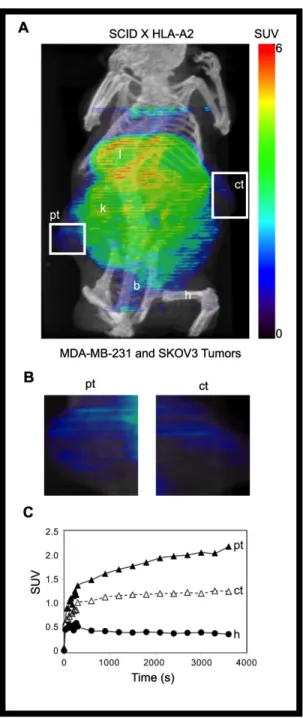
microPET and microCT images were viewed and regions of interest (ROIs) drawn and SUVs quantified using AMIDE [36]. Tumor ROIs were drawn manually to avoid adjacent organs and tumor metastases because the metastases typically had very poorly defined borders. Average SUV was measured for each specified ROI. The student’s t test was used to compare the SUVs from single tumor bearing SCID mice. The paired student’s t test was used to compare the SUVs from the tumors of the double tumor-bearing, HLA-A2-transgenic SCID mice.
2.3 Results
Generation of synthetic antibody-fragments specific to pMHC complexes.
Our goal is to develop tools to target specific tumor cells in vivo for diagnostic testing and treatment. We hypothesized that peptides bound to MHC would be good
32
create monoclonal antibodies (mAb) to specific peptide/MHC complexes, presumably because of the sequence conservation between MHC molecules injected and present in the immunized mouse [42].
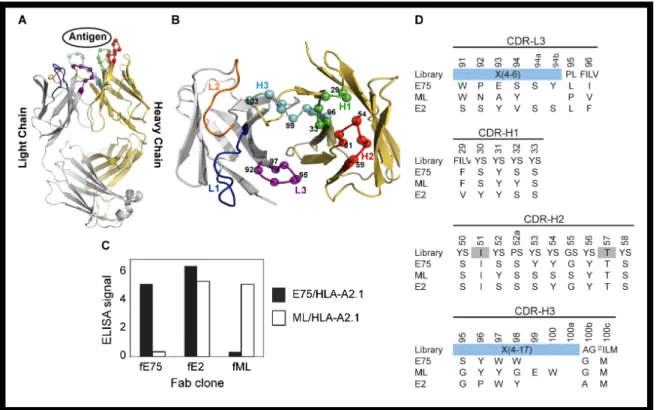
As an alternative to conventional mAb production, we sought to isolate antibody-fragments (Fabs) specific for peptide-bound MHC molecules using phage-display technology. We utilized a “synthetic” antibody library built on a highly stable 4D5 Fab scaffold (Figure 1A) [43] in which the amino acid diversity in the
Complementarity Determining Regions (CDR) was designed using simple but highly tailored amino acid mixtures [27]. Because such synthetic antibody libraries are not subjected to clonal selection against self-antigens, unlike common recombinant antibodies derived from natural immune systems, we felt that synthetic antibody libraries, coupled with phage-display selection, offered a particularly effective
approach to generating antibodies to pMHCs. Based on our previous work [27], we constructed a new antibody library in which four of the six CDRs were diversified, as described in Figure 1B. This library is similar to “Library D” of Fellouse et al. [27], but its amino acid diversity in CDR-L3 was expanded and also the composition of the expanded amino acid diversity, designated as X in Figure 1, was modified based on the analysis of antibody clones isolated from Library D [27].
We used the HER2/neu derived peptide, E75 (KIFGSLAFL), or calreticulin derived peptide, ML (MLSVPLLL), bound to the human MHC molecule HLA-A2 as the targets for sorting the synthetic antibody library. After four rounds of selection, an ELISA was used to test for specificity of the amplified phage Figure 1C. We
Figure 1: Phage-Display isolation of Fabs specific for pMHC molecules. Fabs specific for either E75/HLA-A2 or ML/HLA-A2 molecules were selected by phage-display technology. Each Fab construct was built from the highly stable 4D5 Fab scaffold containing both a heavy and light chain each with a single variable and constant domain (modeled from PDB ID: 1FVD) (A). The diversity of the Complementary Determining Regions (CDR) for the heavy chain; H1, H2, and H3, and the light chain; L3, was restricted in favor of tyrosine, serine, and other small amino acids. The Cα atoms of the synthetically modified H1, H2, H3, and L3 regions are shown as spheres (B). After three rounds of selection, an ELISA was used to test the specificity of the amplified clones (C). Three clones with three different specificities were identified. The Fab clones fE75 and fML bound E75/HLA-A2 and ML/HLA-A2 respectively with no detectable binding to the opposite pMHC molecule. The Fab clone fE2 showed binding to both E75/HLA-A2 and ML-HLA-A2. Following each Fab clone’s specificity determination, the amino acid sequence of each clone was determined (D).
34
selective to ML/HLA-A2, and (iii) cross-reactive to the two pMHCs. Two clones, fE75 and fML, selective to E75/HLA-A2 and ML/HLA-A2 respectively, were chosen for further study based on the phage ELISA, which identified each phage clone to have high affinity and specificity for their respective pMHC. As expected, the two clones have distinct CDR sequences (Figure 1D).
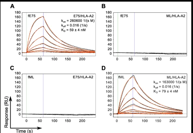
To assess the binding affinity, kinetics and specificity of the two Fabs, they were produced as soluble proteins, with a His-tag at the C-terminus of the heavy chain and a biotinylation tag at the C-terminus of the light chain and characterized using Surface Plasmon Resonance. The results shown in Figure 2 demonstrate that the model appropriately describes the binding responses of each Fab for its cognate pMHC molecule, with curve fitting residuals at or below 1 RU. The KD value for fE75 binding E75/HLA-A2 was determined to be 59 +/- 4 nM based on three separate experiments (Figure 2A). The KD for fML binding ML/HLA-A2 was 79 +/- 4 nM based on three separate experiments (Figure 2D). No binding was observed for fE75 and fML binding the non-cognate pMHC (Figure 2B, 2C), or to other non-cognate pMHC molecules that differed in peptide alone or unrelated peptide bound to unrelated MHC (data not shown).
Fabs bind to cell surface pMHC. In order to test whether these Fabs can be
Figure 2. TCR-like Fabs Bind Cognate pMHC with Nanomolar Affinity. SPR binding response curves of fE75 binding E75/HLA-A2 (A) and ML/HLA-A2 (B) and fML binding E75/HLA-A2 (C) and ML/HLA-A2 (D) are shown. Each Fab was immobilized onto individual flow channels in an NTA-Ni chip. Kinetic data for each Fab binding each pMHC molecule were globally fit to a bimolecular reaction. Green and blue lines designate the start and end respectively of each pMHC injection. Binding curves and curve fits are drawn in black and orange respectively. Each binding curve represents a different concentration of pMHC beginning at 200 nM and decreasing to 1.5 nM in 2 fold dilutions. No binding was observed for either Fab binding non-cognate pMHC molecules up to 400 nM.
36
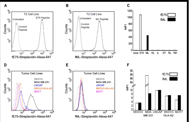
peptides may be loaded onto HLA-A2 by incubating these cells with exogenous peptides [44]. T2 cells were incubated with ML, E75 or control peptides. The biotinylated Fabs were respectively conjugated to fluorescently-labeled streptavidin to form “Fab tetramers”. The fE75 tetramer bound to T2 cells incubated with E75 (Figure 3A), but not to untreated cells. Similarly, the fML tetramer bound to T2 cells incubated with ML (Figure 3B), but not to untreated cells. Additionally, neither Fab bound to T2 cells incubated with any of the control peptides (Figure 3C). Therefore, in agreement with the SPR experiments, these Fabs bound specifically to their cognate pMHC expressed on the cell surface.
The Fabs detect endogenously processed and presented pMHC complexes.
(Figure 3E), but comparable to fE75, neither Fab bound to the HLA-A2 negative control cell line SKOV3 (Figure 3F). In addition, a further control was added with a CHO cell line transfected with HLA-A2. This cell line was HLA-A2 positive but HER2/neu negative. No binding of either fE75 or fML1 was observed for this cell line (data not shown).
Next, we sought to understand the cause for the differences in fE75 binding to the tumor cell lines in order to improve our ability to use the TCR-like Fabs as
diagnostic and treatment tools. The cell surface level of peptide/HLA-A2 complexes is dependent on multiple parameters including: protein antigen availability, peptide generation by the proteasome, antigen processing, and MHC expression [47]. The data allow us to determine which protein plays a more important role in the level of any particular pMHC complex at the cell surface: MHC (HLA-A2) or antigen
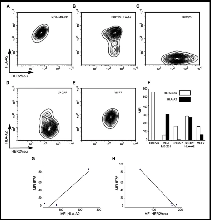
(HER2/neu). We measured HER2/neu and HLA-A2 expression by flow cytometry. There were significant differences in both HLA-A2 expression and HER2/neu levels between the cell lines (Figure 4A-F). The MDA-MB-231 cell line had the highest level of HLA-A2 expression, but also the lowest HER2/neu levels. Both MCF7 and LNCaP cell lines had intermediate levels of HER2/neu, but lower expression of HLA-A2. The SKOV3 HLA-A2 cell line had the highest level of HER2/neu and the second highest MFI of HLA-A2.
Simple two-dimensional linear regression analysis models showed weak correlation of fE75 MFI with HLA-A2 (r2=0.40) and weak, but stronger correlation with HER2/neu expression (r2=0.62) (Figure 4 G-H). Both of the regression
38
Figure 3. Fabs bind specifically to endogenously processed and presented levels of pMHC molecules. Flow cytometric analysis revealed that the Fab tetramer, composed of biotinylated fE75 (A) or fML (B) and streptavidin-Alexa-647, bound to T2 cells incubated with the cognate peptide, but not to control peptides or untreated T2 cells in three separate experiments. In C, a graph of the MFI for several other control peptides against the cognate peptide for each Fab was shown. The staining of HER2/neu positive tumor cell lines by the fE75 and fML tetramers was shown in D and E respectively. The graph of the MFI for one representative experiment from four total was shown in F.
Figure 4. HLA-A2 and HER2/neu expression is high variable on each tumor cell line. The surface expression of HLA-A2 and HER2/neu for each human tumor cell line was measured by flow cytometry. The representative plots of HLA-A2 versus HER2/neu for each tumor cell line were shown in A-E. In F, a graph of the MFI of one representative experiment from four total was shown. G and H were plots of the MFI of fE75 versus the MFI of HLA-A2 or MFI of HER2/neu respectively. Simple two-dimensional linear regression analysis models do not model the system well. The black lines showed weak correlation of fE75 MFI with HLA-A2 (r2=0.40) or HER2/neu expression (r2=0.62).
40
were removed from the analysis, no correlation exists for fE75 MFI and HLA-A2 (r2=0.09) nor HER2/neu (r2=0.00). Since that is not meaningful biologically and we know that fE75 binding did vary for each cell line, we know that either there was another unknown important factor or that both HLA-A2 and HER2/neu influenced fE75 binding. The coordinated involvement of both HLA-A2 and HER2/neu on fE75 levels was modeled using multi-parameter linear regression analysis [48]. This model gave an excellent fit for the correlation of fE75 binding using both HLA-A2 and HER2/neu expression levels (r2=0.97). Removing the MDA-MB-231 cell line from this analysis showed that there was still a significant correlation (r2=0.89),
suggesting that even though MDA-MB-231 appears to dominate the regression, the correlation between fE75 binding and both HLA-A2 and HER2/neu is valid even without the MDA-MB-231 cell line. These data show that both MHC expression levels and the antigen expression affect pMHC expression on tumor cell lines. Both MHC and source of the peptide antigen must be considered in order to accurately predict pMHC expression levels.
The TCR-like Fab recognizes E75/HLA-A2 positive tumors in a
xenotransplanted-tumor mouse model. To address whether the TCR-like Fabs can
computed tomography (SPECT). We routinely obtained an average of 0.7 moles of DOTA per mole of fE75. The yield of 64Cu- fE75-DOTA conjugate was obtained in greater than 95% purity as determined by thin layer chromatography. To confirm that addition of this chelator and the radioactive metal did not alter the binding affinity of fE75 for E75/HLA-A2, in vitro cell binding studies were performed. The ovarian tumor cell line SKOV3 transfected with HLA-A2 was incubated with a range of concentrations of radiolabeled Fab. A Scatchard analysis was used to calculate a binding affinity of 111 nM (95% CI: 63-159), which was in good agreement with the SPR data (59 +/- 4 nM). The 64Cu- DOTA-fE75- conjugate maintained its specificity for E75/HLA-A2, as soluble recombinant E75/HLA-A2 complexes were able to block binding of the radio-labeled fE75 to SKOV3/HLA-A2 cells (Figure S1).
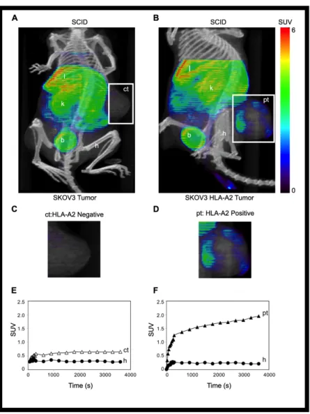
The first test of uptake of the labeled fE75 used severe combined immunodeficiency (SCID) mice. These mice do not express either human
HER2/neu or HLA-A2. The question asked is whether once the Fab is injected into the tail vein, will it be able to accumulate in the tumor. The 64Cu- DOTA-fE75 conjugate was injected via the tail vein of SCID mice bearing either
xenotransplanted SKOV3 or SKOV3 HLA-A2 tumors. MicroPET (color) and