IMPACT OF SUBCELLULAR LOCALIZATION ON ONCOGENIC FUNCTIONS OF THE RHOGEF ECT2 AND ON ITS RHO GTPASE TARGETS
Lauren Parker Huff
A dissertation submitted to the faculty of the University of North Carolina at Chapel Hill in partial fulfillment of the requirements for the degree of Doctor of Philosophy
in the Department of Pharmacology
Chapel Hill 2013
ABSTRACT
Lauren Parker Huff: Impact of subcellular localization on oncogenic functions of the RhoGEF Ect2 and on its Rho GTPase targets
(Under the direction of Adrienne D. Cox)
Rho GTPases are molecular switches that canonically signal from the plasma membrane or endomembranes to control a wide variety of cellular processes. Their
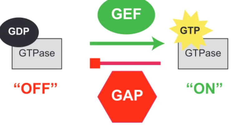
activation is tightly regulated spatiotemporally via regulatory proteins and post-translational modifications. Here, I summarize the recently appreciated consequences of GTPase C-terminal phosphorylation on their localization, effector utilization and biological functions. I also describe in detail the use of recruitment assays for monitoring the subcellular locations of GTPase activity. Misregulation of Rho GTPases is associated with many types of cancer. For example, Rho Guanine nucleotide exchange factors (GEFs), which stimulate exchange of GDP for GTP to turn GTPases “on,” are frequently found overexpressed in tumors and often are necessary for cellular transformation. Here I report validation of a RhoGEF inhibitor with a dose-dependent selectivity for Rho GTPase signaling and anti-transformation activity. I have focused primarily on the RhoGEF Ect2 and its role in ovarian tumors, where it is chromosomally amplified and its mRNA is overexpressed. I observed that Ect2 protein is highly expressed and predominantly nuclear, and that nuclear but not cytoplasmic Ect2 increases with advanced ovarian disease. Knockdown of Ect2 decreased
ACKNOWLEDGEMENTS My advisor -
Adrienne – Thank you for: sharing your enthusiasm towards science, offering constant encouragement, staying patient while editing my less than succinct writing style, and showing me the opportunities that are out there. You have taught me so much, and I will always be grateful for the many ways you enabled me to balance bench-work, non-bench-work, and life.
My committee members -
Channing, Keith, Gary, and Klaus – Thank you for your time, thoughtful input, and guidance.
My lab mates (past and present) -
Jim, Molly, Bingying, Meagan, Chelsea, Margaret, Hong, Marshall, Jamie, Vanessa, Heather, Kelly, Jared, Trey, Meredith, Tony, Velina, Tim, Tigist, Aaron – Thank you for making it a joy to come into lab every day, and for being wonderful listeners and friends. I feel lucky to have met each of you and to have been able to work with such fun, supportive, and intelligent scientists. Additionally, I’d like to thank Jim for challenging me to think deeply about
My collaborators, colleagues, teachers, and mentors –
Pei Fen Kuan, Yuri Trembath, Kay Lund, Bob Bagnell, Steve Ray, Vicky Madden, Mark Olorvida, Ryan Miller, Jinsong Liu, Justin English, all Der lab members (especially Ariella Hanker, Cercina Onesto, Natalia Mitin, Kent Rossman, Jeran Stratford, Danielle Cook), all Major lab members, Chris Welch, Marie Rougié, Scott Slattery, Angelique Whitehurst, Kathy Justice, Alan Fields, Rafael García-Mata, Jen Jen Yeh – Thank you for sharing your expertise, time, reagents, and thoughts.
My dear friends and extended family -
I’ve been blessed with the most wonderful family and friends. Thank you for always being there when I need you. Your encouragement through struggles and cheers through joys, keep me smiling through it all.
Byron, Kelsey, Mom, and Dad -
Thank you for: giving me unwavering support, believing in me beyond my abilities,
TABLE OF CONTENTS
LIST OF TABLES ...XV LIST OF FIGURES ... XVI LIST OF ABBREVIATIONS AND SYMBOLS ... XIX
CHAPTER 1: INTRODUCTION... 1
THE RAS SUPERFAMILY OF SMALL GTPASES ... 1
Definition/structure/function/regulation... 1
Summary of subfamilies ... 2
Subcellular localization and its regulation by prenylation + a second signal... 2
Roles for GTPase prenylation beyond regulation of localization ... 4
RHO FAMILY GTPASES... 5
Definition/family ... 5
Function- cytoskeleton/morphology regulation... 5
Function- transcription... 7
Requirement for unusually precise regulation... 8
RhoGDIs (an extra level of regulation) ... 9
Nuclear GTPases, an emerging field ... 11
RHOGEFS ... 14
Families of RhoGEFs... 14
Dbl family domains- PH ... 15
Dbl family domains- other ... 16
Specificity vs. promiscuity... 16
Regulation ... 17
Subcellular localization ... 17
ECT2 ... 18
Domains ... 18
Expression... 19
Subcellular localization ... 19
Specificity/GEF activity ... 20
Role in cytokinesis/mitosis ... 21
Oncogene- identification... 24
Oncogene- domains required for fibroblast transformation ... 25
Oncogene- expression in tumor cells... 26
Oncogene- multiple transforming functions ... 27
Oncogene- focus on Rac-driven transformation... 31
Oncogene- cytoplasmic mislocalization hypothesis... 32
RAS AND RHO GTPASES IN CANCER... 34
Identification ... 34
Mutants- observed in tumors and used as tools... 34
Rho GTPases and GEFs are necessary and sufficient to drive many tumor phenotypes ... 37
How Rho GTPases drive cancer ... 38
Rho GTPases in ovarian cancer ... 42
TABLES AND FIGURES ... 45
CHAPTER 2: PRENYLATION AND PHOSPHORYLATION OF RHO FAMILY GTPASES... 54
OVERVIEW ... 54
INTRODUCTION ... 55
SMALL GTPASE PRENYLATION ... 56
C-TERMINAL PHOSPHORYLATION OF PRENYLATED RAS FAMILY SMALL GTPASES... 58
K-Ras4b phosphorylation by PKC-alpha alters its protein:protein interactions with calmodulin and galectin-3, and translocates it from the PM, converting it to a death-inducing protein... 59
Rap1 phosphorylation by PKA translocates it from membranes and may induce allosteric conformational changes to alter interactions with downstream effectors ... 62
RalA and RalB are differentially phosphorylated... 64
C-TERMINAL PHOSPHORYLATION OF PRENYLATED RHO FAMILY SMALL GTPASES... 66
RhoA phosphorylation is complex and regulates its protein:protein interactions, activation, and effector binding ... 66
RhoB phosphorylation inhibits its function... 71
RhoG – phosphorylated in vitro only?... 72
RhoE/Rnd3 – phosphorylated everywhere?... 72
Wrch-1/RhoU phosphorylation – an unusual palmitate / tyrosine pair ... 74
TC10/RhoQ – a target for phosphorylation by CDK-5 ... 76
Cdc42 – phosphorylated elsewhere, to regulate GDI binding ... 77
C-TERMNAL PHOSPHORYLATION OF PRENYLATED RAB FAMILY
SMALL GTPASES... 78
CONCLUSIONS ... 79
FIGURES... 81
CHAPTER 3: SUBCELLULAR LOCALIZATION OF RAS AND RHO GTPASE ACTIVITIES AS DETERMINED BY EFFECTOR RECRUITMENT ASSAYS... 84
OVERVIEW ... 84
INTRODUCTION ... 85
MATERIALS... 89
Plating and transfecting cells ... 89
Fixing and staining cells... 90
Imaging and data analysis ... 91
METHODS ... 91
Plating and transfecting cells ... 91
Fixing and staining cells... 93
Imaging and data analysis ... 94
NOTES... 95
TABLES AND FIGURES ... 104
CHAPTER 4: THE ROLE OF ECT2 NUCLEAR RHOGEF ACTIVITY IN OVARIAN CANCER CELL TRANSFORMATION ... 112
OVERVIEW ... 112
INTRODUCTION ... 113
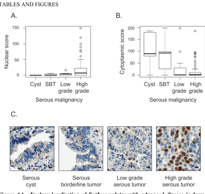
Nuclear localization of Ect2 correlates with advanced disease in
human serous epithelial ovarian cancers ... 115
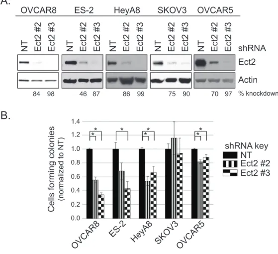
Ect2 is expressed predominantly in the nucleus of ovarian cancer cell lines ... 116
Ect2 is required for transformed growth of ovarian cancer cell lines ... 116
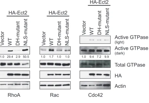
RhoGEF activity is required for Ect2-mediated transformed growth ... 117
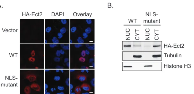
Predominantly nuclear localization is required for Ect2-mediated transformed growth... 119
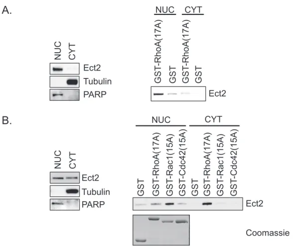
Some nuclear Ect2 is present in an active conformation that has enhanced specificity for Rac1 ... 120
Ect2 activates endogenous Rac in the nucleus and endogenous RhoA in the cytoplasm ... 122
Nuclear Rac1 activity is sufficient to rescue defects in Ect2-mediated transformed growth... 123
DISCUSSION ... 125
MATERIALS AND METHODS ... 129
Molecular constructs and transfections... 129
Cell culture, lentiviral infection, and generation of stable transfectants ... 130
Flow cytometry... 130
Immunoblotting, immunofluorescence, phalloidin staining and microscopy ... 130
Ovarian tumor tissue microarray (TMA) immunohistochemistry ... 131
Anchorage-dependent and -independent growth assays... 131
Cell fractionation... 132
Pulldown and recruitment assays for active Ect2 and Rho GTPases... 132
TABLES AND FIGURES ... 134
OVERVIEW ... 162
INTRODUCTION ... 163
RESULTS ... 166
Screen and hits... 166
In vitro selectivity... 166
RPM compounds dose-dependently inhibit Rho GTPase signaling in cellulo ... 167
RPM811 inhibits anchorage-independent growth of colorectal adenocarcinoma cells ... 169
DISCUSSION ... 170
MATERIALS AND METHODS ... 174
Cell culture... 174
Actin cytoskeleton assays... 174
Anchorage-independent growth assays ... 175
FIGURES... 177
CHAPTER 6: CONCLUSIONS AND FUTURE DIRECTIONS... 183
CONCLUSIONS ... 183
FUTURE DIRECTION 1 – HOW ARE LOCALIZATION AND SPECIFICITY OF ECT2 REGULATED?... 184
1.1 Does phosphorylation regulate the specificity of Ect2? ... 185
1.2 Does the C-terminus regulate the specificity of Ect2?... 188
1.3 How is Ect2 specificity regulated? ... 190
1.4 What determines the localization of Ect2? ... 191
ACTIVITY DRIVE TRANSFORMATION? ... 195
2.1 Is nuclear Rac activity required for Ect2-driven transformation? ... 195
2.2 Does Ect2 regulate mRNA splicing?... 196
FUTURE DIRECTION 3 – HOW AND WHY DOES COMPARTMENTALIZATION OF ECT2 AFFECT PATIENT PROGNOSIS? ... 197
3.1 Does Ect2 need to be able to shuttle between the nucleus and cytoplasm to prevent transformation? ... 199
3.2 Does Ect2 regulate opposing functions in the nucleus and the cytoplasm? ... 199
FUTURE DIRECTION 4 – OTHER QUESTIONS RAISED... 200
4.1 Is activation of Rac1 by Ect2 direct?... 200
4.2 Does Rac1 nucleotide binding affect its localization?... 201
4.3 How does Rac1 localization correlate with Rac1 activity and cellular transformation? ... 202
FIGURES... 205
FUNDING SOURCES ... 216
LIST OF TABLES
Table 1.1. Summary of the controversy surrounding the GTPase specificity of Ect2... 45 Table 3.1. The subcellular locations of active, GTP-bound GTPases are
frequently distinct from the locations of total GTPase pools. ... 104 Table 3.2: GBD probes have varying degrees of GTPase specificity... 108 Supplemental Table 4.1. Primer sequences used to generate NLS-mutant
LIST OF FIGURES
Figure 1.1. GTPases activity is regulated by GEFs and GAPs... 46 Figure 1.2. Examples of RhoA, Rac1, and Cdc42 signaling cascades,
highlighting signaling molecules described in the nucleus and/or involved
in cellular transformation... 47 Figure 1.3. RhoGDI strip GTPases from the plasma membrane and sequester
GTPases from effectors... 48 Figure 1.4. Dbl family RhoGEFs contain a Dbl homology (DH), a pleckstrin
homology (PH), and a variety of other domains... 49 Figure 1.5. Dbl family RhoGEFs vary in specificity. ... 50 Figure 1.6. Domain structure of Ect2... 51 Figure 1.7. The domains and transforming capabilities of the originally
isolated Ect2 truncation mutant cDNAs. ... 52 Figure 1.8. Comparison of models of Ect2-driven cellular transformation... 53 Figure. 2.1. Posttranslational modifications of Ras superfamily small GTPases
in which both prenylation and C-terminal phosphorylation have
been demonstrated. ... 81 Figure. 2.2. C-terminal phosphorylation of prenylated small GTPases
alters subcellular localization and protein:protein interactions, with
consequences for activation state, effector interactions and signaling activities... 83 Figure 3.1. Nuclearly localized, constitutively active Rac1 (Rac1-Q61L-C189S)
recruits GFP-PAK1-PBD to the nucleus... 111 Figure 4.1. Nuclear localization of Ect2 correlates with advanced disease
in human serous epithelial ovarian cancers. ... 134 Figure 4.2. Ect2 is expressed predominantly in the nucleus of ovarian
cancer cell lines... 135 Figure 4.3. Ect2 is required for transformed growth of ovarian cancer cell lines. ... 136 Figure 4.4. RhoGEF activity is required for Ect2-mediated transformed growth. ... 138 Figure 4.5. Predominantly nuclear localization is required for Ect2-mediated
Figure 4.6. Some nuclear Ect2 is present in an active conformation that
has enhanced specificity for Rac... 143 Figure 4.7. Ect2 recruits downstream effectors of Rac to the nucleus and
initiates canonical RhoA signaling in the cytoplasm... 145 Figure 4.8. Nuclear Rac1 activity is sufficient to rescue defects in Ect2-mediated
transformed growth... 150 Supplemental Figure 4.1. Specificity of Ect2 antibody in immunoblot,
immunohistochemistry, and immunofluorescence analyses... 153 Supplemental Figure 4.2. OVCAR8 cells can undergo efficient cytokinesis
upon Ect2 knockdown... 155 Supplemental Figure 4.3. GEF-deficient (DH-mutant, E428A/N608A) Ect2
localizes to the nucleus in a manner indistinguishable from that of WT Ect2. ... 157 Supplemental Figure 4.4. Expression of NLS-mutant Ect2 does not affect
cytokinesis of OVCAR8 cells... 158 Supplemental Figure 4.5. Ect2 activates Rac in the nucleus: Ect2 recruits
PAK-RBD to the nucleus in a GEF-dependent manner... 160 Figure 5.1. RPM compounds dose-dependently inhibit Rho GTPase
signaling in cellulo... 177 Figure 5.2. RPM811 inhibits anchorage-independent growth of colorectal
adenocarcinoma cells. ... 182 Figure 6.1. Ect2 that interacts with RhoA has a different mobility than Ect2
that interacts with Rac1... 205 Figure 6.2. Phosphorylation events regulate the interaction of Ect2 and
nucleotide RhoA, but not Rac1... 206 Figure 6.3. Abolition of the potential phosphorylation site T327 in Ect2 enhances
its cytoplasmic localization... 207 Figure 6.4. Three mechanisms by which localization and specificity may be related... 208 Figure 6.5. High cytoplasmic Ect2 expression trends with positive patient
outcome, whereas high nuclear Ect2 expression trends with poor
Figure 6.6. Restriction of Ect2 to the nucleus or cytoplasm correlates with
poor patient outcome... 210 Figure 6.7. Ect2 regulates both random and directed migration of ovarian
cancer cells... 211 Figure 6.8. Rac1 F28L mutants concentrate in the nucleus more freely and are
expressed more highly than G12V mutants. ... 212 Figure 6.9. Expression of NLS-Rac1 induces vesicle formation in OVCAR8 cells. ... 214 Figure 6.10. Expression of active Rac1 mutants partially rescue the defect
in anchorage-independent growth caused by Ect2 knockdown, except
LIST OF ABBREVIATIONS AND SYMBOLS
a.a. amino acid
Akt protein kinase B
AP-1 activator protein 1
APC adenomatous polyposis coli or anaphase promoting complex
Arf ADP ribosylation factor
ARHGEF6 Rho guanine nucleotide exchange factor 6 ARP2/3 actin-related protein 2/3
ASEF APC-stimulated guanine nucleotide exchange factor
BDK bradykinin
BRCT BRCA1 C-terminus
Brd-U bromodeoxyuridine
C-terminus carboxyl-terminus
CAAX cysteine-aliphatic-aliphatic-unconserved amino acid
CaM calmodulin
cAMP cyclic AMP
CCM1 cerebral cavernous malformation protein 1 Cdc42 cell division cycle 42 small GTPase Cdk-1, -5 cyclin-dependent kinase-1, -5
cDNA complementary deoxyribonucleic acid
CENP-A centromere protein A
CIP calf intestinal alkaline phosphatase
COSMIC catalogue of somatic mutations in cancer CRIB Cdc42- and Rac- interactive binding domain
CYT cytoplasm
DAPI 4',6-diamidino-2-phenylindole
Dbl diffuse B-cell lymphoma
Dbs Dbl's big sister
ddH2O double distilled water
DH Dbl homology
DHR1/DHR2 Dock-homology region-1 and -2
DLC1 deleted in liver cancer 1
DMEM-H high glucose Dulbecco's modified Eagle medium
DMSO dimethyl sulfoxide
DNA deoxyribonucleic acid
DOCK dedicator of cytokinesis
DPBS Dulbecco's phosphate-buffered saline
ECM extracellular matrix
Ect2 epithelial cell transforming sequence #2
EGFR epidermal growth factor receptor
ELM the eukaryotic linear motif resource
ELMO engulfment and cell motility
ERK extracellular-signal-regulated kinase
ESCC esophageal squamous cell carcinoma
FBS fetal bovine serum
FIP3 Rab11 family-interacting protein FRET fluorescence resonance energy transfer
FTase farnesyltransferase
FTI farnesyltransferase inhibitor
Gal3 galectin-3
GalT galactosyl transferase
GAP GTPase-activating protein
GBD GTPase-binding domain
GBM glioblastoma multiforme
GCS GIBCO calf serum
GDF GDI displacement factor
GDI guanine nucleotide-dissociation inhibitor
GDP guanine diphosphate
GEF guanine nucleotide exchange factor
GFP green fluorescent protein
GGTase-I,-II geranylgeranyltransferase-I, -II GGTI geranylgeranyltransferase inhibitor
GPCR G protein-coupled receptor
Grb2 growth factor receptor-bound protein 2
GST glutathione-S-transferase
GTP guanine triphosphate
H-Ras Harvey rat sarcoma virus oncogene homolog small GTPase
HA hemagglutinin
HVR or HVD hypervariable region or hypervariable domain Icmt isoprenylcysteine carboxyl methyltransferase
IHC immunohistochemistry
IM internal membrane
ITSN-1 intersectin-1
JNK c-Jun N-terminal kinase
K-Ras Kirsten rat sarcoma virus oncogene homolog small GTPase
LARG leukemia-associated RhoGEF
LIMK LIM domain kinase
LPA lysophosphatidic acid
MAL megakaryocytic acute leukemia
mDia mammalian diaphanous-related formin-1
MEK mitogen-activated protein kinase kinase MgcRacGAP Rac GTPase-activating protein 1 (RacGAP1)
min minute
MKLP1 mitotic kinesin-like protein 1
MLCK myosin light chain kinase
MLK3 mixed lineage kinase 3
MMP matrix metalloproteinase
MRCK myotonic dystrophy kinase-related CDC42-binding kinase
MTT 3-[4,5-dimethylthiazol-2-yl]-2,5-diphenyltetrazolium bromide MYPT1 myosin-binding subunit of myosin phosphatase 1
N-Ras neuroblastoma-Ras
N-terminus amino-terminus
N-WASP neural Wiskott-Aldrich syndrome protein
NADPH oxidase nicotinamide adenine dinucleotide phosphate oxidase
NES nuclear export signal
Net1 neuroepithelial cell transforming gene 1
NF-κB nuclear factor kappa-light-chain-enhancer of activated B cells
NLS nuclear localization signal
NSCLC non-small cell lung cancer
NT non-targeted
NUC nucleus
PAK p21-activated kinase
PARP poly (ADP-ribose) polymerase
PBD p21-binding domain
PBR polybasic region
PBS phosphate-buffered saline
PDEδ retinal rod rhodopsin-sensitive cGMP 3',5'-cyclic phosphodiesterase subunit delta
PDGF platelet-derived growth factor
PDK1 phosphoinositide-dependent kinase 1
PH pleckstrin homology
PI3K phosphatidylinositol 3-kinase
PIP3 phosphatidylinositol (3,4,5)-triphosphate
PKA cAMP-dependent protein kinases
PKCi protein kinase C iota
PKG cGMP-dependent protein kinase
PLD phospholipase D
Plk-1,-4 polo-like kinase-1, -4
PM plasma membrane
PMA phorbol-12-myristate-13-acetate
POSH plenty of SH3 domains
PRA1 prenylated Rab acceptor 1
PTEN phosphatase and tensin homolog
RA Ras association domain
Rab rat brain small GTPase
Rac-1, -2, -3 Ras-related C3 botulinum toxin substrate-1, -2, -3 small GTPase RacGAP1 Rac GTPase-activating protein 1 (MgcRacGAP)
Raf rapidly accelerated fibrosarcoma kinase
Ral Ras-like small GTPase
Ral BP1 Ral-binding protein 1
RalGDS Ral guanine nucleotide dissociation stimulator Ran Ras-related nuclear protein small GTPase
RBD Ras (or Rho) binding domain
Rce1 Ras and a-factor converting enzyme 1 RhoA,-B,-C,-D,-E,-F,-H Ras homology small GTPase
RhoBTB-1,-2,-3 Rho-related bric-à-brac domain containing small GTPase ROCK/ROK Rho-associated protein kinase
RPMI Roswell Park Memorial Institute cell culture medium RT-PCR reverse transcription polymerase chain reaction SAAX serine-aliphatic-aliphatic-unconserved amino acid
SEM standard error of the mean
shRNA short hairpin ribonucleic acid siRNA small interfering ribonucleic acid
SLAT SWAP70-like adaptor of T cells
SLK Ste20-related kinase
SMART simple modular architecture research tool SmgGDS small GTPase GDP-dissociation stimulator 1
SNRNP200 U5 small nuclear ribonucleoprotein 200 kDa helicase
SOS son of sevenless
SRE serum response element
SRF serum response factor
STAT-3,-5a signal transducer and activator of transcription-3, -5a SV40T simian vacuolating virus 40 large T antigen
SWAP70 switch-associated protein 70
TCF T-cell factor
TCL TC10-like small GTPase
Tiam1 T-cell invasion and metastasis gene 1 TMA tissue microarray or tumor microarray
TPA 12-O-tetradecanoylphorbol-13-acetate
Trio triple functional domain protein
TUNEL terminal deoxynucleotidyl transferase dUTP nick-end labeling Wrch-1, -2 Wnt-regulated Cdc42 homolog-1, -2 small GTPase
WT wild type
YopT Yersinia outer protein T
Zn2+ zinc
µm micron
CHAPTER 1: INTRODUCTION
THE RAS SUPERFAMILY OF SMALL GTPASES
Definition/structure/function/regulation
Summary of subfamilies
Members of the Ras superfamily are categorized into 5 subfamilies based on their structure and function (1). Rabs and Arfs are regulators of vesicular transport (3, 4). The Ran family is comprised of a single human GTPase, Ran (1), that shuttles cargo in and out of the nucleus (5). Ras and Rho family GTPases are the focus of this dissertation. GTPases within these families act as signaling nodes within the cell. A large array of signaling cascades converges on activation of these GTPases, which are then responsible for diverse consequences including regulation of gene expression, the cell cycle, cell polarity, and cell survival (1, 2). Thus it is not surprising that both Ras and Rho family GTPases play critical roles in cellular transformation and tumorigenesis (summarized below). Rho GTPases are also particularly well known for their regulation of the actin cytoskeleton to drive changes in cellular morphology and migration (6-8).
Subcellular localization and its regulation by prenylation + a second signal
In addition to being in the GTP-bound "on" state, small GTPases must be properly localized within the cell to promote signal transduction (2). The founding members of the Ras superfamily (H-Ras and K-Ras) are predominantly (>90% of total protein) localized to the plasma membrane (9, 10) and many Ras superfamily proteins are associated with membranes (plasma or internal) as well (1). For Ras and Rho family GTPases, this
attaches a farnesyl isoprenoid group whereas GGTase-I attaches a geranylgeranyl isoprenoid group to the cysteine of the CAAX box. FTase generally acts on small GTPases terminating in a serine, threonine, alanine, or glutamine, whereas GGTase-I shows preference for
GTPases terminating in a leucine (12). For most small GTPases, addition of either moiety is sufficient to drive downstream signaling (13-15), however the two isoprenoids have opposing downstream consequences when attached to RhoB (16).
Prenylation (isoprenoid modification) of the GTPase targets it for further processing, by Rce1 (Ras and a-factor converting enzyme-1) and Icmt (isoprenylcysteine carboxyl methyltransferase), to cleave the terminal AAX from the GTPase and carboxylmethylate the prenylated cysteine. The modified GTPase is capable of interacting with the plasma
membrane, such that the attached lipid moiety is inserted into the hydrophobic core of the membrane. However, this plasma membrane association occurs only when a second
membrane targeting signal is present (9). The signal is found in the C-terminal hypervariable region of the GTPase and consists either of a sequence of basic amino acids (polybasic region, PBR) that form electrostatic interactions with the plasma membrane, or of the addition of a palmitate fatty acid to a palmitoylatable cysteine residue (9). Phosphorylation of Ras and Rho family GTPases within their hypervariable region allows for regulation of GTPase subcellular localization and function with additional spatio-temporal precision (reviewed in Chapter 2).
myristate fatty acid to the N-terminus of many Arf proteins (18). Ran is not lipidated nor membrane-bound (1).
Roles for GTPase prenylation beyond regulation of localization
The role of prenylation in GTPase signaling cascades is frequently studied using “SAAX mutants”, referring to GTPases in which the prenylated cysteine of the CAAX box has been mutated to a serine and thus cannot be prenylated. Since prenylation is required for membrane association, SAAX mutants are not membrane-bound. As would be expected due to their mislocalization (9, 19), SAAX mutants are impaired in most GTPase functions (9, 13, 14, 16, 20) (although there are reports of Rho family SAAX mutants retaining some ability to regulate certain transcriptional events (14, 16, 20)). Yet the requirement for prenylation vs. the requirement for proper subcellular localization of a GTPase should be considered distinctly.
This is evident when one considers that prenylation of the GTPase is required for full Ras-driven cellular transformation, yet ablation of the second signal impairs but does not ablate its transforming capabilities (9). Since both prenylation and a second signal are required for Ras to localize to the plasma membrane, there must be some functions of Ras that are prenylation dependent, but not dependent on proper plasma membrane localization (second signal independent).
galectins interact with Ras proteins in a farnesyl-dependent manner (24, 25). Binding of a prenyl group to a Rho GTPase destabilizes the GTPase and targets it for degradation (26). This is likely true for other Ras family GTPases as well (27). Therefore unless the prenyl group is inserted within a membrane or another protein, then the GTPase will be unstable. Thus these lipid binding proteins have been proposed to act as chaperones (27).
RHO FAMILY GTPASES
Definition/family
The Rho family of small GTPases is defined by the presence of an extra alpha helix within the G domain, composed of approximately 15 amino acids and termed the “Rho insert” (28, 29). The family includes 20 small GTPases that can be further clustered based on
structural and functional similarity: RhoA-related (RhoA, RhoB, RhoC); Rac1-related (Rac1/1b, Rac2, Rac3); Cdc42-related (Cdc42/G25K, TC10, TCL, Chp/Wrch-2, Wrch-1); Rnd subfamily (Rnd1, Rnd2, Rnd3/RhoE); RhoBTB subfamily (RhoBTB-1, RhoBTB-2, RhoBTB-3); and other (RhoD, Rif, TTF/RhoH) (28). Among these GTPases, the canonical members RhoA, Rac1, and Cdc42 are the best studied.
Function- cytoskeleton/morphology regulation
(32). Since then it has been determined that all Rho GTPases, with the exception of RhoH and RhoBTB1-3, regulate the actin cytoskeleton and cell morphology (28).
These effects on morphology were not only observed via microinjection of Rho proteins. Lysophosphatidic acid (LPA) activates RhoA (33) and bradykinin (BDK) activates Cdc42 (32) through G protein-coupled receptor (GPCR) signaling, whereas platelet-derived growth factor (PDGF) stimulates Rac1 (31) activity through activation of receptor tyrosine kinases (34) to drive the same phenotypes. RhoA, Rac1, and Cdc42 control the actin cytoskeleton through a variety of signaling cascades. In addition to regulating the above described structures, these GTPases also regulate focal adhesion formation/turnover; cell:cell contacts, cell contraction, microtubule assembly, and cell polarity (35). Some of the primary RhoA, Rac1, and Cdc42 signaling pathways controlling these functions are summarized in Figure 1.2. As shown, these three small GTPases converge on many of the same molecules to either amplify or attenuate the action of other GTPases.
sometimes result in the same end phenotype (38). Therefore common experimental
manipulations, e.g., rescue of knockdown, can be technically challenging and require precise levels of expression.
Function- transcription
The function of Rho GTPases is not limited to regulation of cell morphology. Rho GTPases signal to an assortment of effectors. Some of the best-studied pathways for RhoA, Rac1, and Cdc42 are diagrammed in Figure 1.2. As shown, many of these pathways control regulation of gene transcription. For example, most Rho GTPases regulate the transcription factor SRF (serum response factor). When Rho GTPases cause globular actin (G-actin) to polymerize into filamentous actin (F-actin) this releases MAL from its sequestration with G-actin. MAL is then free to translocate to the nucleus, where it acts as a co-activator of SRF and helps drive transcription (39). RhoA, Rac1, and Cdc42 also all regulate the transcription factor NF-κB (40). Overexpression of any of these three GTPases results in phosphorylation of the inhibitory subunit (IκBα), targeting this subunit for degradation and allowing
translocation of active dimers to the nucleus where they can regulate transcription (40). Similarly, multiple Rho GTPases regulate the phosphorylation of cytoplasmic STAT3 and STAT5a at key residues, thereby allowing for their dimerization, nuclear entry, and transcriptional activity (41).
been proposed for Rac1 and β-catenin, in which Rac1 enables β-catenin to regulate transcription via translocation to the nucleus (44).
Because of these roles of Rho proteins in transcription, luciferase reporter assays are commonly used as markers for Rho GTPase activation. In these assays, a promoter regulated by the transcription factor of interest (e.g., the serum response element, SRE, from the
promoter of a gene such as c-fos can be used for SRF) is engineered to control luciferase expression (45). Therefore, a quick spectroscopy readout can be used to determine
transcription factor activity, which correlates with Rho GTPase activity. The transcription factors regulated by Rho GTPases control the expression of a variety of genes, but most of them regulate cyclin D1 expression (41). Therefore, the cyclin D1 promoter is also
commonly engineered to promote luciferase expression, and luminescence then correlates with activation of the cyclin D1 promoter and subsequent cyclin D1 expression (46).
Requirement for unusually precise regulation
The described signaling pathways are essential for most cell functions. Combined actions of Rho GTPases regulate cell migration, cell cycle progression, cytokinesis, apoptosis, cell polarity, and cell adhesion (47). Since many Rho GTPase pathways converge, and
activation of a given GTPase can result in opposing outcomes (see above), proper completion of these functions requires precise spatial and temporal control of Rho GTPases. For
narrow region to initiate cleavage furrow formation, but too broad a region of RhoA activity prevents furrow formation (49). It is therefore not surprising that Rho GTPase activation is very precisely controlled. Even a strong stimulus results in only about 5% of a specific Rho GTPase becoming activated (50, 51). This indicates that even very minor populations of active Rho GTPases can be functionally important.
Even though Rho GTPases must be GTP-bound to interact with downstream effectors, it has been suggested that they are not on/off switches like Ras GTPases. While turning a Ras GTPase “on” is sufficient to drive downstream functions, Rho GTPases (such as Rac1) may require continuous cycling to perform their cellular duties (see section on mutants below). This idea has been described as the “GTPase flux model” (52).
RhoGDIs (an extra level of regulation)
activation. However, emerging evidence suggests that RhoGDIs are also essential for protein stability of prenylated Rho family GTPases (56).
Prenylation of Rho GTPases targets them for degradation through interactions with Hsp90 and the proteasome (26). However, expression of RhoGDI1 inhibits this degradation, presumably by binding to the prenyl moiety, preventing misfolding, and shielding the
GTPase from its proteasome fate (26). This interaction of RhoGDI with Rho GTPases is essential for Rho GTPases to function, since prenylation is both irreversible and a necessary step in targeting Rho GTPases to the plasma membrane. Phosphorylation of both Rho GTPases and RhoGDIs controls their interaction (see Chapter 2). Thus kinase/phosphatase-induced release of GTP-bound GTPases from GDIs may be used by the cell to create a bolus of Rho GTPase activity (11).
The sum of the molar quantities of RhoA, Rac1, and Cdc42 within a cell is
approximately equal to the moles of RhoGDIα (57). The competition of Rho GTPases for limited binding to RhoGDIs determines which GTPases are protected from degradation (27) and again emphasizes the very precisely controlled functions of Rho GTPases. The
phenotypic consequences of experimental overexpression of Rho GTPases within a cell are not only due to signaling from the exogenous GTPase but also due to degradation and loss of signaling from other Rho GTPases that are outcompeted for GDI binding.
cell ((19), see Table 3.1). This is likely due to GDI sequestration of prenylated Rho (but not Ras) GTPases in the cytoplasm. Still, it has been proposed that galectins and/or PDEδ may act similarly to GDIs for Ras family GTPases (22, 58, 59).
Nuclear GTPases, an emerging field
Consistent with their role in cytoskeletal rearrangement, Rho GTPases have been extensively studied for their functions within the cytoplasm and at membranes. Although the vast majority of Rho proteins are found at these locations (57), the existence of a minor pool of nuclear Rho GTPases is beginning to be appreciated as well. As described above, minor populations of Rho GTPases can have important functional consequences.
RhoA (60, 61), Rac1 (62-64), and Cdc42 (65) have all been observed within the cell nucleus, and other Rho GTPases (e.g. RhoC, TCL, Rnd1, etc…) contain nuclear localization signals (NLSs) in the polybasic region of their hypervariable domains (9, 64). NLSs for Rho GTPases might seem unnecessary, since these small GTPases are generally less than 40 kDa (29) and therefore are small enough to passively diffuse through nuclear pores (66).
portion of RhoA is observed in the nucleus (granted, at a lower fraction than Rac1) (62). It is currently unclear how RhoA transits to the nucleus.
Rac1 activity has been observed in the nucleus in response to bacterial infection by
Yersinia pseudotuberculosis (67). The bacterial protein YopT acts as a cysteine protease and
cleaves off the CAAX box of Rac1, de-prenylating Rac1 and allowing for its nuclear import, where it remains active (67). While the function of nuclear Rac1 activity was not explored experimentally, the authors suggested that it may regulate transcription. Indeed, experiments in the Bapat lab suggest that active nuclear (but not cytoplasmic) Rac1 positively regulates Wnt/β-catenin driven transcription in colon cancer cells (44). Active Rac1 enhances nuclear accumulation of β-catenin (44) and also interacts with Wnt-responsive promoters (69). In another system, a constitutively active (Q61L) SAAX mutant of Rac1 was artificially overexpressed in the nucleus of cells (62). This resulted in an increased mitotic index, but did not increase membrane ruffling (62). The mechanism through which active nuclear Rac1 enhances mitosis is unknown. However, the Rac1(Q61L/SAAX) mutant can activate NF-κB transcription to inhibit UV-induced apoptosis (70). It was proposed that Rac1 must be both nuclear and GTP-bound to drive NF-κB transcription, and that Rac1 activation of PAK1 causes nuclear accumulation of the NF-κB p65 protein (70). However these experiments focused on the function of Rac1 SAAX and prenylated nuclear Rac1 was not examined.
of either a GEF or a GAP that act on Cdc42 similarly impaired CENP-A function, thereby making the role of Cdc42 at this location hard to decipher (65).
While it is likely that unique signaling pathways exist within the nucleus, many downstream effectors of Rho GTPases have also been observed there. In the signaling cascades diagrammed in Figure 1.2, proteins that have been observed within the nucleus are highlighted in yellow. As shown, nuclear actin is thought to drive chromatin remodeling and to interact with RNA polymerase to regulate transcription (71). In fact, many signaling molecules downstream of Rho GTPases (such as PAK1, ROCKII, paxillin, and cofilin) have been described to regulate transcription when present in the nucleus (71-75). For example, the Rho effector ROCKII binds to and phosphorylates p300 acetyltransferase when in the nucleus (74). This enhances the ability of p300 to acetylate histones and drive transcription (74). Thus Rho GTPase activation in the nucleus, like its activation in the cytoplasm, may regulate transcription. Consistent with this, there are reports of Rho GTPase SAAX mutants (found to varying degrees in the nucleus) retaining some ability to activate certain
transcriptional events (14, 16, 20).
Regulators of Rho GTPases have also been observed in the nucleus. The Rho GEFs Ect2 (76) and Net1 (77) are predominantly nuclear, and LARG (78), Vav3 (79),
RHOGEFS
Families of RhoGEFs
Within the Ras superfamily of GTPases, each subfamily has its own specific
regulators (58). There are 3 varieties of RhoGEFs, able to catalyze nucleotide exchange on Rho GTPases: Dbl family RhoGEFs, DOCK-related RhoGEFs, and SWAP70/SLAT (85). The majority of RhoGEFs belong to the Dbl family and these will be the focus here. There are also 11 DOCK-related RhoGEFs, each of which activate either Rac1 or Cdc42. The DOCK family Rho GEFs contain 2 well-conserved regions, Dock-homology region-1 and -2 (DHR1 and DHR2), that are composed of a C2 (lipid-binding) domain and an armadillo array (protein-binding) domain (85). Some DOCK-related RhoGEFs require binding of the
scaffolding protein ELMO to gain catalytic activity (86). SWAP70 and SLAT are outliers. They do not show homology with either Dbl or DOCK RhoGEFs, yet still catalyze
nucleotide exchange on Rho GTPases (85, 87, 88).
Dbl family domains- DH
Dbl family domains- PH
With the exception of Tuba, all Dbl-family RhoGEFs contain a pleckstrin homology (PH) domain located adjacent to and C-terminal of the DH domain (85). The PH domain is found in many signaling molecules, and interacts with lipid products of phosphatidylinositol-3-kinase (89). It is widely thought that PH domains aid in association of proteins with membranes via binding to phospholipids (89). However, many RhoGEF PH domains have low affinity for phospholipids and some RhoGEFs localize to plasma membranes despite mutations in their PH domains to prevent phospholipid binding (90, 91). Therefore the plasma membrane association of these GEFs/PH domains may not be due to phospholipid binding, but instead due to binding with other proteins. In fact, GTP-bound (but not GDP-bound) RhoA has recently been shown to bind to the PH domains of many RhoGEFs (such as Lbc, LARG, and GEF-H1) (92). When active RhoA is membrane-bound, it enhances the GEF activity of these RhoGEFs in a positive feedback loop, presumably through allowing the PH domain to bind to membranes such that the GEF can activate other inactive RhoA
proteins (92). Alternatively, PH domain interactions with proteins can target them to other specific subcellular localizations where they activate Rho GTPases. For example, the PH domain of Trio is thought to direct Trio to actin structures via binding of filamin (93).
Whether there is a single global function of the RhoGEF PH domain is unclear. It likely varies among RhoGEFs, as the relative orientation of DH and PH domains differs greatly among them (85).
Dbl family domains- other
Beyond the DH/PH domains, RhoGEFs are an extremely variable class of proteins, containing many different protein:protein interaction and other domains (Fig. 1.4) (85). This diversity allows cells to respond to numerous different signaling inputs by activating a variety of Rho GTPases.
Specificity vs. promiscuity
There are 70 distinct Dbl-family RhoGEFs to regulate the 20 identified Rho family GTPases (99). Some of these Dbl-family GEFs act solely on a single GTPase: however, many have multiple substrates (Fig. 1.5) (85). Examination of crystal structures of GEF/GTPase interactions has allowed identification of key residues that regulate the
Regulation
For many RhoGEFs, the N-terminus is autoinhibitory, and therefore artificial truncation of the GEF activates it (85). In cellulo this autoinhibition is overcome either by post-translational modification or by protein binding. For example, Vav is N-terminally phosphorylated by Src family members. This phosphorylation causes the N-terminus to become unstructured, causing it to release the DH domain and activate Vav (102). Tiam1 is opened/activated by Ras binding to the RBD in its N-terminus (103). Similarly, APC binds the N-terminus of ASEF (104) and Gα13 binds the N-terminus of p115 Rho GEF (105) to activate these RhoGEFs.
Subcellular localization
In general, RhoGEFs are thought to localize similarly to RasGEFs (106). The prototype of GEF localization is the RasGEF Son of Sevenless (SOS). SOS is localized diffusely throughout the cytoplasm, until phosphorylation of receptor tyrosine kinases creates a docking site for the adaptor protein Grb2 (growth factor receptor-bound protein). Grb2 localization to the plasma membrane recruits SOS there, and SOS then activates Ras family GTPases (107). Indeed the RhoGEF Vav also translocates to the plasma membrane upon activation (108), and PDGF stimulation recruits the Rac1-specific GEF Tiam1 to membranes (109).
predominantly localized to the nucleus of interphase cells. It was originally thought that these RhoGEFs were simply sequestered from their Rho GTPase substrates when in the nucleus, (77, 111, 112). However, it has now been shown that Net1 can activate RhoA when in the nucleus (68). In Chapter 4 of this dissertation I discuss a nuclear role for Ect2.
ECT2
Domains
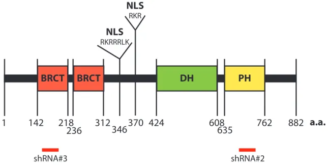
Ect2 is a Dbl family RhoGEF, containing both DH and PH domains. A diagram of the structure of Ect2 is shown in Fig. 1.6. While the DH domain of Ect2 alone is capable of inducing nucleotide exchange, this activity is enhanced by the presence of its PH domain both in vitro and in cellulo (94). Uniquely among RhoGEFs, Ect2 also contains two N-terminal BRCT domains. BRCT domains have been identified in 23 proteins, most of which are involved in DNA damage repair (113). BRCT domains are commonly found in pairs and bind to phospho-serines (114). The BRCT domains of Ect2 bind to its DH domain and autoinhibit its GEF catalytic activity (111, 115). Located between the BRCT domains and DH/PH domains of Ect2 are two nuclear localization signals (NLSs), which are unusual among RhoGEFs.
(111, 116) as well as putative degrons, which are motifs that allow APC binding and polyubiquitination to target Ect2 for degradation (116).
Expression
Initial northern blot analysis revealed that Ect2 is most highly expressed in the testis and also in the kidney, liver, and spleen of adult mice. Ect2 transcript was not detected in lung tissue or in postmitotic (brain, heart, skeletal muscle) tissue of these same animals (117). However, in mouse embryos and tumor cells Ect2 mRNA was more ubiquitously expressed (45). It was therefore hypothesized that Ect2 expression is elevated during cell division. Indeed, after serum starving cells such that >90% were in GO/G1, Ect2 expression at both the mRNA and protein level was increased upon serum stimulation in conjunction with cells re-entering the cell cycle (45). And heightened Ect2 expression has been correlated with S/G2/M phase in a regenerating liver mouse model (118) and by double thymidine block (116). Yet others have not observed dramatic changes in Ect2 expression with the cell cycle and instead showed phosphorylation of Ect2 upon entry into G2/M (described further below) (76). Thus, the rules governing the expression patterns of Ect2 are somewhat unclear.
Subcellular localization
As beautifully captured by Tatsumoto and colleagues, the subcellular localization of Ect2 changes throughout the cell cycle (76). During interphase, Ect2 is one of two RhoGEFs that localizes predominantly to the nucleus, the other being Net1. Approximately 1-2
anaphase and teleophase progress, Ect2 is concentrated at the central spindle (120)/cleavage furrow (76), and in some cells it is also found at the cell cortex (121). Prior to abscission of the two cells, Ect2 is observed at the midbody (76, 122). It translocates to the nucleus as interphase begins again (76).
Within the nucleus, Ect2 has been described as nucleolar (119). I have observed this, but have also observed nucleolar exclusion in some circumstances (LPH, unpublished). Additionally Ect2 has been observed at cell:cell contacts (123), and specifically at the zonula adherens (124).
Specificity/GEF activity
The GTPase specificity of Ect2 has been context-dependent at best and controversial at worst. In the original Ect2 paper (117), the homology of Ect2 to other RhoGEFs was noted. However, Ect2 isolated from insect cells did not catalyze nucleotide exchange on purified RhoA, Rac1, or Cdc42 (117). Since then, it has been shown that full-length Ect2 immunoprecipitated from COS cells (monkey kidney fibroblasts) is capable of activating purified RhoA, Rac1, and Cdc42 (76). In cellulo, cell type and context-specific differences have led to a wide array of specificity profiles for Ect2 (111, 121, 123, 125-128). Table 1.1 summarizes the published specificity of Ect2 towards RhoA, Rac1, and Cdc42. Ect2 is known to bind other GTPases as well, with particularly high affinity for RhoC (117, 129). And expression of truncated Ect2 in COS cells causes activation of RhoB, RhoC, and Rac2 (111).
BRCT-mutated or N-terminally truncated Ect2 drives more dramatic phenotypes than full-length Ect2 (111, 117, 118); and 3) the N-terminus of Ect2 alone acts as a dominant-negative (76, 118, 125). Therefore Ect2 must be activated before it can enhance nucleotide exchange, which may explain some of the conflicting reports about the GEF activity of Ect2.
The mechanism of Ect2 activation remains unclear, but may be achieved by
phosphorylation ((76), see Chapter 6.1). As mentioned above, the C-terminus of Ect2 also regulates its GEF activity, through an unidentified mechanism. It is required for Ect2 to activate Rac1 and Cdc42 and to optimally activate RhoA (94). Thus, further work is required to understand the signals for Ect2 activation and what drives its specificity.
Role in cytokinesis/mitosis
The best-studied function of Ect2 is its role in cytokinesis of normal cells.
Cytokinesis is the last step in cell division, during which a single cell that has replicated its genetic material physically divides into two daughter cells (130). Manipulation of Ect2 levels/activity (via knockout (131)/knockdown (120) or overexpression of constitutively active (118) or dominant-negative (76) Ect2) causes cells to become multinucleated, indicating an inability to complete this process. Thus, fully functional Ect2 is required for proper cytokinesis of many cells.
which acts as a narrow bridge between the two newly distinct cells, is called the midbody. Abscission occurs when the plasma membrane is severed on either side of the midbody, allowing the two cells to separate (130). As detailed below, Ect2 regulates both cleavage furrow formation and abscission.
Centralspindlin is a complex of the proteins MgcRacGAP and MKLP1, that is localized to the central spindle. MgcRacGAP is phosphorylated on serine 157 by Plk1 (132, 133). This phosphorylation creates a docking site for the BRCT domains of Ect2 and recruits Ect2 to the central spindle (120, 132, 133). In addition to properly localizing Ect2, the binding of the BRCT domains of Ect2 to MgcRacGAP also prevents autoinhibition of Ect2, holding it in an active conformation (120). Recruitment of Ect2 to the central spindle allows for activation of RhoA in a narrow area of the cell cortex above the central spindle, which becomes the cleavage furrow (120). This activation of RhoA leads to activation of myosin-II and actin polymerization, causing cleavage furrow ingression and proper execution of
cytokinesis (120).
The onset of cytokinesis is tightly regulated by phosphorylation. Ect2 is
for the BRCT domains of Ect2, and restricting Ect2 to the central spindle where it activates RhoA.
As described above, Ect2 localizes to the midbody in anaphase and early telophase (76, 122). However, for abscission of the two daughter cells to proceed, Ect2 must be released from the midbody (122). Ect2 competes with FIP3 (Rab11 family interacting protein) for binding to MgcRacGAP. Since their binding is mutually exclusive and FIP3 is required for abscission, only upon release of Ect2 from MgcRacGAP is abscission able to proceed (135). Therefore Ect2 localization to the midbody acts as checkpoint for the final step in cytokinesis. Plk-4 is required for proper localization of Ect2 to the midbody (136), thus it is likely that dephosphorylation of sites phosphorylated by Plk-4 allow release of Ect2 from the midbody.
Ect2 has also been implicated in the initiation of mitosis, independent of
MgcRacGAP. One to two minutes prior to nuclear envelope breakdown, Ect2 is released from the nucleus into the cytoplasm. This release causes activation of RhoA at the cell cortex and results in cell rounding (one of the earliest steps in mitosis) (119).
In summary, phosphorylation and protein:protein interactions determine the localization and activation of Ect2 throughout the cell cycle. Ect2 then regulates the
Oncogene- identification
Ect2 was originally identified in a screen for oncogenes performed by Dr. Toru Miki and colleagues (117). NIH 3T3 mouse fibroblast cells were transfected with a cDNA library isolated from mouse keratinocytes. The transfectants were then screened for focus formation, which represents a loss of contact-inhibition and therefore transformed growth (117). The cDNAs capable of creating foci were extracted from cells and identified using a technique they previously developed (139). In this way, Miki and colleagues identified genes from normal epithelial cells that were sufficient to drive transformation.
The original isolate of Ect2, Epithelial cell transforming sequence #2, coded for an N-terminally truncated version of the protein (117). Careful sequence analysis reveals that it lacked both BRCT domains and the first NLS sequence (Fig. 1.7). Therefore, it was not autoinhibited, and represented a constitutively active form of Ect2.
formed tumors within 6 weeks, whereas cells not expressing Ect2 did not create tumors in mice (117). Thus, a constitutively active form Ect2 is capable of acting as potent oncogene.
Oncogene- domains required for fibroblast transformation
Numerous truncation mutants of the original Ect2 isolate were analyzed for their ability to transform NIH3T3 fibroblasts ((117), Fig. 1.7). As would be expected if the GEF activity of Ect2 were required for this phenotype, inclusion of the autoinhibitory BRCT domains or omission of the catalytic DH domain prevented the ability of Ect2 to transform these cells (117). This suggested that the GEF activity of Ect2 was indeed required, which was later confirmed with a point mutation in the DH domain that ablated its catalytic activity (111).
Consistent with its necessity for optimal nucleotide exchange (94), the PH domain is also required for Ect2 to transform fibroblasts (111). Surprisingly, tryptophan 752, a residue that is conserved across all RhoGEF PH domains and is usually necessary for optimal GEF function, is not required for Ect2-driven fibroblast transformation (94).
Multiple labs have determined that a DH/PH/C truncation mutant of Ect2 (activated by removal of the N-terminal BRCT domains, see domain structure in Fig. 1.6) is sufficient to drive cellular transformation in fibroblasts, but upon further removal of the C-terminus, the isolated DH/PH domains cannot recapitulate the phenotype (46, 94, 111). One may expect that this requirement for the C-terminus would provide clues regarding which
nucleotide exchange on RhoA. Therefore it is still unclear which GTPases Ect2 acts through to drive transformation.
It has been proposed that, in addition to activating its GEF activity, Ect2 must also be cytoplasmically mislocalized to drive transformation of fibroblasts (111). Indeed, Saito et al. showed that removal of NLS function (via point mutation or truncation) from Ect2 enhanced the ability of cells expressing it to grow free of contact inhibition (111). Yet, the original Ect2 paper, also from Miki and colleagues, shows that Ect2 with both NLSs, 1 NLS, or 0 NLSs are all transforming ((117), Fig. 1.7), These results suggest that Ect2 may be able to drive transformation from either the nucleus or the cytoplasm, or that all of these mutants shuttle between compartments.
A functional analysis of the domains of Ect2 utilized by tumor cells for transformed growth has not been performed. Since normal fibroblasts require Ect2 to regulate
cytokinesis/mitosis (131) but some tumor cells may not (121, 137), Ect2 could cause transformation of fibroblasts via enhanced cytokinesis/mitosis, but this may not be relevant for tumor cells. Therefore, it is possible that the domains of Ect2 required for transformed growth in tumor cells is different from those sufficient to transform fibroblasts.
Oncogene- expression in tumor cells
mRNA level in a variety of tumor types, including lung (137), pancreatic (141, 142), recurrent liver (143), advanced kidney (144), oral (145), esophageal (146), brain (138, 147, 148), head & neck (149), cervical (149), and ovarian tumors (45, 150, 151). In some tumor types this is due to chromosomal amplification. ECT2 is located on chromosome 3q26.1-3q26.2 (152). This region is frequently amplified in tumors of the: lung (153), pancreas (142), esophagus (154), head & neck (155), cervix (156), and ovaries (151, 157). In fact, 3q26 is the most common amplicon in ovarian tumors (158).
Although protein expression of Ect2 in ovarian tumors had not been investigated, Ect2 was known to be overexpressed at the protein level in lung (137, 146), oral (145), esophageal (146), and brain (138, 147, 148) tumors. In Chapter 4 I describe my analyses of Ect2 expression in ovarian tumors and cell lines.
Oncogene- multiple transforming functions
High expression of Ect2 in lung (146), esophageal (146), and brain (138, 148) tumors is correlated with poor patient outcome. Additionally, chromosomal amplification/mRNA expression of Ect2 in pancreatic ductal adenocarcinoma (PDAC) cell lines correlated with dependence on Ect2 for cell viability (142). Indeed, Ect2 has many functions related to cellular transformation that may explain these correlations (see below). Interestingly, many of these functions appear to be distinct from the role of Ect2 in cytokinesis (121, 137, 138).
tumors with Ect2 knocked down did not show increased multinucleation (138). These results suggest that the phenotype of decreased tumor growth is due to decreased proliferation but not increased apoptosis or cytokinesis defects. The decreased growth rate could also be seen in soft agar colony formation assays of NSCLC cells (137) but Ect2 knockdown did not affect proliferation on plastic of either NSCLC or astrocytoma cells (137, 138). Therefore the role of Ect2 in both of these cell types appears to be related to proliferation during anchorage-independent but not anchorage-dependent growth.
However, there have been conflicting reports about the role of Ect2 in tumor cell proliferation. Hirata and colleagues also found that Ect2 knockdown decreases soft agar growth of NSCLC cells and esophageal squamous cell carcinoma (ESCC) cells. Yet, in their experiments a general proliferation defect was observed, as NSCLC and ESCC cells with Ect2 knocked down became trapped in G1 phase of the cell cycle (146). Similarly
knockdown of Ect2 in oral squamous cell carcinoma cells caused growth arrest in G1 (145), and there are reports in glioblastoma (GBM) cells of Ect2 knockdown causing decreased growth (147, 148) and increased multinucleation (147). Thus it is clear that Ect2 is required for anchorage-independent growth of tumor cells, however, it is less clear if this function is distinct from its role in mitosis/cytokinesis (described above) in all cell types.
stable knockdown of Ect2 in tumor vs. normal cells (137) (and also in two types of tumor cells (121)) shows that cell types vary in their dependence on Ect2 for cytokinesis/mitosis. A much lower percentage of A549 and H1703 (NSCLC) cells showed multinucleation upon stable knockdown of Ect2 when compared directly with MDCK (canine kidney) cells (137).
As described above, overexpression of Ect2 in NIH 3T3 cells can induce soft agar colony formation (117). Together with the data from the previously described knockdown experiments, these results show that Ect2 can be both necessary and sufficient for anchorage-independent growth. Overexpression of activated Ect2 can also induce focus formation (46, 94, 111, 117), indicating that Ect2 is also sufficient to allow cells to bypass contact-inhibition and continue to grow. In summary, Ect2 is responsible for multiple aspects of transformed growth.
Beyond regulation of tumor cell growth, Ect2 also regulates migration and invasion. Intracranial injection of astrocytoma cells expressing endogenous Ect2 created tumors with more invasive borders, compared to the rare and discrete tumors formed when Ect2
given GTPase can lead to opposite phenotypes (discussed above), perhaps these seemingly contradictory results should not be surprising.
There are two major types of single cell migration: amoeboid and mesenchymal, both of which require precise activation of Rho family GTPases (160). Mesenchymal migration consists of a leading edge ruffling, extending forward, and adhering to a matrix, followed by the cell contracting to bring the rest of the cell forward and finally releasing its adhesions to a previous location (160). Amoeboid migration requires much less adhesion and is powered by cycles of cellular expansion and contraction (160). During amoeboid migration through a matrix the cell rounds into a ball, squeezing and blebbing its way through a matrix (37). In contrast, during mesenchymal migration within a matrix, tumor cells typically degrade the matrix to create a tunnel through which they migrate (160). Tumor cells utilize different forms of motion based on cell-type and the properties of the surrounding extracellular matrix (37). Amoeboid migration is significantly faster than mesenchymal migration (160). It has been proposed that overexpression of Ect2 can speed cell migration by transforming the preferred cellular migration pattern from mesenchymal to amoeboid (138). Alternatively, for cells consistently moving in a mesenchymal fashion, Ect2 overexpression can slow migration by increasing focal adhesions and preventing forward motion (121). Still, some level of Ect2 activation appears to be required for proper membrane ruffling and optimal mesenchymal migration (159).
Oncogene- focus on Rac-driven transformation
Although the importance of the DH domain/GEF activity of Ect2 in tumor cells has not been examined, the function of Ect2 in tumor cells has been correlated with its ability to activate Rac1. In NSCLC, decreased anchorage-independent growth that results from Ect2 knockdown can be rescued by expression of constitutively active Rac1 (137). While this experiment does not prove that Ect2 acts through Rac1 to drive transformation, it shows that activated Rac1 is sufficient to drive transformation in these cells. Further correlative data indicates that knockdown of Ect2 attenuates Rac1 activity (137, 138). And a T327 (T328 in reference, see Fig. 1.6 legend) phospho-deficient mutant of Ect2 that cannot rescue Rac1 activity also does not rescue anchorage-independent growth. In contrast, WT and T327 phospho-mimetic Ect2 rescue both Rac1 activity and anchorage-independent growth in NSCLC cells (161). Therefore it has been hypothesized that Ect2 drives NSCLC cellular transformation through activation of Rac1 (137).
Oncogene- cytoplasmic mislocalization hypothesis
Based on work in NIH 3T3 mouse fibroblasts (described above), the current model of Ect2 oncogenic function requires mislocalization of Ect2 from the nucleus to the cytoplasm in order to activate Rho GTPases and drive cellular transformation (111). Consistent with this idea Ect2 has been observed in the cytoplasm of a variety of tumor cells.
Immunohistochemistry (IHC) has been used to stain normal lung and NSCLC tumors for Ect2. In normal lung epithelia Ect2 was observed in the nucleus, whereas in tumor epithelia it appeared to be nuclear excluded with high cytoplasmic expression (137). Although
example, the initial characterization of Ect2 subcellular localization was performed in HeLa cells, which were derived from a cervical tumor (162), and in these cells Ect2 is
predominantly nuclear during interphase (76).
Nuclear Ect2 has been assumed to be in an autoinhibited state, and sequestered from Rho GTPases (111). Consistent with this idea the majority of Rho GTPases are localized in the cytoplasm (47). However, pools of multiple Rho GTPases have also been observed in the nucleus (described above). Additionally, not all nuclear Ect2 is in an autoinhibited state, since, at least in HEK293T cells, a portion of nuclear Ect2 can interact with nucleotide-free RhoA (68). Therefore, the previous model requires revising (Fig. 1.8).
Also, the mechanism behind cytoplasmic mislocalization of Ect2 in tumor cells remains unclear. Exogenous Ect2 localizes to both the nucleus and cytoplasm of astrocyte progenitor cells, astrocytes, and astrocytomas, but only to the nucleus of some
non-astrocytoma cell lines (138). Thus it has been hypothesized that overexpression alone is not sufficient to induce cytoplasmic mislocalization of Ect2, but instead that cytoplasmic
mislocalization is regulated in some way (138). The Fields lab proposed that this regulation is conferred by PKCi phosphorylation of Ect2 on residue T327, and that the negatively charged phosphate opposes the adjacent basic NLSs to enhance cytoplasmic localization of Ect2 (137, 161). However, it is unclear if/how phosphorylation of Ect2 regulates its
RAS AND RHO GTPASES IN CANCER
Identification
The study of Ras superfamily GTPases began in the 1960s when 2 viruses capable of inducing tumors in mice were identified (163, 164). These viruses contained genes from rodent cells that drove cellular transformation, which were later identified as the GDP/GTP binding proteins that we today call H-Ras (Harvey-Rat sarcoma) and K-Ras (Kirsten-Rat sarcoma) (2). Thus it is not surprising that misregulation of Ras and Rho GTPases drives tumor formation and many aspects of the transformed phenotype (2, 47).
Mutants- observed in tumors and used as tools
Ras family GTPases are frequently mutated in tumor cells (165). Alterations in residues G12, G13, and Q61 account for 97-99% of all observed Ras mutations in cancer (2). A variety of amino acids replace the WT residues in tumors (2), which is not surprising, because many amino acid substitutions result in the same phenotype. For example, mutation of G12 to any residue other than proline is sufficient to drive cellular transformation (166). Mutation of G12, G13, or Q61 creates a constitutively active GTPase by further impairing its intrinsically low ability to hydrolyze GTP and by conferring GAP insensitivity (167).
optimal Rho GTPase activity (52). However, recent deep sequencing studies have allowed the identification of Rac mutations in melanoma tumor cells and a variety of tumor cell lines (168-170). In fact, Rac1(P29S) is the third most common mutation in sun-exposed
melanomas (168, 169). This mutation is located in the switch I region of GTPase and activates Rac (168-170). The mechanism by which P29S activates Rac1 is distinct from the activation of Ras by the mutations described above. P29S causes rapid nucleotide
dissociation from Rac1 (170, 171) (hence, it is referred to as a "fast-cycling" mutant), which results in a GTPase that is frequently GTP-bound due to the higher prevalence of cellular GTP compared to GDP. Additionally, mutation of this residue may destabilize the GDP-bound conformation of Rac1 (169). The P29S mutation promotes proliferation (168); has an anti-apoptotic effect (170); is sufficient to drive anchorage-independent and tumor growth (170); enhances membrane ruffling (171); and increases speed of migration (168) of cells expressing mutant Rac1. Interestingly, Rac1(P29S) did not cause a dramatic increase in multinucleation whereas expression of an artificial Q61L Rac1 mutant did (171), suggesting that a fast cycling mutant allows progression through cytokinesis that is disrupted by a GTPase-deficient Rac1. N92I and C157Y mutations of Rac have also been observed in cancer cell lines, are fast cycling, and can drive anchorage-independent and tumor growth (170). These recent advances suggest that more Rho GTPase mutations may await discovery.
homologous to Ras(F28L) cause rapid nucleotide dissociation from GTPases without
affecting GTP hydrolysis (172, 173). Although the engineered fast-cycling mutants result in the same effect that is now understood for the Rac1(P29S) mutation identified in melanomas, the structures of the two fast-cycling proteins are distinct (171).
In a screen for engineered Ras mutations that inhibit its activity, Ras(S17N) (174) and Ras(G15A) (175) were found. These mutants act as dominant-negatives. They have a low affinity for downstream effectors or nucleotides, but bind to GEFs with higher affinity than WT GTPases (176). Therefore expression of these mutants outcompetes endogenous Ras for GEFs and prevents activation of endogenous Ras (176). The G15A mutation creates the strongest interaction between Ras and GEFs, which has been described as irreversible (176). Yet, G15A is not stable in cells, so the less dramatic mutant, S17N, is commonly used as a dominant-negative in cellulo (176). Experiments utilizing these dominant-negative mutants must be interpreted with caution, especially for Rho GTPases which have very promiscuous GEFs (85). Since these mutants bind the GEF to prevent further activation of GTPases, expression of a dominant-negative GTPase (e.g. Rac(S17N)) will likely inhibit signaling to other GTPases as well (e.g. RhoA and Cdc42 GEF-dependent activation) (176).