Review
KRAS Mutant Pancreatic Cancer: No Lone Path to an
Effective Treatment
Daniel Zeitouni, Yuliya Pylayeva-Gupta, Channing J. Der * and Kirsten L. Bryant
Lineberger Comprehensive Cancer Center, University of North Carolina at Chapel Hill, Chapel Hill, NC 27599, USA; [email protected] (D.Z.); [email protected] (Y.P-G.); [email protected] (K.L.B.) * Correspondence: [email protected]; Tel.: +1-919-966-5634
Academic Editor: Jonas Cicenas
Received: 23 February 2016; Accepted: 11 April 2016; Published: 18 April 2016
Abstract:Pancreatic ductal adenocarcinoma (PDAC) is among the deadliest cancers with a dismal 7% 5-year survival rate and is projected to become the second leading cause of cancer-related deaths
by 2020. KRASis mutated in 95% of PDACs and is a well-validated driver of PDAC growth and
maintenance. However, despite comprehensive efforts, an effective anti-RAS drug has yet to reach the clinic. Different paths to inhibiting RAS signaling are currently under investigation in the hope of finding a successful treatment. Recently, direct RAS binding molecules have been discovered, challenging the perception that RAS is an “undruggable” protein. Other strategies currently being pursued take an indirect approach, targeting proteins that facilitate RAS membrane association or downstream effector signaling. Unbiased genetic screens have identified synthetic lethal interactors of mutant RAS. Most recently, metabolic targets in pathways related to glycolytic signaling, glutamine utilization, autophagy, and macropinocytosis are also being explored. Harnessing the patient’s immune system to fight their cancer is an additional exciting route that is being considered. The “best” path to inhibiting KRAS has yet to be determined, with each having promise as well as potential pitfalls. We will summarize the state-of-the-art for each direction, focusing on efforts directed toward
the development of therapeutics for pancreatic cancer patients with mutatedKRAS.
Keywords:RAS; pancreatic; cancer; therapeutics
1. MutantKRASDrives PDAC Development and Maintenance
Approximately 90% of pancreatic cancers are pancreatic ductal adenocarcinoma (PDAC), which
is almost universally fatal [1]. Major advancement in the treatment of PDAC has been lacking [2].
Currently, surgery remains the lone curative option. To be eligible for surgery with curable intent the
tumor must be resectable, meaning there are no signs of distant metastasis [3]; however, most patients
are diagnosed with late-stage disease, and hence less than 20% of patients are eligible. Recent exome
sequencing has provided a detailed genetic profile of PDAC, with mutational activation of theKRAS
oncogene found in ~95% of patients [4–7]. With significant and compelling evidence that aberrant
KRAS protein function is critical for PDAC growth and maintenance [8–10], the Pancreatic Cancer
Working Group (NCI) identified targeting KRAS as one of four key priorities for pancreatic cancer
research [11]. However, despite more than three decades of intensive effort, an effective anti-RAS
therapy has yet to reach the clinic [12–14].
The RAS family of small GTPases includes three genes:HRAS,NRAS, andKRAS. These three
loci encode four different protein isoforms: HRAS, NRAS, KRAS4A, and KRAS4B. The two KRAS
isoforms differ due to the alternative splicing of exon 4 in theKRASlocus, with KRAS4B being the
predominant isoform expressed in most tissues [15]. Each RAS protein is comprised of two major
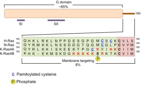
domains, the G domain and the membrane targeting domain (Figure1). All of the isoforms are similar
in the amino acid sequence of the G domain (~80%) with major differences being restricted to the
hypervariable region of their C-terminal domains [16]. Mutations in RAS occur in residues 12, 13
and 61, and inhibit GTP hydrolysis activity [17]. The threeRASgenes constitute the most frequently
mutated oncogene family in human cancers [14,18]; however, the specific isoform and amino acid
mutation varies among cancers. Mutations inHRASare most frequently found in melanoma, bladder
and mammary carcinoma;NRASmutations are found in melanoma and thyroid carcinoma; andKRAS
mutations are most prevalent in cancers of the bladder, ovary, thyroid, lung, colon and pancreas.
In pancreatic cancer, mutations in codon 12 ofKRASoccur the most frequently.
hypervariable region of their C‐terminal domains [16]. Mutations in RAS occur in residues 12, 13 and 61, and inhibit GTP hydrolysis activity [17]. The three RAS genes constitute the most frequently mutated oncogene family in human cancers [14,18]; however, the specific isoform and amino acid mutation varies among cancers. Mutations in HRAS are most frequently found in melanoma, bladder and mammary carcinoma; NRAS mutations are found in melanoma and thyroid carcinoma; and KRAS mutations are most prevalent in cancers of the bladder, ovary, thyroid, lung, colon and pancreas. In pancreatic cancer, mutations in codon 12 of KRAS occur the most frequently.
Figure 1. Human RAS proteins are composed two functional domains, the G domain and the
membrane targeting domain. The G domain spans residues 1–164 and includes the regions of the
protein responsible for binding and hydrolyzing GTP. Specifically, residues in the switch I (SI = amino
acids 30–38) region and switch II (SII = amino acids 60–76) region experience a conformational change
during GDP‐GTP cycling. The membrane targeting domain is comprised of the remaining 24/25 C‐
terminal residues. The first 20–21 amino acids are referred to as the hypervariable region and this is
where the three RAS isoforms exhibit the greatest diversity in protein sequence. The hypervariable
region contains elements important for membrane association including cysteines (blue, underlined)
that are covalently modified by the addition of a palmitate fatty acid, and stretches of polybasic amino
acids. Additionally KRAS4B contains a serine (181) that can be phosphorylated and regulates the
association of this protein with the plasma membrane or endomembranes. The four most C‐terminal
residues of the membrane‐targeting domain comprise the CAAX motif, where C = cysteine, A = any
aliphatic residue, and X = the terminal amino acid. A C15 farnesyl group is covalently attached to the
cysteine residue by farnesyltransferases and this lipid moiety aids in membrane association.
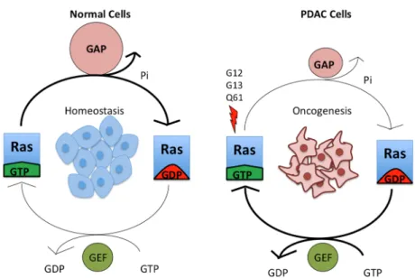
RAS proteins play an active role in cell differentiation, proliferation, migration, and apoptosis, making them important in cancer signaling [19]. Individual RAS proteins are activated when they are bound to guanosine triphosphate (GTP) and are inactive when they are bound to guanosine diphosphate (GDP) (Figure 2). Intrinsic RAS GTP‐GDP cycling is regulated by guanine nucleotide exchange factors (GEFs) that stimulate nucleotide exchange and by GTPase activating proteins (GAPs) that accelerate the intrinsic GTP hydrolysis activity of RAS (Figure 2). Once activated, RAS‐GTP preferentially interacts with a spectrum of catalytically diverse downstream effectors that then regulate a myriad of cytoplasmic signaling networks.
Figure 1. Human RAS proteins are composed two functional domains, the G domain and the membrane targeting domain. The G domain spans residues 1–164 and includes the regions of the protein responsible for binding and hydrolyzing GTP. Specifically, residues in the switch I (SI = amino acids 30–38) region and switch II (SII = amino acids 60–76) region experience a conformational change during GDP-GTP cycling. The membrane targeting domain is comprised of the remaining 24/25 C-terminal residues. The first 20–21 amino acids are referred to as the hypervariable region and this is where the three RAS isoforms exhibit the greatest diversity in protein sequence. The hypervariable region contains elements important for membrane association including cysteines (blue, underlined) that are covalently modified by the addition of a palmitate fatty acid, and stretches of polybasic amino acids. Additionally KRAS4B contains a serine (181) that can be phosphorylated and regulates the association of this protein with the plasma membrane or endomembranes. The four most C-terminal residues of the membrane-targeting domain comprise the CAAX motif, where C = cysteine, A = any aliphatic residue, and X = the terminal amino acid. A C15 farnesyl group is covalently attached to the cysteine residue by farnesyltransferases and this lipid moiety aids in membrane association.
RAS proteins play an active role in cell differentiation, proliferation, migration, and apoptosis,
making them important in cancer signaling [19]. Individual RAS proteins are activated when they
are bound to guanosine triphosphate (GTP) and are inactive when they are bound to guanosine
diphosphate (GDP) (Figure2). Intrinsic RAS GTP-GDP cycling is regulated by guanine nucleotide
exchange factors (GEFs) that stimulate nucleotide exchange and by GTPase activating proteins (GAPs)
that accelerate the intrinsic GTP hydrolysis activity of RAS (Figure2). Once activated, RAS-GTP
Figure 2. Mutant KRAS is continuously in a GTP‐bound, active state. Wild‐type KRAS cycles between
an active, GTP‐bound and an inactive, GDP‐bound state, and it exists largely in an inactive state in
non‐dividing cells. Upon growth factor stimulation, normal KRAS is activated by RAS guanine
nucleotide exchange factors (RASGEFs), which facilitate the binding of GTP to KRAS. KRAS‐GTP
then binds downstream effectors. This signaling is attenuated due to the action of RAS GTPase‐
activating proteins (RASGAPs), which promote the hydrolysis of the bound GTP to GDP and hence
formation of inactive KRAS‐GDP. Mutation of residues G12, G13 or Q61 constitutively activates
KRAS by preventing the formation of van der Waals interactions between RAS and RASGAPs [20]
and interfering with the position of a water molecule necessary for GTP hydrolysis [21], respectively.
The arrow thickness and relative size of the symbols for GEFs and GAPs indicate the level of signaling.
KRAS mutation is the initiating genetic event for PDAC, with KRAS mutations found in ~95% of pancreatic intraepithelial neoplasias (PanINs), the earliest pre‐neoplastic stages of pancreatic cancer progression [22,23]. Progression to invasive, malignant PDAC involves a step‐wise accumulation of additional genetic alterations, in particular, the inactivation of tumor suppressor genes [24]. Loss of the cyclin‐dependent kinase inhibitor 2A (CDKN2A) tumor suppressor gene function by mutation or promoter methylation is found in 95% of pancreatic tumors [25]. CDKN2A encodes p16/Ink4a and p14/Arf, inhibitors of cyclin‐dependent kinases 4 and 6 (CDK4/6) and MDM2‐mediated p53 tumor suppressor degradation, respectively. CDK4/6 hyperactivation in turn inactivates the RB tumor suppressor, promoting tumor progression. Later stage steps involve missense, loss‐of‐function mutations in TP53 and the SMAD4 tumor suppressor genes. TP53 is mutated in 75% of PDAC. Smad4 functions as a downstream component of the tumor growth factor ‐signaling network. In pancreatic cancer, a mutation in Smad4 is often associated with metastatic disease [26].
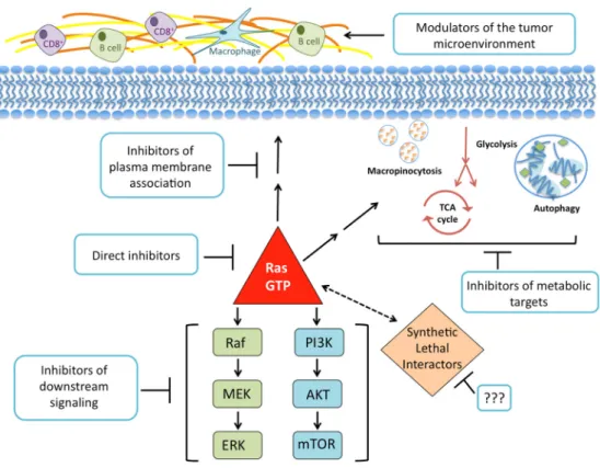
KRAS plays a vital role in PDAC and is believed to be a key target for treatment. Decades of research have shaped our understanding of the biochemistry, structure, and cellular signaling of KRAS in cancer. This foundation of knowledge can be viewed in two ways: support for the need to find different routes to silence KRAS, or fodder for the notion that KRAS is “undruggable”. In this review, the most promising paths taken in an attempt to suppress the effects of KRAS in cancer are discussed. We will examine efforts to target KRAS directly, prevent KRAS from binding to cellular membranes, inhibit its downstream effectors, search for synthetic lethal interactors of mutant KRAS, disrupt the metabolic pathways KRAS regulates, and exploit the ways KRAS signaling influences the tumor microenvironment (Figure 3).
Figure 2.Mutant KRAS is continuously in a GTP-bound, active state. Wild-type KRAS cycles between an active, GTP-bound and an inactive, GDP-bound state, and it exists largely in an inactive state in non-dividing cells. Upon growth factor stimulation, normal KRAS is activated by RAS guanine nucleotide exchange factors (RASGEFs), which facilitate the binding of GTP to KRAS. KRAS-GTP then binds downstream effectors. This signaling is attenuated due to the action of RAS GTPase-activating proteins (RASGAPs), which promote the hydrolysis of the bound GTP to GDP and hence formation of inactive KRAS-GDP. Mutation of residues G12, G13 or Q61 constitutively activates KRAS by preventing the formation of van der Waals interactions between RAS and RASGAPs [20] and interfering with the position of a water molecule necessary for GTP hydrolysis [21], respectively. The arrow thickness and relative size of the symbols for GEFs and GAPs indicate the level of signaling.
KRASmutation is the initiating genetic event for PDAC, withKRASmutations found in ~95% of
pancreatic intraepithelial neoplasias (PanINs), the earliest pre-neoplastic stages of pancreatic cancer
progression [22,23]. Progression to invasive, malignant PDAC involves a step-wise accumulation of
additional genetic alterations, in particular, the inactivation of tumor suppressor genes [24]. Loss of
the cyclin-dependent kinase inhibitor 2A (CDKN2A) tumor suppressor gene function by mutation
or promoter methylation is found in 95% of pancreatic tumors [25]. CDKN2Aencodes p16/Ink4a
and p14/Arf, inhibitors of cyclin-dependent kinases 4 and 6 (CDK4/6) and MDM2-mediated p53 tumor suppressor degradation, respectively. CDK4/6 hyperactivation in turn inactivates the RB tumor suppressor, promoting tumor progression. Later stage steps involve missense, loss-of-function
mutations inTP53and theSMAD4tumor suppressor genes.TP53is mutated in 75% of PDAC. Smad4
functions as a downstream component of the tumor growth factorβ-signaling network. In pancreatic
cancer, a mutation in Smad4 is often associated with metastatic disease [26].
KRASplays a vital role in PDAC and is believed to be a key target for treatment. Decades of
research have shaped our understanding of the biochemistry, structure, and cellular signaling of KRAS in cancer. This foundation of knowledge can be viewed in two ways: support for the need to find
different routes to silenceKRAS, or fodder for the notion thatKRASis “undruggable”. In this review,
the most promising paths taken in an attempt to suppress the effects ofKRASin cancer are discussed.
We will examine efforts to target KRAS directly, prevent KRAS from binding to cellular membranes,
inhibit its downstream effectors, search for synthetic lethal interactors of mutantKRAS, disrupt the
metabolic pathways KRAS regulates, and exploit the ways KRAS signaling influences the tumor
Figure 3. The current paths in the pursuit of an anti‐KRAS therapy. There have been past and ongoing
efforts to synthesize molecules that bind directly to the RAS protein and inhibit its GDP‐GTP
regulation or effector signaling. Disrupting RAS membrane localization by inhibiting farnesylation
showed promising preclinical effects but no anti‐tumor activity in clinical trials. Attempts to inhibit
downstream effector signaling have generated a large number of inhibitors currently under clinical
evaluation. Unbiased genetic functional RNAi screens have identified genes that may act as synthetic
lethal interactors. However, these studies have been limited by reproducibility or the transition of hits
to a therapeutic strategy. The broken line represents the functional relationship in the absence of a
linkage via a specific signaling network. The elucidation of the many metabolic processes that KRAS
regulates may result in new therapies for patients with PDAC. Likewise, the discovery of ways to
degrade the dense stroma associated with PDAC tumors and employ the immune response may lead
to novel therapies for PDAC.
2. Direct Inhibition of RAS
Inhibiting RAS directly is the most obvious approach and arguably the most attractive for developing a treatment for KRAS mutant PDAC. Since a treatment aimed at targeting all RAS isoforms would be deleterious to normal as well as neoplastic tissue, studies have focused on specifically targeting KRAS. Unfortunately, RAS activation and signaling is accomplished primarily through protein‐protein interactions and such interfaces have traditionally been difficult to target with small molecules because they lack well‐defined binding pockets [27]. Although some deemed KRAS “undruggable”, recent data have revived the hope to target RAS directly.
The first small molecules identified as direct binders that altered RAS function targeted the site on RAS involved in its recognition by the RASGEF, SOS1. SOS1 catalyzes the exchange of GDP to GTP, the rate‐limiting step in RAS activation, and thus regulates RAS activity. Fragment‐based lead discovery (FBLD) provided a starting point to finding compounds that bind to RAS targets, leading to the discovery of small molecules that bound to KRAS GDP and inhibited SOS‐mediated nucleotide exchange [28]. Independently, a second group identified small molecules that bind to RAS and also inhibit SOS1‐catalyzed nucleotide exchange [27]. These small molecules bind to the hydrophobic pocket on the CDC25 domain of SOS. The structure not only demonstrated how these small molecules bind, but also revealed other potential binding sites that have yet to be targeted by existing compounds [29].
Figure 3. The current paths in the pursuit of an anti-KRAS therapy. There have been past and ongoing efforts to synthesize molecules that bind directly to the RAS protein and inhibit its GDP-GTP regulation or effector signaling. Disrupting RAS membrane localization by inhibiting farnesylation showed promising preclinical effects but no anti-tumor activity in clinical trials. Attempts to inhibit downstream effector signaling have generated a large number of inhibitors currently under clinical evaluation. Unbiased genetic functional RNAi screens have identified genes that may act as synthetic lethal interactors. However, these studies have been limited by reproducibility or the transition of hits to a therapeutic strategy. The broken line represents the functional relationship in the absence of a linkage via a specific signaling network. The elucidation of the many metabolic processes that KRASregulates may result in new therapies for patients with PDAC. Likewise, the discovery of ways to degrade the dense stroma associated with PDAC tumors and employ the immune response may lead to novel therapies for PDAC.
2. Direct Inhibition of RAS
Inhibiting RAS directly is the most obvious approach and arguably the most attractive for
developing a treatment forKRASmutant PDAC. Since a treatment aimed at targeting all RAS isoforms
would be deleterious to normal as well as neoplastic tissue, studies have focused on specifically
targetingKRAS. Unfortunately, RAS activation and signaling is accomplished primarily through
protein-protein interactions and such interfaces have traditionally been difficult to target with small
molecules because they lack well-defined binding pockets [27]. Although some deemed KRAS
“undruggable”, recent data have revived the hope to target RAS directly.
The first small molecules identified as direct binders that altered RAS function targeted the site on RAS involved in its recognition by the RASGEF, SOS1. SOS1 catalyzes the exchange of GDP to GTP, the rate-limiting step in RAS activation, and thus regulates RAS activity. Fragment-based lead discovery (FBLD) provided a starting point to finding compounds that bind to RAS targets, leading to the discovery of small molecules that bound to KRAS GDP and inhibited SOS-mediated
nucleotide exchange [28]. Independently, a second group identified small molecules that bind to
RAS and also inhibit SOS1-catalyzed nucleotide exchange [27]. These small molecules bind to the
small molecules bind, but also revealed other potential binding sites that have yet to be targeted by
existing compounds [29].
Shortly after, another study identified molecules that inhibited RAS protein-protein interactions, the Kobe0065-family compounds were found to bind to RAS-GTP and inhibit interactions with
downstream effectors [30]. Shimaet al.suggests that once these compounds are structurally optimized,
they could be used to develop RAS inhibitors with the high potency and specificity, as well as the low toxicity necessary for clinical application [30].
A third class of RAS-binding small molecules was developed to selectively recognize the G12C
missense mutant of KRAS [31]. Targeting the S-IIP binding site affects KRAS signaling through
two mechanisms. It shifts the nucleotide affinity from GTP to GDP, which leads to more RAS molecules
in the inactive state, and it diminishes interactions with effectors [31]. SML-8-73-1, a GDP analogue
has been developed to specifically target cancers with a KRASG12Cmutation. SML-8-73-1 competes
with GTP and GDP for active site binding and treatment with SML-8-73-1 stabilizes an inactive form
of KRAS [32]. Although SML-8-73-1 can penetrate the cell membrane, it requires high concentrations,
which may result in a loss of selectivity and potential off-target activities. Although the KRASG12C
mutation is prevalent in non-small-cell lung cancer, this mutation is rarely found in PDAC (3%) [14].
To date, mutation specific compounds targeting the more common KRAS G12D or G12V mutations have yet to be developed.
Using RNA interference (RNAi) to suppressKRASexpression has been validated as therapeutic
strategy inRAS mutant-driven mouse models of cancer [33,34]. RNAi mediated suppression of
expression of mutantKRASin pancreatic cancer cells reduced proliferation, anchorage-independent
growth, and tumorigenic growth [10,35]. The effects ofKRASsiRNA on PDAC suggest that RNAi
can be explored as a potential drug forKRASmutant PDAC [36]; however, the delivery of siRNA
in vivois a challenge because of enzymatic breakdown, renal clearance, and precise targeting to the tissue of interest. Improved delivery methods have been developed, such as a Local Drug EluteR, (LODER), a miniature biodegradable polymetric matrix that protects the siRNA and enables stable,
local release of the siRNA for months within the tumor tissue [37]. Using this technology, delivery
ofKRASG12D-specific siRNA clearly dampenedKRASexpression and inhibited thein vivogrowth of
pancreatic tumors in both subcutaneous and orthotopic mouse models [37]. A clinical phase I/IIa study
of siG12D-LODER in combination with chemotherapy was recently completed in patients with locally
advanced PDAC [38]. The LODER was inserted into the tumor using a standard endoscope ultrasound
biopsy and was thus able to provide local, continuous treatment in the tumor for several months. The treatment was given in combination with FOLFIRINOX, a standard of care chemotherapy cocktail
commonly used in advanced pancreatic cancer patients in good health, and was tolerated well [38]. The
results of the combination treatment showed a median overall survival of 15.13 months, and a median
time to metastasis of 8.25 months [38]. A drawback to this technology is the LODER must be directly
implanted in the tumor. Pecotet al.achieved systemicin vivodelivery ofKRAS-targeting siRNA using
a nanoliposomal delivery platform made of 1,2-dioleoyl-sn-glycero-3-phosphatidylcholine (DOPC). Mice with KRAS mutant lung cancer treated with the DOPC-mediated siRNA showed decreased
downstream signaling, inhibited proliferation, and a decrease in metastatic burdenin vivo[39]. Finally,
since wild type and mutantRASdiffer by just one missense mutation, an additional challenge is the
development of siRNA that targets the mutant gene selectively.
3. Disruption of RAS Plasma Membrane Localization
farnesyl isoprenoid to the cysteine of the CAAX-motif. Next, RAS-converting enzyme 1 (RCE1) catalyzes the proteolytic removal of the AAX peptide, and finally isoprenylcysteine methyltransferase (ICMT) catalyzes the carboxylmethylation of the now terminal farnesylated cysteine. Pharmacological inhibitors of all three CAAX-modifying enzymes have been developed, but FTase has been the most
favored target since it is the first step of the three modifications [14]. Preclinical cell culture and
mouse model studies showed farnesyl transferase inhibitors (FTIs) are potent, non-toxic inhibitors
of HRAS-driven growthin vitroandin vivo. FTIs can also block the farnesylation and membrane
association of the RAS isoforms more commonly mutated in cancer (KRAS and NRAS); however, when FTase activity is blocked, these RAS isoforms can undergo modification by a related lipid geranylgeranyl transferase enzyme (GGTase-I), overcoming the actions of FTI treatment. A logical solution to this limitation of FTIs is the use of GGTIs in combination treatment with FTI. However, since there may be up to 300 additional substrates of FTase and GGTase-I aside from RAS proteins, off-target effects are a concern for normal tissue toxicityin vivo.
Clinical trials of FTIs to treat PDAC and other cancers with prevalentKRASmutations did not
show significant anti-tumor activity or impact on patient survival [41,42]. A phase II study of tipifarnib
in patients with surgically incurable or advanced PDAC showed no benefit [43]. The problem was
initially thought to be the dose of tipifarnib, but further studies demonstrated that the dose was
appropriate as inhibition of farnesyl transferase in peripheral blood monocytes was observed [44].
Since higher doses could not be tolerated, the possibility of combination therapies was explored, but a Phase III study with the combination of tipifarnib and the cytotoxic chemotherapy gemcitabine showed no benefits [45].
Another strategy to prevent the interaction of the RAS farnesyl group with the cell membrane is the use of a farnesyl-cysteine mimetic that would compete with RAS proteins for association
with RAS anchorage cites [46]. Farnesyl thiosalicylic acid (FTS/salirasib) inhibits RAS signaling,
in part, by dislodging RAS from the cell membrane and rendering it susceptible to proteolytic
degradation [47]. Salirasib showed some promise in mice, as salirasib treatment inhibited cell growth
in HRAS transformed rat fibroblasts with drug concentrations that did not affect processes like
farnesylation and carboxyl methylation [48]. While salirasib showed activity towards active RAS,
it is certain that salirasib will have non-RAS targets. Salirasib has undergone phase II clinical trials
in non-small cell lung carcinoma (NSCLC) patients with mutated KRAS. The monotherapy was
determined to be insufficient at the current dose with modest toxicity, presenting another disappointing result [49].
The discovery of proteins that facilitate the trafficking of RAS to the plasma membrane has provided an alternative approach to interfering with RAS membrane association. Phosphodiesterase
6 delta (PDEδ) is important for photoreceptor signaling and is responsible for the trafficking of the
PDE6 complex (which contains farnesylated and geranylgeranylated substrates) [50]. PDEδcan also
recognize KRAS4B and enhance its transit to the plasma membrane, and thus, interfering with the
binding of PDEδto KRAS provides an opportunity to disrupt RAS function [51]. Inhibition of the
PDEδ-KRAS interaction using small molecules provides an opportunity to suppress KRAS and effect
pancreatic cancer tumor development. Deltarasin, which is a high affinity PDEδ-KRAS interaction
inhibitor, had a negative effect on the plasma membrane association of KRAS4B and reduced the
growth ofKRAS-dependent PDAC cell lines [51].
RAS controls many downstream pathways and this could be due to its compartmentalization in cells. In addition to the plasma membrane, RAS also signals from endosomes, the endoplasmic reticulum, the Golgi apparatus, and mitochondria. In T lymphocytes, the location of RAS signaling
dictates the biological outcome [52]. Phosphorylation by protein kinase C (PKC) at serine 181 (S181) in
the polybasic region of KRAS4B results in its release from the plasma membrane and accumulation
of KRAS4B on internal membranes, representing a farnesyl-electrostatic switch [53]. After the switch
is engaged, phospho-KRAS4B translocates from the cell membrane to the endoplasmic reticulum,
antiproliferative effects as phospho-KRAS4B signaling through inositol-triphosphate receptors at
the ER promotes cell death, suggesting a possible strategy for anti-RAS treatments [54].
Bryostatins are PKC agonists, and therefore are capable of triggering KRAS dissociation from the
plasma membrane [55]. Mice bearing orthotopic tumors derived from the human pancreatic cancer
cell line, KC1-MOH1 [56], responded very well to a combination of bryostatin-1 and gemcitabine
with remission in only one of every seven animals [57]. However, to date, there have been more than
20 clinical studies using bryostatin-1 monotherapy or combination therapies in numerous different
cancer types, and none have been successful [58]. In addition to not being clinically effective, there
were toxic effects like myalgia, local phlebitis, fatigue, nausea, and thrombocytopenia [59]. Further
studies determining the functionality of RAS based on its subcellular localization would be a critical step toward finding a drug that targets specific RAS pathways.
4. Searching for Synthetic Lethal Interactors
Synthetic lethality arises when a combination of mutations in two or more genes leads to cell death. Thus, synthetic lethal interactors of mutant RAS would be genes for which the loss of function would be lethal to the cell only in the presence of mutant RAS. The existence of oncogene-specific synthetic lethal interactions is supported by the notion that oncogenic transformation profoundly changes the
phenotype of the cell [60,61]. Potential targets may exist is pathways that aid theRAS-transformed
cell in coping with the cellular stress associated with persistent proliferation or the nutrient-supply pathways that fuel this proliferation. Several studies have identified synthetic lethal interactors with
mutantKRASthrough the use of RNAi screens in human cancer cell lines [62]; unfortunately, this first
generation of screens yielded only new information about the biology of mutantKRAS-harboring cells,
not new therapeutic targets.
The hits from screens for synthetic lethal interactors for mutantKRAS span many different
cellular processes including: Cell cycle/mitosis, cell survival, gene transcription, and cell growth.
Therapeutically, targeting cell cycle regulators such as survivin,CDK1[63], orTPX2[64] would most
likely be similarly toxic to normal and neoplastic cells. Additionally, transcription factors such as GATA2[65,66] are largely considered undruggable. A potentially druggable hit, serine/threonine
protein kinase 33 (STK33), was initially considered a tractable target [67]; however, follow-up studies
have determined that both genetic depletion and pharmacological inhibition of STK33 has no effect
on cell growth [68]. Likewise, genetic and pharmacological validation of the hit TBK1 [69] found no
reproducible requirement for TBK1 in the growth ofKRAS-mutant tumor cell linesin vitro[70].
There were a number of limitations with the first generation of mutantKRASsynthetic lethal
screens that could be improved upon in future studies. First, many of these screens relied on isogenic
matched pairs of cells lines harboringKRASmutations, and a matched counterpart in which the
KRAS allele is genetically ablated. Acute ablation ofKRAS causes apoptosis and severe growth
impairment [71], and thus proliferating cells that eventually arise must acquire additional, adaptive
alterations and are therefore not truly isogenic. Currently, efforts are underway to screen large panels
of cancer cell lines that are more representative of the heterogeneity that exists in humanKRAS-mutant
cancers. Additionally, all previously described screens have utilizedin vitroanchorage-dependent
culture conditions. Second generation screens would benefit from anchorage-independent culture
systems, such as organoid cultures orin vivoxenograft tumor assays, which more accurately model
tumor biology. Such methods have already been utilized in screens for other purposes [72–74].
Furthermore, the first generation screens serve as a reminder that as with any high-throughput approach, hits must be rigorously validated.
5. Targeting RAS Downstream Signaling Pathways
Eleven RAS effector families have been identified to date, with six of these families having
validated roles in contributing to RAS-dependent cancer initiation and/or maintenance [17,75].
for targeting RAS signaling is through targeting its downstream effectors’ signaling. Herein, we will focus on the most intensely targeted pathways: the RAF-MEK-ERK mitogen-activated protein kinase (MAPK) cascade and the PI3K-AKT-mTOR cell-survival signaling pathway.
5.1. RAF-MEK-ERK
Active RAS can engage three highly related RAF serine/threonine kinases: ARAF, BRAF, and
CRAF. The largely mutually exclusive frequency ofBRAFandRASmutations supports the notion
that RAF is a critical driver inKRAS-mutant pancreatic cancer. RAS-GTP binds preferentially to RAF,
resulting in the translocation of RAF to the plasma membrane where subsequent events promote its activation. Active RAF phosphorylates and activates the MEK1 and MEK2 dual specificity kinases, which in turn phosphorylate and activate ERK1 and ERK2 serine-threonine MAPKs. Activated ERKs
phosphorylate a diverse spectrum of more than 200 cytoplasmic and nuclear substrates [76,77].
There are currently two BRAF-selective inhibitors in the clinic, vemurafenib and dabrafenib.
Vemurafenib is approved for the treatment ofBRAF-mutant melanoma. Vemurafenib proved effective
in melanoma patients harboringBRAFmutations with a response rate of greater than 50% and a rapid
improvement in quality of life. Unexpectedly, when these RAF inhibitors were used inRAS-mutant
cancers, activation rather than inactivation of ERK was observed [78–80]. This is due to the propensity
of these first-generation RAF inhibitors to induce RAF dimerization, which causes activation of
CRAF [81]. Recently, pan-RAF inhibitors have entered clinical evaluation [82]. This new class of
inhibitors is not subject to the paradoxical activation seen with the BRAF-selective inhibitors, and may be more effective inRAS-mutant cancers [83,84].
In addition to BRAF inhibitors for melanoma, MEK inhibitors have also be developed. Trametinib
(GSK112021) is a selective allosteric inhibitor of MEK1/2 activation and kinase activity [85]. In a
Phase III trial of patients with advanced or metastaticBRAFV600E/K-positive melanoma, the response
rate for the trametinib monotherapy was 22%, but in combination with dabrafenib the response rate
increased to 64% [85]. This combination therapy likely delays pathway alterations that lead to ERK
reactivation and the resistance that occurs in response to BRAF or MEK monotherapy. While MEK
inhibitors have shown success in the treatment ofBRAF-mutant melanoma, they have shown limited
efficacy inRAS-mutant human tumor cell lines [86] andRAS-driven mouse models of cancer [87].
A recent study suggests that the mechanism by which MEK is activated inRAS-versusBRAF-mutant
cancers is different, thus explaining the different responses in different systems [88]. Resistance to MEK
inhibitors inRAS-driven cancers occurs due to upregulation or amplification of upstream activators
that restore ERK activity [82].
As dynamic kinome reprogramming [89] in response to RAF and MEK inhibitors eventually
leads to increased ERK signaling inRAS-driven cancers, ERK has become an attractive target [81,82].
Four ERK inhibitors (BVD-523, MK8353, GDC-0994, and CC-90003) have entered Phase I studies, and
MK8353 (an analog of SCH772984) has been described preclinically [90], where it showed promising
results in BRAF- or MEK-inhibitor resistant cell line models [90]. Additionally, concurrent targeting
of the RAF-MEK-ERK cascade at multiple nodes is currently under investigation as an effective strategy to achieve prolonged ERK suppression. A recent study focused on directly targeting ERK as a treatment for pancreatic cancer identified the degradation of the MYC oncoprotein and the induction of a senescence-like phenotype as the predominant growth suppression mechanism of ERK
inhibitors [71]. This study also identified PI3K-AKT-mTOR signaling as a critical determinant of
ERK inhibitor sensitivity, and PI3K, Notch, and p38 as potential modulators of ERK resistance [71].
This suggests that multiple inhibitor-based combinations will be necessary to treat across multiple
KRAS-mutant PDAC populations.
5.2. PI3K-AKT-mTOR
The catalytic subunits of class I PI3K lipid kinases (α, β,δ andγ) comprise the second-best
is often mutationally activated in cancers, which supports its role as a cancer driver. PI3K signaling pathways are important for the regulation of cellular functions such as metabolism, growth, proliferation, survival, transcription and protein synthesis. Mutant RAS activates PI3K. Once PI3K is
activated, it binds to PIP2(phosphatidylinositol 4,5-bisphosphate), a component of the cell membrane
and phosphorylates PIP2to PIP3(phosphatidylinositol 3,4,5-triphosphate), which in turn can regulate
the activities of many signaling proteins, in particular the 3-phosphoinositide-dependent protein
kinase-1 (PDPK1 or PDK1) and AKT serine/threonine kinase. Upon PIP3-dependent recruitment to
the plasma membrane, AKT is phosphorylated by PDK1, which itself is associated with PIP3at the
plasma membrane. AKT promotes activation of the Rheb small GTPase, which then activates mTOR, a protein that is involved in growth factor signaling, the energy state of the cell, and nutrient and oxygen availability.
There are four main pharmacologic approaches for inhibition of PI3K signaling: PI3K inhibitors,
AKT inhibitors, and mTOR inhibitors, and dual PI3K-mTOR inhibitors [14,91]. PI3K inhibitors can be
isoform-specific or pan-PI3K inhibitors, which target all class I PI3Ks. Whether pan- or isoform-specific PI3K inhibitors will be more effective is not clear. Isoform-selective treatment may exhibit less toxicity, which means it may be tolerated at the higher doses necessary for more complete target inhibition
with fewer adverse effects [92]. The realization that different PI3K isoforms play non-redundant roles
in different tumor types has attracted increasing interest in isoform-specific inhibitors [93]. However,
since RAS can utilize multiple PI3K isoforms, more effective suppression may require a pan-PI3K inhibitor. More work must be done to understand the mechanisms underlying drug resistance and escape of PI3K dependency following isoform-specific therapies. AKT inhibitors are typically either ATP mimetics or allosteric, non-catalytic site inhibitors. Allosteric AKT inhibitors block the attachment of AKT to the membrane by interfering with the binding of the PH (pleckstrin homology) domain to phosphoinositides. Mislocalization of AKT in turn diminishes its ability to signal. A potential drawback of this class of inhibitors is that they will not block the non-AKT effectors of PI3K signaling and hence paradoxically increase the PI3K-dependent activation of those effectors via the loss of negative feedback [92].
Mammalian target of rapamycin (mTOR) exists as two distinct complexes, mTOR complex 1 (mTORC1; which contains the regulatory-associated protein of TOR1 (RAPTOR)) and mTORC2 (which contains the rapamycin-insensitive companion of mTOR (RICTOR)). Rapamycin and its analogues (also known as rapalogues, which include everolimus, ridaforolimus and temsirolimus) are selective for mTORC1, forming an inactive complex with mTOR and FKBP12. Second-generation mTOR catalytic site inhibitors directly inhibit both mTOR complexes, mTORC1 and mTORC2, and are more effective inhibitors of downstream signaling and ultimately RNA translation than the first generation rapalogues [94,95]. A concern with these inhibitors is that feedback activation of PI3K from
mTOR inhibition may result in hyperactivation of AKT-independent effectors of PI3K signaling [92].
Since the p110 subunits of PI3K and mTOR have similar structures, the inhibition of p110 often
results in the inhibition of mTOR [96]. This dual inhibition of PI3K-mTOR is expected to shut down
PI3K-AKT-mTORC1 signaling; however, it is still unclear whether a dose that sufficiently blocks cellular signaling will be tolerable.
The recent observation that downstream of PI3K, PDK1 is a key effector of oncogenic KRAS
signaling in the pancreas has led to enhanced interest in specifically targeting this PI3K effector [97].
PHT-427 is a novel AKT/PDK1 pleckstrin homology domain inhibitor, which is capable of binding to both AKT and PDK1; however, inhibition of PDK1 was more closely correlated to its antitumor activity
than AKT inhibition [98]. Furthermore, when PHT-427 was encapsulated in poly (lactin-co-glycolic)
acid (PLGA) nanoparticles drug delivery was improved and tumor volume was reduced by 4-6 fold in
preclinical mouse models [99].
Monotherapies targeting PI3K, AKT, and/or mTOR have been largely disappointing in
RAS-mutant cancers. However, in mouse models, potent synergistic activity has been observed
Specifically, in mice with PDAC, treatment with MEK (GDC-0973) or PI3K inhibitors (GDC-0941) alone showed slight tumor growth inhibition and had no significant effect on survival. However, in comparison to the monotherapies, the combination of the two treatments resulted in a survival
advantage [100]. Furthermore, combined inhibition of MEK and AKT showed synergistic activity in
PDAC cell linesin vitro[101]. There are now numerous clinical trials evaluating the effect of combined
inhibition of PI3K and RAF [102]. Recently, a novel approach for targeting these two pathways was
described. Van Dort et al. designed a single compound that is a hybrid of the ATP competitive
pan-PI3K inhibitor ZSTK474 [103] and the allosteric MEK inhibitor RO5126766 [104]. Western blot
analysis showed a dose-dependent decrease of pERK and pAKT in treated PANC-1 cells, verifying the
compound is cell permeable and effective [104]. Although this therapy must be optimized in order
to achieve MEK and PI3K inhibition in mouse models, it is an important framework for creating a
combination therapy with a single compound [104].
6. KRAS-Regulated Metabolic Targets
OncogenicKRAShas been implicated in controlling a number of metabolic processes including
induction of glucose uptake, unique utilization of glucose intermediates, repurposed glutamine
metabolism, and increased autophagy and macropinocytosis [13]. Further elucidation of the links
between oncogenic KRAS and deregulated PDAC metabolism has the potential to result in the
formulation of new anti-KRAS therapies.
6.1. Glucose Utilization and Glutamine Metabolism
PDAC cells have altered metabolic processes consistent with increased aerobic glycolysis [105].
Interestingly, it has been demonstrated that in PDAC, mutantKRASis responsible for orchestrating
this phenotype by enhancing the expression of the glucose transporter GLUT1, as well as many
other genes that encode rate-limiting glycolytic enzymes, including hexokinase I and 2 (HK1, HK2),
phosphofrutokinase-1 (PFK1), and lactate dehydrogenase A (LDHA) [8]. This regulation could be
exploited therapeutically by targeting LDHA, as was demonstrated using a small molecule (FX-11),
which caused increased ROS production and cell death [106]. Hk2 has been identified as an attractive
target forKRAS-driven lung cancers as whole-body deletion ofHk2in the mouse selectively targets
tumor cells [107]. The small molecule 3-bromopyruvate (3BP) inhibits Hk2 and has shown potent
anticancer activity in a number of animal models [108], as well as promising results in a human case
study [109]. Additionally, mutantKRASexpression leads to the shunting of glycolytic intermediates
through the non-oxidative arm of the pentose phosphate pathway (PPP), which leads to the generation
of ribose 5-phosphate, a necessary component for nucleic acid biosynthesis [8]. Downstream of mutant
KRAS, the ERK-MAPK pathway, which culminates with the MYC transcription factor, is the major
driver of glucose metabolism adjustments [8].
In addition to altered levels of glycolysis, cancer cells also display an increased dependence
on glutamine [110], which contributes to cancer cell proliferation by providing carbon to fuel the
tricarboxylic acid (TCA) cycle and nitrogen for nucleotide, nonessential amino acid, and hexosamine
biosynthesis [111]. Glutamine is catabolized toα-ketoglutarate (αKG), a TCA cycle intermediate,
through two deamination reactions, the first requiring glutaminase (GLS) to generate glutamate and the second occurring via glutamate dehydrogenase (GDH) or transaminases. mTORC1 has been shown to positively regulate GLS and glutamine flux through this pathway through the S6K1-dependent
regulation of MYC [112]. KRASmutant PDAC has been shown to utilize glutamine metabolism
to regulate redox balance by increasing the NADPH/NADP+ratio in the cell through an aspartate
transaminase (GOT1)-dependent mechanism [113]. As treatment with glutamine analogs is profoundly
toxic [114] the current strategy for targeting glutamine utilization as a cancer treatment, is to target
those processes that cancer cells are specifically addicted to. Thus, as GOT1 is dispensable for normal cells while PDAC cells rely on this enzyme for redox homeostasis, it could be an enticing
and bis-2-(5-phenylacetamido-1,2,4-thiadiazol-2-yl)ethyl sulfide (BPTES) [116], have demonstrated a growth suppressive effect on PDAC cells that is enhanced when combined with hydrogen peroxide
treatment [113]. Finally, as inhibition of GOT1 or GLS ultimately leads to a disruption of redox
homeostasis in PDAC, such inhibition may synergize with therapies that increase reactive oxygen
species, such as chemotherapy and radiation [117].
6.2. Macropinocytosis and Autophagy
To fuel metabolic processes,KRASsignaling leads to the scavenging of extracellular proteins
and lipids and activates self-eating and recycling of proteins through autophagy [118]. PDAC
cells specifically expressing oncogenic KRAS utilize macropinocytosis to transport extracellular
protein into the cell [119] and use it as a source of essential amino acids (EAAs) in order to sustain
survival and proliferation [120]. Consistent with these studies, active macropinocytosis has been
observed in primary human PDAC specimens [121]. This scavenging phenotype appears to be a
general property ofRAS-driven cancers asRAS-transformed cells have also been shown to scavenge
lysophospholipids, which contributes to their metabolic robustness [121]. The regulation of the
degradation of the EAAs taken up by macropinocytosis may shed light on the disappointing lack of efficacy of mTOR inhibitors as PDAC therapeutics. It has been demonstrated that in mammalian cells, mTORC1 signaling suppresses lysosomal catabolism of proteins that were taken up from
the extracellular environment [120]. Thus, mTORC1 inhibition may enhance cell proliferation that
depends on extracellular proteins, such as PDAC cells inhabiting a poorly vascularized area [120].
A specific inhibitor of macropinocytosis has yet to reach the clinic; however, preclinical studies in
which heterotopic tumor bearing mice were treated with the tool compound 5-(N-ethyl-N-isopropyl)
amiloride (EIPA) showed attenuation of tumor growth and in some cases, regression [119].
Autophagy is a highly conserved mechanism to degrade intracellular components and promote the survival of stressed cells by providing energy in the form of ATP and building blocks such as
amino acids, lipids, sugars, and nucleosides [122]. The role of autophagy in cancer is extremely
complex [123] and while it appears clear that PDAC cells depend on autophagy for growth, the
role of oncogenicKRASin this dependence remains unclear. When tissue samples from 71 PDAC
patients were analyzed via immunohistochemical staining for LC3 protein (a component of the autophagosome), it was determined that high expression was correlated with large tumor size,
short-disease free period, and overall poor patient outcome [124]. Additional studies have revealed
that pancreatic cancers have a clear dependence on autophagy. Genetic or pharmacological inhibition of autophagy results in increased reactive oxygen species, elevated DNA damage, and mitochondrial defects that lead to decreased proliferation of pancreatic cancer cell linesin vitro, as well as substantial
tumor regression and sustained survival inin vivomodels of pancreatic cancer [125,126]. In support
of a cooperative role betweenRAS expression and proliferation fueled by autophagy, immortal,
non-tumorigenic baby mouse kidney epithelial (iBMK) cells ectopically expressing oncogenicHRASor
KRASexperienced defects in mitochondrial respiration upon autophagy inhibition [126]. However,
a cooperative relationship between RAS and autophagy is not universally reported. For example,
acute expression of oncogenic HRASG12Vin immortalized human ovarian surface epithelial cells was
associated with caspase-independent cell death, rather than increased proliferative capacity [127].
Furthermore, autophagy was implicated as a facilitator ofRAS-induced senescence in a study in which
oncogenic HRAS was overexpressed in IMR90 human diploid fibroblasts [128]. Additionally, a very
recent study assayed a panel of 47 different cancer cell lines comprised of bothKRASmutant and
KRASwild-type lines and found that theKRAS-mutated cells were no more dependent on autophagy
than their wild-type counterparts [129]. Thus, the specific role thatKRASplays in the upregulation of
autophagy in PDAC remains a controversy.
A recent study that used a mouse model of PDAC harboring an embryonic homozygousTrp53
deletion paradoxically demonstrated that loss of autophagy accelerates tumor onset [130]. However,
mutations in human PDAC, as well as patient-derived xenographs showed that p53 status does not
affect the response of a patient to autophagy inhibition [131]. Interestingly, an inhibitor of autophagy,
hydroxychloroquine (HCQ), has been available clinically for quite some time as the FDA approved it for the treatment of malaria and rheumatic disorders years ago. Hence, there are multiple early phase studies exploring the use of HCQ in as a treatment for pancreatic cancer. However, early observations have been disappointing, likely due to the limited potency of HCQ to block autophagy in vivo[132]. Notably, HCQ does not specifically inhibit autophagy, it is an inhibitor of lysosomal acidification, and thus hinders all pathways that terminate in the lysosome. Therefore, processes such as macropinocytosis are similarly inhibited with HCQ. This is supported by a recent study that found that while HCQ treatment is antiproliferative and synergizes with targeted anticancer drugs, these
effects may be independent of autophagy inhibition [129].
7. Harnessing the Immune Response
Recent successes of cancer immunotherapy, such as antibody blockade of cytotoxic T lymphocyte antigen-4 (CTLA-4), have generated a lot of excitement, but unfortunately this form of treatment has
been less successful in patients with pancreatic cancer [133]. A major barrier for immunotherapeutic
approaches is thought to be profound immune suppression associated with the pancreatic tumor
microenvironment [134]. It is now well understood that formation of PDAC is accompanied by
pronounced alterations in stromal responses and immune surveillance programs, and there is an increasing appreciation that signaling by oncogenic RAS plays a direct role in orchestrating some of these changes in tumor microenvironment. Multiple signaling mechanisms downstream of RAS may account for this effect. Activation of oncogenic RAS has been shown to down regulate expression
of major histocompatibility complexes (MHC) [135–138], resulting in decreased antigen presentation
by tumor cells and reduced recognition by the immune system [139,140]. Oncogenic RAS has also
been shown to upregulate expression of immunomodulatory cytokines, such as IL-8 and GM-CSF. Experimental perturbation in tumor-derived cytokine levels resulted in reduced inflammation and
increased anti-tumor cytotoxic T cell response [141,142]. Significantly, pharmacologic inhibition of
either ERK or AKT downregulated cytokine expression in KRAS transformed cells. These observations propel the hypothesis that abrogation of signaling pathways downstream of activated RAS may improve anti-tumor immune response. In support of this idea, inhibition of MEK or BRAF in melanoma correlated with reduced levels of immunosuppressive cytokines and an increase in infiltrating
T cells [143–145]. A rational extension of this hypothesis is the idea that inhibition of RAS signaling
may yield better responses to immune checkpoint blockade agents. Indeed, recent studies in TBNC featuring activation of RAS signaling pathways demonstrated that co-targeting of MEK and PD-L1
results in upregulation of MHC I and II on tumor cells and increase in CD8 T cell infiltration [146].
While inhibition of RAS-driven signaling has so far been shown to have positive immunomodulatory function, it will also be helpful to evaluate its potential effects on T cell activity [147]. Overall, strategies aimed at combining RAS-targeted therapy and immunotherapy hold significant promise as clinically feasible and effective therapeutic modalities.
PDAC is distinctive as desmoplastic stroma accounts for 70%–80% of the tumor volume [148,149].
Stromal accumulation isKRASdriven and is initiated in PanIN lesions, suggesting that its early onset
is important to tumor growth and progression [150]. The dense stromal compartment is thought to
prevent the delivery of drugs to the target tissue; therefore, the targeting of the stroma itself may be a way to improve current chemotherapeutics. The stroma is composed of cancer-associated fibroblasts, extracellular matrix, inflammatory cells and blood vessels [151]. Stroma and cancer cells both contribute to the extracellular matrix, which is composed of macromolecules like collagens and hyaluron and
regulatory components like secreted protein acidic and rich in cytosine (SPARC) [152]. Hyaluronic acid
and supporting drug delivery [154]. In combination with gemcitabine, PEGPH20 increased response
rate, decreased metastasis and increased median survival in the KPC mouse model of PDAC [154].
Clinical trials have suggested the co-treatment provides improved progression-free survival for PDAC
patients with high hyaluron expression, while increasing chances of thromboembolic events [155].
Although the pursuit of stromal depletion appears promising, other recent data suggest that the stroma
could also function to protect the cancer from quickly spreading [152]. Clearly, PDAC has a unique
microenvironment due to its unusual stromal content, which requires a better understanding.
8. Conclusions
In summary, we have highlighted six promising paths to finding an effective treatment for
KRAS-mutant pancreatic cancer: targeting KRAS directly, upsetting its membrane association,
exploiting synthetic lethal interactions, targeting the pathways downstream ofKRAS, pursuing the
metabolic processes thatKRASregulates, and harnessing the immune response. Although each of
these approaches has shown promise in cells and even animal studies, none of these treatments have been very successful in the clinic. Thus, pancreatic cancer is projected to overcome breast cancer to
become the third leading cause of cancer deaths in the U.S. in 2016 [156], and then surpass colorectal
cancer to become the second leading cause of cancer deaths in the U.S. by around 2020 [157]. The
major challenge with treatments that seem successful preclinically is that a patient quickly develops resistance, making combination therapies an attractive new direction. Despite the lack of clinical
treatments to date, studies to better understandRASmutant cancers continue to move in promising
directions, which will hopefully lead to benefits for patients with PDAC.
Acknowledgments: Kirsten L. Bryant is supported by a Pancreatic Cancer Action Network-AACR Pathway to Leadership grant (15-70-25-BRYA). Yuliya Pylayeva-Gupta is supported by a Pancreatic Cancer Action Network-AACR Pathway to Leadership grant (13-70-25-PYLA). Channing J. Der is supported by grants from the NIH (CA042978, CA179193, and CA199235), Department of Defense (CA140731), Lustgarten Foundation and the Pancreatic Cancer Action Network-AACR.
Author Contributions: Daniel Zeitouni, Yuliya Pylayeva-Gupta, Channing J. Der, and Kirsten L. Bryant contributed to the manuscript text; Daniel Zeitouni, Channing J. Der, and Kirsten L. Bryant prepared the figures; and Channing J. Der and Kirsten L. Bryant conceived the structure of the review.
Conflicts of Interest:The authors declare no conflict of interest.
Abbreviations
The following abbreviations are used in this manuscript:
3BP 3-bromopyruvate
BPTES bis-2-(5-phenylacetamido-1,2,4-thiadiazol-2-yl)ethyl sulfide
CDK cyclin-dependent kinase
CDKN2A cyclin-dependent kinase inhibitor 2A CTLA-4 cytotoxic T lymphocyte antigen-4
DOPC 1,2-dioleoyl-sn-glycero-3-phosphatidylcholine
EAA essential amino acid
EIPA 5-(N-ethyl-N-isopropyl)amiloride
ER Endoplasmic Reticulum
FBLD fragment-based lead discovery
FTase farnesyltransferase
FTI farnesyl transferase inhibitor FTS farnesyl thiosalicylic acid
GAP GTPase activating proteins
GDP guanosine diphosphate
GEF guanine nucleotide exchange factors
HA hyaluronic acid
HBS hydrogen bond surrogate
HCQ hydroxychloroquine
Hk1 hexokinase 1
Hk2 hexokinase 2
ICMT isoprenylcysteine methyltransferase
LDHA lactate dehydrogenase A
LODER Local Drug EluteR
MHC major histocompatibility complexes
mTOR mammalian target of rapamycin
mTORC1 mTOR complex 1
mTORC2 mTOR complex 2
NSCLC Non-small cell lung cancer PanIN pancreatic intraepithelial neoplasia PDAC Pancreatic Ductal Adenocarcinoma
PDEδ Phosphodiesteraseδ
Pfk1 phosphofructokinase
PFS progression free survival
PH pleckstrin homology
PIP2 phosphatidylinositol 4,5-bisphosphate
PKC protein kinase C
PPP pentose phosphate pathway
RCE1 RAS converting enzyme 1
RNAi RNA interference
Serine 181 S181
SOS Son of Sevenless
TP53 tumor protein 53
References
1. Ryan, D.P.; Hong, T.S.; Bardeesy, N. Pancreatic adenocarcinoma. N. Engl. J. Med. 2014,371, 2140–2141. [CrossRef] [PubMed]
2. Wolfgang, C.L.; Herman, J.M.; Laheru, D.A.; Klein, A.P.; Erdek, M.A.; Fishman, E.K.; Hruban, R.H. Recent progress in pancreatic cancer.CA Cancer J. Clin.2013,63, 318–348. [CrossRef] [PubMed]
3. Stark, A.; Hines, O.J. Endoscopic and operative palliation strategies for pancreatic ductal adenocarcinoma. Semin. Oncol.2015,42, 163–176. [CrossRef] [PubMed]
4. Biankin, A.V.; Waddell, N.; Kassahn, K.S.; Gingras, M.C.; Muthuswamy, L.B.; Johns, A.L.; Miller, D.K.; Wilson, P.J.; Patch, A.M.; Wu, J.; et al. Pancreatic cancer genomes reveal aberrations in axon guidance pathway genes.Nature2012,491, 399–405. [CrossRef] [PubMed]
5. Jones, S.; Zhang, X.; Parsons, D.W.; Lin, J.C.; Leary, R.J.; Angenendt, P.; Mankoo, P.; Carter, H.; Kamiyama, H.; Jimeno, A.;et al.Core signaling pathways in human pancreatic cancers revealed by global genomic analyses. Science2008,321, 1801–1806. [CrossRef] [PubMed]
6. Waddell, N.; Pajic, M.; Patch, A.M.; Chang, D.K.; Kassahn, K.S.; Bailey, P.; Johns, A.L.; Miller, D.; Nones, K.; Quek, K.;et al.Whole genomes redefine the mutational landscape of pancreatic cancer.Nature2015,518, 495–501. [CrossRef] [PubMed]
7. Witkiewicz, A.K.; McMillan, E.A.; Balaji, U.; Baek, G.; Lin, W.C.; Mansour, J.; Mollaee, M.; Wagner, K.U.; Koduru, P.; Yopp, A.;et al. Whole-exome sequencing of pancreatic cancer defines genetic diversity and therapeutic targets.Nat. Commun.2015. [CrossRef] [PubMed]
8. Ying, H.; Kimmelman, A.C.; Lyssiotis, C.A.; Hua, S.; Chu, G.C.; Fletcher-Sananikone, E.; Locasale, J.W.; Son, J.; Zhang, H.; Coloff, J.L.;et al. Oncogenic Kras maintains pancreatic tumors through regulation of anabolic glucose metabolism.Cell2012,149, 656–670. [CrossRef] [PubMed]
10. Brummelkamp, T.R.; Bernards, R.; Agami, R. Stable suppression of tumorigenicity by virus-mediated RNA interference.Cancer Cell2002,2, 243–247. [CrossRef]
11. Abbruzzese, J.L. Pancreatic cancer: Scanning the horizon for focused interventions. Available online: http://deainfo.nci.nih.gov/advisory/ctac/archive/0313/PCwgReport.pdf 1-22 (accessed on 5 January 2016). 12. Stephen, A.G.; Esposito, D.; Bagni, R.K.; McCormick, F. Dragging RAS back in the ring.Cancer Cell2014,25,
272–281. [CrossRef] [PubMed]
13. Bryant, K.L.; Mancias, J.D.; Kimmelman, A.C.; Der, C.J. KRAS: Feeding pancreatic cancer proliferation. Trends Biochem. Sci.2014,39, 91–100. [CrossRef] [PubMed]
14. Cox, A.D.; Fesik, S.W.; Kimmelman, A.C.; Luo, J.; Der, C.J. Drugging the undruggable RAS: Mission possible? Nat. Rev. Drug Discov.2014,13, 828–851. [CrossRef] [PubMed]
15. Tsai, F.D.; Lopes, M.S.; Zhou, M.; Court, H.; Ponce, O.; Fiordalisi, J.J.; Gierut, J.J.; Cox, A.D.; Haigis, K.M.; Philips, M.R. K-Ras4A splice variant is widely expressed in cancer and uses a hybrid membrane-targeting motif.Proc. Natl. Acad. Sci. USA2015,112, 779–784. [CrossRef] [PubMed]
16. Cox, A.D.; Der, C.J.; Philips, M.R. Targeting RAS Membrane Association: Back to the Future for Anti-RAS Drug Discovery?Clin. Cancer Res.2015,21, 1819–1827. [CrossRef] [PubMed]
17. Vigil, D.; Cherfils, J.; Rossman, K.L.; Der, C.J. RAS superfamily GEFs and GAPs: Validated and tractable targets for cancer therapy?Nat. Rev. Cancer2010,10, 842–857. [CrossRef] [PubMed]
18. Prior, I.A.; Lewis, P.D.; Mattos, C. A comprehensive survey of RAS mutations in cancer.Cancer Res.2012,72, 2457–2467. [CrossRef] [PubMed]
19. Cox, A.D.; Der, C.J. Ras history: The saga continues.Small GTPases2010,1, 2–27. [CrossRef] [PubMed] 20. Scheffzek, K.; Ahmadian, M.R.; Kabsch, W.; Wiesmüller, L.; Lautwein, A.; Schmitz, F.; Wittinghofer, A.
The Ras-RasGAP complex: Structural basis for GTPase activation and its loss in oncogenic RAS mutants. Science1997,277, 333–338. [CrossRef] [PubMed]
21. Scheidig, A.J.; Burmester, C.; Goody, R.S. The pre-hydrolysis state of p21 (RAS) in complex with GTP: New insights into the role of water molecules in the GTP hydrolysis reaction of ras-like proteins.Structure1999,7, 1311–1324. [CrossRef]
22. Kanda, M.; Matthaei, H.; Wu, J.; Hong, S.M.; Yu, J.; Borges, M.; Hruban, R.H.; Maitra, A.; Kinzler, K.; Vogelstein, B.; Goggins, M. Presence of somatic mutations in most early-stage pancreatic intraepithelial neoplasia.Gastroenterology2012,142, 730–733. [CrossRef] [PubMed]
23. Hong, S.M.; Vincent, A.; Kanda, M.; Leclerc, J.; Omura, N.; Borges, M.; Klein, A.P.; Canto, M.I.; Hruban, R.H.; Goggins, M. Genome-wide somatic copy number alterations in low-grade PanINs and IPMNs from individuals with a family history of pancreatic cancer. Clin. Cancer Res. 2012,18, 4303–4312. [CrossRef] [PubMed]
24. Vincent, A.; Herman, J.; Schulick, R.; Hruban, R.H.; Goggins, M. Pancreatic cancer.Lancet2011,378, 607–620. [CrossRef]
25. Schutte, M.; Hruban, R.H.; Geradts, J.; Maynard, R.; Hilgers, W.; Rabindran, S.K.; Moskaluk, C.A.; Hahn, S.A.; Schwarte-Waldhoff, I.; Schmiegel, W.;et al.Abrogation of the Rb/p16 tumor-suppressive pathway in virtually all pancreatic carcinomas.Cancer Res.1997,57, 3126–3130. [PubMed]
26. Malkoski, S.P.; Wang, X.J. Two sides of the story? Smad4 loss in pancreatic cancer versus head-and-neck cancer.FEBS Lett.2012,586, 1984–1992. [CrossRef] [PubMed]
27. Sun, Q.; Burke, J.P.; Phan, J.; Burns, M.C.; Olejniczak, E.T.; Waterson, A.G.; Lee, T.; Rossanese, O.W.; Fesik, S.W. Discovery of small molecules that bind to K-Ras and inhibit Sos-mediated activation. Angew. Chem. Int. Ed. Engl.2012,51, 6140–6143. [CrossRef] [PubMed]
28. Maurer, T.; Garrenton, L.S.; Oh, A.; Pitts, K.; Anderson, D.J.; Skelton, N.J.; Fauber, B.P.; Pan, B.; Malek, S.; Stokoe, D.;et al.Small-molecule ligands bind to a distinct pocket in Ras and inhibit SOS-mediated nucleotide exchange activity.Proc. Natl. Acad. Sci. USA2012,109, 5299–5304. [CrossRef] [PubMed]
29. Burns, M.C.; Sun, Q.; Daniels, R.N.; Camper, D.; Kennedy, J.P.; Phan, J.; Olejniczak, E.T.; Lee, T.; Waterson, A.G.; Rossanese, O.W.;et al. Approach for targeting Ras with small molecules that activate SOS-mediated nucleotide exchange.Proc. Natl. Acad. Sci. USA2014,111, 3401–3406. [CrossRef] [PubMed] 30. Shima, F.; Yoshikawa, Y.; Ye, M.; Araki, M.; Matsumoto, S.; Liao, J.; Hu, L.; Sugimoto, T.; Ijiri, Y.;
31. Ostrem, J.M.; Peters, U.; Sos, M.L.; Wells, J.A.; Shokat, K.M. K-RAS (G12C) inhibitors allosterically control GTP affinity and effector interactions.Nature2013,503, 548–551. [CrossRef] [PubMed]
32. Lim, S.M.; Westover, K.D.; Ficarro, S.B.; Harrison, R.A.; Choi, H.G.; Pacold, M.E.; Carrasco, M.; Hunter, J.; Kim, N.D.; Xie, T.;et al. Therapeutic targeting of oncogenic K-RAS by a covalent catalytic site inhibitor. Angew. Chem. Int. Ed. Engl.2014,53, 199–204. [CrossRef] [PubMed]
33. Yuan, T.L.; Fellmann, C.; Lee, C.S.; Ritchie, C.D.; Thapar, V.; Lee, L.C.; Hsu, D.J.; Grace, D.; Carver, J.O.; Zuber, J.;et al. Development of siRNA payloads to target KRAS-mutant cancer. Cancer Discov. 2014,4, 1182–1197. [CrossRef] [PubMed]
34. Xue, W.; Dahlman, J.E.; Tammela, T.; Khan, O.F.; Sood, S.; Dave, A.; Cai, W.; Chirino, L.M.; Yang, G.R.; Bronson, R.;et al. Small RNA combination therapy for lung cancer.Proc. Natl. Acad. Sci. USA2014,111, 3553–3661. [CrossRef] [PubMed]
35. Lim, K.H.; Counter, C.M. Reduction in the requirement of oncogenic RAS signaling to activation of PI3K/AKT pathway during tumor maintenance.Cancer Cell2005,8, 381–392. [CrossRef] [PubMed]
36. Fleming, J.B.; Shen, G.L.; Holloway, S.E.; Davis, M.; Brekken, R.A. Molecular consequences of silencing mutant K-RAS in pancreatic cancer cells: Justification for K-RAS-directed therapy.Mol. Cancer Res.2005,3, 413–423. [CrossRef] [PubMed]
37. Zorde Khvalevsky, E.; Gabai, R.; Rachmut, I.H.; Horwitz, E.; Brunschwig, Z.; Orbach, A.; Shemi, A.; Golan, T.; Domb, A.J.; Yavin, E.;et al. Mutant KRAS is a druggable target for pancreatic cancer. Proc. Natl. Acad. Sci. USA2013,110, 20723–20728. [CrossRef] [PubMed]
38. Golan, T.; Khvalevsky, E.Z.; Hubert, A.; Gabai, R.M.; Hen, N.; Segal, A.; Domb, A.; Harari, G.; David, E.B.; Raskin, S.;et al.RNAi therapy targeting KRAS in combination with chemotherapy for locally advanced pancreatic cancer patients.Oncotarget2015,6, 24560–24570. [CrossRef] [PubMed]
39. Pecot, C.V.; Wu, S.Y.; Bellister, S.; Filant, J.; Rupaimoole, R.; Hisamatsu, T.; Bhattacharya, R.; Maharaj, A.; Azam, S.; Rodriguez-Aguayo, C.;et al.Therapeutic silencing of KRAS using systemically delivered siRNAs. Mol. Cancer Ther.2014,13, 2876–2885. [CrossRef] [PubMed]
40. Ahearn, I.M.; Haigis, K.; Bar-Sagi, D.; Philips, M.R. Regulating the regulator: Post-translational modification of RAS.Nat. Rev. Mol. Cell Biol.2012,13, 39–51. [CrossRef] [PubMed]
41. Basso, A.D.; Kirschmeier, P.; Bishop, W.R. Lipid posttranslational modifications. Farnesyl transferase inhibitors.J. Lipid Res.2006,47, 15–31. [CrossRef] [PubMed]
42. Berndt, N.; Hamilton, A.D.; Sebti, S.M. Targeting protein prenylation for cancer therapy.Nat. Rev. Cancer 2011,11, 775–791. [CrossRef] [PubMed]
43. Macdonald, J.S.; McCoy, S.; Whitehead, R.P.; Iqbal, S.; Wade, J.L.; Giguere, J.K.; Abbruzzese, J.L. A phase II study of farnesyl transferase inhibitor R115777 in pancreatic cancer: A Southwest oncology group (SWOG 9924) study.Investig. New Drugs2005,23, 485–487. [CrossRef] [PubMed]
44. Cohen, S.J.; Ho, L.; Ranganathan, S.; Abbruzzese, J.L.; Alpaugh, R.K.; Beard, M.; Lewis, N.L.; McLaughlin, S.; Rogatko, A.; Perez-Ruixo, J.J.;et al.Phase II and pharmacodynamic study of the farnesyltransferase inhibitor R115777 as initial therapy in patients with metastatic pancreatic adenocarcinoma.J. Clin. Oncol.2003,21, 1301–1306. [CrossRef] [PubMed]
45. Van Cutsem, E.; van de Velde, H.; Karasek, P.; Oettle, H.; Vervenne, W.L.; Szawlowski, A.; Schoffski, P.; Post, S.; Verslype, C.; Neumann, H.;et al. Phase III trial of gemcitabine plus tipifarnib compared with gemcitabine plus placebo in advanced pancreatic cancer. J. Clin. Oncol. 2004,22, 1430–1438. [CrossRef] [PubMed]
46. Blum, R.; Cox, A.D.; Kloog, Y. Inhibitors of chronically active ras: Potential for treatment of human malignancies.Recent Pat. Anticancer Drug Discov.2008,3, 31–47. [PubMed]
47. Haklai, R.; Weisz, M.G.; Elad, G.; Paz, A.; Marciano, D.; Egozi, Y.; Ben-Baruch, G.; Kloog, Y. Dislodgment and accelerated degradation of Ras.Biochemistry1998,37, 1306–1314. [CrossRef] [PubMed]
48. Marom, M.; Haklai, R.; Ben-Baruch, G.; Marciano, D.; Egozi, Y.; Kloog, Y. Selective inhibition of Ras-dependent cell growth by farnesylthiosalisylic acid.J. Biol. Chem.1995,270, 22263–22270. [CrossRef] [PubMed]