PHENYLALANINE- AND TYROSINE-FREE AMINO ACID MIXTURE
Sarah Christine Taylor
Principle Investigator: Dr. Donita Robinson Biology Advisor: Dr. Gidi Shemer
A thesis submitted to the faculty at the University of North Carolina at Chapel Hill in partial fulfillment of the requirements for graduating with Honors in the Department of Biology in the
College of Arts and Sciences. Chapel Hill
2014
ABSTRACT
Dopamine is an important neurotransmitter modulating neuronal mechanisms of human and animal behavior. In humans, one way to study the role of dopamine in neuronal mechanisms is by temporary depletion of dopamine synthesis and release. The depletion can be accomplished via administration of an amino acid (AA) mixture lacking the precursors to dopamine:
phenylalanine (Phe) and tyrosine (Tyr). By using the same methodological approach in an animal model, this study examined the neurobiological consequences of this AA depletion on dopamine concentrations in the nucleus accumbens (NAc). Specifically, we used fast scan cyclic
voltammetry (FSCV) to test whether the Phe-/Tyr- AA mixture reduced real-time dopamine transient activity in the NAc of rats. We hypothesized that administration of the mixture would reduce dopamine synthesis and lead to less dopamine available for release. Thus, we predicted that the Phe-/Tyr- AA mixture would decrease both the frequency and amplitude of dopamine transients in the NAc of rats. Data indicate that systemic administration of Tyr-/Phe- AA mixture, but not a control AA mixture containing Phe and Tyr, decreased frequency and caused a shift towards lower concentrations of dopamine transients in the NAc. We conclude that Phe-/Tyr- AA mixture decreases dopamine release in the NAc due to reduced dopamine synthesis.
INTRODUCTION
Dopamine has been shown to play a key role in stimulus-reward learning. During
Projections from these VTA neurons extend into several other brain areas, including the
prefrontal cortex and nucleus accumbens (NAc), where dopamine is released. These target areas of dopamine neurons have been implicated in signal-reward processes during learning. In
humans, fMRI imaging has shown increased activity in the NAc in response to reward-prediction error in a monetary reward task (Knutson et al, 2003). In primates, increased firing of dopamine neurons in the VTA occurs at the presentation of a cue predicting a reward (Schultz et al. 1997). In addition, measurements taken in the NAc of rats have shown that phasic dopamine release can predict the expected magnitude of reward (Beyen, et al. 2010; Gan et al. 2010).
We hypothesized that administration of a tyrosine- and phenylalanine-free amino-acid mixture to awake rats would result in a decrease in both spontaneous transients as well as electrically stimulated dopamine release. In order to test this hypothesis, we first monitored spontaneous dopamine release by using FSCV in the NAc core of awake animals before and after systemic administration of an amino acid mixture either lacking tyrosine and phenylalanine (Tyr-/Phe- AA) or a control mixture containing tyrosine and phenylalanine (Tyr+/Phe+ AA).
METHODS
Animals
Adult, male Sprague-Dawley rats were purchased from Charles River Laboratories and weighed 330 g ± 11 g at the time of surgery (n=12). Rats were double housed until surgery, after which rats were single-housed for the remainder of the experiment. Rats were housed in a temperature (25o C) and light controlled room (12h light/12h dark) and with access to food and water ad libitum. All experimental procedures were approved by the Institutional Animal Care and Use Committees at the University of North Carolina, in accordance with the Public Health Service policy on Humane Care and Use of Laboratory Animals.
Surgery
secured using stainless steel screws and dental acrylic. After surgery, rats were given ibuprofen (15 mg/kg, once/day for 3 days) and allowed 4-5 days to recover.
Fast scan cyclic voltammetry in awake rats
Spontaneously occurring dopamine transients were measured in awake animals,
previously described (Robinson et al. 2011). Animals were handled 1 day prior to experiments. On the day of the experiment a carbon fiber electrode (7 µm in diameter and 80-120 µm length) was lowered into the guide cannula and a triangle waveform (-0.4 V to 1.3 V and back to -0.4 V vs Ag/AgCl reference electrode) was applied at 60 Hz for 20-30 minutes to condition the carbon fiber (Hermans et al. 2008). During experiments the same waveform was used at a frequency of 10 Hz to allow dopamine measurement every 100 ms. Electrical stimulation (60 Hz, 24 pulses and 124 µA) was delivered in the VTA in order to evoke dopamine in the NAc. Once evoked and/or spontaneous dopamine release (with signal-to-noise ratio > 5) was obtained, the carbon fiber was secured in place and voltammetric recording began. Basal level measurements to observe spontaneous dopamine release events (dopamine transients) were collected for 2 min. After collection of basal levels, an injection of the Tyr-/Phe- AA mixture or the Tyr+/Phe+ AA mixture was given i.p. and voltammetric data was collected for one hour on the same schedule. Next, a second injection of the same volume and solution was given (McTavish et al. 1999). Voltammetric data was collected using the same schedule for an additional two hours.
Amino Acid Solutions
acids were added: 415.5 mg isoleucine methyl ester HCl and 623.2 mg leucine methyl ester HCl. The solution was brought to a final volume of 12.3 mL using distilled water. The pH was
adjusted to physiological pH (~7.4). For the amino acid complete mixture (Tyr+/Phe+ AA), the depletion mixture was prepared to a final volume of 6.5 mL. In a separate vial, 250 mg of tyrosine methyl ester HCl and 250 mg phenylalanine were added to 9 mL of distilled water and stirred for 5 min. The two solutions were combined and stirred for an additional 10 min. The pH was adjusted to physiological pH (~7.4). After solvation, solutions were allowed to sit until time of injection. Solutions were made fresh daily. All amino acids were obtained from
Sigma-Aldrich (St. Louis, Missouri). All rats were injected with a total volume 6.74 mL/kg to deliver 1 g/kg of amino acids, although the composition of the specific amino acids differed between the Tyr-/Phe- AA and Tyr+/Phe+ AA solutions.
Electrode calibration
Post experimental calibration of electrodes used during experiments was performed using known concentrations of dopamine (0.5 µM and 1.0 µM) in Tris buffer (2.5 mM KCl, 2.4 mM CaCl2, 1.2 mM MgCl2, 2.0 mM Na2SO4, 1.2 mM NaH2PO4, 15 mM TRIS HCl, 126 mM NaCl, pH=7.4), as in previous studies (Robinson et al. 2009).
Histological Analysis
After experiments, rats were anesthetized with urethane (≥1.5 g/kg) and cardiovascularly perfused with saline followed by 10% formalin in saline. Brains were removed and stored in 10% formalin followed by cryoprotectant (10% formalin, 25% sucrose, 0.9% NaCl).
Statistical Analysis
Dopamine transients were analyzed by comparison to a cyclic voltammogram template of dopamine (Robinson et al. 2003), and voltammograms that correlated with the template at r > 0.866 were accepted as dopamine transients. Current data from transients was converted to concentration based on data from electrode calibration and each transient’s maximum concentration ([DA]max) was determined.
RESULTS
Effect of a tyrosine- and phenylalanine-free amino acid mixture on the frequency of
dopamine transients in the nucleus accumbens
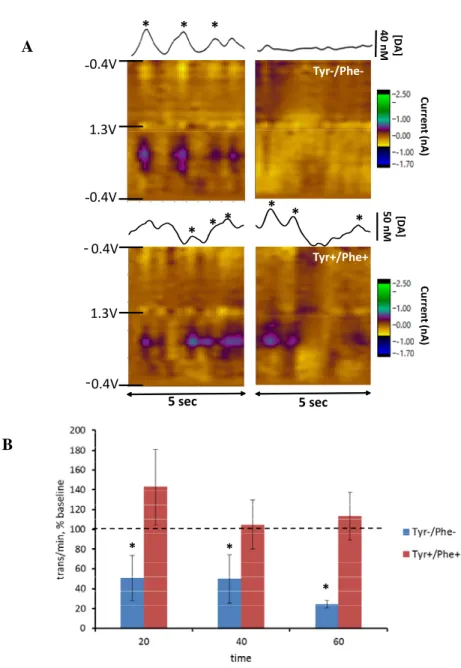
We hypothesized that the frequency of spontaneous transients would decrease after administration of the Tyr-/Phe- AA mixture during the second hour after the second injection, based on previous microdialysis data (McTavish et al. 1999). Figure 1 shows the effect of the
Tyr-/Phe- and Tyr+/Phe+ AA mixtures on the frequency of dopamine transients in the NAc. In
Figure 1A, representation of voltammetric signals obtained from a single rat are shown.
Corresponding color plots indicate changes in current at each applied potential and show the characteristic oxidative and reductive current for dopamine. Concentration versus time traces demonstrate changes in dopamine concentration across time. Administration of the Tyr-/Phe- AA mixture decreased the number of transients indicated in the color plot and the concentration vs time traces during a 5-second interval compared with baseline. In contrast, administration of the Tyr+/Phe+ mixture to a separate rat did not decrease the number of transients during same time interval. Figure 1B shows average dopamine transients per minute at baseline (20 min) and 20
group, the number of transients was 1.85 ± 0.35 at baseline and 2.04 ± 0.78 during the last 20 min after the second injection. A two-way RM ANOVA revealed non-significant main effects of time and group (p=0.1) but a significant time by group interaction (F3,30=4.1, p<0.05). Post-hoc analysis revealed a significant decrease in transient frequency from baseline in the Tyr-/Phe- group during all three intervals in the second hour after the second injection (p<0.05).
Effect of a tyrosine- and phenylalanine-free amino acid mixture on the concentration of
dopamine transients in the nucleus accumbens
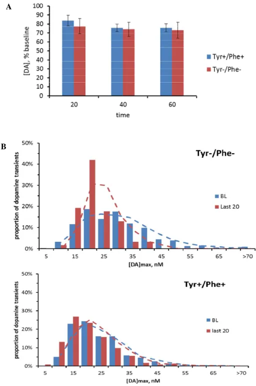
Figure 2 shows the [DA]max of transients in the NAc before and after either Tyr-/Phe-
or Tyr+/Phe+ AA injection. Figure 2A shows average data for both the Tyr-/Phe- and
Tyr+/Phe+ groups. Initially, the peak [DA]max was estimated for each transient and averaged over baseline (20 minutes) and over 20 min intervals during the second hour after the second injection. The average [DA]max was 33.9 ± 6.3 nM at baseline for the Tyr-/Phe- group and 22.5 ± 1.9 nM during the last 20 min of the second hour after the second injection. The Tyr+/Phe+ group had an average [DA]max of 23.0 ± 2.8 nM at baseline and 17.6 ± 2.5 nM during the last 20 min during the second hour after the second injection. 2-way RM ANOVA revealed a significant decrease in concentration of dopamine between baseline and the second hour after the second injection (p<0.05) but no significant group by time interaction (p=0.2). In order to further evaluate the effect of both mixtures on dopamine concentrations of individual transients, we plotted the distribution of transients across concentrations. Figure 2B shows the
mixture. Future analysis will statistically analyze the distribution curves with parametric multivariate regression analysis (Robinson et al. 2011).
In conclusion, administration of the Tyr-/Phe- AA mixture, but not the Tyr+/Phe+ AA mixture caused as significant decrease in DA transient frequency during the second hour after the second injection. Additionally, there was a shift in distribution towards lower [DA]max with the Try-/Phe- mixture but not the Tyr+/Phe+ mixture.
DISCUSSION
We found that administration of an amino acid mixture lacking tyrosine and
phenylalanine led to a decrease in the frequency and concentration of spontaneous dopamine in the nucleus accumbens of awake rats. This effect is consistent with previous experiments involving anesthetized rats (McTavish et al., 1999).
Administration of the Tyr-/Phe- mixture led to a decrease in the frequency of dopamine transients as well as a shift in distribution towards lower concentrations. Because the mixture is designed to deplete the amount of dopamine available for release, the neurons would still fire when presented with the proper stimulus, but less dopamine will be released as a result. The number of dopamine transients decreased over time, indicating that after administration of the depletion mixture, the amount of dopamine released into the synapse decreased. Some transients may have been too small to reach the correlation threshold of 0.867 used to indicate the presence of dopamine by our criteria (Robinson et al. 2003), which would have resulted in a decrease in the number of transients. Administration of a control mixture did not lead to a decrease in transient frequency over time, and resulted in no shift in distribution across concentrations, indicating that availability of tyrosine and phenylalanine for synthesis of transients prevented loss of dopamine over the time course of the experiment. The frequency data agree with microdialysis results found by McTavish and colleagues in anesthetized rats (McTavish et al., 1999). They found that administration of a depletion mixture led to lower amphetamine-evoked dopamine concentrations in the NAc during the same time interval (during the second hour after the second injection). In future studies, the ability to measure individual transient events with FSCV will allow for the use of the depletion mixture to study the role of dopamine in real-time during behavior, such as attention to a conditioned cue or consumption of a reward.
Tyr-/Phe- AA mixture results in a net decrease in transient number after the second hour after the second injection, the Tyr+/Ph+ AA mixture results in the number of transients returning to basal levels.
Though no effect on average [DA]max was observed, administration of the Tyr-/Phe- AA mixture led to a shift in distribution of transients towards lower concentrations. This shift was not seen after administration of the Tyr+/Phe+ AA mixture. The changes in transient distribution have not been yet analyzed for statistical significance. However, the shift towards lower
concentrations indicates a loss of transients with high [DA]max after administration of Tyr-/Phe- AA mixture.
This experiment demonstrated the use of a tyrosine-free amino acid mixture to deplete both concentration and frequency of naturally occurring dopamine transients in the nucleus accumbens of awake rats. Administration of a control mixture containing tyrosine and
Appendix I: Figures * * * ‐0.4V ‐0.4V 1.3V
Tyr‐/Phe‐
Curre nt (nA) [D A] 40 nM
5 sec 5 sec * * * * * * Tyr+/Phe+ [D A] 50 nM Cur ren t (nA) ‐0.4V ‐0.4V 1.3V * * * A B
Figure 1. Effect of Tyr-/Phe- and Tyr+/Phe+ AA mixtures on frequency of spontaneous dopamine transients in NAc. (A) Representation of dopamine signals in the NAc of individual rat before and after Tyr-/Phe- and Tyr+/Phe+ AA mixtures. In color plots, current is expressed in color. The time is plotted on the x axis and the potential applied to the carbon fiber electrode is plotted on the y axis.
Concentration-versus-time traces are shown above color plots with concentration converted from current after in vitro
electrode calibration. * indicates transient found using a DA template match with r > 0.866. (B)
Frequency of DA transients recorded in Tyr-/Phe- (n=6 rats) and Tyr+/Phe+ (n=6 rats) groups, presented as transients per minute in percent of baseline (indicated by the dotted line). Data are mean±SE grouped into 20-min bins for basal level (BL) and during second hour after the second injection (20, 40, 60). 2-way RM ANOVA yielded a non-significant effect of time (p=0.1) and a significant group X time interaction (F3,30=4.1, p<0.05). * indicates significant difference from baseline. Statistical analysis was
A
B
Appendix II: References
References
Beyene M, Carelli RM, Wightman RM. (2010) Cue-evoked dopamine release in the nucleus accumbens shell tracks reinforcer magnitude during intracranial self-stimulation. Neuroscience 169:1682.
Cacciapaglia F, Saddoris MP, Wightman RM, Carelli RM. (2012) Differential dopamine release dynamics in the nucleus accumbens core and shell track distinct aspects of goal-directed behavior for sucrose. Neuropharmacology
62:2050.
Day J and Carelli R. (2007) The nucleus accumbens and pavlovian reward learning. Neuroscientist 13:148.
Flagel SB, Clark JJ, Robinson TE, Mayo L, Czuj A, Willuhn I, Akers CA, Clinton SM, Phillips PEM, Akil H. (2011) A selective role for dopamine in stimulus-reward learning. Nature 469:53.
Gan JO, Walton ME, Phillips PEM. (2010) Dissociable cost and benefit encoding of future rewards by mesolimbic dopamine. Nature Neuroscience 10:1038.
Garris PA, Budygin EA, Phillips PE, Venton BJ, Robinson DL, Bergstrom BP, Rebec GV, et al. (2003) A role for presynaptic mechanisms in the actions of nomifensine and haloperidol. Neuroscience118:819–829.
Hermans A, Keithley RB, Kita JM, Sombers LA, Wightman RM. (2008) Dopamine detection with fast-scan cyclic
voltammetry used with analog background subtraction. Anal Chem80:4040-8.
Knutson B, Fong GW, Bennet SM, Adams CM, Hommer D. (2003) A region of mesial prefrontal cortex tracks monetarily rewarding outcomes: Characterization with rapid event-related fMRI. Neuroimage 18:263.
Kuhar MJ, Couceyro PR, Lambert PD. (1999) Biosynthesis of catecholamines, in Basic Neurochemistry: Molecular,
Cellular and Medical Aspects (Siegal GJ, Agranoff BW and Albers RW eds)Lippincott-Raven, Philadelphia.
McTavish SFB, Cowen PJ, Sharp T. (1999) Effect of a tyrosine-free amino acid mixture on regional brain catecholamine synthesis and release. Psychopharmocology 141:182.
Robinson DL, Hermans A, Seipel AT, Wightman RM. (2008) Monitoring rapid chemical communication in the brain. Chem Rev 108:2554.
Robinson DL, Howard EC, McConnell S, Gonzales RA, Wightman, RM. (2009) Disparity between tonic and phasic ethanol-induced dopamine increase in the nucleus accumbens of rats. Alcohol Clin Exp Res33:1187-1196.
Robinson DL, Venton BJ, Heien ML, Wightman RM. (2003) Detecting subsecond dopamine release with fast-scan cyclic voltammetry in vivo. Clin Chem49:1763–1773.
Robinson DL, Zitzman DL, Smith KJ, Spear LP. (2011) Fast dopamine release events in the nucleus accumbens of early adolescent rats. Neuroscience 176:296-307.
Schultz W, Dayan P, Montague PR. (1997) A neural substrate of prediction and reward. Science 275:1593.