Manipulation of Corticotropin-Releasing Factor Signaling Using DREADDs Reduces Binge-Like Ethanol Consumption of CRF-Cre Transgenic Mice
By Suzahn Ebert
Senior Honors Thesis Department of Biology
University of North Carolina at Chapel Hill March 24, 2016
This thesis has been prepared in conjunction with BIOL692H and in partial satisfaction of the requirements for graduation from UNC-CH with research honors.
Abstract
Binge-like ethanol consumption is characterized by the rapid achievement of blood ethanol concentrations (BEC) of ≥80 mg/dl and is associated with an increased risk of ethanol dependence.
Evidence of this association is based on an overlap between neurobiological mechanisms active during ethanol dependence and those activated during excessive ethanol consumption prior to dependence. One such contributor is the corticotropin-releasing factor (CRF) system. Our laboratory has shown that
antagonism of CRF receptors within the central amygdala (CeA) reduces binge-like ethanol consumption. The current study was designed to extend previous research by measuring ethanol consumption following the manipulation of CRF signaling in the CeA using Cre-induced inhibitory designer receptors
exclusively activated by designer drugs (DREADDs) technology. Additionally, we tested whether manipulation of CRF signaling in the CeA modulates general rewarding behavior or specifically reduces binge-like ethanol consumption. Ethanol or sucrose, consumption of which is indicative of general rewarding behavior, was administered using a 4-day procedure that mimics binge-like drinking. On the first two test days, mice received drug injections to activate the DREADDs, thereby inhibiting CRF neurons within the CeA. Statistical analysis revealed that Gi-DREADD activation in the CeA reduced ethanol consumption but had no significant effect on sucrose consumption. These data suggest that the reduced ethanol consumption during the first two cycles was not merely a general rewarding behavior but was instead specific to ethanol consumption. Moreover, they suggest that inhibition of the CRF signaling pathway in the CeA remains a viable target for manipulating binge-like ethanol consumption.
Introduction
voluntary consumption (McBride, 1998). However, rodents do not voluntarily consume enough ethanol under standard conditions to produce pharmacologically relevant BECs or to generate behavioral intoxication (Spanagel, 2000), both of which characterize human binge drinking. Rodent models have since been developed to more accurately model human binge drinking. One such model is the Drinking in the Dark (DID) paradigm which regularly produces BECs greater than 80 mg/dl and evidence of
behavioral intoxication (Rhodes et al., 2005). Thus, the DID model simultaneously serves as a rodent correlate of human binge drinking as well as a useful tool for studying the neurobiological mechanisms that underlie binge-like ethanol consumption in mice (Sprow and Thiele, 2012).
the CRF system, but also that antagonism of CRF receptors within the CeA reduces binge-like ethanol consumption (Lowery et al., 2010).
The current study extends previous research by measuring ethanol consumption
following the manipulation of CRF signaling in the CeA using Cre-induced Designer Receptors Exclusively Activated by Designer Drugs (DREADDs) technology. DREADDs are G-protein coupled receptors which are activated by small, drug-like molecules. They are particularly useful as a means of investigating and manipulating discrete populations of neurons in the brain, as they have reduced off-target effects and heightened functional selectivity compared to other
psychopharmological methods, including agonist and antagonist ligands (Fortress et al., 2015; Lee and Roth, 2014). For this study, the Gi-DREADD, which induces neuronal silencing, was used to selectively inhibit CRF signaling in the CeA.
Materials and Methods
Animals
CRF-Cre transgenic mice, backcrossed with C57s, were bred in house and were 10-18 weeks
of age at the onset of the study. These mice expressed the exogenous Cre-recombinase only in cells
that expressed endogenous CRF (Martin et al., 2010). All mice were housed in individual plastic
cages with constant access to Purina RMH 3000 chow. Water was available at all times except when
the mice were given access to alcohol or sucrose. The animal colony room was kept at a temperature
of approximately 22⁰C and operated on a 12 hour light: 12 hour dark cycle (lights on at 08:00 hours).
All procedures were performed in accordance with the National Institute of Health Guidelines for the
Care and Use of Laboratory Animals and were approved by the University of North Carolina
Institutional Animal Care and Use Committee (National Research Council, 2011).
The study used surgically delivered Cre-inducible adeno-associated viruses to encode for
designer receptors exclusively activated by designer drugs (DREADDs) as a means of investigating
specific neurological changes associated with binge-like alcohol consumption (Fortress et al., 2015).
The DREADDs were incorporated into the viral genome, so the neural cells of interest were exposed
to the virus and subsequently expressed the designer receptor. Prior to surgery, mice received an
anesthetic dose of a ketamine cocktail as well as a topical anesthetic. Stereotactic surgery was used to
bilaterally infuse either the inhibitory Gi-DREADD (AAV8-hSyn-DIO-hM4D-mCherry) or a control
virus with a fluorescent tag (AAV8-hSyn-DIO-mCherry) at the following coordinates in the central
amygdala: AP: -1.05 ML:+/-2.5 DV:-4.64. A Hamilton syringe was used to inject a total of 0.3 µl
bilaterally at a rate of 0.1 µl per minute. After each injection, the virus was allowed to diffuse for ten
minutes.
Drinking-in-the-Dark Procedure
Ethanol was administered using the DID procedure, a well-established 4-day binge-like
drinking model (Rhodes et al., 2005, 2007; Sparta et al., 2008; Lowery et al., 2010). Animals were
given access to a 20% (v/v) ethanol solution three hours into the dark cycle on each of the 4 days.
Tap water and 95% ethyl alcohol were used to prepare the 20% (v/v) ethanol solution. The ethanol
the ethanol was available for 4 hours. Animals underwent 3 consecutive weeks of ethanol testing
followed by an open field test and one week of sucrose testing.Sucrose was administered using the
DID procedure and served as a control to examine the specificity of the inhibition of CRF neurons to
ethanol consumption. Immediately preceding the DID procedures in week 1 and 2 of the ethanol test
and the sucrose test, mice were injected intraperitoneally with 0.1 mg/kg clozapine-N-oxide (CNO)
to induce Gi activation which in turn induced neuronal silencing in CRF neurons of the CeA
expressing the DREADDs. However, immediately preceding week 3 of ethanol testing, mice were
injected with saline instead of CNO to measure ethanol consumption in the absence of manipulation
of CRF signaling.
Approximately 60 µl of tail blood was collected from each animal immediately following the
end of the binge drinking period on day 4 of each test week. The samples were used to determine
individual BECs using an Analox Analyzer (Analox Instruments).
Open Field Testing
Mice used in the DID tests were split into two groups, receiving either intraperitoneal CNO
or saline, and subsequently subjected to field locomotor activity tests. The purpose of the
open-field tests is to measure anxiety-like behavior, as avoidance of the central area of the chamber reflects
a heightened state of anxiety (Choleris et al., 2001; Fee et al., 2004). The open field tests were
conducted identical to previous reports using locomotor activity chambers made of clear Plexiglass
and measuring 40.64 x 40.64 x 30.48 cm (Sparta et al., 2008). Testing sessions were 4 hours long,
with measurements taken every 5 minutes. Total distance traveled (cm) was measured to assess the
effect of CNO induced Gi activation in the CeA on locomotor activity, and center distance (cm) and
time were used to examine the effects of silencing CeA CRF neurons on anxiety.
Placement Checks
At the conclusion of the study, mice were euthanized with a sodium pentobarbital overdose
The brains were extracted, sliced in 40 µm sections using a vibrating microtome (Leica VT1000S;
Wetzlar, Germany), and mounted. The placement of the virus was visualized and confirmed to be in
the CeA using an Olympus BX-51 fluorescent microscope set to a 100x magnification.
Data Analysis
Two tailed t-tests were performed to determine if Gi-DREADD activation had a significant
effect on ethanol consumption. Two-way ANOVAs were used to determine if CNO induced Gi
activation in the CeA had an effect on locomotor activity and to examine the effects of silencing CeA
CRF neurons on anxiety.
Results
DID Trials
During the first two weeks of ethanol testing, mice received CNO injections, inhibiting CRF
neurons within the CeA. T-tests were used to determine if Gi-DREADD activation had a significant
effect on ethanol consumption. Gi-DREADD activation in the CeA resulted in reduced ethanol
consumption ([Test 1: Gi = 2.875 ± 0.3775 g/kg ethanol; control = 5.036 ± 0.3765 g/kg ethanol;
p=0.0006]; [Test 2: Gi = 3.384 ± 0.2911 g/kg ethanol; control = 5.009 ± 0.4624 g/kg ethanol;
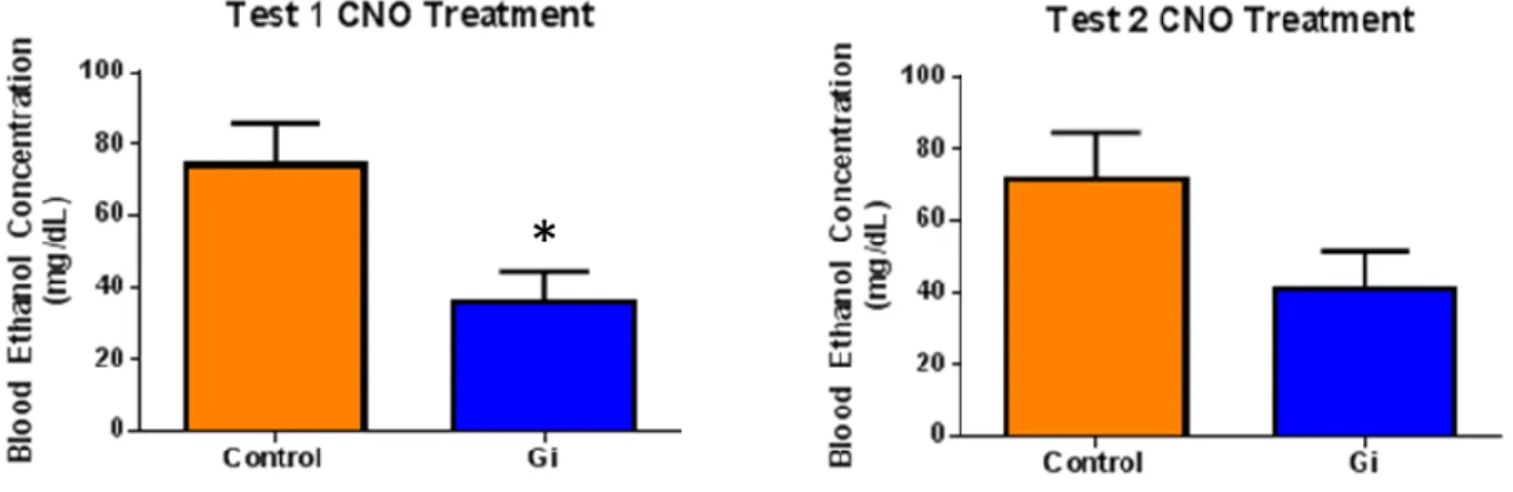
p=0.0075]). Accordingly, there was also a significant reduction in BECs in test 1; however, there was
no significant reduction in BECs in test 2 ([Test 1: Gi = 35.87 ± 8.71 mg/dl; control = 73.92 ± 11.68
Figure 1. Two tailed t-tests indicated a significant reduction in ethanol consumption upon Gi activation in the CeA for test 1 (p = 0.0006; n=11) and test 2 (p=0.0075; n=11).
Figure 2. Two tailed t-tests indicated a significant reduction in BECs upon Gi activation in the CeA for test 1 (p=0.0167; n=11), but no significant reduction for test 2 (p=0.0787; n=11).
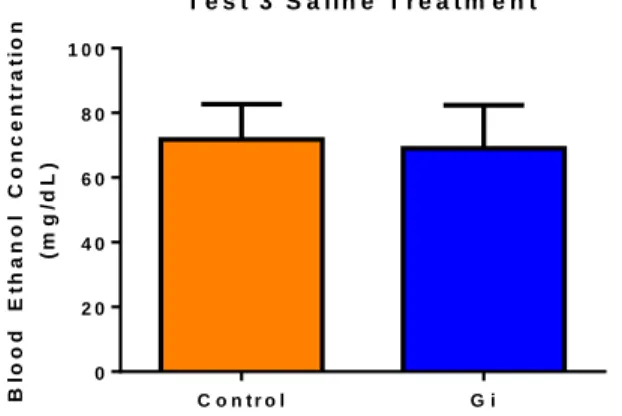
To determine if CNO had an effect independent of the virus, mice received a saline injection
prior to the third round of ethanol testing. No significant difference in ethanol consumption was
observed between the experimental and control group under these conditions (Test 3: Gi = 4.250 ±
0.2980 g/kg ethanol; control = 4.541 ± 0.5232 g/kg ethanol; p= 0.6339). Additionally, there was no
significant difference in BECs under these conditions (Test 3: Gi = 69.07 ± 13.27 mg/dl; control =
71.83 ± 10.88 mg/dl; p=0.8743).
T e s t 3 S a lin e T r e a t m e n t
C o n t r o l G i
0 2 4 6
E
th
a
n
o
l
C
o
n
s
u
m
p
ti
o
n
(
g
/k
g
Figure 3. A two tailed t-test indicated no significant difference in ethanol consumption in mice treated with saline (p= 0.6339; n=10).
T e s t 3 S a lin e T r e a t m e n t
C o n t r o l G i
0 2 0 4 0 6 0 8 0 1 0 0
B lo o d E th a n o l C o n c e n tr a ti o n (m g /d L )
Figure 4. A two tailed t-test indicated no significant difference in BECs in mice treated with saline (p=0.8743; n=10).
In the final round of testing, mice were given access to sucrose solution in the place of
ethanol to test the specificity of Gi DREADD manipulations in the CeA and to determine if
manipulating CRF signaling had an effect on general rewarding tendencies. A t-test showed no
significant effect of Gi DREADD activation on sucrose consumption (Test 4: Gi = 8.997 ± 1.503
g/kg sucrose; control = 8.333 ± 1.225 g/kg sucrose; p= 0.7436).
T e s t 4 C N O T r e a t m e n t
C o n t r o l G i
0 4 8 1 2 S u c r o s e C o n s u m p ti o n ( g /k g )
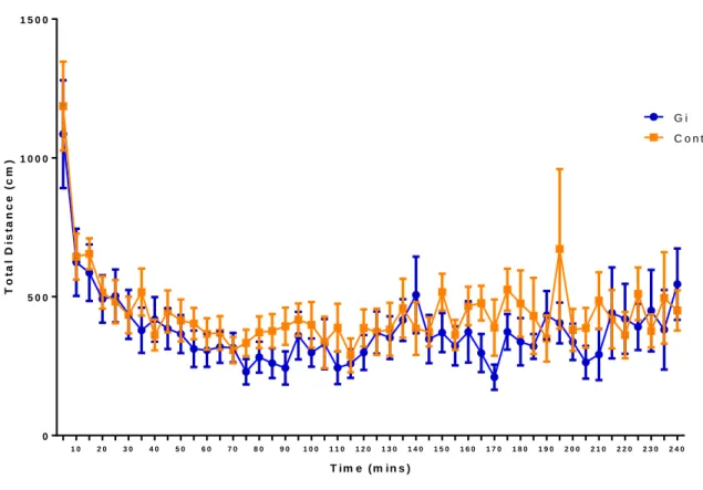
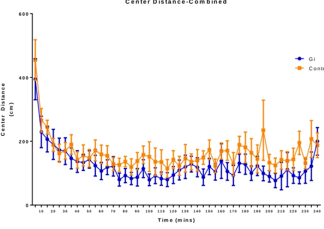
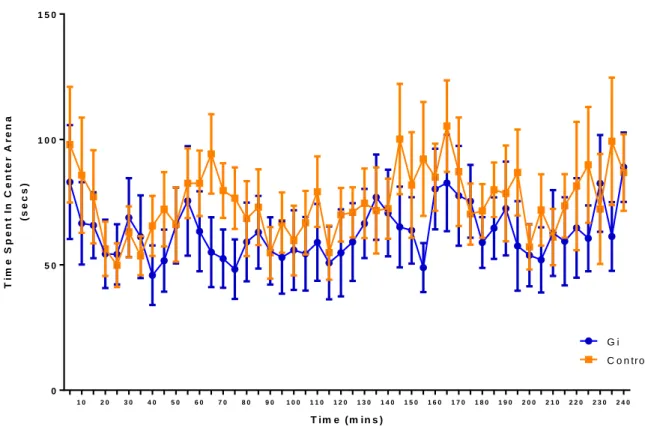
Open Field Tests
Mice received either an intraperitoneal CNO or saline injection and were subjected to
open-field locomotor activity tests. Total distance traveled was measured to assess the effect of CNO
induced Gi activation in the CeA on locomotor activity, and center distance and time were used to
examine the effects of silencing CeA CRF neurons on anxiety. For center distance and total distance,
a main effect of time was observed [(Center distance: F=6.36; p<0.0001); (Total distance: F=5.80;
p<0.0001)], but there was no main effect of Gi activation in the CeA [(Center distance: F=2.31;
p=0.1451); (Total distance: F=0.79; p=0.3859)]. For center time, no main effect of Gi activation
(F=1.35; p=0.2599) or of time was observed (F=1.01; p=0.4478).
1 0 2 0 3 0 4 0 5 0 6 0 7 0 8 0 9 0 1 0 0 1 1 0 1 2 0 1 3 0 1 4 0 1 5 0 1 6 0 1 7 0 1 8 0 1 9 0 2 0 0 2 1 0 2 2 0 2 3 0 2 4 0 0
5 0 0 1 0 0 0 1 5 0 0
T o t a l D is t a n c e - C o m b in e d
T im e ( m in s )
T
o
ta
l
D
is
ta
n
c
e
(
c
m
)
C o n tr o l G i
1 0 2 0 3 0 4 0 5 0 6 0 7 0 8 0 9 0 1 0 0 1 1 0 1 2 0 1 3 0 1 4 0 1 5 0 1 6 0 1 7 0 1 8 0 1 9 0 2 0 0 2 1 0 2 2 0 2 3 0 2 4 0 0
2 0 0 4 0 0 6 0 0
C e n t e r D is t a n c e - C o m b in e d
T im e ( m in s )
C
e
n
te
r
D
is
ta
n
c
e
(c
m
)
C o n tr o l G i
1 0 2 0 3 0 4 0 5 0 6 0 7 0 8 0 9 0 1 0 0 1 1 0 1 2 0 1 3 0 1 4 0 1 5 0 1 6 0 1 7 0 1 8 0 1 9 0 2 0 0 2 1 0 2 2 0 2 3 0 2 4 0 0
5 0 1 0 0 1 5 0
C e n t e r T i m e - C o m b in e d
T im e ( m in s )
T im e S p e n t In C e n te r A r e n a (s e c s )
C o n tr o l G i
Figure 8. No interaction or main effects were observed in center time. Discussion
The current study confirms previous findings that modulation of the CRF system in the CeA
plays a role in alcohol consumption. The results showed that CRF neuronal inhibition in the CeA
using Cre-induced DREADD technology reduced binge-like ethanol consumption. There was no
significant difference in ethanol consumption in mice treated with saline, indicating that CNO did not
have an effect independent of the virus. Additionally, there was no significant effect of Gi DREADD
activation on sucrose consumption, suggesting that the effects of CRF receptor antagonism in the
CeA is specific to ethanol consumption and does not affect general rewarding tendencies.
The results of the open field tests indicated that CRF receptor antagonism in the CeA had no
effect on locomotor activity or anxiety. Together, the results of the DID tests and the open field tests
ethanol consumption without decreasing activity or increasing anxiety. Thus, it remains a viable
target for manipulating binge-like ethanol consumption without other negative consequences.
The mounting evidence for the role of CRF in excessive, binge-like ethanol consumption
suggests an overlap between the neurobiological systems involved in the general mammalian stress
response and those involved in the regulation of excessive alcohol consumption and dependence. The
fundamental role of CRF receptors in the regulation of stress and alcohol dependence is
well-documented, but recent studies have shown that peptides other than CRF, including a variety of
urocortins, may also act on CRF receptors to modulate both stress and alcohol consumption
(Ryabinin et al., 2012). Among others, the aforementioned studies demonstrate that stress is a risk
factor for alcoholism. Thus, the neurobiological relationship between stress and alcohol consumption
merits further study, as it could provide new insight into why people drink, how alcohol affects their
behavior, and why some people become dependent on alcohol.
The current findings are limited to the role of CRF in the CeA, so it would be interesting to
investigate the potential of manipulating CRF signaling in other regions of the amygdala to influence
ethanol consumption. Specifically, the basal lateral amygdala (BLA) is known to have glutamatergic
projections to the CeA (Ren et al., 2010), which could allow for upstream targeting of the CRF
system. In conclusion, the current study sheds light on the role of the CRF signaling in the CeA in
ethanol consumption. On a broader scale, it implicates CRF signaling in other parts of the brain as
well as additional signaling pathways involved in the stress response in the regulation of excessive
alcohol consumption.
Acknowledgements
The author declares no conflict of interest. This work was supported by National Institutes of Health grants AA022048, AA013573, AA015148, AA021611, AA011605, & GM000678.
References
1. Bahi A. Increased anxiety, voluntary alcohol consumption and ethanol-induced place preference in mice following chronic psychosocial stress. Stress. 2013;16(4):441-451. 2. Choleris E, Thomas AW, Kavaliers M, Prato FS. A detailed ethological analysis of the
mouse open field test: effects of diazepam, chlordiazepoxide and an extremely low frequency pulsed magnetic field. Neuroscience & Biobehavioral Reviews.
2001;25(3):235-260.
3. Council NIAAA. NIAAA council approves definition of binge drinking. NIAAA Newsletter; 2004.
4. Fee JR, Sparta DR, Knapp DJ, Breese GR, Picker MJ, Thiele TE. Predictors of High Ethanol Consumption in RIIβ Knock-Out Mice: Assessment of Anxiety and Ethanol-Induced Sedation. Alcoholism, clinical and experimental research. 2004;28(10):1459-1468.
5. Fortress AM, Hamlett ED, Vazey EM, et al. Designer Receptors Enhance Memory in a Mouse Model of Down Syndrome. The Journal of Neuroscience. 2015;35(4):1343-1353. 6. Gilpin NW. Neuropeptides in Central Amygdala: Role in Anxiety- and Alcohol-Related
Behaviors. Alcohol (Fayetteville, N.Y.). 2012;46(4):329-337.
8. Lee H-M, Giguere PM, Roth BL. DREADDs: novel tools for drug discovery and development. Drug discovery today. 2014;19(4):469-473.
9. Lowery EG, Spanos M, Navarro M, Lyons AM, Hodge CW, Thiele TE. CRF-1 Antagonist and CRF-2 Agonist Decrease Binge-Like Ethanol Drinking in C57BL/6J Mice Independent of the HPA Axis. Neuropsychopharmacology. 2010;35(6):1241-1252. 10.Martin EI, Ressler KJ, Jasnow AM, et al. A novel transgenic mouse for gene-targeting
within cells that express corticotropin-releasing factor. Biological psychiatry. 2010;67(12):1212-1216.
11.McBride WJW. Animal models of alcoholism: neurobiology of high alcohol-drinking behavior in rodents. Critical reviews in neurobiology. 1998;12(4):339-369.
12.McCarty CA, Ebel BE, Garrison MM, DiGiuseppe DL, Christakis DA, Rivara FP. Continuity of Binge and Harmful Drinking From Late Adolescence to Early Adulthood. Pediatrics. 2004;114(3):714-719.
13.National Research Council (US) Committee for the Update of the Guide for the Care and Use of Laboratory Animals. Guide for the Care and Use of Laboratory Animals. 8th edition. Washington (DC): National Academies Press (US); 2011. Available from: http://www.ncbi.nlm.nih.gov/books/NBK54050/
14.Ren W, Neugebauer V. Pain-related increase of excitatory transmission and decrease of inhibitory transmission in the central nucleus of the amygdala are mediated by mGluR1. Molecular Pain. 2010;6:93-93.
15.Rhodes JS, Best K, Belknap JK, Finn DA, Crabbe JC. Evaluation of a simple model of ethanol drinking to intoxication in C57BL/6J mice. Physiology & Behavior.
16.Rhodes JS, Ford MM, Yu CH, et al. Mouse inbred strain differences in ethanol drinking to intoxication. Genes, Brain and Behavior. 2007;6(1):1-18.
17.Roberto M, Cruz MT, Gilpin NW, et al. Corticotropin Releasing Factor–Induced Amygdala Gamma-Aminobutyric Acid Release Plays a Key Role in Alcohol Dependence. Biological psychiatry. 2010;67(9):831-839.
18.Ryabinin AE, Tsoory MM, Kozicz T, et al. Urocortins: CRF’s siblings and their potential role in anxiety, depression and alcohol drinking behavior. Alcohol (Fayetteville, N.y.). 2012;46(4):349-357.
19.Spanagel, R. Recent animal models of alcoholism. Alcohol Research and Health. 2000;24(2):124-31. Retrieved from:
http://libproxy.lib.unc.edu/login?url=http://search.proquest.com/docview/222452098?acc ountid=14244
20.Sparta DR, Sparrow AM, Lowery EG, Fee JR, Knapp DJ, Thiele TE. Blockade of the Corticotropin Releasing Factor Type Receptor Attenuates Elevated Ethanol Drinking Associated With Drinking in the Dark Procedures. Alcoholism, clinical and experimental research. 2008;32(2):259-265.