STRUCTURAL ABNORMALITIES WITHIN THE EPISODIC PROSPECTION AND DECISION MAKING CIRCUITRY IN CIGARETTE SMOKERS: A DTI AND
SMRI ANALYSIS
Audrey Rose Verde
A dissertation submitted to the faculty of the University of North Carolina at Chapel Hill in partial fulfillment of the requirements for the degree of Doctor of Philosophy in the Neurobiology Curriculum.
Chapel Hill 2014
c
2014
ABSTRACT
AUDREY ROSE VERDE: STRUCTURAL ABNORMALITIES WITHIN THE EPISODIC PROSPECTION AND DECISION MAKING CIRCUITRY IN
CIGARETTE SMOKERS: A DTI AND SMRI ANALYSIS. (Under the direction of Charlotte Boettiger and Martin Styner)
ACKNOWLEDGMENTS
I would first like to thank my advisors, Martin Styner and Charlotte Boettiger, who challenged, encouraged, and supported me for the past 2.75 years of my PhD. I was in a tough position switching labs and research fields a year into my PhD studies, but joining their labs was the best decision I made in graduate school. Through their mentoring I have developed into the scientist and critical thinker I am today. I am incredibly grateful for their guidance and support.
I would also like to thank my thesis committee, Drs. Kelly Giovanello, J.C. Garbutt, Hongtu Zhu, and Donita Robinson, for their advice, guidance, and honest feedback during the course of my project. I am particularly thankful for my committee chair, Dr. Kelly Giovanello, whose positivity, advice, and belief in my abilities kept me going when times were tough.
There are so many people that made this project possible. I would not have suc-ceeded in this endeavor without all of you. I would like to thank all of my lab mates in Martin’s and Charlotte’s labs. Particularly, Aditya Gupta and Francois Budin, my success in the lab is largely due to the time you spent with me over the years teaching me the ways of scripting, how our tools work, and how to fix them when they break. Thank you Amanda Lyall for always keeping me in check anytime I was too stressed out, and for teaching me how to relax. A provider of unwavering support, a big thank you to Ipek Oguz for your friendship, guidance, and words of encouragement. Chris Smith, you are a beam of positivity and I am so grateful to have been in Charlotte’s lab with you. Your upbeat attitude always centered me and reminded me that there are big deals and there are little deals; most things are little deals. Thank you to Mihye Ahn for your help and support with FADTTS and statistics on this project. And a HUGE thank you to my two undergraduate students, Nicole Seider and Kirsten Con-sing, without whom I would have been far less productive. Thank you both for your hard work and partnership.
I came to UNC for the incredibly supportive environment of the UNC MD−PhD program. The program developed and led by Dr. Orringer, Dr. Rathmell, Dr. Desh-muhk, Alison Regan, and Carol Herion, is truly one where all of our students are encouraged to reach their full potential as a scientist, as a doctor, and as an individual. Their support, and the amazing students in our program have made the past 6 years an incredibly fun journey. A special place in my heart is held for my MD−PhD classmates; going through this experience with you all is an honor. You all have taught me how to enjoy life and knowing you all has made me a better person.
My friends have kept me going over the years, and they always make sure I remember to laugh and enjoy life. I would particularly like to thank Kate Hacker, Chris O’Conor, Melody Chou, Nick Taylor, Rachel Blasiak, Ryan Gessner, Kari Hacker, Justin Brown, Melissa Gough, Sal Gough, my friends from undergrad, and everyone else who I don’t have enough space to list. Also, a special thanks to one of my undergraduate research mentors and close friend, Dr. Darnell Graham. Thank you for always challenging me and supporting me in my goals.
I would not be who I am today without the support of my parents, Rose-Marie and Jeffrey Nelson, and my family. While we had meager means, my parents instilled in me that I could achieve anything I put my mind to with hard work and determination. I am infinitely grateful to them for the support they have given me throughout life, for all of my different goals and passions.
Lastly I would like to thank my wonderful husband, Barrett Green. It has been so hard living apart during my PhD and I so look forward to when you can come home from the Navy. I truly could not have done this without your incredible love, support, and belief in me. Thank you, and I love you very much.
TABLE OF CONTENTS
Table of Contents . . . ix
List of Figures . . . xiii
List of Tables . . . xv
List of Abbreviations . . . xvi
1 Introduction . . . 1
1.1 Cigarettes − a global epidemic . . . 1
1.2 Diagnosis and treatment of cigarette dependence . . . 3
1.3 Neurobiology of cigarette dependence . . . 6
1.4 Nicotine’s effect − greater than just dopamine release . . . 10
1.5 There is more to cigarettes than nicotine . . . 11
1.6 Smoking and Neuroimaging . . . 11
1.7 Measurements of smoking consumption and addiction severity . . . 12
1.8 Measuring behavioral impulsivity −the delay discounting task . . . 13
1.9 The circuitry of episodic memory . . . 14
1.10 Imagining future events reduces delay discounting − Implications for cigarette dependence . . . 15
1.11 Prior neuroimaging findings in smokers within the circuitry implicated in episodic prospection and decision−making . . . 16
1.11.2 Structural MRI (sMRI) . . . 20
2 UNC−Utah NA−MIC Framework for DTI Fiber Tract Analysis . . 23
2.1 Foreword . . . 23
2.2 Introduction . . . 25
2.3 Material and Methods . . . 31
2.3.1 DWI and DTI Quality Control . . . 31
2.3.2 Atlas Building . . . 33
2.3.3 Tractography . . . 37
2.3.4 Property profiles and statistical analysis . . . 39
2.4 Results . . . 44
2.5 Discussion and Conclusion . . . 45
2.6 Software and Example Dataset . . . 46
3 White matter microstructure abnormalities in the fornix and cingulum of cigarette smokers: a tractography based analysis . 48 3.1 Introduction . . . 48
3.2 Methods . . . 51
3.2.1 Participant sample and demographics . . . 51
3.2.2 Measurements . . . 52
3.2.3 Magnetic resonance imaging . . . 52
3.2.4 Image analysis framework . . . 52
3.2.5 Statistical analysis . . . 56
3.2.6 Result visualizations . . . 56
3.3 Results . . . 57
3.3.1 Group differences in white matter microstructure . . . 57
3.3.2 WM and measures of cigarette consumption . . . 58
3.3.4 Summary of Findings . . . 61
3.4 Discussion . . . 62
3.4.1 Differences between smokers and nonsmokers . . . 62
3.4.2 Findings within smokers . . . 63
3.4.3 Limitations . . . 65
3.4.4 Open questions . . . 66
3.5 Conclusions . . . 66
4 Structural alterations in cigarette smokers: implications for cognitive control, episodic prospection, and decision−making . . . 68
4.1 Introduction . . . 68
4.2 Methods . . . 70
4.2.1 Participants . . . 70
4.2.2 Measures . . . 71
4.2.3 Image Acquisition . . . 71
4.2.4 Image processing . . . 71
4.2.5 Statistics . . . 72
4.3 Results . . . 74
4.3.1 Comparison of smokers to non−smokers . . . 75
4.3.2 Within smokers analysis . . . 75
4.4 Discussion . . . 77
4.4.1 Structural differences between cigarette smokers and non−smokers . . . 77
4.4.2 Grey matter thickness and volume negatively correlates with cigarette consumption and addiction severity . . . 78
4.4.3 Limitations . . . 80
5 Discussion . . . 81
5.1 Aim of this dissertation . . . 81
5.2 Brief results summary . . . 82
5.3 Circuit components with significant results in both DTI and sMRI analysis . . . 85
5.3.1 Group differences in the fornix and hippocampus . . . 85
5.3.2 How measurements of smoking consumption and severity affect the cingulum and cingulate . . . 86
5.4 Implications of findings on clinical practice . . . 87
5.5 How our findings in cigarette smokers relate to other forms of addiction . . . 88
5.6 All things considered . . . 89
5.7 Novel components of this study . . . 89
5.8 Limitations . . . 91
5.9 Remaining questions and future studies in an ideal world . . . 92
5.10 In conclusion . . . 95
LIST OF FIGURES
1.1 Distribution of nicotinic acetylcholine receptors (nAChRs) . . . 6
1.2 Distribution of nicotinic acetylcholine receptors (nAChRs) . . . 9
1.3 Nicotine promotes the release of neurotransmitters . . . 11
1.4 Measurements of diffusion used in DTI analysis . . . 16
1.5 Prior methods used for DTI analysis . . . 18
2.1 UNC−Utah NA−MIC DTI Framework . . . 27
2.2 Schematic of DTI Processing Workflow . . . 29
2.3 Artifacts encountered during DWI and DTI quality control . . . 30
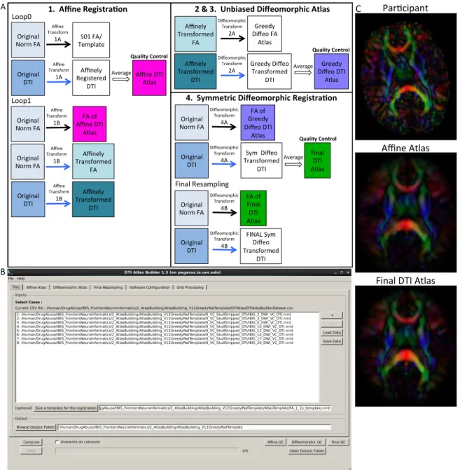
2.4 DTIAtlasBuilder steps, GUI, and registration progression . . . 34
2.5 Slicer label map tractography . . . 38
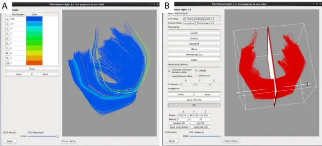
2.6 FiberViewerLight clustering, cleaning, and plane creation . . . 39
2.7 DTIAtlasFiberAnalyzer GUI − Tract parameterization and diffusion profiles . . . 41
2.8 Functional Analysis of Diffusion Tensor Tract Statistics (FADTTS) and MergeStatWithFiber Plots . . . 43
2.9 Our tools as Slicer extensions . . . 46
3.1 UNC−Utah NA−MIC framework for DTI fiber tract analysis . . . 53
3.2 Anatomy of analyzed fiber tracts . . . 54
3.3 Participant fornix fractional anisotropy (FA) profiles . . . 55
3.5 Effect of cigarettes per day on white matter integrity . . . 58 3.6 Effect of breath carbon monoxide (CO) on white matter integrity . . . 59 3.7 Effect of cigarette dependence on white matter integrity . . . 60
4.1 Cortical regions investigated that are implicated in
episodic prospection and decision−making . . . 72 4.2 Volume and thickness differences between smokers and non−smokers . 74 4.3 Volume and thickness findings within smokers and how
they correlate with measurements of cigarette consumption
LIST OF TABLES
1.1 DSM−V Tobacco Use Disorder . . . 2
3.1 DTI Participant demographics and smoking measures . . . 51
4.1 sMRI Participant demographics and smoking measures . . . 70 4.2 Significant results from cortical and subcortical volume,
LIST OF ABBREVIATIONS
ACC Anterior cingulate cortex
AD Axial diffusivity
ANTS Advanced normalization tools
AUDIT Alcohol use disorders identification test
BMI Body mass index
CC Corpus callosum
CDS Cigarette dependence scale
CO Carbon monoxide
COMT Catechol-O-methyl transferase
CSV Comma separated values
DA Dopamine
dACC Dorsal anterior cingulate cortex
DAT Dopamine transporter
dlPFC Dorsolateral prefrontal cortex
DSM Diagnostic and statistical manual
DTI Diffusion tensor imaging
DWI Diffusion weighted imaging
EM Episodic memory
FA Fractional anisotropy
FADTTS Functional analysis of diffusion tensor tract statistics
fMRI Functional magnetic resonance imaging
GABA Gamma-aminobutyric acid
GM Gray matter
GUI Graphical user interface
IBIS Infant brain imaging study
IDWI Isotropic diffusion weighted image
IFG Inferior frontal gyrus
LDTn Laterodorsal tegmental nucleus
MD Mean diffusivity
ml Milliliters
mm Millimeters
MNI Montreal neurological institute
mPFC Medial prefrontal cortex
MRI Magnetic resonance imaging
NA-MIC National alliance for medical image computing
NAc Nucleus accumbens
nAChR Nicotinic acetylcholine receptor
nBM Nucleus basalis of meynert
NITRC The neuroimaging informatics tools and resources clearinghouse
NRT Nicotine replacement therapy
PCC Posterior cingulate cortex
PFC Prefrontal cortex
PHC Parahippocampal cortex
PPn Pedunculopontine nucleus
QC Quality control
ROI Region of interest
sMRI Structural magnetic resonance imaging
SNP Single nucleotide polymorphism
SNR Signal to noise ratio
TBSS Tract based spatial statistics
UNC University of North Carolina at Chapel Hill
VBA Voxel based analysis
VBM Voxel based morphometry
vlPFC Ventrolateral prefrontal cortex
VTA Ventral tegmental area
CHAPTER 1 INTRODUCTION
1.1 Cigarettes − a global epidemic
This year marks the 50−year anniversary of the first US public health warning from the Surgeon General stating that cigarettes play a causative role in cancer and disease. Since then research has continued to uncover the role cigarettes play in causing disease and death. However, even after half a century of research and public health warnings, one billion people continue to smoke cigarettes worldwide [1]. Cigarettes are the number one preventable cause of death and disease, killing more than five million people worldwide each year [2]. One person dies from a tobacco related disease every six seconds, accounting for one in ten deaths worldwide [1] and one in five deaths in the US [3]. More deaths are caused by cigarettes than alcohol use, illegal drug use, HIV, motor vehicle accidents, murders, and suicides combined [4; 3; 5]. Ultimately, half of smokers will die due to a tobacco related illness [6].
to smoke cigarettes, and to help those who are currently addicted to quit.
Of those that try cigarettes once, 32% will progress to dependence. This experiment to addiction ratio for cigarettes is higher than that of heroin (23%), cocaine (17%), or alcohol (15%), indicating the habit−forming power of cigarettes [8]. Of current smokers, approximately 70% report wishing to quit and 50% report a past year quit attempt, but only 5% successfully abstain at one year out from their quit date [9; 10]. With the use of available therapies quit success can be raised to 30% [11], however, 95% of smokers will not seek help in their quit attempt. While new, more effective therapies are needed, we also need to begin to understand why currently available therapies are underused. Further understanding the neurobiology of cigarette addiction may help to shed light on both of these pressing issues.
DSM-V
Tobacco Use Disorder
maladaptive pattern of substance use leading to clinically significant impairment or distress, as manifested by 2 (or more) of the following, occurring within a 12-month period:
1. Recurrent substance use resulting in a failure to fulfill major role obligations at work, school, or home 2. Recurrent substance use in situations in which it is physically hazardous
3. Continued substance use despite having persistent or recurrent social or interpersonal problems caused or exacerbated by the effects of the substance
4. Tolerance, as defined by either of the following: a) need for markedly increased amounts of the substance to achieve intoxication or desired effect b) markedly diminished effect with continued use of the same amount of the substance
5. Withdrawal, as manifested by either of the following: a) the characteristic withdrawal syndrome for the substance; b) the same (or a closely related) substance is taken to relieve or avoid withdrawal symptoms 6. The substance is often taken in larger amounts or over a longer period than was intended
7. There is a persistent desire or unsuccessful efforts to cut down or control substance use
8. A great deal of time is spent in activities necessary to obtain the substance, use the substance, or recover from its effects
9. Important social, occupational, or recreational activities are given up or reduced because of substance use 10. The substance use is continued despite knowledge of having a persistent or recurrent physical or
psychological problem that is likely to have been caused or exacerbated by the substance 11. Craving or a strong desire or urge to use a specific substance
1.2 Diagnosis and treatment of cigarette dependence
The extremely low success rate of smoking cessation attempts may in part be due to the fact that cigarettes are both physiologically and psychologically addictive. Phys-iologic or somatic addiction refers to the development of tolerance and withdrawal symptoms. Psychological addiction refers to changes in behavior characterized by com-pulsive use of the substance, craving, addiction memory, loss of control, and continuing to use the substance despite negative consequences and the desire to quit [12]. While the physical symptoms of withdrawal will cease after a few weeks, the psychological symptoms of addiction such as craving and addiction memory can last a lifetime. It is this mental aspect of addiction that leads many to relapse.
The diagnosis of cigarette dependence is made using clinical criteria from the newest version of the Diagnosis and Statistical Manual of Mental Disorders (DSM). In the DSM−V, the diagnosis terminology was changed from a two stepped Nicotine Abuse and Nicotine Dependence, to just Tobacco Use Disorder. Further, the criteria for diag-nosis were changed to now only require two or more symptoms within the past twelve months from a list of 11 criteria as seen in Table 1.1. Notably, the new criteria list includes craving, as much research has centered on this aspect of cigarette addiction and how it relates to relapse.
the younger a smoker quits the better their health outcomes [15].
However, most smokers who try to quit prefer to attempt cessation cold turkey and do not seek out professional help [10; 9]. Unfortunately, this technique results in a one−year cessation success rate of 5%. Long−term cessation success rates can be improved through physician assistance and the use of available over the counter and prescription therapies [9]. Treatments available consist of informational counseling, cognitive behavioral therapy, nicotine replacement therapy (NRT), and pharmacological agents.
Today there is broad array of ways to start a quit attempt. Resources are available online such as smokefree.gov, and by telephone via quit lines like 1−800−Quit Now that allow patients a way to seek counseling from the safety of their own home [16]. Also, physicians can provide individual or group cognitive behavioral therapy sessions meant to create a community of support, construct a quit plan, and to outline strategies for how to deal with cravings and relapse. While both providing information and formal therapy improve the chance of successfully quitting, the most benefit comes from combining these efforts with one or more pharmacological agents for smoking cessation.
when combining multiple forms of NRT [17; 18]. Effectiveness of NRT options varies though by BMI, number of cigarettes consumed daily, nicotine metabolism, and sex [19; 20; 21; 22; 23].
After NRT, other first line pharmacological agents used for the treatment of cigarette dependence are bupropion and varenicline. Bupropion was first approved for use as an antidepressant and is thought to be a weak inhibitor of dopamine and norepinephrine reuptake, although the true therapeutic mechanism is unknown [24]. It has been shown to be as effective as NRT in aiding smoking cessation [25]. Varenicline is another first line pharmacological agents for the treatment of nicotine dependence that works as a partial agonist for the α4β2 nicotinic acetylcholine receptor [26]. As varenicline binds to the same receptor as does nicotine, varenicline is thought to reduce withdrawal symptoms, and to also reduce the euphoric effect of nicotine by blocking the receptors, preventing nicotine from binding [26]. Of the pharmacological agents available, Vareni-cline has been found to have the best cessation success rates, and treatment outcome further improves by combining it with NRT [27].
Second line pharmacological agents for smoking cessation are theα2 adrenoreceptor agonist clonidine, and the tricyclic antidepressant nortriptyline. While both medica-tions yielded cessation rates similar to NRT and bupropion respectively, the side effect profile for these two medications makes them rarely prescribed [28; 29]. Other medi-cations investigated for use in treating cigarette dependence but that have had mixed results are naltrexone, fluoxetine, mecamylamine, and monoamine oxidase inhibitors [29; 30; 31].
rewarding effects of smoking. While the vaccine has shown promising results in small trials, stage III clinical trials failed to show efficacy [32]. Further work to improve immune response to the vaccine is ongoing.
Feduccia et al. nAChRs: neuroplastic changes underlying addiction
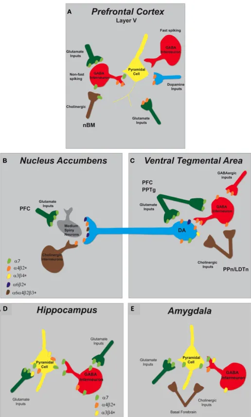
FIGURE 1 | Neuronal nicotinic acetylcholine receptors (nAChRs) are widely distributed in different brain regions that include the ventral tegmental area (VTA), nucleus accumbens (NAc), hippocampus, prefrontal cortex (PFC), and amygdala.Activation of nAChRs in these
brain areas significantly contribute to the rewarding effects of ethanol and nicotine and play a role in modulating synaptic plasticity. GABAergic (red), glutamatergic (green), and dopaminergic (blue) connections between these structures constitute a major neural circuitry underlying addictive disorders.
Much of the NMDAR-dependent plasticity has focused on mech-anisms responsible for the initial increase in synaptic strength, however the long-lasting biological effects likely require new pro-tein synthesis and gene transcription (Lynch, 2004). There are quantifiable alterations in the morphology of dendrites and den-dritic spines that accompany LTP (Andersen and Soleng, 1998; Yuste and Bonhoeffer, 2001; Matsuzaki et al., 2004) and together these long-lasting changes have been implicated as an essen-tial mechanism and molecular basis for learning and memory. Additionally, there are other modulators of plasticity, such as metabotropic glutamate receptors and endocannabinoids, that have been discovered and reviewed extensively elsewhere [see review,Kauer and Malenka(2007)].
Alcohol and tobacco addiction are among the highest causes of preventable death worldwide (Mokdad et al., 2004) and the comorbidity of these two substance abuse disorders is striking (DiFranza and Guerrera, 1990; Batel et al., 1995; Falk et al., 2006). While the easy availability and low social stigma of alcohol and cigarettes provides an explanation of their high prevalence
common link between these two substances (de Fiebre et al., 1990; Smith et al., 1999; Gould et al., 2001; Marubio et al., 2003; Tizabi et al., 2007). Neuronal nicotinic acetylcholine recep-tors (nAChRs) are widely expressed throughout the brain (Gotti et al., 2007) and are suggested to be the common biological tar-get of nicotine and ethanol (Tapper et al., 2004; Funk et al., 2006; Steensland et al., 2007; Bito-Onon et al., 2011). nAChRs are pentameric ligand-gated ion channels, consisting of various heteromeric or homomeric combinations ofα(α2–α10) andβ (β2–β4) subunits (Albuquerque et al., 2009; Gotti et al., 2009). Most neuronal nAChRs are heteromeric receptors with just two binding sites, but some subunits, such as theα7, form functional homomeric receptors with five binding sites (Changeux, 2009; Gotti et al., 2009). The most abundant of nAChR subtypes in the brain are theα4β2∗(∗indicates the possibility of other sub-units) followed by theα7; correspondingly, the mRNA of these subtypes are found throughout the entire brain. The vast regional distribution and location of nAChRs are thoroughly reported in the following reviews (Gotti and Clementi, 2004; Gotti et al.,
Figure 1.1: Neuronal nicotinic acetylcholine receptors (nAChRs) are widely distributed in different brain regions that include the ventral tegmental area (VTA), nucleus ac-cumbens (NAc), hippocampus, prefrontalcortex (PFC), and amygdala. Activation of nAChRs in these brain areas significantly contribute to the rewarding effects of nico-tine and play a role in modulating synaptic plasticity. GABAergic (red), glutamatergic (green), and dopaminergic (blue) connections between these structures constitute a major neural circuitry underlying addictive disorders. Printed with permission from c [33]
1.3 Neurobiology of cigarette dependence
is a volatile alkaloid naturally occurring in the tobacco leaf and other plants of the nightshade family. When the end of a cigarette is lit, the air within the cigarette heats up volatilizing the nicotine from the dried tobacco leaf. As the air is pulled through the cigarette towards the smokers mouth the air cools and condenses the nicotine on to particles of smoke for efficient travel to the lungs. Here nicotines small size and lipophilic nature allows the alkaloid to pass across the alveolar space to enter the blood stream and reach the brain in 15−20 seconds after being inhaled [16].
Once in the brain, the partial agonist nicotine binds acetylcholine receptor subtypes distributed throughout the brain termed nicotinic acetylcholine receptors (nAChRs) [34; 35]. nAChR are ligand gated ion channels composed of hetero or homo pentameric combinations of alpha (α2−10) and beta (β 2−4) subunits [36; 37]. The most common nAChR in the brain is the high affinityα4(3)β2(2) heteromeric pentamer, with the next most prevalent being the low affinity α7(5) homomeric receptor. Receptor subtypes and functions vary by brain region, cell type, location on the cell, type of stimulus, and time course of synaptic events (Figure 1.1). Most nAChRs are located on the soma, perisynaptically, or presynaptically and play a modulatory role in synaptic transmission [38]. When nicotine binds the nAChR alpha subunits there is a conformational change in the receptor subunits that allows for the influx of cations (Na2+ and Ca2+) into the
cell. This influx of cations causes the cell to depolarize and to fire an action potential, causing the release of neurotransmitter [39].
the euphoric and reinforcing aspect of cigarettes, as well as the formation of addiction memory [40; 41; 42; 43].
Regulating this release of DA from the VTA DA neurons are inputs from gluta-matergic, gabaergic, and cholingeric cells from other regions of the brain, and within the VTA (Figure 1.2). Within the VTA, nicotine binds the presynapticα4β2 nAChRs on VTA GABA interneurons and α7 nAChRs on VTA glutamatergic neurons, both of which take part in regulating dopamine release. The VTA DA neurons receive further excitatory glutamatergic inputs from the PFC, NAc, amygdala, bed nucleus of stria terminalis, and pontomesencephalic tegmental area [33]. While the α4β2 receptors quickly desensitize within seconds, the α6 and α7 receptors continue to exert their ef-fect on the VTA DA neurons for minutes, leading to long−term plasticity of behavior and reinforcement of the addiction [44; 45; 46].
Feduccia et al. nAChRs: neuroplastic changes underlying addiction
FIGURE 2 | Schematic representation of nAChR subtypes and circuit function in the mesolimbic dopaminergic system. (A)Pyramidal cells in layer V of the PFC lack nAChRs but their activity is modulated by excitatory and inhibitory neurons that do express them. There are two types of GABAergic interneurons, fast spiking and non-fast spiking, with only the latter bearing nAChRs (α7 andα4β2∗). Distinct populations of glutamatergic inputs express eitherα7 orα4β2∗nAChRs while DA terminals projecting from the VTA containα4β2∗nAChRs. Cholinergic inputs into the PFC arise from the nucleus basalis of Meynert (nBM).(B)In the NAc, nAChRs (α4β2∗,α6β2∗,
andα6α4β2∗) expressed on DAergic terminals from the VTA mediate DA release based on the neuronal activity firing rate. A small population of tonically active cholinergic interneurons (∼2%) is synchronized with DA cell firing. Glutamatergic inputs from the PFC endowα7 nAChRs.(C)The VTA receives cholinergic innervation from the pedunculopontine (PPn) and laterodorsal tegmental nuclei (LDTn). In addition to the nAChRs localized on DA cell bodies, DAergic cell firing is modulated byα4β2∗(and possiblyα7) nAChRs expressed on GABAergic interneurons and excitatory glutamatergic afferents from the PFC and the PPn.
2000); however, β2∗-containing receptors rapidly desensitize. Simultaneously, nicotine acts on α4β2∗ nAChRs located on GABAergic neurons to induce a transient inhibition of DA activ-ity. These nicotinic receptors will also subsequently desensitize in the presence of nicotine with a net result being a reduction in the inhibitory control of GABA on dopaminergic transmis-sion (Mansvelder and McGehee, 2002). On the same time scale, nicotine binds to α7 nAChRs present on glutamatergic termi-nals in the VTA, whose activation increases glutamate release onto NMDA-type glutamate receptors located on dopaminergic cell bodies and increases the frequency of spontaneous excitatory postsynaptic currents (sEPSCs) (Mansvelder and McGehee, 2000; Schilstrom et al., 2000; Marchi et al., 2002; Pidoplichko et al., 2004). Hence, the cumulative result of nicotine acting on nAChRs in the VTA is enhanced excitatory input onto DA neurons which triggers reward-related high frequency burst firing resulting in increased accumbal DA outflow (Corrigall et al., 1994; Nisell et al., 1994; Schilstrom et al., 2000; Pidoplichko et al., 2004).
Several labs have investigated the specific subunit composi-tions of the nAChRs that may be critical in the development of nicotine dependence using cell-based heterologous expression systems, transgenic mouse lines, and pharmacological manipu-lations using nAChR ligands in animal behavior models (Tapper et al., 2004; Steensland et al., 2007; Chatterjee and Bartlett, 2010; Cahir et al., 2011). Theα4β2∗andα6β2∗nAChR subtypes play an essential role in mediating the rewarding effects of nicotine (Dani and De Biasi, 2001; Nashmi et al., 2007), demonstrated by a lack of nicotine-elicited DA release inβ2 andα4 knockout mice and a decrease in nicotine self-administration by mice lacking theα4,
α6, orβ2 subunits compared to wild-types (Picciotto et al., 1998; Marubio et al., 2003; Pons et al., 2008). By contrast, the roleα7 nAChRs play in the reinforcing properties of nicotine is less clear. Deletion of theα7 did not affect nicotine conditioned place pref-erence (Walters et al., 2006) or self-administration (Pons et al., 2008); however, high doses of theα7 nAChR antagonist methylly-caconitine attenuated nicotine self-administration (Markou and Paterson, 2001) and reduced the rewarding effects of nicotine when infused directly into the VTA (Laviolette and van der Kooy, 2003).
The effects of ethanol in the VTA are more complex than the effects of nicotine. Ethanol, unlike nicotine, is not a direct ago-nist at nAChRs (Cardoso et al., 1999; Zuo et al., 2002) but can modulate DA release by influencing the function of nAChRs in both the VTA and NAc (Ericson et al., 1998, 2003; Larsson et al., 2005). While local perfusion of ethanol into the VTA does not increase DA release in the accumbens, infusion of ethanol into the accumbens does elevate extracellular DA to a similar degree as systemic administration (Ericson et al., 2003; Tuomainen et al., 2003). However, ethanol infused into both regions simultane-ously resulted in higher DA levels than when injected into the NAc alone (Lof et al., 2007). Furthermore, perfusion of mecamy-lamine (MEC, non-selective nAChR antagonist) into the VTA, but not into the NAc, blocks DA release stimulated by systemic administration of ethanol. It has been postulated that ethanol’s actions in the NAc may facilitate the release of endogenous ACh in the VTA, leading to activation of nAChRs and consequently elevating accumbal DA release (Larsson et al., 2005). In sup-port, voluntary ethanol intake in rats has been shown to cause
Frontiers in Molecular Neuroscience www.frontiersin.org August 2012 | Volume 5 | Article 83|4
Feduccia et al. nAChRs: neuroplastic changes underlying addiction
FIGURE 3 | Schematic representation of nAChR subtypes and circuit function in the hippocampus and amygdala. (A)In the
hippocampus,α7 andα4β2∗nAChRs are abundantly expressed on pyramidal cells and inhibitory interneurons. GABAergic interneurons have pre-synaptic α7 nAChRs and somato-dendritic expression ofα7 andα4β2∗nAChRs. Glutamatergic afferents have predominately pre-synapticα7 nAChRs and only
low levels ofα3β4∗.(B)In the amygdala, cholinergic inputs from the basal forebrain synapse in proximity to pre-synaptic nAChRs that modulate both excitatory and inhibitory synaptic transmission. Glutamatergic afferents and pyramidal neurons endowα7 nAChRs and GABAergic interneurons express multiple nAChRs (α7,α4β2∗, and α3β4∗).
amygdala, and PFC (Kelley, 2004). Therefore, alterations in structure or function in the hippocampus may be translated by other brain regions that drive maladaptive behaviors associ-ated with addiction. Most studies investigating the involvement of nAChRs in synaptic plasticity have been conducted in the hippocampus.
The α7 and α4β2∗ nAChRs (see Figure 3) are abundantly expressed on GABAergic interneurons and pyramidal cells within the hippocampus and are capable of modulating intracellu-lar signaling molecules and downstream effectors that gov-ern plasticity (Jones and Yakel, 1997; Vizi and Lendvai, 1999).
GABAergic interneuron populations express α7 nAChRs on
pre-synaptic terminals, whereas somato-dendritic compartments endow bothα7 andα4β2∗ nicotinic receptors (Radcliffe et al., 1999; Alkondon and Albuquerque, 2001). Furthermore, mod-ulation of glutamate synaptic transmission to pyramidal
neu-rons in the CA1 region is attributed to predominately α7
nAChRs but also to a minimal number of α3β4∗ nAChRs
(Gray et al., 1996; Ji et al., 2001; Alkondon and Albuquerque, 2002).
nAChR-mediated synaptic plasticity. Nicotinic receptors exert a temporally- and spatially-dependent bidirectional control over synaptic plasticity, bothin vitroandin vivo(Table 2). For exam-ple, in the CA1 region of hippocampal slices ACh and nico-tine can act on post-synaptic receptors of pyramidal neurons to increase intracellular Ca2+which facilitates the conversion of short-term potentiation to LTP by reducing the threshold needed for induction (Fujii et al., 1999; Ji and Dani, 2000; Nakauchi et al., 2007) or by attenuating the inhibitory input of interneu-rons to pyramidal cells (Ji and Dani, 2000; Yamazaki et al., 2005); these effects are mediated by both the activation of non-α7
Furthermore, blunting the evoked release of inhibitory GABA onto pyramidal cells to facilitate nicotine-induced LTP induction was shown to rely on desensitization of non-α7 nAChRs (Fujii et al., 2000b; Yamazaki et al., 2005; Nakauchi et al., 2007). Additionally, activation of nAChRs on hippocampal interneu-rons can induce LTP or LTD depending on the exact timing of agonist application in respect to the pre-synaptic stimula-tion (Ji et al., 2001; Ge and Dani, 2005). Furthermore, acti-vation of α7 nAChRs on pre-synaptic glutamatergic terminals can increase the frequency of miniature EPSCs and enhance glutamate release onto pyramidal neurons offering yet another mechanism for the modulation of plasticity (Gray et al., 1996; Radcliffe and Dani, 1998). In the CA3 region of hippocam-pal slices, bath application of nicotine can drive the pyrami-dal cells above threshold in the absence of an action poten-tial by activating pre-synaptic nAChRs located on glutamater-gic terminals. Activation of these receptors enhances miniature EPSCs and glutamate release through mobilization of intra-cellular calcium stores by CICR (Sharma and Vijayaraghavan, 2003).
Within the dentate gyrus, induction of LTP by nicotine required activation of mGLuR5 and L-type Ca2+ channels, as
well as Ca2+release from ryanodine-sensitive stores and wasα7
nAChR-dependent (Welsby et al., 2006, 2009). in vivostudies in mice showed nicotine or epibatidine, an α4β2 nAChR ago-nist, dose-dependently induced synaptic plasticity in the dentate gyrus and importantly, required intact midbrain DA signaling (Matsuyama et al., 2000; Matsuyama and Matsumoto, 2003; Tang and Dani, 2009). In the developing brain, long-lasting changes in synaptic transmission were observed following a single expo-sure to nicotine in the hippocampus. nAChR signaling facilitated the conversion of pre-synaptic silent synapses into functional
D" E"
Feduccia et al. nAChRs: neuroplastic changes underlying addiction
FIGURE 2 | Schematic representation of nAChR subtypes and circuit function in the mesolimbic dopaminergic system. (A)Pyramidal cells in layer V of the PFC lack nAChRs but their activity is modulated by excitatory and inhibitory neurons that do express them. There are two types of GABAergic interneurons, fast spiking and non-fast spiking, with only the latter bearing nAChRs (α7 andα4β2∗). Distinct populations of glutamatergic inputs express eitherα7 orα4β2∗nAChRs while DA terminals projecting from the VTA containα4β2∗nAChRs. Cholinergic inputs into the PFC arise from the nucleus basalis of Meynert (nBM).(B)In the NAc, nAChRs (α4β2∗,α6β2∗,
andα6α4β2∗) expressed on DAergic terminals from the VTA mediate DA release based on the neuronal activity firing rate. A small population of tonically active cholinergic interneurons (∼2%) is synchronized with DA cell firing. Glutamatergic inputs from the PFC endowα7 nAChRs.(C)The VTA receives cholinergic innervation from the pedunculopontine (PPn) and laterodorsal tegmental nuclei (LDTn). In addition to the nAChRs localized on DA cell bodies, DAergic cell firing is modulated byα4β2∗(and possiblyα7) nAChRs expressed on GABAergic interneurons and excitatory glutamatergic afferents from the PFC and the PPn.
2000); however, β2∗-containing receptors rapidly desensitize. Simultaneously, nicotine acts on α4β2∗ nAChRs located on GABAergic neurons to induce a transient inhibition of DA activ-ity. These nicotinic receptors will also subsequently desensitize in the presence of nicotine with a net result being a reduction in the inhibitory control of GABA on dopaminergic transmis-sion (Mansvelder and McGehee, 2002). On the same time scale, nicotine binds toα7 nAChRs present on glutamatergic termi-nals in the VTA, whose activation increases glutamate release onto NMDA-type glutamate receptors located on dopaminergic cell bodies and increases the frequency of spontaneous excitatory postsynaptic currents (sEPSCs) (Mansvelder and McGehee, 2000; Schilstrom et al., 2000; Marchi et al., 2002; Pidoplichko et al., 2004). Hence, the cumulative result of nicotine acting on nAChRs in the VTA is enhanced excitatory input onto DA neurons which triggers reward-related high frequency burst firing resulting in increased accumbal DA outflow (Corrigall et al., 1994; Nisell et al., 1994; Schilstrom et al., 2000; Pidoplichko et al., 2004).
Several labs have investigated the specific subunit composi-tions of the nAChRs that may be critical in the development of nicotine dependence using cell-based heterologous expression systems, transgenic mouse lines, and pharmacological manipu-lations using nAChR ligands in animal behavior models (Tapper et al., 2004; Steensland et al., 2007; Chatterjee and Bartlett, 2010; Cahir et al., 2011). Theα4β2∗andα6β2∗nAChR subtypes play an essential role in mediating the rewarding effects of nicotine (Dani and De Biasi, 2001; Nashmi et al., 2007), demonstrated by a lack of nicotine-elicited DA release inβ2 andα4 knockout mice and a decrease in nicotine self-administration by mice lacking theα4,
α6, orβ2 subunits compared to wild-types (Picciotto et al., 1998; Marubio et al., 2003; Pons et al., 2008). By contrast, the roleα7 nAChRs play in the reinforcing properties of nicotine is less clear. Deletion of theα7 did not affect nicotine conditioned place pref-erence (Walters et al., 2006) or self-administration (Pons et al., 2008); however, high doses of theα7 nAChR antagonist methylly-caconitine attenuated nicotine self-administration (Markou and Paterson, 2001) and reduced the rewarding effects of nicotine when infused directly into the VTA (Laviolette and van der Kooy, 2003).
The effects of ethanol in the VTA are more complex than the effects of nicotine. Ethanol, unlike nicotine, is not a direct ago-nist at nAChRs (Cardoso et al., 1999; Zuo et al., 2002) but can modulate DA release by influencing the function of nAChRs in both the VTA and NAc (Ericson et al., 1998, 2003; Larsson et al., 2005). While local perfusion of ethanol into the VTA does not increase DA release in the accumbens, infusion of ethanol into the accumbens does elevate extracellular DA to a similar degree as systemic administration (Ericson et al., 2003; Tuomainen et al., 2003). However, ethanol infused into both regions simultane-ously resulted in higher DA levels than when injected into the NAc alone (Lof et al., 2007). Furthermore, perfusion of mecamy-lamine (MEC, non-selective nAChR antagonist) into the VTA, but not into the NAc, blocks DA release stimulated by systemic administration of ethanol. It has been postulated that ethanol’s actions in the NAc may facilitate the release of endogenous ACh in the VTA, leading to activation of nAChRs and consequently elevating accumbal DA release (Larsson et al., 2005). In sup-port, voluntary ethanol intake in rats has been shown to cause
Frontiers in Molecular Neuroscience www.frontiersin.org August 2012 | Volume 5 | Article 83|4
Figure 1.2: Schematic representation of nAChR subtypes for areas important for cigarette dependence. Caption on following page.
1.4 Nicotine’s effect − greater than just dopamine release
The presence of nicotine in the brain and body does more than just release DA. The influx of nicotine with each cigarette causes an increase in signaling for multiple other neurotransmitter systems including acetylcholine, glutamate, γ−aminobutyric acid (GABA), serotonin, norepinephrine, and beta−endorphin (Figure 1.3) [49; 50]. The upregulation of all of these other neurotransmitter systems, along with that of DA combine to make the rewarding and reinforcing properties of cigarette addiction. Fur-ther, it is this upregulation and alteration in receptor expression across neurotransmit-ter systems that creates the physiological and psychological symptoms of withdrawal. These symptoms include agitation, irritability, anxiety, depression, decreased cognition, headaches, hunger, and cigarette craving.
Figure 1.2. Schematic representation of nAChR subtypes for areas important for cigarette dependence A. Pyramidal cell activity in layer V of the PFC is modulated by excitatory and inhibitory neurons expressing nAChRs. Non−fast spiking GABAer-gic interneurons bear nAChRs (α7 and α4β2). Distinct populations of glutamatergic inputs express either α7 or α4β2 nAChRs while DA terminals projecting from the VTA contain α4β2 nAChRs. Cholinergic inputs into the PFC arise from the nucleus basalis of Meynert (nBM). B. In the NAc, nAChRs (α4β2, α6β2, and α6α4β2) ex-pressed on DAergic terminals from the VTA mediate DA release based on the neuronal activity firing rate. A small population of tonically active cholinergic interneurons (2%) is synchronized with DA cell firing. Glutamatergic inputs from the PFC endow
α7 nAChRs. C. The VTA receives cholinergic innervation from the pedunculopon-tine (PPn) and laterodorsal tegmental nuclei (LDTn). In addition to the nAChRs localized on DA cell bodies, DAergic cell firing is modulated by α4β2 (and possibly
α7) nAChRs expressed on GABAergic interneurons and excitatory glutamatergic af-ferents from the PFC and the PPn. D. In the hippocampus, α7 and α4β2 nAChRs are abundantly expressed on pyramidal cells and inhibitory interneurons. GABAer-gic interneurons have pre−synaptic α7 nAChRs and somato−dendritic expression of
inant in the human brain and is believed to be the main receptor mediating nicotine dependence. In mice, knocking
out the!2subunit gene eliminates the behavioral effects of
nicotine, including self-administration.23Reinserting the!
2
subunit gene into the ventral tegmental area of a!2
knock-out mouse restores behavioral responses to nicotine.24The
"4subunit appears to be an important determinant of
sen-sitivity to nicotine. In mice, a single nucleotide point mu-tation in the pore-forming region results in a receptor that is
hypersensitive to the effects of nicotine.25 This mutation
made mice much more sensitive to nicotine-induced reward behaviors, as well as to effects on tolerance and
sensitiza-tion. The "3!4 and possibly the "7 homomeric receptor
subtypes are believed to mediate the cardiovascular effects
of nicotine.26The"
7subtype is also thought to be involved
in rapid synaptic transmission and may play a role in
learn-ing27and sensory gating.28
Biology of Nicotine Reinforcement
Brain imaging studies demonstrate that nicotine acutely increases activity in the prefrontal cortex, thalamus, and visual system, consistent with activation of corticobasal
ganglia-thalamic brain circuits.29 Stimulation of central
nAChRs by nicotine results in the release of a variety of neurotransmitters in the brain, most importantly dopamine. Nicotine causes the release of dopamine in the mesolimbic area, the corpus striatum, and the frontal cortex. Of partic-ular importance are the dopaminergic neurons in the ventral tegmental area of the midbrain and the release of dopamine in the shell of the nucleus accumbens, as this pathway
appears to be critical in drug-induced reward.22,30 Other
neurotransmitters, including norepinephrine, acetylcholine,
serotonin, #-aminobutyric acid (GABA), glutamate, and
endorphins, are released as well, mediating various
behav-iors of nicotine (Figure 1).5
It is believed that most of the release of neurotransmitters occurs via modulation by presynaptic nAChRs, although
direct release of neurotransmitters also occurs.31Dopamine
release is facilitated by nicotine-mediated augmentation of glutamate release and, with long-term treatment, by
inhibi-tion of GABA release. In addiinhibi-tion to direct and indirect stimulation of neurotransmitter release, chronic cigarette smoking (but not acute nicotine administration) reduces brain monoamine oxidase A and B activity, which would be expected to increase monoaminergic neurotransmitter levels such as dopamine and norepinephrine in synapses, thus augmenting the effects of nicotine and contributing to
ad-diction.29
Dopamine release signals a pleasurable experience and is critical to the reinforcing effects of nicotine and other drugs
of abuse.30Chemically or anatomically lesioning dopamine
neurons in the brain prevents nicotine self-administration in
rats.32When intracranial self-stimulation is used as a model
for brain reward in rats, nicotine acutely lowers the
thresh-old for self-stimulation.33Thus, through its effects on
do-pamine release, acute nicotine administration increases brain reward function. Likewise, nicotine withdrawal is associated with significant increases in intracranial self-stimulation reward threshold, consistent with deficient
do-pamine release and reduced reward.34This is similar to the
effects of withdrawal from other drugs of dependence. The decrease in brain reward function experienced during nico-tine withdrawal is an essential component of niconico-tine ad-diction and a key barrier to abstinence.
With repeated exposure to nicotine, there is
neuroadap-tation to some, but not all, of the effects of nicotine.35
Concurrent with this neuroadaptation is an increase of nAChRs in the brain. This increase is believed to represent upregulation in response to nicotine-mediated desensitiza-tion of receptors. This desensitizadesensitiza-tion may play a role in nicotine tolerance and dependence. It has been suggested that craving and withdrawal symptoms begin in chronic
smokers when previously desensitized "4!2 nAChRs
be-come unoccupied and recover to a responsive state during
periods of abstinence, such as during nighttime sleep.36
Thus, nicotine binding and desensitization of these recep-tors during smoking may alleviate craving and withdrawal. This is supported by clinical evidence that cigarette smok-ing in amounts used by typical daily smokers maintains near-complete saturation—and, thus, desensitization— of
Figure 1 Nicotine receptor activation promotes the release of neurotransmitters, which may then mediate various effects of nicotine use.
GABA!#-aminobutyric acid. (Adapted with permission fromPrim Care.5)
S6 The American Journal of Medicine, Vol 121 (4A), April 2008
Figure 3 |
Figure 1.3: Nicotine receptor activation promotes the release of neurotransmitters, which then mediate various effects of nicotine use. GABA = γ−aminobutyric acid.
Printed with permission from c [49]
1.5 There is more to cigarettes than nicotine
Most of what we know about the neurobiology of cigarette addiction has come from work in animal models of nicotine addiction, as nicotine is identified as the primary addictive ingredient in cigarettes [49]. However, recently the cigarette dependence research community has stepped back to acknowledge the existence of the 7000 other chemicals in cigarette smoke and that studying just the effect of nicotine on the brain and behavior is not the same as studying the effects of cigarettes on the brain and behavior. To study the effect cigarette smoke has on neurobiology, investigators are turning to the use of actual cigarette smoke in experiments or by creating cigarette smoke extract via the condensation of cigarette smoke into a liquid. This new direction of study is promising, and will help to paint a more realistic and complete understanding of cigarette dependence, and how cigarettes affect brain biology, structure, and function.
1.6 Smoking and Neuroimaging
to the large comorbidity of cigarette smoking with other human disorders (alcohol use disorders, schizophrenia, depression, etc), imaging literature looking singularly at the effects of cigarette dependence in otherwise healthy individuals is scarce. Of the exist-ing literature in just cigarette smokers, most investigations utilize functional imagexist-ing to understand the anatomical underpinnings of craving, attention to smoking cues, memory, cognition, and impulsivity. Studies looking at structural abnormalities in the brains of cigarette smokers using anatomical magnetic resonance imaging (sMRI) or diffusion tensor imaging (DTI) are few, and have been largely limited by the analytical methods employed. Thankfully, due to advancements in image acquisition and analysis techniques, the field is at an exciting time to study the structural abnormalities in the brains of cigarette smokers.
1.7 Measurements of smoking consumption and addiction severity
While clinical diagnosis of nicotine dependence is made using a structured interview and DSM criteria, in the lab it is useful to have shorter questionnaires with continuous scales in order to capture a range of addiction severity. In almost all cigarette depen-dence studies investigators use the Fagerstrom Test for Nicotine Dependepen-dence (FTND) [52]. While several other similar measures exist, even some that have been shown to perform superiorly to the FTND, the field continues to use the FTND. This may be in part due to tradition, and in part due to the fact that at this point some 30 years of research has been performed using this test where it has been found to correlate with many important aspects of cigarette addiction. One of the available tests that has been shown to perform better than the FTND on measures of construct and pre-dictive validity is the Cigarette Dependence Scale (CDS) [53; 54]. With the exception of tolerance, the CDS covers the main criteria for ICD−10 and DSM−IV definitions of dependence: withdrawal symptoms, compulsion, loss of control, time allocation, neglect of other activities, and persistence despite harm [55]. We believe the CDS provides a more accurate measurement of cigarette addiction severity and thus it is the measure that we use in this study.
1.8 Measuring behavioral impulsivity − the delay discounting task
This impulsive choice behavior holds true for those dependent on cigarettes as well [56; 57; 58]. While this task originated on paper, it has since been modified to work on a computer, as well as for use in a MRI scanner to allow for functional imaging during the task [59].
1.9 The circuitry of episodic memory
needed for episodic prospection [78; 68; 75; 71; 79; 78].
1.10 Imagining future events reduces delay discounting − Implications for cigarette dependence
Two recent studies have shown that episodic prospection (imagining future events) reduces discounting of delayed rewards in healthy control participants via enhanced connectivity between the frontal lobe and mediotemporal regions mediating episodic prospection [80; 81]. As noted above, cigarette smokers have a heightened tendency to discount delayed rewards; however the neural bases of this tendency are not fully understood. Thus, a possible neural mechanism underlying enhanced delay discounting in cigarette smokers is impaired connectivity between frontal regions and the circuitry engaged during episodic prospection. Moreover, behavioral studies have demonstrated that those with cigarette dependence commonly exhibit impaired episodic memory, sug-gesting possible specific structural deficits in the episodic circuitry [82; 83]. Together, these observations have led us to the following hypothesis:
Hypothesis: Individuals with cigarette dependence show variations in the structural properties of the episodic memory circuitry that correlate with cigarette addiction severity, particularly its connections to frontal elements of decision−making circuits.
AD = axial diffusion = λ
1RD = radial diffusion = (λ
2+ λ
3)/2
λ
1λ
2λ
3MD = mean (overall) diffusion
= = (λ
1+ λ
2+ λ
3)/3
FA = fractional anisotropy (tensor shape)
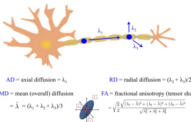
Figure 1.4: Measurements of diffusion used in DTI analysis. Diffusion parameters for DTI analysis are calculated from the first three primary eigenvalues (λ1, λ2,λ3). The top image shows how the three primary eigenvalues would describe diffusion for a single water molecule moving down a neuronal axon. The primary direction of diffusion is defined as the first eigenvalue (λ1). A combination of the second and third eigenvalues describe water diffusion orthogonal to the axon (λ2, λ3). Together, the first three eigenvalues of diffusion are used to compute the fractional anisotropy (FA), which describes the shape of the diffusion tensor (bottom center image). Adapted and printed with permission from: http: // www. clker. com/ clipart-9828. html
1.11 Prior neuroimaging findings in smokers within the circuitry impli-cated in episodic prospection and decision−making
1.11.1 Diffusion Tensor Imaging (DTI)
axon will have their diffusion limited by the dense macrostructure of many surround-ing axons in a white matter tract, as well as the microstructure of myelin surroundsurround-ing axons, and organelles and microtubules within individual axons making their diffusion described as more anisotropic (linear) in nature. Under these assumptions we can then measure to what degree water diffusion is linear, or the fractional anisotropy (FA), in re-gions of interest in the brain. Composing the measurement of FA are the measurements of axial, radial, and mean diffusivity. Axial diffusivity (AD) is the primary direction of water diffusion, or the first eigenvalue, and is also described as the diffusion of water that is parallel to the fiber bundle or axon. Radial diffusivity (RD) is the average of the second and third eigenvalues, which describe the magnitude of diffusion orthogonal to the axon or fiber bundle. Mean diffusivity (MD), is then an average of the first three eigenvalues, and describes the overall amount of diffusion. FA combines all three of these other measurements to describe the overall shape of the diffusion tensor (Figure 1.4).
+ Robust against imperfect registration
- Multiple tracts/tissues within one ROI + Good for hypothesis generation - Requires PERFECT registration
B
+ Works well with imperfect registration - Multiple tracts combined on skeleton - Max FA is a less stable measurement C
A
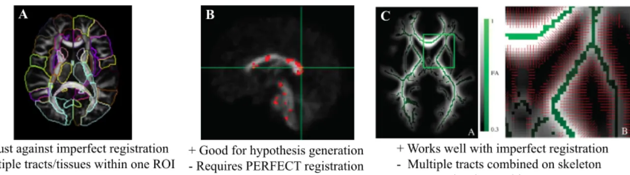
Figure 1.5: Prior methods used for DTI analysis. A. Region of interest (ROI) analysis displayed here with ROIs from the Susumu Mori atlas. DTI analysis performed with large ROIs as displayed here often average the diffusion within the entire ROI for a single measurement, combining readings from multiple white matter tracts into one region, as well as combining together diffusion in white matter and grey matter. This leads to results that are not localizable to a specific tract or a specific region on a tract.
Adapted and printed with permission from [99]. B. Whole brain voxel based analysis used to find single voxels of significance. This method assumes perfect registration on the voxel level between participants, which is often not possible. This method is a great way to generate hypotheses for future studies using a more specific method of analysis.
Prior investigations of DTI in smoking
To date, little work has investigated the relationship between cigarette smoking and white matter (WM) integrity of the brain using DTI. Of the studies that exist, most utilize voxel wise analysis, TBSS (a form of voxel wise analysis), or large nonspecific regions of interest (ROIs), and do not perform specific along the tract tractography analysis (Figure 1.5) [101; 99; 100; 102]. Thus previously reported DTI findings in smokers are not truly localizable to specific tracts or to exact locations along tracts (Figure 1.5). When comparing smokers to nonsmokers using these DTI analysis tech-niques, findings have been mixed. While most studies report smokers to have increased FA in regions of the brain compared to nonsmokers [103; 104; 105; 106; 107], oth-ers find smokoth-ers to have decreased FA [108; 109], or no difference in FA [110; 111]. Likewise there are varying conclusions as to how cigarette consumption and addiction severity relate to WM integrity in smokers. Studies have found a negative correlations [104; 110; 106; 107], positive correlations [103], or no correlation [105; 109] between measures of addiction severity and FA.
There may be several reasons for these discrepancies in findings, one of which is the use of nonspecific voxel wise or ROI−based analyses. With these analytical techniques, single average measurements of diffusion parameters (FA, MD, RD, AD) are used to represent entire fiber bundles, when the regions analyzed may consist of multiple WM tracts, or may contain both white and grey matter. In order to better understand how cigarette smoking addiction affects WM integrity, a more specific and localizable technique needs to be applied.
1.11.2 Structural MRI (sMRI)
Study of structural (anatomical) MRI images aids in determining differences in brain region volumes between smokers and non−smokers. Two forms of methods avail-able for this form of analysis are voxel based morphometry (VBM) and surface based morphometry (FreeSurfer). VBM uses a previously segmented atlas to compute tissue probability maps for each subject in order to segment the brain into white matter, grey matter, and cerebrospinal fluid (CSF). VBM does this segmentation and the follow-ing statistical tests on a voxel by voxel basis [112]. Results from VBM are typically reported as either measurements of volume or density.
In addition to segmenting cortical and subcortical brain volumes like VBM, FreeSurfer is also capable of computing cortical thickness and white matter surface area measure-ments. The publicly available FreeSurfer1 software assesses bilateral regional cortical and subcortical volumes, and regional cortical thickness [85; 86; 87; 88; 89; 90; 91; 92; 93; 94; 95; 96; 97]. Briefly, this processing includes T1 motion correction [93], removal of non−brain tissue using a hybrid watershed/surface deformation procedure [86], automated Talairach transformation, segmentation of the subcortical white matter and deep gray matter volumetric structures [87; 94], intensity normalization Sled1998, tessellation of the gray matter white matter boundary, automated topology correction [89; 90], and surface deformation following intensity gradients to optimally place the gray/white and gray/cerebrospinal fluid borders at the location where the greatest shift in intensity defines the transition to the other tissue class [85; 91; 92]. Once the corti-cal models are complete, a number of deformable procedures can be performed for in further data processing and analysis including surface inflation [95], registration to a
spherical atlas which utilized individual cortical folding patterns to match cortical ge-ometry across subjects [96], parcellation of the cerebral cortex into units based on gyral and sulcal structure [97], and creation of a variety of surface based data including maps of curvature and sulcal depth. This method uses both intensity and continuity infor-mation from the entire three dimensional MR volume in segmentation and deforinfor-mation procedures to produce representations of cortical thickness, calculated as the closest distance from the gray/white boundary to the gray/CSF boundary at each vertex on the tessellated surface [92]. The maps are created using spatial intensity gradients across tissue classes and are therefore not simply reliant on absolute signal intensity. The maps produced are not restricted to the voxel resolution of the original data thus are capable of detecting submillimeter differences between groups.
Prior investigations of sMRI in smoking
and decision−making. Thus to add to existing literature, we utilized FreeSurfer to investigate the cortical and subcortical volumes, and cortical thickness of brain regions implicated in episodic memory and decision−making (Chapter 4).
Benefits of multi−modal analysis
CHAPTER 2
UNC−UTAH NA−MIC FRAMEWORK FOR DTI FIBER TRACT ANALYSIS
1
2.1 Foreword
Dr. Styner’s lab is specialized in creating cutting edge, open source processing and analysis tools for use in the investigation of diffusion tensor images (DTI), anatomical magnetic resonance images (MRI), and MRI shape analysis. When I joined the lab in the summer of 2011 many tools for the processing and analysis of DTI were available in command line format, but were not accessible to investigators without a computer science background. In order to make these tools usable by any researcher that collects DTI data, graphical user interfaces (GUIs) and advanced visualization options had to be created for the existing tools, and new programs were required to make the analysis pipeline seamless. Computer science students and research staff in Dr. Styner’s lab set to work coding these new tool interfaces and as they finished testing the tools from a development standpoint, I then assisted in testing them from the viewpoint of a nave user providing design feedback, option ideas to improve usability, and help in
1This chapter is modified from Audrey R Verde, Francois Budin, Jean−Baptiste Berger, Aditya
Gupta, Mahshid Farzinfar, Adrien Kaiser, Mihye Ahn, Hans J Johnson, Joy Matsui, Heather C Hazlett, Anouja Sharma A, Casey Goodlett, Yundi Shi, Sylvian Gouttard, Clement Vachet, Joseph Piven,
Hongtu Zhu, Guido Gerig, Martin A. Styner,UNC−Utah NA−MIC Framework for DTI Fiber Tract
troubleshooting bugs that became apparent in processing.
I helped with the development, testing, and debugging of our DTI analysis frame-work tools for the first two years of my thesis frame-work. While the Styner lab focuses mostly on neonatal and infant neurodevelopment analysis, my project is the only one to study a disease of adulthood. Thus, since our goal was to create a DTI analysis framework that any investigator could use, regardless of participant group age range, it was to our advantage for me to test our proven infant DTI analysis framework with an adult dataset. Assuring the ability of our tools to be broadly applied in the field of DTI analysis.
While Chapter 2 illustrates our DTI analysis framework through the use of a neona-tal dataset, the programs in our framework were first developed with and tested on my smoking dataset, proving these tools to be powerful analysis techniques for both infant and adult DTI. The resulting analysis of the smoking dataset with our DTI analysis framework is showcased in Chapter 3. Thus with this framework freely accessible at
http://www.nitrc.org/projects/namicdtifiber, researchers in any field, cigarette smoking or otherwise, have the tools and an example dataset available to them to help in the successful processing and analysis of their own DTI data.
2.2 Introduction
Since its invention in the 1980s diffusion weighted imaging has become increasingly popular for the analysis of brain pathologies and development. It is now standard practice for investigators to collect diffusion weighted images (DWI) concurrently with other modalities of interest without any expertise on how to preprocess or analyze DWI. To address this growing wealth of DWI data, we have created a coherent framework of tools for the pre−processing and analysis of DWI and diffusion tensor images (DTI) that is accessible for the non−technical user. It is the aim of this paper to describe our DTI processing workflow and to provide example data processed with this framework for increased workflow clarity.
Characterizing the properties of diffusion can inform us about the integrity of white matter micro−structure and provides insight on the mechanisms and progression of dis-ease. To date DTI has been used in research to further the understanding of pathology in many neurologic diseases including multiple sclerosis [119], Alzheimer’s [120], and Parkinson’s [121], as well as normal neurodevelopment [122] and aging [123]. The most common properties measured in DWI/DTI are axial (AD,λ k), radial (RD, λ ⊥), and mean diffusivity (MD), as well as fractional anisotropy (FA). Axial diffusivity has been shown to decrease in the case of cell death, while radial diffusivity will increase in the case of myelin damage [98]. Alterations in fractional anisotropy reflect changes of AD, RD, and MD and describe the overall pattern of diffusion.