Detection of breast cancer using non-invasive X-ray
diffraction technique of hair:
A preliminary study
INTRODUCTION
Early diagnosis of breast cancer is important for making decision on proper treatment planning. Because of high and also increasing worldwide morbidity and mortality rates of
breast cancer (1-3), there are many researchers
who interested in diagnosis and treatment of this type of neoplasm. Since the cure rate of cancer is highly dependent on stage of the disease, early detection of disorder is an important subject to choose an effective treatment strategy. To obtain an accurate and also proper treatment plan and strategy, there is strong need for reliable and also early diagnosis methods. The usual methods for detection of breast cancer after physical examination are mammography; which is gold standard, magnetic resonance imaging (MRI) and
ultrasound (3, 4). Since detection of dense breasts
is dif*icult in mammography(5), the radiographer
and also the physician should be pro*icient (6).
The sensitivity for mammography is about 70 percent, and for de*inite diagnosis of cancer there is a need of pathology and biopsy con*irmation. It seems that a simple, less dangerous and safe method with high sensitivity could be initiated and implemented.
There are many methods for studying human tissue as an indicator of breast cancer such as molecular structure, protein crystallography or
biomarkers evaluation (7, 8). Diffraction effect
produces a characteristic scattering pattern of
the tissue which has been irradiated. This signature is dependent upon the molecular composition of the target and hence could be
used to characterize the tissue (8-10).
James presented a method using
A. Maziar
1, D. Shahbazi-Gahrouei
1*, M.B. Tavakoli
1, V. Changizi
2,
Z. Ghasemian
11Department of Medical Physics, School of Medicine, Isfahan University of Medical Sciences, Isfahan, Iran 2Department of Medical Physics, Tehran University of Medical Sciences, Tehran, Iran
ABSTRACT
Background: An early diagnosis of breast cancer relates directly to an accurate treatment plan and strategy. Early detec on of breast cancer before its development would be a significant reduc on of morbidity and mortality rates. The aim of this preliminary study is to inves gate the sensi vity of Wide Angle X-ray diffrac on (WAXRD) method on women hair samples of healthy and breast cancer pa ents in comparison with other modali es such as synchrotron based XRD beam and mammography. Materials and Methods: Hair samples were taken from occipital region of skull from healthy and breast cancer pa ents (43 women) were analyzed using X-ray diffrac on and the results were analyzed and compared with mammography and pathology reports. Results: The results of analyzed samples showed the sensi vity for purposed WAXRD method was 86% in comparison with synchrotron based XRD beam (64%) and also with mammography (70%).
Conclusion: This non-invasive method is less harmful and is more sensi ve than the two other methods and help the physicians for choosing accurate treatment plan.
Keywords: X-ray diffraction (XRD), breast cancer, hair, detection, non-invasive.
*Corresponding author:
Dr. Daryoush Shahbazi-Gahrouei,
Fax: +98 313 7929032
E-mail: [email protected] Revised: Feb. 2015
Accepted: March 2015
Int. J. Radiat. Res., April 2016; 14(2): 153-158
► Technical note
DOI: 10.18869/acadpub.ijrr.14.2.153
synchrotron based X-Ray diffraction (XRD) which produced diffraction patterns of hair and showed a good correlation between healthy state of hair samples and related pattern.
Saengkaew et al. (12) presented an analysis of
human-hair microstructures by wide-angle X-ray diffractions (WAXRD) and small-angle X-X-ray
scattering (SAXS). In another study, Corino et al.
(13) made a research with a special sample holder
and get the related results between molecular
structure of hair and presence of disease. They
reported that synchrotron-derived X-ray
diffraction has the potential ability to provide a
non-invasive method to show the presence of breast cancer(13).
Several studies by other researchers con*irmed an association between the XRD hair
patterns and the presence of breast cancer (14).
Limitations including of the complexity of
interpretation the data and lack of
synchrotron based on X-ray diffraction apparatus in all medical experiments centers, caused evaluation of XRD patterns using a facility other than synchrotron. For this reason, investigation of changes in diffraction pattern of hair samples of healthy and breast cancer patients using Wide Angle X-ray Diffraction (WAXRD) was proposed. At the end, advantages and disadvantages of the purposed method against previous synchrotron based method and mammography was investigated.
MATERIALS
AND METHODS
In this study, hair samples of 43 women
(including 14 healthy, 17 patient and 12 suspicious) individuals in four cancer treatment
centers of Tehran hospitals were used. Healthy
individuals were women those physical examinations and mammography reported as
healthy, and cancer cases were those patients
that had positive physical examination, mammography and also pathology reports. The
suspicious individuals whom mammography reports were reported normal but the clinical evidences and examinations were suspicious.
All the sample donors were Iranian with Arian origin and with the age between 33-75
years old with average of 51.3 ± 11.5 years. The hair samples obtained from women who had no
colored hair for 6 weeks or more and hair samples of cancer cases were collected from
patients who had not delivered chemotherapy
and medicine therapy. Hairs were cut from occipital region of skull and taking through sample preparation method introduced by the
International Atomic Energy Association (IAEA)
(15). In order to cleaning of samples from any
kind of external contamination, coded samples were taking washing method with ethanol and distilled water for three times and then dried in
oven about 30 minutes at 80 oC temperature.
Then, the samples were cut with surgical stainless steel scissors into *ine pieces about 2 mm and using a mortar the samples became powdered and drying in oven for about 10 hours
at an 80 oC temperature (16).
Wide Angle X-Ray Diffraction (WAXRD) was used as measurement method. The procedure of sample preparation was as follow. The amount of 0.4 g of each powdered sample *illed in special sample holders of XRD measuring system with
10 × 10 mm2 exposure window and measured
with X’ Pert Pro MPD (PANalytical Inc., Netherlands) XRD system with copper anode in
40 kV and 40 mA at 25 oC temperature. The
detector was high resolution Germanium (Ge)
semiconductor PIXcel (PANalytical Inc.,
Netherlands) detector. Starting and ending positions of each exposure were 2.02 and 89.97
degrees, respectively with 0.02o step size and 48
seconds step scan time. The proportional curves in intensity (counts) versus 2θ (exposure angle) were drawn and studied.
The WAXRD results of coded hair samples
were analyzed by XRD experts. These results were matched with donors’ health state
data. The results of healthy and cancer patient groups were compared with mammography and also pathology reports and based on the results,
the sensitivity of the method versus
mammography were also evaluated.
XRD curve of healthy and also breast cancer patients were proceed and statistics parameters such as mean and standard deviation values of peak heights (counts) and peak positions (θ) were analyzed with IBM SPSS (version 22.0,
2013) software.
To show normal distribution of data,
Shapiro-Wilk test was used. Students’ t-test and
non-parametric statistical tests were used to compare the data. To check the assumption of correlation data were used Pearson correlation coef*icient was used to show correlation of data. P value <0.05 was considered as signi*icant level.
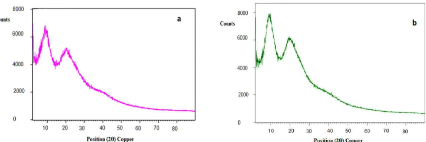
An example of X-ray diffraction pattern of
healthy woman hairs and hairs from patient with
breast cancer is shown in *igure 1. As this *igure
shows, two distinguishable peaks with different
heights (counts) are presented. Also, diffraction pattern of normal human hair and hair from a patient with breast cancer using synchrotron beam XRD method is shown in *igure 2.
RESULTS
Results of WAXRD experiment of hair samples belong to healthy and breast cancer
patients are shown in *igure 3. As it seen, 1st and
2nd peaks height of breast cancer patients which
are shown in counts (height) is more than
counts related to healthy samples. So, X-ray
diffraction patterns of hair from breast cancer patients have more intensity than that of normal.
Figure 3 also demonstrates the comparison
between healthy and cancer patients. In addition, in each group, the results were compared with mammography and also pathology report of each individual and they are
shown in table 1.
Figure 2. Diffrac on pa/ern of (A) normal human hair and (B) hair from a pa ent with breast cancer using synchrotron beam XRD. The diffuse ring that has shown in (B) indicates the presence of tumor.
Figure 1.Examples of X-ray diffrac on pa/ern of healthy human hairs (a) and hairs from pa ent with breast cancer (b) which showing two dis nguishable peaks but in different heights (counts).
breast cancer with changes in hair structure using WAXRD method were considered.
In this study, the average age of 43 women was 51.3 ± 11.5, which is less than
screening populations (16). Since in
mammography there is radiation hazards to
patients, and the sensitivity of
Table 1. Comparison of obtained results and their sensi vi es of different studied groups among three methods of WAXRD, mammography and pathology.
# WAXRD Mammography Pathology
Sensi vity
P* N** P N P N
Healthy 14 2 12 - 14 - 14 85.7
Suspicious 12 9 3 7 5 9 3 58.3
Cancer 17 17 - 17 - 17 - 100
Total 43
Note: *Pa ent, **Normal
DISCUSSION
This study was performed to evaluate the ef*iciency of WAXRD in detection of breast cancer as a safe and non-invasive method. In addition, comparing the results of pathology as gold standard and mammography in detecting
Figure 3. Comparison charts of normal (N) and breast cancer pa ents (P) for 1st peak (a) and 2nd peak (b).
mammography is decreases with increase of age
(16), the goal of this study was to
determine if differences between XRD curve of
healthy women and breast cancer
patients can lead to early detection of breast cancer or prediction of disease without radiation hazards or not.
In this research, the difference between XRD curve of healthy and cancer patients were studied and compared. As *igure 1 showed, there are two separate and distinguishable
peaks in 9.8 AE and 4.4 AE which re*lects d-spacing of hair structure. These data were
compatible with those obtained by previous study using synchrotron source which, rings related to d-spacing of hair structure have been
reported (9-13). But, as shown in *igure 3, there
was an additional ring re*lects 4.7 nm d-spacing
in cancer patients (12, 14). The 9.8 AEre*lection
d-spacing refers to structure pattern of the alpha-helix diameter and the 4.4 AE re*lection is referring to the periodic distance of the
3.6-alpha-helix structure (18).
As it is shown in *igure 3, the overall height of
1st and also 2nd peak in patient group is higher
than that of normal women and difference in the
height means of two group for 1st peak is
signi*icant (P<0.05). Of the *irst 17 bars of patient related bars in *igure 3-a, 15 of them has greater height in comparison with healthy individuals. Hence, it can be assumed that presence of disease can make differences in hair structure, especially the molecular structure of
alpha-helix (18). For mean of 2nd peak height in
the two examined groups, the difference was not signi*icant (P<0.05).
The results in table 1 indicated that WAXRD
could provide a proper diagnostic test for patient with breast cancer. In addition, in two
groups of suspicious and cancer, the results were completely the same as results of WAXRD
and pathology reports. In cancer group, the results were the same among all three studied
methods. In comparison between results of
WAXRD and pathology, the detection rate of
breast cancer by WAXRD was 60.4%. Also, sensitivity of approximately 86% was achieved.
The results obtained here were more accurate
than those reported by Corino et al. (64%) (6).
When comparing the age of donors with results of the method, there was no correlation
between age and average height 1st peak of
WAXRD. This result is against the results which
reported by Corino et al. (17). It can be duo to
less number of samples that were tested in this work.
Comparing with mammography, the total procedure time, includes sample preparation;
XRD examination time and the curve interpretation, were taking about 48 hours
which is longer than mammography. From expense point of view, it was clear that the
price of XRD procedure is half of that for mammography. As it is shown in table 1, the
sensitivity of XRD (86%) was higher than that of mammography (64%).
Therefore, identi*ication of breast cancer at
an early stage, using XRD technique may be provided an alternative early low-cost, without
radiation hazards, non-invasive and more sensitive method. Ultimately, there are look
promising in detecting breast cancer by XRD method.
Of course, further study should be done to examine more hair samples of breast cancer patients. In addition, the need to provide more
experiments is of utmost importance. In conclusion, the *indings of this work may be
open a new era to investigate whether WAXRD or other modalities such as mammography and also synchrotron based methods are useful to
diagnosis of early stages of breast cancer patients.
ACKNOWLEDGMENT
This work is a part of PhD thesis which -inancially supported by Isfahan University of
Medical Sciences (MUI) (Grant No. 391456). The authors would like to acknowledge the
Radiation Oncology Department of Cancer Institute, Tehran University of Medical Sciences
for supporting this research.
Con lict of Interest: Declared none.
Applied Radia on and Isotopes, 68(12): 2237–45.
10. Kidane G, Speller RD, Royle GJ, Hanby AM (1999) X-ray
sca/er signatures for normal and neoplas c breast ssues. Phys Med Biol, 44: 1791–1802.
11. James V, Kearsley J, Irving T, Amemiya Y, Cookson D (1999) Using hair to screen for breast cancer. Nature, 398 (6722): 33-4.
12. Saengkaew P, Ussawawongaraya W, Khaweerat S, Rugmai S, Ouajai S, Luengviriya J, et al. (2011) A preliminary X-ray study on human-hair microstructures for a health-state
indicator. World Academy of Science, Eng Technol,
59: 1945-9.
13. Corino GL and French PW (2008) Diagnosis of breast cancer by X-ray diffrac on of hair. Interna onal Journal of
Cancer, 122(4): 847-56.
14. Mistry DAH, Haklani J, French PW (2012) Iden fica on of
breast cancer associated lipid in scalp hair.
Breast Cancer: Basic and Clinical Research, 6: 113-123. 15. Interna onal Atomic Energy Agency (IAEA) TECDOC-950
(1997) Sampling, storage and sample prepara on
procedures for X-ray fluorescence analysis of
environmental materials.
16. Gholizadeh N, Kabiri Z, Kakuee O, Saleh-Kotahi M, Changizi V, Fathollahi V, et al. (2013) Feasibility of breast cancer screening by PIXE analysis of hair. Biological Trace Element Research, 153 (1-3): 105-10.
17. Corino GL, French PW, Lee M, Ajaj MM, Haklani J, Mistry
DA, et al. (2009) Characteriza on of a Test for Invasive
Breast Cancer Using X-ray Diffrac on of Hair-Results of a Clinical Trial. Breast Cancer: Basic and Clinical Research, 3: 83-90.
18. Pauling L, Corey RB, Branson HR (1951) The structure of proteins; two hydrogen-bonded helical configura ons of
the polypep de chain. Proceedings of the Na onal
Academy of Sciences of the United States of America, 37(4): 205-11.
REFERENCES
1. WHO World cancer report 2012
2. Shahbazi-Gahrouei D (2003) Possible effect of background radia on on cancer incidence in Chaharmahal and Bakh ari province. Iran J Radiat Res, 1(3): 171-174. 3. Sasieni PD, Shelton J, Ormiston-Smith N, Thomson CS,
Silcocks PB (2011) What is the life me risk of developing
cancer? The effect of adjus ng for mul ple primaries. Br J Cancer, 105(3): 460-5.
4. Hagen AI, Kvistad KA, Maehle L, Holmen MM, Aase H, Styr
B, et al. (2007) Sensi vity of MRI versus conven onal
screening in the diagnosis of BRCA-associated breast
cancer in a na onal prospec ve series. The Breast, 16(4):
367-74.
5. Carney PA, MiglioreN DL, Yankaskas BC, Kerlikowske K,
Rosenberg R, Ru/er CM, et al. (2003) Individual and
combined effects of age, breast density, and hormone replacement therapy use on the accuracy of screening
mammography. Annals of Internal Medicine,
138(3): 168-75.
6. Théberge I, Chang SL, Vandal N, Daigle JM, Guer n MH,
Pelle er E, Brisson J (2014) Radiologist interpre ve volume
and breast cancer screening accuracy in a Canadian
organized screening program. J Natl Cancer Inst,
106(3): djt461.
7. Rawashdeh MA, Lee WB, Bourne RM, et al. (2013) Markers
of good performance in mammography depend on number of annual readings. Radiology, 269(1): 61-7.
8. Maziar Asghar, Shahbazi-Gahrouei D, Tavakoli Mohammad Bagher, Changizi Vahid (2015) Non invasive XRF analysis of human hair for health state determina on of breast ssue. Iran J Cancer Prev, 8(6): e3983.
9. Chaparian A, Oghabian MA, Changizi V, Farquharson MJ (2010) the op miza on of an energy-dispersive X-ray
diffrac on system for poten al clinical applica on.