1
IN-SITU FORMING IMPLANT SYSTEM FOR LONG ACTING HIV PROPHYLAXIS Shelby L. Hudson, MS
PharmD Candidate
UNC Eshelman School of Pharmacy November 17, 2015
2 Abstract
Although its prevention and treatment has progressed significantly since HIV’s first known case of human infection in 19591, with a worldwide prevalence of 36.9 million and incidence of 2.0 million in the
year 2014 alone there remains significant room for improvement. Along with this persistence of infection comes an ongoing drive for better preventative measures. The current pre-exposure prophylaxis (PrEP) method available, once daily oral Truvada, exhibits variable efficacy due to
differences in individual patient absorption and mandates consistent patient utilization for prevention of infection.2 This project aims to improve upon these PrEP methods by providing a long acting injectable
for the introduction of one or more antiretrovirals (ARVs) prior to the exposure for the prevention of infection in such an event.
The following is a description of a subcutaneous injection which will introduce an in-situ forming implant (ISFI) allowing the prolonged release of drug through diffusion from and degradation of the resulting solid depot. Our goal is to achieve plasma drug concentration/s in appropriate animal models greater than or equal to four times the IC90 of each drug for at least three months. Also, we would like to ultimately demonstrate that the subcutaneous route of ISFI administration will allow for emergency removal of the system if needed. This promotes safety above the currently offered oral PrEP method as well as the intramuscular nanopartical suspension based injections currently in phase II clinical trials.3
Introduction
According to the CDC, there are currently more than 1.2 million people in the United States that are at high enough risk for HIV infection to warrant daily oral pre-exposure prophylaxis (PrEP). Unfortunately, most of these individuals are not receiving treatment contributing to the around 40,000 yearly new HIV diagnoses in the US. A primary obstacle to use is knowledge of PrEP availability. Since its approval in 2012, oral Truvada has been widely used for the prevention of HIV infection upon exposure, however, according to the CDC only about one third of primary care physicians and nurses are aware of PrEP for HIV prevention.4 When Truvada is implemented, variability in adherence and absorption results in a
wide range of efficacy ranging from 0% to 75% in six clinical trials during the time period from 2010 through 2015.5 There is a widely accepted correlation between adherence and efficacy of PrEP for HIV.
Even small variations in administration can result in a significant effect on disease contraction upon exposure. Overall adherence has been shown to range from 21 to 86% with the latter resulting in a 96% reduction in incidence of infection and efficacy decreasing with decreasing usage.5
There are many barriers to adherence when it comes to HIV PrEP. Patients may have trouble
3
intramuscular injection. Their intramuscular administration and suspension based nature of their formulation will not allow for their removal in the event of an adverse reaction. Also, the nanocrystal approach has not proven flexible enough to accommodate two or more drugs at this time. HIV has proven to be a highly resistant virus which is prone to mutation, the treatment and prevention of which requires a multidrug regimen. Any inability to formulate multiple drugs into a prophylactic system presents a large disadvantage.
A simple formulation which can accommodate multiple antiretrovirals and allow the removal of the drug release system in the event of an adverse reaction is much needed. We intend to provide an in situ forming implant (ISFI) injection based on a simple formulation of a polymer and biocompatible solvent into which drug is added as easily as a solution is made. The system will be introduced subcutaneously and solidify into a depot upon contact with the aqueous environment. This method of administration permits the surgical removal of the depot should any adverse reaction occur. It can also easily accommodate multiple antiretrovirals with the potential to deliver at least 4 times the IC90 of each antiretroviral for at least 30 days following injection providing long term protection against infection in the event of HIV exposure. The portion of the project described in this paper pertains to the production of various ISFI formulations, the characterization of these ISFI formulations (including stability and drug saturated concentration) and their in vitro release of drug over time.
Methods
Formulation
Depots were prepared at varying concentrations of drug in various ratios of polymer to solvent. All are based on the dissolution of polylactide-co-glycolide (PLGA) in N-methyl-2-pyrrolidone (NMP). PLGA to NMP mass ratios of 1:2, 1:4, 1:8, and 1:16 were investigated with the addition of one of three
antiretrovirals: MK-2048, dolugetravir (DTG), or rilpivirine (RPV). To prepare the ISFIs, an appropriate mass of NMP was added to a 10 mL scintillation vial followed by an appropriate mass of PLGA (depending on the mass ratio desired). Dissolution of the polymer was then assisted with immediate vortexing followed by 5 minutes in a sonicating water bath at 40oC. Subsequent vortexing and 40oC
water bath sonication was performed as needed until complete dissolution is achieved. The desired amount of drug was then added to the formulation with vortexing followed by sonication in a 40oC
water bath which was repeated again as often as needed until drug was completely dissolved.
Density and Stability
The density of various formulations with and without drug were determined and noted (Tables 1-3). Stability of various formulations and various concentration of drug was tested at both room
temperature and 4oC. For stability testing formulations were prepared at various drug concentrations
and PLGA/NMP (w/w) ratio then kept at constant room temperature or 4oC while in 10 mL scintillation
vials wrapped in aluminum foil to protect from light. They were then observed over time for precipitation of drug to occur (Table 4).
In-vitro Release
4
investigation into the profile of this release. Drug depots were injected into 200 mL of medium and kept at 37oC. One mL aliquots of medium were removed over time. Prior to sampling, the medium was mixed
by pulling up and releasing with the 1 mL pipettor 3 times before keeping the 4th mL as a sample. 1 mL of
fresh medium was then added to the sample jar. Experiments are done in triplicate (n=3) for each drug. All experiments are kept at 37o C. Resulting samples were analyzed using HPLC UV/Visible
spectrophotometry to determine the analytical amount of drug released over time. Media
experimented with included 0.01 M phosphate buffered saline (PBS) with 2% (w/w) solutol adjusted to a pH of 7.0, 0.01 M PBS with 2% Tween 80 (w/w) adjusted to a pH of 7.0, 25% (w/w) liquid PEG 400 in 0.10 M PBS adjusted to a pH of 7.0, and 50% IPA and water (Figures 1-11).
The influence of the use of a foam support as a skin tissue simulant on the release of drug from an ISFI system was investigated (Figure 10). Using a 20 gauge 1 inch long needle fitted to a 20 µL pipettor set at 20.00 µL, depot was injected into the center of a pre-cut and pre-wetted 1 cm3 cut from polytech foam.
The foam cube was wetted by repeatedly squeezing the foam cube with flat tip forceps while
submerging it into the release medium (0.01 M phosphate buffered saline with 2% solutol, pH 7.4) until air was no longer released upon squeezing. Prior to pulling depot solution from sample vial, the pipettor was pushed to the second stop to obtain greater suction. Injection was done just outside of the release medium jar with the ISFI containing foam them immediately dropped into the jar. Time was started as soon as the depot containing foam was placed into the release medium and the first sample was taken at t = 0. One milliliter samples were removed from medium at intervals throughout a 35 day period using the same sampling method as described above followed by the same subsequent analysis.
Results
Table 1: Saturated DTG depot: Placebo formulation
PLGA/NMP (w/w)
Density (g/mL) DTG (mg) in ISFI Conc % (w/w) Conc (mg/mL)
1:2 1.1132 208.0 8.2022 91.304
1:4 1.0933 265.6 11.205 122.50
1:8 1.0911 331.1 15.605 170.27
1:16 1.0768 387.1 18.979 204.38
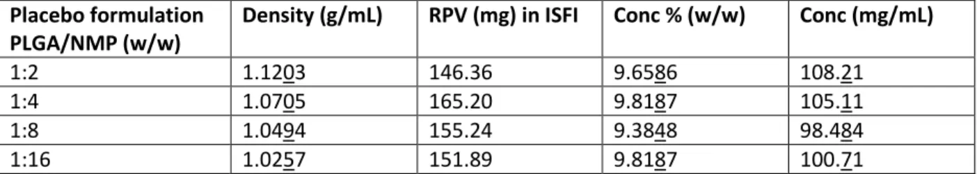
Table 2: Saturated RPV depot: Placebo formulation
PLGA/NMP (w/w)
Density (g/mL) RPV (mg) in ISFI Conc % (w/w) Conc (mg/mL)
1:2 1.1203 146.36 9.6586 108.21
1:4 1.0705 165.20 9.8187 105.11
1:8 1.0494 155.24 9.3848 98.484
1:16 1.0257 151.89 9.8187 100.71
Table 3: Placebo formulations: Placebo formulation
PLGA/NMP (w/w)
Density (g/mL)
1:4 1.0720
5 Table 4: Stability:
Formulation Date Initiated Room Temperature (RT): Date of precipitation
4oC: Date of precipitation
Days in solution (RT)
Days in solution (4oC)
DTG: 9.0909 % in 1:2 (w/w)
7/17/14 9/21/14 7/20/14 66 3
DTG: 6.994% (w/w) in 1:2 PLGA/NMP
12/15/14 2/14/15 12/19/14 61 4
DTG: 5.997 % in 1:2 (w/w)
8/5/14 None as of 11/2/15
9/14/14 >1 yr 21
RPV: 4.842 % (w/w) in 1:2 PLGA/NMP
2/16/15 None as of 11/2/15
6/2/15 > 8 mo 107
SATURATED SOLNS
DTG: 11.205 % (w/w) in 1:4 PLGA/NMP
7/16/15 7/23/15 7/17/15 7 1
DTG: 15.605 % (w/w) in 1:8 PLGA/NMP
7/16/15 7/23/15 7/17/15 7 1
DTG: 18.979 % (w/w) in 1:16 PLGA/NMP
10/9/15 None as of 12/15/15
10/10/15 67+
(last observed 12/15/15)
1
RPV: 9.659 % (w/w) in 1:2 PLGA/NMP
10/15/15 10/28/15 10/16/15 13 1
RPV: 9.819 % (w/w) in 1:4 PLGA/NMP
10/20/15 12/15/15 10/21/15 56 1
RPV: 9.385 % (w/w) in 1:8 PLGA/NMP
10/20/15 11/1/15 10/21/15 12 1
RPV: 9.066 % (w/w) in 1:16 PLGA/NMP
6
Figure 1 (above): Cumulative % drug release over time from 0.8:1:2 (w/w/w) MK-2048/PLGA/NMP ISFI by in 200 mL 0.01 M PBS with 2% solutol at pH 7.0 and 37oC.
Figure 2 (above): Cumulative percent drug release over time from 9:1:30 (w/w/w) MK-2048/PLGA/NMP ISFI by in 200 mL 0.01 M PBS with 2% solutol at pH 7.0 and 37oC.
0 5 10 15 20 25 30 35
0 20 40 60 80 100 120
%
Rel
ease
Days
Average % MK 2048 release from MK 2048/PLGA/NMP
0.8:1:2 ISFI in 2% (w/w) Solutol in 0.1 M PBS (n = 3)
0 20 40 60 80 100 120 140
0 1 2 3 4 5 6 7
%
Rel
ease
Days
7
Figure 3 (above): Cumulative percent drug release over time from 9:1:30 (w/w/w) MK-2048/PLGA/NMP ISFI in 200 mL 2% Tween 80 in 0.01 M PBS at pH 7.0 and 37oC.
Figure 4 (above): Cumulative percent drug release over time from 0.2:1:2 (w/w/w) DTG/PLGA/NMP ISFI by in 200 mL 0.01 M PBS with 2% solutol at pH 7.0 and 37oC.
-20 0 20 40 60 80 100 120
-20 0 20 40 60 80 100 120 140 160
%
Rel
ease
Days
Average % MK 2048 release from MK 2048/PLGA/NMP
9:1:30 ISFI in 2% (w/w) Tween 80 in 0.01 M PBS (n = 3)
0 5 10 15 20 25 30 35 40 45 50
0 10 20 30 40 50 60 70
Av
era
ge
%
Rel
ease
Days
8
Figure 5 (above): Cumulative percent drug release over time from 0.2:1:2 (w/w/w) DTG/PLGA/NMP ISFI in 200 mL of 25% (w/w) liquid PEG 400 in 0.01 M PBS at pH 7.0 and 37oC.
Figure 6 (above): Cumulative percent drug release over time from 0.2:1:2 (w/w/w) DTG/PLGA/NMP ISFI in 200 mL of 50% IPA/H2O at 37oC.
0 5 10 15 20 25
0 2 4 6 8 10 12 14 16
%
Rel
ease
Days
Average % DTG release from DTG/PLGA/NMP 0.2:1:2 ISFI
in 200 mL 25% (w/w) liquid PEG 400 in 0.01 M PBS (n = 3)
0 10 20 30 40 50 60 70
0 2 4 6 8 10 12 14 16
%
Rel
ease
Days
9
Figure 7 (above): Cumulative concentration of release (mcg/mL) of DTG over time from saturated ISFIs of various PLGA/NMP (w/w) ratios (1:2, 1:4, 1:8, and 1:16) in 0.01 M PBS with 2% solutol at pH 7.0 and 37oC.
Figure 8 (above): Cumulative percent release of DTG over time from saturated ISFIs of various PLGA/NMP (w/w) ratios (1:2, 1:4, 1:8, and 1:16) in 0.01 M PBS with 2% solutol at pH 7.0 and 37oC.
0
5
10
15
0
10
20
30
40
C
on
cen
tr
at
ion
(mc
g
/mL)
Days
1:2, 1:4, 1:8, 1:16 PLGA/NMP DTG Saturated ISFI release
in 2% (w/w) solutol in 0.01M PBS (n = 3):
Average Concentration over Time
1:2
1:4
1:8
1:16
0
5
10
15
20
0
10
20
30
40
%
R
ele
ase
Days
1:2, 1:4, 1:8, 1:16 PLGA/NMP DTG Saturated ISFI release in
2% (w/w) solutol in 0.01M PBS (n = 3):
Average Percent Release over Time
1:2
1:4
1:8
10 Formulation PLGA:NMP (w/w) Average DTG Concentration at 10 days (mcg/mL)
Multiplicative
comparator of release to previous formulation at 10 days (mcg/mL)
Average % DTG Release at 10 days
Multiplicative
comparator of release to previous formulation (%) at 10 days
1:2 1.195 NA 1.307 NA
1:4 2.310 1.933 x [1:2] 3.598 2.752 x [1:2]
1:8 5.911 2.559 x [1:4] 8.241 2.290 x [1:4]
1:16 12.803 2.166 x [1:16] 16.341 1.983 x [1:8]
Table 5 (above): A comparison of the average concentration of DTG in release medium for various PLGA:NMP (w/w) formulations saturated with DTG (Table 1 and Figures 7-8).
PLGA:NMP (w/w)
Multiplicative
comparator of release to previous formulation at 10 days (mcg/mL)
Explanation of "multiplicative
comparator" (mcg/mL)
SD from 2
(representitive of a relative doubling)
Average SD from 2 of comparators
1:4 1.9330 [1:4] = 1.9330 x [1:2] 0.0474 0.1867
1:8 2.5590 [1:8] = 2.5590 x [1:4] 0.3953
1:16 2.1660 [1:16] = 2.1660 x [1:8] 0.1174
Table 6 (above): The influence of doubling of the mass of NMP relative to PLGA mass in the ISFI of saturated DTG formulations on the average concentration of DTG in release medium by multiplicative comparator (Table 1 and Figures 7-8).
PLGA:NMP (w/w)
Multiplicative
comparator of release to previous formulation at 10 days (%)
Explanation of "multiplicative comparator" (%)
SD from 2
(representative of a relative doubling)
Average SD from 2 of comparators
1:4 2.7520 [1:4] = 2.7520 x [1:2] 0.5317 0.2496
1:8 2.2900 [1:8] = 2.2900 x [1:4] 0.2051
1:16 1.9830 [1:16] = 1.9830 x [1:8] 0.0120
11
Figure 9 (above): Cumulative percent drug release over time from 0.2:1:2 (w/w/w) RPV/PLGA/NMP ISFI by in 200 mL 0.01 M PBS with 2% solutol at pH 7.0 and 37oC. (note: standard deviations are too small for
error bars to be visible beyond data points)
Figure 10 (above): Cumulative percent drug release over time from 0.2:1:2 (w/w/w) RPV/PLGA/NMP ISFI suspended in foam support by in 200 mL 0.01 M PBS with 2% solutol at pH 7.0 and 37oC. (note: standard
deviations are too small for error bars to be visible beyond data points) 0
5 10 15 20 25
0 5 10 15 20 25 30
%
Rel
ease
Days
Average % RPV release from 0.2:1:2 RPV/PLGA/NMP ISFI in
200 mL 2% (w/w) solutol in 0.1 M PBS (n = 3)
0 5 10 15 20 25
0 5 10 15 20 25 30 35 40
%
Rel
ease
Days
12
Figure 11 (above): Cumulative percent drug release over time from 0.8:1:2 (w/w/w) RPV/PLGA/NMP ISFI in 200 mL of 50% IPA/H2O at 37oC.
Conclusions and Discussion
All saturated solutions of drug in ISFI (prepared at room temperature) precipitated within 24 hours at 4oC, as would be expected (Table 4). For unsaturated solutions, length of stability of DTG dissolved in
ISFI (1:2 mass ration of PLGA:NMP) increased with decreasing DTG concentration at both room temperature and 4oC, with the exception of 9.0909% versus 6.994% at room temperature which
precipitated at 66 and 61 days respectively. All unsaturated and saturated solutions for both DTG and RPV in ISFI remained in solution for a greater length of time when kept at room temperature versus 4oC
(Table 4). This combined with the non-aqueous nature of all formulations indicate that room temperature is a superior, safe, and preferred storage condition.
Figure 1 and Figure 2 demonstrate that increasing the relative amount of MK-2048 in the formulation while decreasing the relative amount of PLGA to NMP will result in a faster and overall much more complete release of MK-2048 from ISFI (> 99% versus < 5% at 5 days). Although results which were comparable to that of 2% solutol in 0.1 M PBS were initially observed, further experimentation with 2% (w/w) Tween 80 in 0.01 M PBS (formulation used in Figure 3) produced inconsistent and unpredictable data. It was also found that DTG release from 1:2 (w/w) PLGA/NMP ISFI into 50% (w/w) IPA/H2O
demonstrated an incomplete release of drug with a profile resembling saturation at 2 days (Figure 6). RPV release from the same ISFI formulation into the same medium, however, produced complete release at 63 days with a desirable profile (Figure 11). As for experimentation with foam support as a skin simulant, Release profiles for 0.2:1:2 RPV in 200 mL of 2% (w/w) solutol in 0.1 M PBS for free release ISFI and foam supported ISFI were very similar (Figure 9-10). Both achieved 20% release within 30 days (between days 25 to 30).
-20 0 20 40 60 80 100 120
0 10 20 30 40 50 60 70 80 90
%
Rel
ease
Days
13
Of the various media formulations, 2% solutol (w/w) in 0.1 M PBS produced the most favorable and consistent results for DTG release (Figures 4-6) when compared to 0.01 M PBS with 2% Tween 80 (w/w), 25% (w/w) liquid PEG 400 in 0.10 M PBS, and 50% IPA and water. As a result of these findings, a 2% solutol (w/w) in 0.1 M PBS medium was used in the subsequent experiments performed to analyze the effects of varying the mass of PLGA present relative to NMP in a DTG containing ISFI (Table 1; Figure 7-8). Following this investigation, a trend was observable in the comparison of 1:2, 1:4, 1:8, and 1:16 PLGA:NMP (w/w) formulations of saturated DTG ISFIs. There was a relative doubling of both cumulative drug concentration and % drug release compared to total drug in depot at 10 days following ISFI release into medium with each doubling of the mass of NMP used relative to the mass of PLGA used in the ISFI formulation. There was an average standard deviation of 0.2 from a multiplicative comparator of 2 (Table 5 and Table 6) for each corresponding doubling of NMP mass relative to PLGA. This same standard deviation of 0.2 from an exact doubling is apparent in both the average concentration versus time data (Figure 7 and Table 5) as well as the average % drug release relative to the total drug present in each depot data (Figure 8 and Table 6). The presence of the trend in the latter experiment (Figure 8) demonstrates its conservation even with the normalization of drug release based on the concentration of drug in depot. This corrects for the variability in the saturation concentrations of DTG for each PLGA:NMP (w/w) formulation (Table 1).
Future Plans
There is much investigation required in preparation for clinical trials. Future investigative plans include the Scanning Electron Microscopy analysis of in vitro depot degradation. Also, the pharmacokinetics of DTG release from ISFI in non-human primate models will be assessed. In preparation for multi-drug containing ISFIs in order to provide effective prophylaxis from HIV’s ability to readily mutate, the In vivo efficacy of DTG/RPV combination ISFI in BLT mouse model should be determined. Optimization of ISFI to accommodate drug concentrations for human dosing will also be required for feasibility of providing serum concentrations that can protect against infection of HIV in the event of exposure.
References
1. Pence, G E2. Preventing the Global Spread of AIDS. In Medical Ethics Accounts of the Cases That Shaped and Define Medical Ethics.2008. New York, NY:McGraw;330
2. Hill.http://www.who.int/features/factfiles/hiv/en/, WHO - 10 Facts on HIV/AIDS;2015 3. Castel AD, Magnus M, Greenberg AE. Pre-exposure prophylaxis for human immunodeficiency
virus: the past, present, and future. Infect Dis Clin North Am.2014;28(4):563-83
4. McCarthy, M. HIV pre-exposure prophylaxis could help 1.2 million in US.2015;351:h6384 5. Haberer JE. Current concepts for PrEP in the PrEP revolution: from clinical trials to routine