A Sterile Sperm Caste Protects Brother
Fertile Sperm from Female-Mediated Death
in
Drosophila pseudoobscura
Luke Holman1and Rhonda R. Snook1,*
1Department of Animal and Plant Sciences University of Sheffield
Sheffield S10 2TN United Kingdom
Summary
Spermicide (i.e., female-mediated sperm death) is an under-studied but potentially widespread phenomenon that has important ramifications for the study of sexual conflict, post-copulatory sexual selection, and fertility [1, 2]. Males are predicted to evolve adaptations against spermicide, but few antispermicidal mechanisms have been definitively identified. One such adaptation may be the enigmatic infer-tile sperm morphs or ‘‘parasperm’’ produced by many spe-cies, which have been hypothesized to protect their fertile brother ‘‘eusperm’’ from spermicide[2, 3]. Here, we show that femaleDrosophila pseudoobscurareproductive tracts are spermicidal and that the survival of eusperm after expo-sure to the female tract is highest when males produce many parasperm. This study clarifies the adaptive significance of infertile sperm castes, which has remained elusive in Dro-sophilaand other taxa despite much recent interest[2–8]. We suggest that spermicide and male countermeasures against it are more common than is appreciated currently and discuss how spermicide could drive the evolution of several key male traits, including sperm size and number. Results and Discussion
Males of theDrosophila obscuragroup are sperm heteromor-phic; that is, they produce two distinct morphologies of sperm, short and long. The long sperm fertilize ova and are called eusperm[9], whereas the shorter parasperm type is fertiliza-tion incompetent[10, 11]and comprises 34%–94% of the ejac-ulate among species[3]. Parasperm are likely to be adaptive
[3]but do not function to provision the female[12] or delay her remating[3, 4]. Sperm heteromorphism occurs in insects, mollusks, vertebrates, and other taxa, but its evolutionary significance remains a conundrum[8]. Parasperm were sug-gested recently to be a male counteradaptation to spermicide, i.e., female-mediated sperm death, protecting their ‘‘brother’’ eusperm in the female reproductive tract[2]. Game theory models have demonstrated that sperm heteromorphism could indeed have evolved to maximize postmating sperm survival, assuming parasperm are cheaper to produce and spermicide is substantial[2].
The current study empirically tested two key predictions of the antispermicide hypothesis for the evolution of parasperm using the fruitflyDrosophila pseudoobscura: (1) that the female reproductive tract is spermicidal and (2) that the infertile parasperm protect brother fertile eusperm from spermicide. Spermicide is defined here as female-mediated sperm death
resulting from chemical damage (e.g., by enzymes, adverse pH, or the immune system) or phagocytosis[1, 2]and is not necessarily adaptive to the female[2]. We tested each predic-tion by using both in vivo and in vitro techniques to quantify sperm viability and its relationship to parasperm production.
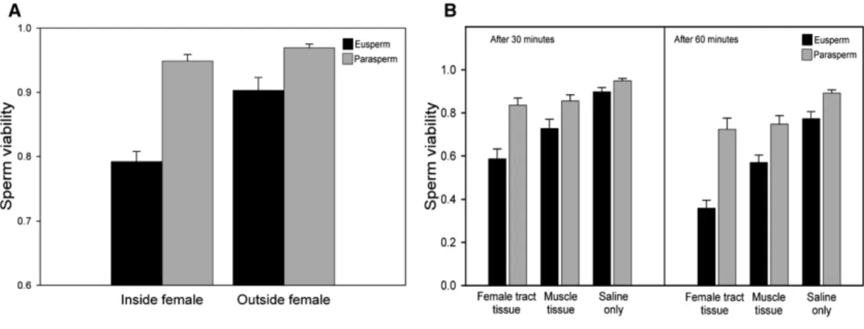
To test whether femaleD. pseudoobscuraare spermicidal, we first measured sperm viability (the proportion of live sperm) in an in vivo experiment with natural matings. Mated females were dissected 30 min after the cessation of copulation to measure the viability of the sperm in the uterus. As a control, females were dissected immediately after mating, and then the sperm were stored for 30 min before viability scoring. We found that eusperm viability was significantly lower when sperm were left inside the female (Figure 1A; F1,27 = 20.34, p < 0.0001, effect size 95% confidence limits: r = 0.39–0.79). In contrast, parasperm viability was unaffected by exposure to the female (Figure 1A; treatment was absent from the minimum adequate model of parasperm viability). Eusperm viability also was lower than parasperm viability, irrespective of when sperm were removed from the female (Figure 1A). Copulation duration had no significant effect on eusperm or parasperm viability and was excluded from the minimum adequate models.
We also used an in vitro approach to test whether the fe-male reproductive tract caused sperm death by measuring the effect of different female tissues on sperm viability[13]. Sperm from male seminal vesicles (n = 30) were incubated with extracts prepared by grinding and centrifuging (1) female reproductive tract tissue, (2) thoracic muscle tissue, or (3) saline. Sperm viability was measured after either 30 min or 60 min incubation by staining and counting sperm as before. The three solutions had significantly different effects on sperm viability (Figure 1B;Table 1). Eusperm viability was sig-nificantly lower when exposed to female reproductive tract extract rather than muscle tissue (t132= 4.57, p < 0.0001, r = 0.21–0.50), and muscle tissue reduced eusperm viability more than did exposure to saline (t132= 6.54, p < 0.0001, r = 0.36–0.60). Similarly, parasperm viability was higher after exposure to saline rather than muscle tissue (t132 = 2.85, p = 0.005, r = 0.07–0.39); however, there was no strong differ-ence between the female reproductive tract and the muscle tissue solutions (t132= 1.78, p = 0.077, r =20.02–0.31). The viability of both eusperm and parasperm was lower when sperm were incubated for 60 min compared to 30 min (Figure 1B;Table 1). There were no interactions between treat-ment and time, indicating that the effects of the treattreat-ment solutions on sperm viability were consistent at both time inter-vals. Eusperm viability again was markedly lower than para-sperm viability (Figure 1B).
The results of both experiments strongly suggested that the reproductive tract of femaleD. pseudoobscuracauses sperm death. Therefore, we proceeded to test whether parasperm alleviate the effects of this spermicide on eusperm as hypoth-esized[2, 3]. In addition to measuring sperm viability in the in vivo and in vitro experiments described above, we also mea-sured the proportion of parasperm produced by each male. If parasperm protect eusperm from female spermicide, then par-asperm proportion should be related positively to eusperm *Correspondence:[email protected]
viability after exposure to the female reproductive tract. We also determined the mean length of eusperm and parasperm produced by each male in the in vitro experiment in order to test whether sperm length influences sperm survival as hypothesized in sperm competition theory (reviewed in[14]).
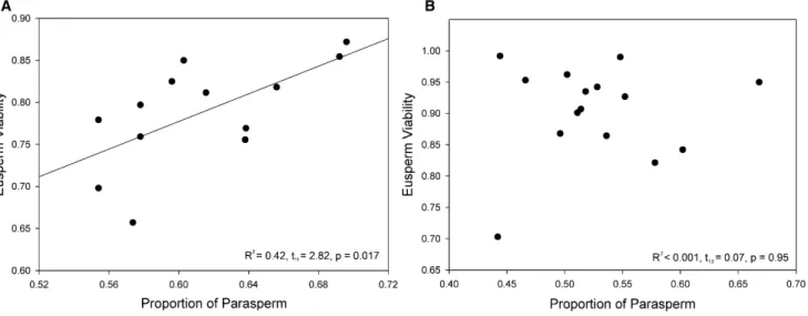
The in vivo experiment revealed the predicted positive relationship between eusperm viability and parasperm propor-tion after sperm had been exposed to the live female reproduc-tive tract for 30 min (Figure 2A; R2= 0.42, t
11= 2.82, p = 0.017, r = 0.15–0.84). When sperm were removed from the female immediately after mating, no relationship between parasperm proportion and eusperm viability was observed (Figure 2B; R2< 0.001, t
13= 0.07, p = 0.95, r =20.46–0.49), implying that the female reproductive tract may have mediated the relation-ship between investment in parasperm and eusperm survival. However, the design of this experiment meant that, by neces-sity, parasperm proportion was measured at the same time as sperm viability. Since parasperm proportion may have been altered as a result of exposure to the female, the measures of sperm survival and parasperm proportion were noninde-pendent. The observed results are nevertheless compelling.
The in vitro experiment did not encounter this design limita-tion. We were able to take subsamples of a male’s sperm for
independent determination of sperm viability and parasperm proportion. The in vitro experiment also provided support for the hypothesis that parasperm protect eusperm from spermi-cide; we observed a significant interaction between para-sperm proportion and exposure time in their effects on eusperm survival (Table 1). Eusperm viability was affected more positively by parasperm proportion in the 30 min group than in the 60 min group. We therefore tested whether para-sperm proportion predicted eupara-sperm viability after 30 min exposure to the female reproductive tract and found a strong positive relationship (Figure 3; multiple R2 = 0.39, F2,24 = 7.51, p = 0.003; parasperm proportion: t = 2.54, p = 0.018, r = 0.09–0.66; latency: t = 3.10, p = 0.005), replicating the result from the in vivo assay. Also, in the in vitro trial, we found an in-teraction between treatment group and parasperm proportion in the model of parasperm viability (Table 1), which indicated that parasperm proportion had a more positive effect on parasperm viability when exposed to reproductive tract rather than muscle tissue (t132 = 2.70, p = 0.008, r = 0.17–0.67). Eusperm and parasperm length had no effect on either eu-sperm or paraeu-sperm viability in the in vitro trial (Table 1).
Taken together, our in vivo and in vitro results strongly suggest that sperm are killed by the female reproductive tract inD. pseudoobscuraand that parasperm lessen the effects of this spermicide on eusperm viability. Although we cannot be sure that the mechanism of spermicide was the same in both experiments, we suggest that the observed sperm death was chemically mediated. The protocol of the in vitro trial involved homogenizing the female tissues and then removing all non-soluble material, strongly suggesting the presence of a non-soluble, cytotoxic chemical in or around the cells of the female repro-ductive tract. The results of the in vivo experiment are also most consistent with chemically-mediated spermicide: Phagocytosis of live sperm within the 30 min timeframe seems unlikely because the sperm are much larger than hemocytes. Also, sperm, seminal fluid and mating itself are known to induce a multitude of changes in mated female D. mela-nogaster [15–17], including elevated secretory activity [17]
and upregulation of enzyme and defense genes in the lower reproductive tract[16].
Our experiments supported the hypothesis that parasperm function to protect eusperm from spermicide. In both assays, Figure 1. Mean Eusperm and Parasperm Survival after In Vivo and In Vitro Exposure to the Female
(A) In the in vivo trial, sperm were either left inside a live female for 30 min after insemination or removed from the uterus immediately after mating and stored for 30 min (n = 14 inside, n = 15 outside).
(B) In the in vitro trial, sperm were exposed to extracts containing female reproductive tract tissue, thoracic muscle, or saline only. Sperm viability was mea-sured after both 30 and 60 min (n = 30 males). Error bars represent 1 SEM.
Table 1. Two Mixed-Effect Models of the In Vitro Experiment Sperm Morph Model Component F d.f. p Eusperm Treatment group 58.1 2,132 <0.0001
Exposure time 42.9 1,132 <0.0001
Latency 6.26 1,132 0.014
Parasperm proportion 0.47 1,27 0.50 Exposure time3
parasperm proportion
6.43 1,132 0.012 Parasperm Treatment group 19.2 2,132 <0.0001
Exposure time 24.5 1,132 <0.0001
Latency 12.2 1,132 0.0007
Parasperm proportion 0.25 1,26 0.62 Treatment group3
parasperm proportion
4.10 2,132 0.019
These are the minimum adequate models created by backward stepwise removal of nonsignificant terms. In this way eusperm length, parasperm length, and the other interaction terms were excluded. Male identity is the random factor, n = 30 males; d.f. = degrees of freedom.
Sperm Heteromorphism and Sperm Death 293
we observed a positive relationship between eusperm viability and the proportion of parasperm after exposing sperm to the female reproductive tract. This relationship was not detected when sperm had either no (in vitro) or momentary (in vivo) contact with the female reproductive tract, implying that the female tract may have mediated this result. The precise mech-anism by which parasperm protect eusperm is not clear, but we showed in a game-theory model that parasperm can in-crease eusperm survival simply by being present[2]. If fewer eusperm die as a result of parasperm helping to saturate the spermicidal chemicals/cells and parasperm are sufficiently cheap to produce, then parasperm do not necessarily need to perform any special function, e.g., delivering sperm-bound peptides[18]that protect eusperm. The parasperm ofD. pseu-doobscuraare almost four times shorter than the eusperm, so they are likely to be less costly[3]. Parasperm might even
‘‘buffer’’ the uterus of mated females to the benefit of subse-quent males, as suggested for seminal fluid[19].
As well as measuring parasperm proportion, we also examined sperm length and its relationship to sperm viability. The nature of the relationship between sperm length and sperm survival is predicted to drive sperm length evolution
[14, 20–22]. Although this study found no effect of length on eusperm or parasperm viability, we did find that the viability of eusperm was substantially lower than that of parasperm. Eusperm might be more vulnerable than parasperm to chemi-cal damage, perhaps because they present a greater target. Alternatively, the lower viability of eusperm could have been an artifact; for example, eusperm may have been differentially damaged by shear forces when the coverslip was applied despite measures to prevent damaging sperm. This constraint was unavoidable because it is not possible to know if sperm Figure 2. The Proportion of Parasperm in a Male’s Ejaculate Predicted Eusperm Viability In Vivo but Only after Exposure to the Female Reproductive Tract (A) When sperm were left inside the female for 30 min, the proportion of parasperm predicted eusperm viability (n = 13).
(B) When sperm were removed from the female after mating and incubated for 30 min, there was no such relationship (n = 15).
Figure 3. Parasperm Proportion Predicted Sperm Viabil-ity after 30 Min In Vitro Exposure to a Female Reproduc-tive Tract
Statistics are from a multiple regression controlling for latency, n = 29.
were killed during staining and mounting because sperm viability cannot be directly measured in any other way[23]. However, there is no reason to think that sperm death caused by the viability assay itself occurred at a different rate among treatments, so this limitation does not diminish the reliability of our main conclusions.
Studies have indicated that spermicide may be common in insects, mammals, and other taxa (reviewed in[1, 2]), and there is growing interest in the evolutionary significance of this phe-nomenon (e.g.,[1, 2, 13, 19, 22, 24]). Spermicide might benefit the female, for example, by providing nutriment from digested sperm or by weeding out damaged or aged sperm[1, 22, 25]. Spermicide also could facilitate cryptic female choice. For example, female Utetheisa ornatrix moths have a sperm-digesting organ that is especially full after remating to a smaller male[24]. Alternatively, spermicide may occur as a nonadap-tive by-product of other functions such as immunity[2, 25]. Whatever the function and potential adaptive significance of spermicide in females, spermicide can potentially reduce male fitness by affecting the number of viable sperm reaching storage (e.g.,[20, 21, 26]). Spermicide, therefore, may be a trait over which there is sexual conflict, with the potential to pro-duce sexually antagonistic co-evolution. A model of the evolu-tion of antispermicidal parasperm predicted that males would escalate their investment in the antispermicidal trait if females became more spermicidal, suggesting that an arms race over sperm survival could occur[2].
There are likely to be other male counteradaptations to female spermicide in addition to sperm heteromorphism. For example, a model considering sperm-monomorphic species showed that spermicide can select for the production of addi-tional sperm to compensate for those that die[1], so insemina-tion of multitudinous sperm could be viewed as an adaptainsemina-tion against spermicide. Spermicide also might affect sperm morphology because a trade-off is thought to exist between sperm size and number[20, 21]and because different sperm morphologies might vary in their vulnerability to spermicide
[1]. In support of the latter idea, this study found evidence that the long eusperm are more vulnerable than parasperm. Additionally, spermicide by the immune system could select for reduced antigenicity of sperm-cell membrane receptors.
In addition to varying the number, morphology, and bio-chemistry of sperm, males could mitigate spermicide by using seminal fluid, which is chemically diverse and performs a mul-titude of functions[27–29]. In mammals, seminal fluid contains compounds that cause immunosuppression and immune tolerance[30, 31]. Seminal fluid also contains antioxidants, which may defend sperm from oxidative damage after mating
[28, 32–34], and protease inhibitors, which could ameliorate proteolytic damage[28, 29, 32, 35–37]. At least two of the seminal antiproteases ofD. melanogasterare toxic to females
[36, 37], highlighting the fascinating possibility that male coun-termeasures to spermicide may be a source of sexual conflict. In summary, we found strong evidence that the reproductive tract of femaleD. pseudoobscurais a hazardous environment for sperm. Sperm death occurred after exposure to both the live reproductive tract and to an extract of soluble reproduc-tive-tract compounds, indicating that sperm may have been killed chemically. Moreover, we twice observed a relationship between the amount of parasperm transferred and the survival of eusperm, and this relationship only was observed in the presence of the female reproductive tract. These results are consistent with the hypothesis that males produce parasperm to reduce the number of eusperm lost to spermicide.
Supplemental Data
Supplemental Experimental Procedures are available at http://www. current-biology.com/cgi/content/full/18/4/292/DC1/.
Acknowledgments
We are very grateful to M.F. Wolfner and two anonymous referees for pro-viding comments on the manuscript and to A. Beckerman for statistical help. We thank N.S. Badcock, H.S. Crudgington, K.J. Hutchence, R.A. Nay-lor, and A. Turner for their assistance during this project. This work was sup-ported by a National Environment Research Council studentship to L.H. and a National Science Foundation grant (DEB-0093149) to R.R.S.
Received: December 6, 2007 Revised: January 11, 2008 Accepted: January 15, 2008 Published online: February 21, 2008 References
1. Greeff, J.M., and Parker, G.A. (2000). Spermicide by females: what should males do? Proc. R. Soc. Lond. B. Biol. Sci.267, 1759–1763. 2. Holman, L., and Snook, R.R. (2006). Spermicide, cryptic female choice
and the evolution of sperm form and function. J. Evol. Biol. 19, 1660–1670.
3. Holman, L., Freckleton, R.P., and Snook, R.R. (2008). What use is an infertile sperm? A comparative study of sperm-heteromorphic Drosophila. Evolution Int. J. Org. Evolution62, 374–385. Published online December 5, 2007. 10.1111/j.1558–5646.2007.00280.x. 4. Snook, R.R. (1998). The risk of sperm competition and the evolution of
sperm heteromorphism. Anim. Behav.56, 1497–1507.
5. Presgraves, D.C., Baker, R.H., and Wilkinson, G.S. (1999). Coevolution of sperm and female reproductive tract morphology in stalk-eyed flies. Proc. R. Soc. Lond. B. Biol. Sci.266, 1041–1047.
6. Cook, P.A., and Wedell, N. (1999). Non-fertile sperm delay female remat-ing. Nature397, 486.
7. Oppliger, A., Naciri-Graven, Y., Ribi, G., and Hosken, D.J. (2003). Sperm length influences fertilization success during sperm competition in the snailViviparus ater. Mol. Ecol.12, 485–492.
8. Till-Bottraud, I., Joly, D., Lachaise, D., and Snook, R.R. (2005). Pollen and sperm heteromorphism: convergence across kingdoms? J. Evol. Biol.18, 1–18.
9. Healy, J.M., and Jamieson, B.G.M. (1981). An ultrastructural examina-tion of developing and mature paraspermatozoa inPyrazus ebeninus (Mollusca, Gastropoda, Potamididae). Zoomorphology98, 101–119. 10. Snook, R.R., Markow, T.A., and Karr, T.L. (1994). Functional
nonequiva-lence of sperm inDrosophila pseudoobscura. Proc. Natl. Acad. Sci. USA 91, 11222–11226.
11. Snook, R.R., and Karr, T.L. (1998). Only long sperm are fertilization-competent in six sperm-heteromorphicDrosophilaspecies. Curr. Biol. 8, 291–294.
12. Snook, R.R., and Markow, T.A. (1996). Possible role of nonfertilizing sperm as a nutrient source for female Drosophila pseudoobscura Frolova (Diptera: Drosophilidae). Pan Pacific Entomol.72, 121–129. 13. Bernasconi, G., Hellriegel, B., Heyland, A., and Ward, P.I. (2002). Sperm
survival in the female reproductive tract in the flyScathophaga stercora-ria(L). J. Insect Physiol.48, 197–203.
14. Snook, R.R. (2005). Sperm in competition: Not playing by the numbers. Trends Ecol. Evol.20, 46–53.
15. McGraw, L.A., Gibson, G., Clark, A.G., and Wolfner, M.F. (2004). Genes regulated by mating, sperm, or seminal proteins in mated female Drosophila melanogaster. Curr. Biol.14, 1509–1514.
16. Mack, P.D., Kapelnikov, A., Heifetz, Y., and Bendr, M. (2006). Mating-responsive genes in reproductive tissues of female Drosophila melanogaster. Proc. Natl. Acad. Sci. USA103, 10358–10363. 17. Heifetz, Y., and Wolfner, M.F. (2004). Mating, seminal fluid components,
and sperm cause changes in vesicle release in theDrosophilafemale reproductive tract. Proc. Natl. Acad. Sci. USA101, 6261–6266. 18. Peng, J., Chen, S., Busser, S., Liu, H.F., Honegger, T., and Kubli, E.
(2005). Gradual release of sperm bound sex-peptide controls female postmating behavior inDrosophila. Curr. Biol.15, 207–213.
19. Hodgson, D.J., and Hosken, D.J. (2006). Sperm competition promotes the exploitation of ejaculates. J. Theor. Biol.243, 230–234.
Sperm Heteromorphism and Sperm Death 295
20. Parker, G.A. (1993). Sperm competition games: Sperm size and number under adult control. Proc. R. Soc. Lond. B. Biol. Sci.253, 245–254. 21. Parker, G.A. (1998). Sperm competition and the evolution of ejaculates:
towards a theory base. In Sperm Competition and Sexual Selection, T.R. Birkhead and A.P. Møller, eds. (London: Academic Press), pp. 3–54. 22. Reinhardt, K. (2007). Evolutionary consequences of sperm cell aging. Q.
Rev. Biol.82, 375–393.
23. Stewart, A.D., Hannes, A.M., and Rice, W.R. (2007). An assessment of sperm survival in Drosophila melanogaster. Evolution Int. J. Org. Evolution61, 636–639.
24. Curril, I.M., and LaMunyon, C.W. (2006). Sperm storage and arrangement within females of the arctiid mothUtetheisa ornatrix. J. Insect Physiol.52, 1182–1188.
25. Birkhead, T.R., Møller, A.P., and Sutherland, W.J. (1993). Why do females make it so difficult for males to fertilize their eggs? J. Theor. Biol.161, 51–60.
26. Garcia-Gonzalez, F., and Simmons, L.W. (2005). Sperm viability matters in insect sperm competition. Curr. Biol.15, 271–275.
27. Chapman, T., and Davies, S.J. (2004). Functions and analysis of the seminal fluid proteins of male Drosophila melanogaster fruit flies. Peptides25, 1477–1490.
28. Poiani, A. (2006). Complexity of seminal fluid: A review. Behav. Ecol. Sociobiol.60, 289–310.
29. Ram, R.M., and Wolfner, M.F. (2007). Seminal influences:Drosophila Acps and the molecular interplay between males and females during reproduction. Integr. Comp. Biol.47, 427–445.
30. Alexander, N.J., and Anderson, D.J. (1987). Immunology of semen. Fertil. Steril.47, 192–205.
31. Robertson, S.A. (2007). Seminal fluid signaling in the female reproduc-tive tract: Lessons from rodents and pigs. J. Anim. Sci.85, E36–E44. 32. Mueller, J.L., Ram, K.R., McGraw, L.A., Qazi, M.C.B., Siggia, E.D., Clark,
A.G., Aquadro, C.F., and Wolfner, M.F. (2005). Cross-species examina-tion ofDrosophilamale accessory gland protein genes. Genetics171, 131–143.
33. Breque, C., Surai, P., and Brillard, J.-P. (2003). Roles of antioxidants on prolonged storage of avian spermatozoa in vivo and in vitro. Mol. Reprod. Dev.66, 314–323.
34. Collins, A.M., Williams, V., and Evans, J.D. (2004). Sperm storage and antioxidative enzyme expression in the honey bee,Apis mellifera. Insect Mol. Biol.13, 141–146.
35. Chapman, T. (2001). Seminal fluid-mediated fitness traits inDrosophila. Heredity87, 511–521.
36. Lung, O., Tram, U., Finnerty, C.M., Eipper-Mains, M.A., Kalb, J.M., and Wolfner, M.F. (2002). TheDrosophila melanogasterseminal fluid protein Acp62F is a protease inhibitor that is toxic upon ectopic expression. Genetics160, 211–224.
37. Mueller, J.L., Page, P.L., and Wolfner, M.F. (2007). An ectopic expres-sion screen reveals the protective and toxic effects of Drosophila seminal fluid proteins. Genetics175, 777–783.