Prospective-triggered high-pitch spiral versus
sequential dual-source CT coronary angiography:
comparison of image quality and radiation dose
INTRODUCTION
The recent rapid developments in multi-detector CT have made it an important
tool in the assessment of coronary arterial stenosis (1, 2). Retrospectively ECG-triggered
CCTA is associated with high radiation exposure (8-18 mSv) (3). As one of the most promising radiation saving techniques, prospective triggered sequential CCTA reduced the radiation
dose to 1.5-2.5 mSv for 64-slice DSCT (3-6). For 128-slice DSCT, prospectively ECG-triggered CCTA can be performed with a sequential or high-pitch spiral scan mode. A signi.icant 75% reduction in the radiation dose was achieved with the sequential scan mode compared with the retrospectively ECG-triggered scan mode (7-9). Prospectively ECG-triggered high-pitch spiral CCTA is a scan mode unique to DSCT. This
method can acquire complete data in only one
Y. Zhuang
1, 2, W. Huang
2#, Y. Shi
3, G. Bo
2, D. Lu
2, J. Zhang
2, D. Kong
2,
Y. Shi
2,
B.Wang
4*1Shandong Medical Imaging Research Institute, Shandong University, No. 324, JingWu Road, Jinan 250021,
Shandong, China
2Department of Medical Imaging, Huai’an First People’s Hospital, Af,iliated to Nanjing Medical University No.6,
Western Beijing Road, Huanyin District, Huai’an 223300, Jiangsu, China
3Digestive Department, Huai’an First People’s Hospital, Af,iliated to Nanjing Medical University No.6, Western
Beijing Road, Huanyin District, Huai’an 223300, Jiangsu, China
4Binzhou Medical University, No. 346, Guanhai Road, Laishan District, Yantai 264003, Shandong, China
ABSTRACT
Background: Prospec vely electrocardiography (ECG)-triggered high-pitch spiral coronary computed tomography angiography (CCTA) is a unique scan mode for dual -source CT (DSCT). Our reports aim to compare image quality and radia on dose of CCTA using high-pitch spiral or sequen al acquisi on mode in pa ents with low and stable heart rates. Materials and Methods: Pa ents with low and stable heart rates (HR) (HR ≤ 70 beats per minute [bpm]; heart rate variability [HRV] < 10 bpm) were randomly assigned to high-pitch spiral mode (group A; n = 80) or sequen al acquisi on mode (group B; n = 80). Image quality scores, image noise, effec ve radia on dose and influencing factors on image quality were assessed. Results: Mean image quality scores were 1.51 ± 0.32 and 1.70 ± 0.38 for groups A and B (P < 0.05), respec vely. Image noises of the two groups were 19.05±4.70 Hu and 27.21±8.88 Hu (P < 0.05). Contrast media cost in group A was lower than group B (P < 0.05). No sta s cal difference was found in the rate of diagnos c pa ents between the two groups (P = 0.416). The es mated radia on dose of group A was 26.0% reduced compared with group B (0.74 ± 0.34 mSv vs. 1.00 ± 0.48 mSv, P < 0.05). Conclusion: In pa ents with regular and low heart rates, the prospec vely high-pitch spiral acquisi on mode can reduce radia on dose and contrast media cost while maintaining image quality compared with the prospec vely sequen al mode.
Keywords: Tomography, X-ray computed, coronary angiography, image quality, radiation dose.
*Corresponding authors:
Dr. Bin Wang,
Fax: +860 535 691 3002
E-mail:
[email protected] Revised: July 2017
Accepted: August 2017
Int. J. Radiat. Res., April 2018; 16(2): 225-233
► Original article
DOI: 10.18869/acadpub.ijrr.16.2.225
heart beat with a remarkable reduction of radiation dose while maintaining similar image
quality for patients with low and stable heart
rates compared with retrospectively ECG-triggered CCTA (6-9). However, to the best of
our knowledge, little information is known
about the superiority of prospectively ECG-triggered high-pitch spiral CCTA compared
with sequential CCTA. The purpose of our study was to evaluate image quality and radiation dose of prospectively ECG-triggered high-pitch spiral or sequential CCTA in patients with low and stable heart rates using a 128-slice DSCT.
MATERIALS AND METHODS
Study populationWe prospectively enrolled 170 consecutive patients with low (≤ 70 bpm) and stable (HRV, de.ined as the maximum HR minus the minimum HR, < 10 bpm) heart rates from April 2015 to February 2016. The patients were all suspected of coronary artery disease and underwent CCTA examination. Exclusion criteria were as follows: arrhythmia, renal insuf.iciency, unstable clinical condition, contraindications to beta-blockers and inability to follow breath-hold instructions.
Six patients with arrhythmia and four with bypass grafts were excluded. A total of 160 patients were randomly assigned to the high-pitch group (group A, n = 80) or sequential
group (group B, n = 80). In general, patients with negative CCTA or good response to medical treatment were not referred to conventional coronary angiography (CCA). Finally, 71 patients (group A, n=35; group B, n=36) underwent CCA
within 4 weeks after CCTA. The study was
226
approved by the local ethics committee, and written informed consent was obtained from all patients prior to examination.CT protocol and image reconstruction
All imaging was performed using a 128-slice
DSCT system (Somatom De.inition Flash, Siemens Healthcare, Forchheim, Germany).
CCTA data in group A were acquired in a prospectively ECG-triggered high-pitch mode in
one heartbeat. The acquisition window was
started at 60% R-R interval (.igure 1). A prospectively ECG-triggered sequential mode
was used for data acquisition in group B. The acquisition window was 70%-70% R-R interval. The entire heart was covered in three or four heart beats. Gantry rotation time was 0.28 s. Detector collimation was 2×64×0.6 mm. A z-axis .lying focal spot was applied, resulting in an acquisition of 2×128-slice images per rotation. An automatic tube potential selection algorithm (CARE kV) was used. All patients received 0.5 mg nitroglycerin sublingually 3 minutes prior to scanning. Patients with an HR > 65 bpm received oral beta-blockers (50 mg metoprolol tartrate; AstraZeneca) one hour prior to examination to control heart rate.
The amount of Iohexol injection (Omnipaque 350, 350 mg I/mL; GE Healthcare) was applied according to the body weight (0.8 mL/kg for
group A and 1 mL/kg for group B) of an individual patient with a .low rate of 4.0-4.5 mL/
s (group A) or 4.5-5.0 mL/s (group B) followed by 40 mL of 20% blended contrast with saline
with the same .low rate. A bolus tracking technique was used to trigger scanning with a
threshold of 120 Hu at the root of the ascending aorta.
Figure 1. The ECG signal demonstrated a regular heart rate, and the data acquisi on was exclusively performed in only one cardiac cycle.
All CCTA images were reconstructed with a slice thickness of 0.6 mm and an increment of 0.5 mm. An iterative reconstruction algorithm (sonogram-af.irmed iterative reconstruction,
SAFIRE, strength 3) was used with a medium-soft convolution kernel (I26f) and an
additional sharp convolution kernel (I46) in patients with coronary calcium.
Image evaluation
Reconstructed images were sent to an of.line post-processing workstation (SOMATOM De.initon; SIEMENS, Forchheim, Germany) Syngo.Via, Siemens Forchheim, Germany). Two
blinded observers with at least 5 years of experience in cardiac imaging assessed image
quality independently on per-segment and per-patient bases. A 16-segment model of the
American Heart Association was used to evaluate image quality with a 4-point scale as
follows: 1 = excellent, no artifacts; 2 = good, minor artifacts; 3 = moderate, moderate artifacts, but still diagnostic; 4 = poor, severe
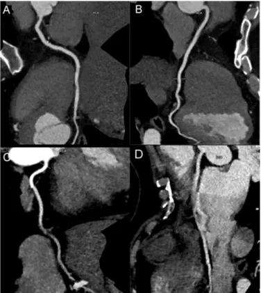
artifacts, non-diagnostic (.igure 2) (9, 10). Only
segments with a diameter of ≥1.5 mm were
evaluated. Discrepancy was resolved by consensus of the two observers. Furthermore,
the objective image quality parameters were measured. Signal intensity was measured at the root of ascending aorta (AA), the proximal part of left main artery (LM), the right coronary artery (RCA), the left ascending artery (LAD), the left circum.lex artery (LCX) and mediastinal fat. Image noise was de.ined as the corresponding standard deviation at the aortic root. The signal-to-noise ratio (SNR) was calculated by dividing the mean signal intensity by image noise. The contrast-to-noise ratio (CNR) was calculated by dividing contrast values (vessel attenuation – fat attenuation) by image noise. One cardiologist (5 years of experience in CCA) evaluated all coronary angiograms. All available segments were assessed and categorized (greater than 50% or not greater than 50%)
using a validated quantitative coronary angiography (QCA) algorithm. The stenoses
were classi.ied as signi.icant if the lumen diameter reduction was ≥50%.
227
Figure 2. Subjec ve image quality was evaluated using a 4-point scale: 1 = excellent, no ar facts (A); 2 = good, minor ar facts (B); 3 = moderate, moderate ar facts but s ll diagnos c (C); 4 = poor, severe ar facts, non-diagnos c (D).
Radiation dose estimation
The study investigators obtained the parameters relevant to radiation dose, including
volume CT dose index (CTDIvol) and dose-length product (DLP) from the scan protocol generated by the CT system after each
coronary CTA study. The estimated radiation dose (ED) was calculated by multiplying the DLP by the conversion coef.icient (0.014 mSv · mGy-1 · cm-mGy-1) at the chest (mGy-1mGy-1). This weighting factor
is considered to be derived from the most self-consistent and reliable dataset.
Statistical analysis
All statistical analyses were performed with using SPSS software version 17.0 (SPSS Inc. Chicago, USA). Quantitative variables were expressed as the means ± standard deviations, and categorical variables were expressed as frequencies and percentages. The Student’s t-test, Mann-Whitney U test, Chi-square test and Fisher’s exact test were performed to analyze the differences in patient characteristics, image acquisition parameters, objective image quality parameters, and estimated radiation dose. The inter-observer agreement between the two observers was evaluated with kappa analysis (κ > 0.75, excellent agreement; κ = 0.4-0.75, good agreement). Mean image quality grade between groups A and B was assessed using the Mann-Whitney U test. The rates of diagnostic segments and patients were compared using the Chi-square test. Statistical signi.icance was de.ined as a P-value of < 0.05.
The diagnostic performance of CTCA for the diagnosis of signi.icant coronary artery disease compared with the reference standard was determined using sensitivity, speci.icity, positive predictive value (PPV) and negative predictive value (NPV) and their 95% con.idence intervals.
RESULTS
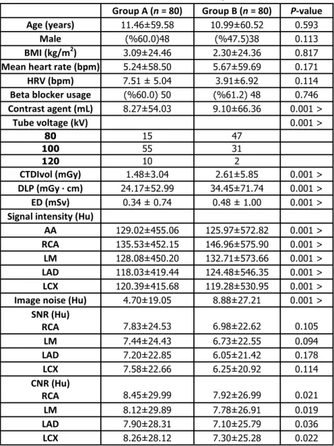
The patient characteristics and scan parameters are summarized in table 1. Signi.icant differences in image noise, contrast
agent cost, kV ratio and CNR (P < 0.05) were
noted. The estimated radiation dose of group A was 26.0% reduced compared with group B (0.74 ± 0.34 mSv vs. 1.00 ± 0.48 mSv, P < 0.001). No signi.icant differences in mean HR, HRV, and
other patient characteristics and scan parameters were noted (P > 0.05).
Image quality scores of coronary artery segments in the two groups are summarized in
table 2. In group A, a total of 980 segments were included in the analysis, of which 99.4% (974/980) segments were diagnostic (.igure 3). In group B, 978 segments were evaluated, of which 98.9% (967/978) was diagnostic. No signi.icant difference was observed in the rate of diagnostic segments between groups A and B (P = 0.222). Mean image quality scores were 1.51 ±
0.32 and 1.70 ± 0.38 for groups A and B, respectively. A statistical difference in image
quality scores was noted between groups A and B (P < 0.001). On a per-patient basis, the rates of diagnostic patients were 92.5% (74/80) and 88.8% (71/80) for groups A and B, respectively. No statistical difference was observed regarding the rate of diagnostic patients between the two groups (P = 0.416). On a per-segment basis, the rates of diagnostic segments between the two groups were 98.7% (312/316) and 98.7% (306/310), 100% (412/412) and 99.3% (415/418), 100% (250/250) and 98.4% (246/250) in RCA, LAD (including LM) and LCX, respectively. No statistical difference was found in the rate of diagnostic segments in RCA and LAD (P=0.551, P=0.085). A statistical difference in the diagnostic segments in LCX (P=0.045) was noted between the two groups.
There was excellent agreement of image quality grading between the two reviewers (κ= 0.530). Six segments had non-diagnostic images due to motion artifacts (4/6), poor cooperation in breath holding (1/6) and blurring artifacts (1/6) in group A, and these events all occurred in
RCA. In group B, there were eleven non-diagnostic segments caused by severe
stair-step artifacts (8/11) and blurring artifacts (3/11). Four segments occurred in RCA, 3 in LAD and an additional 4 in LCX.
Using CCA .indings as the reference standard,
a total of 35 (413 segments) and 36 (425 segments) patients were con.irmed in the
228
229
high-pitch and sequential acquisition group,
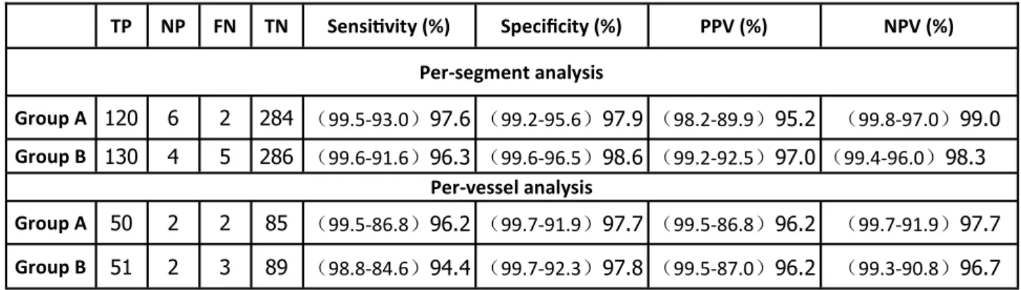
respectively. The diagnostic accuracy, sensitivity, speci.icity, PPV and NPV on per-segment and per-vessel bases are presented
in table 3. In the 71 patients, 258 segments stenoses with ≥50% lumen diameter reduction
were detected by CCA, whereas 260 were observed by DSCT. Of the coronary segments, 10
were classi.ied as false-positive by DSCT: 6 were
invaluable and thus estimated as having signi.icant stenosis but did not exhibit
signi.icant stenosis on CCA, and 4 segments were evaluated as false-positive given that the degree of lumen reduction was overestimated. Eight segments were classi.ied as false negative due to the non-diagnostic image. The diagnostic
accuracy values of the high-pitch group and sequential group were 97.8% (404/413) and
97.9% (416/425), respectively. No signi.icant difference in the diagnostic accuracy was noted between the two groups (p=0.951).
Group A (n = 80) Group B (n = 80) P-value
Age (years) 11.46±59.58 10.99±60.52 0.593
Male (%60.0)48 (%47.5)38 0.113
BMI (kg/m2) 3.09±24.46 2.30±24.36 0.817
Mean heart rate (bpm) 5.24±58.50 5.67±59.69 0.171
HRV (bpm) 7.51 ±5.04 3.91±6.92 0.114
Beta blocker usage (%60.0)50 (%61.2)48 0.746
Contrast agent (mL) 8.27±54.03 9.10±66.36 0.001 >
Tube voltage (kV) 0.001 >
80 15 47
100 55 31
120 10 2
CTDIvol (mGy) 1.48±3.04 2.61±5.85 0.001 >
DLP (mGy · cm) 24.17±52.99 34.45±71.74 0.001 >
ED (mSv) 0.34 ±0.74 0.48 ±1.00 0.001 >
Signal intensity (Hu)
AA 129.02±455.06 125.97±572.82 0.001 >
RCA 135.53±452.15 146.96±575.90 0.001 >
LM 128.08±450.20 132.71±573.66 0.001 >
LAD 118.03±419.44 124.48±546.35 0.001 >
LCX 120.39±415.68 119.28±530.95 0.001 >
Image noise (Hu) 4.70±19.05 8.88±27.21 0.001 >
SNR (Hu) RCA
24.53 ± 7.83
22.62 ± 6.98
0.105
LM 7.44±24.43 6.73±22.55 0.094
LAD 7.20±22.85 6.05±21.42 0.178
LCX 7.58±22.66 6.25±20.92 0.114
CNR (Hu) RCA
29.99 ± 8.45
26.99 ± 7.92
0.021
LM 8.12±29.89 7.78±26.91 0.019
LAD 7.90±28.31 7.10±25.79 0.036
LCX 8.26±28.12 7.30±25.28 0.022
Table 1. Pa ent characteris cs and scan parameters
230
Int. J. Radiat. Res., Vol. 16 No. 2, April 2018
Image quality Group A (n = 980) Group B (n = 978)
1 (%56.9)557 (%43.1)421
2 (%36.9)362 (%45.5)445
3 (%5.6)55 (%10.3)101
4 (%0.6)6 (%1.1)11
Table 2. Image quality scores of the two groups
Figure 3. High-pitch spiral CCTA of a 65-year-old man exhibited excellent volume rendering images (A) and curve mulitplanar reforma on imaged of RCA (B) and LAD (C). Mixed plaques and soF plaques were observed in the right coronary artery and leF
anterior descending artery. The lesion was confirmed by CCA (D and E). The dose-length product was 51 mGy.cm (F), and the es mated effec ve dose was 0.71 mSv.
A four-point grading scale was used with grading defined as follows: 1, excellent; 2, good; 3, moderate, diagnos c; 4, poor image quality, non-diagnos c.
TP NP FN TN Sensi7vity (%) Specificity (%) PPV (%) NPV (%)
Per-segment analysis
Group A 120 6 2 284 (99.5-93.0)97.6 (99.2-95.6)97.9 (98.2-89.9)95.2 (99.8-97.0)99.0
Group B 130 4 5 286 (99.6-91.6)96.3 (99.6-96.5)98.6 (99.2-92.5)97.0(99.4-96.0)98.3
Per-vessel analysis
Group A 50 2 2 85 (99.5-86.8)96.2 (99.7-91.9)97.7 (99.5-86.8)96.2 (99.7-91.9)97.7
Group B 51 2 3 89 (98.8-84.6)94.4 (99.7-92.3)97.8 (99.5-87.0)96.2 (99.3-90.8)96.7 Figure 3. High-pitch spiral CCTA of a 65-year-old man exhibited excellent volume rendering images (A) and curve mulitplanar reforma on imaged of RCA (B) and LAD (C). Mixed plaques and soF plaques were observed in the right coronary artery and leF
anterior descending artery. The lesion was confirmed by CCA (D and E). The dose-length product was 51 mGy.cm (F), and the es mated effec ve dose was 0.71 mSv.
Table 3. Diagnos c performance of high-pitch mode and sequen al mode on 128-slice DSCT angiography.
DISCUSSION
The recent development of new-generation dual-source CT systems allowed a high-pitch spiral acquisition protocol with a very high-pitch value of 3.4 (1-8). In our study, the high-pitch mode acquired better diagnostic performance at a low radiation dose and contrast agent cost compared with sequential mode. The high-pitch mode can be applied as the .irst-line protocol for patients with low and stable heart rates.
The high-pitch spiral mode and sequential mode with diastolic reconstruction have been used successfully in patients with heart rates less than 70 bpm (9-13). During late diastole, most of left ventricular .illing has already occurred,
and the myocardium is relaxed. Thus, late diastole is relatively quiescent compared with
other phases of the cardiac cycle, making it optimal for image acquisition due to reduced coronary motion. However, the duration of late diastole decreases with increasing heart rate. For patients with heart rates greater than 80 bpm, late diastole becomes negligible in length,
and diastolic coronary motion increases signi.icantly (14, 15). Our results were consistent
with previous studies. The heart rates in the two
groups were less than 70 bpm, and image quality scores were good to excellent.
The most challenging issue in CCTA is non-diagnostic imaging. The high-pitch spiral
mode of DSCT enables data acquisition for one
heart beat with continuous and fast table movement (16, 17). In high-pitch mode, non-diagnostic images are mainly attributed to
motion artifact, predominantly affecting the mid to distal RCA. Minimal motion artifacts occur in a
phase of the cardiac cycle with the least coronary artery motion (6). For sequential CCTA,
stair-step artifacts are the main reason for non-diagnostic images. Coronary CT angiography performed on wide detector offers
a large z-coverage that reduces the scan blocks (17-19). Stair-step artifacts typically do not occur in high-pitch mode given the high speed of data acquisition within a single cardiac cycle. In our study, four segments had non-diagnostic images due to motion artifacts (4/6) in group A, and all segments occurred in RCA. In group B, eight non
-diagnostic segments were caused by severe stair-step artifacts (8/11): 3 segments occurred in RCA, 3 in LAD and 2 in LCX. The high-pitch mode has an advantage in the manifestation of LCX.
Numerous factors affect CCTA radiation dose,
including scan length, scan speed and tube voltage. The main improvement of DSCT using
high-pitch mode was the increased thread pitch from 0.2-0.4 to 3.4. Thus, the total scan time and
the radiation dose were subsequently signi.icantly reduced. An automatic tube voltage
selection technique was used in the two groups. The technique was based on modulating the tube voltage and maintaining constant image noise from patient to patient and over the entire
scan. The reference voltage was adjusted automatically according to the anatomy of the
patient’s body and organs (2). This technique was
used to reduce radiation exposure while preserving diagnostic image quality in our study.
Group A used a higher tube voltage, and there
was no additional radiation exposure. Our results suggested that radiation exposure could
be signi.icantly reduced with the high-pitch protocol. Two main reasons explain this .inding. First, the sequential technique requires an overlap of approximately 10% of block images due to the cone-beam geometry. However, this overlap is not required for the high-pitch spiral mode. Second, the sequential scan technique requires “beam on” time for each block, whereas the high-pitch mode requires the extra “beam on” only once. Thus, the cumulative “beam on”
time is reduced for the high-pitch spiral technique (8).
From a clinical perspective, reducing the amount of iodine as much as possible is helpful
to avoid the risk of contrast-induced nephropathy (20-22). However, a lower iodine concentration will lead to reduced vascular attenuation, which may result in degraded image quality and thus affect diagnostic accuracy. In our study, we reduced the contrast
agent cost in group A, which only required one
cardiac cycle. Group A had a lower mean attenuation. This .inding may be explained by
the reduced contrast media cost and 80 kV ratio in group A. The high-pitch mode achieved
231
signi.icant reductions in image noise and radiation dose and improvements in image quality and CNR. CCTA is becoming the .irst-line diagnostic examination in patients with low-to-intermediate risk of coronary heart disease (23). High-pitch mode provides high diagnostic accuracy for the detection of coronary artery stenoses in patients with low and stable heart rates (23-25). Our results were in contrast with the results reported by Nee.jes et al.(23).
The strength of CCTA is its high negative predictive value. As a .irst-line test, CCTA should be as safe and non-invasive as possible because the vast majority of patients do not have signi.icant CAD. The introduction of radiation-lowering techniques has boosted the
implementation of CCTA in routine clinical settings (23-25). Achembach et al. reported that the high-pitch mode provided high diagnostic accuracy for the detection of coronary artery
stenoses with a sensitivity of 100% and a negative predictive value of 100% in patients
with low and stable heart rates (25). Our results were consistent with previously reported work, but most of patients did not need to undergo conventional coronary angiography. Thus, we will collect more materials and perform more studies in the future.
Study limitations
There are several limitations. First, we excluded patients with high and irregular heart rates. Second, the image quality grading may have been in.luenced by a subjectivity bias. Third, image diagnostic accuracy was compared with conventional CCA in part patients. Finally, we used a 128-slice DSCT, and the results cannot be generalized to other CT systems.
CONCLUSION
Our study suggested that the prospectively ECG-triggered high-pitch spiral acquisition CCTA reduces radiation doses and contrast agent costs while improving image quality compared with prospectively ECG-triggered sequential mode.
Con licts of interest: Declared none.
REFERENCES
1. Paul JF, Amato A, Rohnean A (2013) Low-dose coronary-CT angiography using step and shoot at any heart rate: com-parison of image quality at systole for high heart rate and diastole for low heart rate with a 128-slice dual-source machine. Int J Cardiovasc Imaging, 29: 651-657.
2. Pan CJ, Qian N, Wang T, Tang XQ, Xue YJ (2013) Adap ve prospec ve ECG-triggered sequence coronary angiography in dual-source CT without heart rate control: Image quality and diagnos c performance. Exp Ther Med, 5: 636-642. 3. Hausleiter J, Meyer T, Hermann F, Hadamitzky M, Krebs M,
Gerber TC, McCollough C, Mar noff S, Kastra A, Schomig A, Achenbach S (2009)Es mated radia on dose associat-ed with cardiac CT angiography. JAMA, 301: 500-507. 4. Yin WH, Lu B, Gao JB, Li PL, Sun K, Wu ZF, Yang WJ, Zhang
XQ, Zheng MW, McQuiston AD, Meinel FG, Schoepf UJ (2015) Effect of reduced x-ray tube voltage, low iodine concentra on contrast medium, and sinogram-affirmed itera ve reconstruc on on image quality and radia on dose at coronary CT angiography: results of the
prospec-ve mul center REALISE trial. J Cardiovasc Comput
Tomogr, 9: 215-224.
5. Sabarudin A, Md Yusof AK, Tay MF, Ng KH, Sun Z (2013) Dual-source CT coronary angiography: effec veness of radia on dose reduc on with lower tube voltage. Radiat
Prot Dosimetry, 153: 441-447.
6. Sun K, Han RJ, Ma LJ, Wang LJ, Li LG, Chen JH (2012) Pro-spec vely electrocardiogram-gated high-pitch spiral acqui-si on mode dual-source CT coronary angiography in pa-ents with high heart rates: comparison with retrospec ve electrocardiogram-gated spiral acquisi on mode. Korean J
Radiol 13: 684-693.
7. Muenzel D, Noel PB, Dorn F, Dobritz M, Rummeny EJ, Hu-ber A (2011) Step and shoot coronary CT angiography using 256-slice CT: effect of heart rate and heart rate vari-ability on image quality. Eur Radiol, 21: 2277-2284. 8. Nie P, Wang X, Cheng Z, Ji X, Duan Y, Chen J (2012)
Accura-cy, image quality and radia on dose comparison of high-pitch spiral and sequen al acquisi on on 128-slice dual-source CT angiography in children with congenital heart disease. Eur Radiol, 22: 2057-2066.
9. Kim HY, Lee JW, Hong YJ, Lee HJ, Hur J, Nam JE, Choi BW, Kim YJ (2012) Dual-source coronary CT angiography in pa ents with high heart rates using a prospec vely ECG-triggered axial mode at end-systole. Int J Cardiovasc
Imag-ing, 28: 101-107.
10. Xu L, Yang L, Zhang Z, Wang Y, Jin Z, Zhang L, Lu G (2013) Prospec vely ECG-triggered sequen al dual-source coro-nary CT angiography in pa ents with atrial fibrilla on: comparison with retrospec vely ECG-gated helical CT. Eur
Radiol, 23: 1822-1828.
11. Lu B, Lu JG, Sun ML, Hou ZH, Chen XB, Tang X, Wu RZ, Johnson L, Qiao SB, Yang YJ and Jiang SL (2011) Compari-son of diagnos c accuracy and radia on dose between prospec ve triggering and retrospec ve gated coronary
232
233
angiography by dual-source computed tomography. Am J
Cardiol, 107: 1278-1284.
12. Stolzmann P, GoeT R, Baumueller S, Plass A, Falk V, Scheffel H, Feuchtner G, Marincek B, Alkadhi H, Leschka S (2010) Prospec ve and retrospec ve ECG-ga ng for CT coronary angiography perform similarly accurate at low heart rates. Eur J Radiol, 79: 85-91.
13. Lee AM, Beaudoin J, Engel LC , Sidhu MS, Abbara S, Brady TJ, Hoffmann U, Ghoshhajra BB (2013) Assessment of image quality and radia on dose of prospec vely ECG-triggered adap ve dual-source coronary computed tomog-raphy angiogtomog-raphy (cCTA) with arrhythmia rejec on algo-rithm in systole versus diastole: a retrospec ve cohort study. Int J Cardiovasc Imaging, 29: 1361-1370.
14. Achenbach S, Ropers D, Holle J, Muschiol G, Daniel WG, Moshage W (2000) In-plane coronary arterial mo on ve-locity: measurement with electron-beam CT. Radiology,
216: 457-463.
15. Adler G, Meille L, Rohnean A, Sigal-Cinqualbre A, Capderou A, Paul JF (2009)Robustness of end-systolic
reconstruc-ons in coronary dual-source CT angiography for high heart rate pa ents. Eur Radiol, 20: 1118-1123.
16. Leschka S, Stolzmann P, Desbiolles L, Baumueller S, GoeT R, Schertler T, Scheffel H, Plass A, Falk V, Feuchtner G, Marincek B, Alkadhi H (2009) Diagnos c accuracy of high-pitch dual-source CT for the assessment of coronary steno-ses: first experience. Eur Radiol 19: 2896-2903.
17. Araoz PA, Kirsch J, Primak AN, Braun NN, Saba O, William-son EE, Harmsen WS, Mandrekar JN, McCollough CH (2009) Op mal image reconstruc on phase at low and high heart rates in dual-source CT coronary angiography.
Int J Cardiovasc Imaging, 25: 837-845.
18. Husmann L, Herzog BA, Burkhard N, Tatsugami F, Valenta I, Gaemperli O, Wyss CA, Landmesser U and Kaufmann PA (2009) Body physique and heart rate variability determine the occurrence of stair-step artefacts in 64-slice CT coro-nary angiography with prospec ve ECG-triggering. Eur
Radiol, 19: 1698-1703.
19. Stolzmann P, Leschka S, Scheffel H, Krauss T, Desbiolles L, Plass A, Genoni M, Flohr TG, Wildermuth S, Marincek B, Alkadhi H (2008) Dual-source CT in step-and-shoot mode: noninvasive coronary angiography with low radia on dose. Radiology, 249: 71-80.
20. McDonald JS, McDonald RJ, Comin J, Williamson EE, Katzberg RW, Murad MH and Kallmes DF (2013) Frequency of acute kidney injury following intravenous contrast me-dium administra on: a systema c review and meta-analysis. Radiology, 267: 119-128.
21. Newhouse JH and RoyChoudhury A (2013) Quan ta ng contrast medium-induced nephropathy: controlling the controls. Radiology, 267: 4-8.
22. McDonald RJ, McDonald JS, Bida JP, Carter RE, Fleming CJ, Misra S, Williamson EE, Kallmes DF (2013) Intravenous contrast material-induced nephropathy: causal or coinci-dent phenomenon? Radiology, 267: 106-118.
23. NeeVes LA, Rossi A, Genders TS, Nieman K, Papadopoulou SL, Dharampal AS, Schultz CJ, Weus nk AC, Dijkshoorn ML, Ten Kate GJ, Dedic A, van Straten M, Cademar ri F, Hunink MG, Kres n GP, de Feyter PJ, Mollet NR (2012) Diagnos c accuracy of 128-slice dual-source CT coronary angi-ography: a randomized comparison of different acquisi on protocols. Eur Radiol, 23: 614-622.
24. Weus nk AC, Mollet NR, NeeVes LA, Meijboom WB, Gale-ma TW, van Mieghem CA, Kyrzopoulous S, Eu RN, NieGale-man K, Cademar ri F, van Geuns RJ, Boersma E, Kres n GP, de Feyter PJ (2010) Diagnos c accuracy and clinical u lity of noninvasive tes ng for coronary artery disease. Ann Intern
Med, 152: 630-639.
25. Achenbach S, Goroll T, Seltmann M, Pflederer T, Anders K, Ropers D, Daniel WG, Uder M, Lell M, Marwan M (2011) Detec on of coronary artery stenoses by low-dose, pro-spec vely ECG-triggered, high-pitch spiral coronary CT angiography. JACC Cardiovasc Imaging, 4: 328-337.