Dittapong Songsaeng, M.D.*, Sunsiree Soodchuen, M.D.*,**, Pornpim Korpraphong, M.D.*, Anek Suwanbundit., Ph.D.*
*Department of Radiology, Faculty of Medicine Siriraj Hospital, Mahidol University, Bangkok 10700, **Department of Radiology, Faculty of Medicine, Srinakaharinwirot University, Nakhon Nayok 26120, Thailand.
Siriraj Thyroid Imaging Reporting and Data System
and Its Efficacy
Correspondence to: Dittapong Songsaeng E-mail: [email protected]
ABSTRACT
Objective: To standardize a modified practical thyroid imaging reporting and data system (Siriraj-TIRADS). Methods: 196 thyroid nodules underwent ultrasound and FNAB and were retrospectively pathologically proven. Data design was divided into 2 groups of benign and malignant. Sensitivity and specificity of cancer prediction were calculated for the comparison of Siriraj-TIRAD and TIRAD. Interobserver agreement was calculated by kappa statistics.
Results: Sensitivity and specificity of Siriraj’s TIRADS on cancer prediction were 95% and 64.8%, respectively, and sensitivity and specificity of TIRADS were 45% and 92%, respectively.
Conclusion: The Siriraj- TIRADS classification is easy, practical and has better sensitivity than TIRADS for the diagnosis of thyroid carcinoma. TIRADS is good for the screening management strategy but Siriraj-TIRADS has benefit to cancer management strategy.
Keywords: Siriraj thyroid imaging reporting and data system; Siriraj-TIRADS (Siriraj Med J 2017;69: 262-267)
BACKGROUND
In 2009, Thyroid imaging reporting and data system (TIRADS) (Table 1) was introduced by Horvath et al4 to
ultrasonographic screening of thyroid nodule patients before they underwent diagnosis by fine needle aspiration biopsy (FNAB) to protect them from unnecessary complications.5,7 Since then it has been widely used for
the ultrasonographic criteria of thyroid nodule for clinical management which is the aim of cancer screening, in which only 7% are malignant.2 Several ultrasonographic
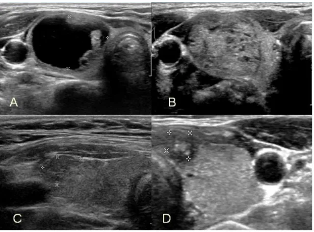
characteristics have been proposed to identify nodules at risk for being malignant (Fig 1, 2), such as solid hypoechoic nodules, containing microcalcification and central hypervascularization.3 The diagnostic challenge
was to categorize thyroid lesions with a percentage of malignancy. In 2011, Kwak et al8, complemented this
classification by adding one subtype whereas the use of the TIRADS criteria has reduced the number of FNABs.
TABLE 1. TIRAD Clasification (Horvath et al, 2009).
Grade Interpretation Meaning
1 Normal thyroid gland Normal
2 Benign conditions 0% malignancy
3 Probably benign nodules 5% malignancy
4 Suspicious nodules 5-80% malignancy
4a 5-10% malignancy
4b 10-80% malignancy
5 Probably malignant nodules 80% malignancy
6 Proven malignancy Category included biopsy proven malignant nodules
TABLE 2. Siriraj-TIRAD system.
Fig 2. Example of ultrasonographic characteristics of TIRAD (Horvath et al, 2009).
A: Type 1 colloid nodule (TIRADS 2) B: Type 2 colloid nodule (TIRADS 2) C: Type 3 colloid nodule (TIRADS 2)
D: Hashimoto thyroiditis with a pseudo-nodule (arrow) (TIRADS 3)
E: A simple neoplastic pattern (TIRADS 4A)
F: A hypoechoic area with ill-defined borders, without calcifications (TIRADS 4A)
G: A suspicious neoplastic pattern (TIRADS 4B)
H: A malignant pattern A: solid hypoechoic, irregular nodules with ill-defined margins, with calcifications (thin arrow) or without calcifications (thick arrow) (TIRADS 4B)
I: A malignant pattern B: solid, nonencapsulated, isoechoic, ill-defined nodule with peripheral microcalcifications (TIRADS 5)
J: A malignant pattern C: a mixed, isoechoic, vascularized, nonencapsulated nodule with calcifications and no hyperechoic spots (TIRADS 6)
Grade Interpretation Meaning
0 Normal thyroid gland without thyroid nodule Normal with no action
1 Cyst, colloidal cyst with comet tail Highly suggestive of benign nodule 2 Spongioform pattern : heterogenous echo, Advised management is to follow up US
macrocalcification and benign calcification, next 6 months cystic nodule with intramural solid component
3 Well-defined mass with microcalcification Indeterminate. Advised management is
to go on FNAB
4 Ill-defined mass with or without microcalcification Probably malignant. Advised management
is to FNAB and immediate re-aspiration
if non diagnostic FNA result
5 Spiculated mass, outgrowth lesion Highly suggestive of malignancy.
Advised management is to consider
surgery regardless of FNAB result
MATERIALS AND METHODS
This study was conducted at the Siriraj-imaging Center of the Department of Radiology, Faculty of Medicine Siriraj Hospital, Mahidol University between June 2011 and May 2013. The study was approved by the institutional Medical Ethics Committee Board (Si 526/2013). Informed consent was obtained from all patients before recruitment. A total of 289 thyroid nodules submitted for fine needle aspiration biopsy were image reviewed and scored using both Siriraj-TIRAD and TIRAD. The ultrasound equipment was the Philips iU22 (Philips Healthcare, Andover, MA) with advanced breast mode protocol with a 5-12 MHz linear probe. Then FNAB was performed in compliance and tissues obtained received routine histological studies. However, only 196 samples were analyzed completely. To divide benign and malignant nodule with Siriraj TIRADS, the cut-off scores were Siriraj-TIRAD 1, 2, 3 was benign type and Siriraj-TIRAD score 4, 5 was malignant type. In contrast, TIRADS score 1, 2, 3, 4A as benign type and TIRAD score 4B, 5 as malignant type nodule.
Interobserver reliability was checked by a third year
radiology resident and a 12-yr experienced radiologist, blinded to the histopathological result, who reviewed the images and scored the nodule grade on the basis of TIRAD and Siriraj-TIRAD.
Statistical analysis
Interobserver reliability was analyzed by kappa statistics. Sensitivity and specificity were calculated for the Siriraj-TIRAD and TIRAD between benign and malignant group. A p-value less than 0.05 vs considered to indicate a significant difference.
RESULTS
Of the 196 nodules, 176 were benign and 20 were malignant. The kappa statistic of interobserver reliability was 1.0 (Siriraj-TIRAD) and 0.8 (TIRAD) respectively. The number and percentage of benign and malignant nodules in each Siriraj-TIRAD and TIRADS categories were shown in Table 3 and 4. Summarily, sensitivity and specificity of Siriraj-TIRADS were 95% and 64.8% as shown in Table 5 and TIRADS were 45% and 92% as shown in Table 6, respectively.
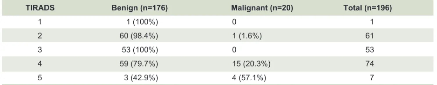
TABLE 3. Classification of thyroid nodule evaluated by Siriraj- TIRADS.
TIRADS Benign (n=176) Malignant (n=20) Total (n=196)
1 1 (100%) 0 1
2 60 (98.4%) 1 (1.6%) 61
3 53 (100%) 0 53
4 59 (79.7%) 15 (20.3%) 74
5 3 (42.9%) 4 (57.1%) 7
TABLE 4. Classification of thyroid nodule evaluated by TIRADS.
TIRADS Benign (n=176) Malignant (n=20) Total (n=196)
2 60 (98.4%) 1 (1.6%) 61
4A 102 (91.1%) 10 (8.9%) 112
4B 14 (66.7%) 7 (33.3%) 21
5 0 2 (100%) 2
TABLE 5. Sensitivity and specificity of Siriraj TIRADS.
Final pathological diagnosis Total
Benign Malignant
Siriraj-TIRADS Score 1,2,3 114 (64.8%) 1 (5.0%) 115
Score 4,5 62 (35.2%) 19 (95.0%) 81
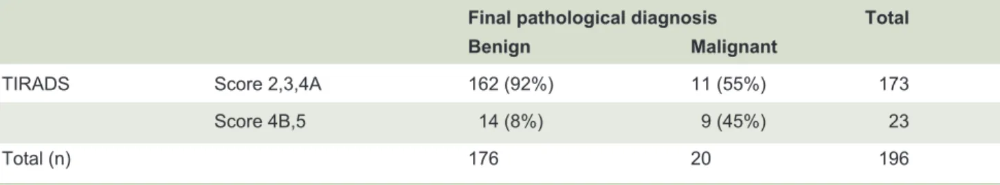
TABLE 6. Sensitivity and specificity of TIRADS.
Final pathological diagnosis Total
Benign Malignant
TIRADS Score 2,3,4A 162 (92%) 11 (55%) 173
Score 4B,5 14 (8%) 9 (45%) 23
Total (n) 176 20 196
DISCUSSION
Ultrasonography is commonly used to check for the prevalence of thyroid nodule1 and TIRAD is a tool which is widely utilized by radiologists to select high risk nodulws for FNAB in the screening with the concept of avoiding unnecessary FNAB and cost-effectiveness since the sensitivity of benign prediction was high (88%) and specificity was low (49%)4 which aligned to the clinical
management strategies. However, Siriraj Hospital, Thailand is a university hospital, which needs high accuracy of diagnosis of cancer. High sensitivity value of cancer prediction was required, but TIRAD by itself has not supported our goal. Even though, modified TIRAD is done by many groups of researchers8,9,10 some inaccuracy
still exists.
The data design of this research was reversed to Horvart in which TIRAD showed the low sensitivity of cancer prediction (45%) while Siriraj-TIRAD showed the high
sensitivity (95%) and moderate specificity (64.8%) which was sufficient benefit to apply the new classification to the clinical management for high impact. Our classification is simplified and has high sensitivity of malignancy prediction while it enrolled more number of patients to the FNAB. Therefore, the Siriraj-TIRADS score of 1 to 3 patients had a low probability of malignancy, which may indicate that it is comfortable to follow a wait and see strategy.
CONCLUSION
The Siriraj-TIRADS classification of thyroid nodules based on a score according to the most relevant sonographically suspicious criteria for malignancy was more easily applied in daily practice. High sensitivity of cancer prediction and moderate specificity is sufficient for benign prediction. Strategically, the clinical management has to take a role. If the strategy is the screening who does not need FNAB, our classification does not fit, but if the strategy changed to be detection and optimized manipulation of thyroid cancer, our classification is preferred.
REFERENCES
1. Ezzat S, Sarti DA, Cain DR, Braunstein GD. Thyroid incidentalomas: prevalence by palpation and ultrasonography. Arch Intern Med 1994; 154(16):1838-40.
2. Papini E, Guglielmi R, Bianchini A, Crescenzi A, Taccogna S, Nardi F, et al. Risk of malignancy in nonpalpable thyroid nodules: predictive value of ultrasound and color-Doppler features. J Clin Endocrinol Metab 2002; 87(5): 1941-6.
3. Nam-Goong IS, Kim HY, Gong G, Lee HK, Hong SJ, Kim WB, et al. Ultrasonography-guided fine-needle aspiration of thyroid incidentaloma: correlation with pathological findings.
Clin Endocrinol (Oxf) 2004; 60(1): 21-8.
4. Horvath E, Majlis S, Rossi R, Franco C, Niedmann JP, Castro A, et al. An ultrasonogram reporting system for thyroid nodules stratifying cancer risk for clinical management. J Clin Endocrinol Metab 2009; 94(5):1748-51.
5. Khoo TK, Baker CH, Hallanger-Johnson J, Tom AM, Grant CS, Reading CC, et al. Comparison of ultrasound-guided fine- needle aspiration biopsy with core-needle biopsy in the evaluation of thyroid nodules. Endocrine Practice 2008; 14(4): 426-31.
6. Gursoy A, Ertugrul DT, Sahin M, Tutuncu NB, Demirer AN, Demirag NG, et al. The analgesic efficacy of lidocaine/prilocaine (EMLA) cream during fine-needle aspiration biopsy of thyroid nodules. Clinical Endocrinolog 2007; 66: 691-694.
7. Stergios A. Polyzos, Athanasios D. Anastasilakis AD. Clinical Complications Following Thyroid Fine-needle Biopsy: A Systematic Review. Clin Endocrinol 2009; 71(2): 157-165.
8. Kwak JY, Han KH, Yoon JH, Moon HJ, Son EJ, Park SH, et al. Thyroid imaging reporting and data system for US fratures of nodules: A step in estaplishing better stratification of cancer risk. Radiology 2011; 260(3): 892-99.
9. Russ G, Royer B, Bigorgne C, Rouxel A, Bienvenu-Perrard M, Leenhardt L. Prospective evaluation of thyroid imaging reporting and data system on 4550 nodules with and without elastography. Eur J Endocrinol 2013; 168: 649-655.