Original Research Article
A study of C reactive proteins in patients with stable chronic
obstructive pulmonary disease
Ashok Kumar Gudagunti
1*, Sudhir Chaudari
2INTRODUCTION
Chronic obstructive pulmonary disease (COPD) is a preventable n treatable disease with significant extra-pulmonary manifestations. There is progressively worsening limitation of airflow which can be partially but not fully reversible with medications. Exposure to noxious particles or gases can lead to abnormal inflammatory response in the lungs as well as systemic
inflammation. The inflammatory response is the mainstay of this disease.1 COPD causes the major burden not only
on the patient but also on his family and the community. The prevalence is increasing globally. It can also contribute to death of the patient.2 It has been estimated
by world health organization (WHO) that globally COPD ranks at fourth place as the causes of death. Every year it has been estimated that nearly 2.75 million people die due to COPD all over the world. The mortality and
ABSTRACT
Background: Whenever there is damage to the tissue or inflammatory process, the hepatocytes synthesize a protein which is an acute phase reactant, and this is the C reactive protein. Studies have shown that serum C reactive protein levels are usually elevated during acute exacerbations of COPD. The objective of this study was studying the baseline serum C reactive protein levels in patients with stable chronic obstructive pulmonary disease.
Methods: During the study period, 90 subjects were studied. Out of them, 47 had stable COPD and 43 were without COPD. The subjects were either admitted in indoor wards or were attending OPD of department of tuberculosis and respiratory diseases, Dr. Murari Lal Chest Hospital, G.S.V.M Medical College, Kanpur, Uttar Pradesh, and were recruited if they fulfilled the criteria for inclusion. Informed consent was taken from all study subjects and the study protocol was approved by the board of faculty of medicine.
Results: Of the 47 patients in the stable COPD group, 23 were found to be active smokers. Pearson’s correlation coefficient showed that lower limit of serum CRP levels were significantly negatively correlating with 6 MWD (r= -0.707; p=0.0001), FEV1% (r=-0.635; p=0.0001), PaO2 (r=-0.592; p=0.0001), and BMI (r= -0.534; p=0.0001).
Pearson’s correlation coefficient showed that upper limit of serum CRP levels were significantly negatively correlating with 6 MWD (r= -0.707; p=0.0001), FEV1% (r= -0.633; p=0.0001), PaO2 (r= -0.61; p=0.001) and BMI
(r= -0.520; p=0.0001).
Conclusions: The circulating levels of inflammatory marker, baseline serum CRP are significantly elevated in patients with stable chronic obstructive pulmonary disease.
Keywords: Chronic obstructive pulmonary disease, C reactive proteins, Stable
1Department of Pulmonary Medicine, Karnataka Institute of Medical Sciences, Hubballi, Karnataka, India 2Department of Pulmonary Medicine, Murari Lal Chest Hospital, GSVM Medical College, Kanpur, Uttar Pradesh,
India
Received: 27 February 2019
Accepted: 02 March 2019
*Correspondence:
Dr. Ashok Kumar Gudagunti, E-mail: [email protected]
Copyright: © the author(s), publisher and licensee Medip Academy. This is an open-access article distributed under the terms of the Creative Commons Attribution Non-Commercial License, which permits unrestricted non-commercial use, distribution, and reproduction in any medium, provided the original work is properly cited.
prevalence rate are going on increasing. It has been predicted that COPD can advance in the rankings of leading causes of the death to the third place very soon.3
C reactive protein is a marker which is used to indicate inflammatory responses in patients with COPD in recent years. Use of CRP as a marker is on increase. Whenever there is damage to the tissue or inflammatory process, the hepatocytes synthesize a protein which is an acute phase reactant, and this is the C reactive protein. Studies have shown that serum C reactive protein levels are usually elevated during acute exacerbations of COPD.4 The
overall course of the COPD can be predicted by the use of serum C reactive proteins. The C reactive protein has been found to be related to the degree of obstruction of the airflow.5 Present study was carried out to establish
baseline serum C reactive protein levels in patients with stable chronic obstructive pulmonary disease.
METHODS
A present study was carried out at G.S.V.M Medical College, Kanpur, Uttar Pradesh. The study period was from July 2009 to October 2010.
Sample size
During the study period, 90 subjects were studied. Out of them, 47 had stable COPD and 43 were without COPD. The subjects were either admitted in indoor wards or were attending OPD of Department of tuberculosis and respiratory diseases, Dr. Murari Lal Chest Hospital, G.S.V.M Medical College, Kanpur, Uttar Pradesh, and were recruited if they fulfilled the criteria for inclusion. Informed consent was taken from all study subjects and the study protocol was approved by the board of faculty of medicine.
Inclusion criteria
• Patients having post bronchodilator FEV1/FVC ratio of <0.7 after 400 micrograms of inhaled salbutamol,
• Clinically stable COPD patients,
• Apparently normal healthy persons, patients with bronchial asthma and bronchiectasis were included as controls.
Exclusion criteria
• Known chronic systemic infection or inflammatory condition such as SLE, rheumatoid arthritis etc.
• Chronic renal failure, history of ischemic heart disease in past 3 months,
• History of cerebro-vascular accidents in past 2 months.
• COPD patients with acute exacerbations.
A hospital based cross sectional comparative study. Detailed history was taken, and thorough clinical examination was carried out for all subjects. Chest X ray PA view and spirometry was done. Stage of COPD was recorded. Investigations like hemoglobin, total leukocyte count, differential leukocyte count, urine for albumin and sugar, fasting blood sugar, serum creatinine, SGPT, serum protein (gm%), sputum microscopy for AFB staining, ABG analysis, pulse oximetry, HRCT thorax, serum C reactive protein were done.
The subjects included in this study were classified into four groups. Group 1, the study group consisted of only stable COPD patients. The other three groups namely two, three and four were controls. Group 2 had apparently normal healthy persons, group 3 had 13 patients of bronchial asthma and group 4 had 7 patients of Bronchiectasis with no other illness. Now in group 1, depending upon the severity of airflow obstruction, stage of all patients was defined according to GOLD criteria.
Statistical analysis
The data was analyzed using proportions.
RESULTS
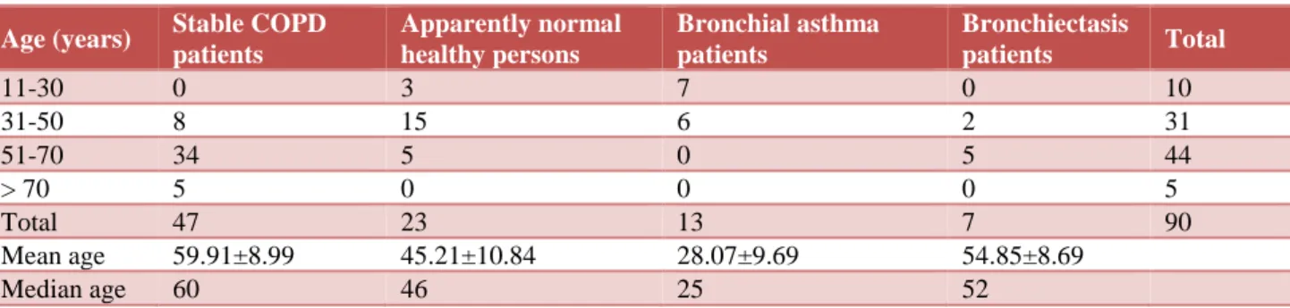
Table 1 shows age distribution of subjects in study and different control groups. The mean age of stable COPD patients (study subjects) was 59.91 years ±8.99, apparently healthy normal subjects was 45.21 years ±10.84, in group 3 28.07±9.69 years and in group 4 54.85±8.69 years.
Table 1: Age distribution of subjects in study and different control groups.
Age (years) Stable COPD patients
Apparently normal healthy persons
Bronchial asthma patients
Bronchiectasis
patients Total
11-30 0 3 7 0 10
31-50 8 15 6 2 31
51-70 34 5 0 5 44
> 70 5 0 0 0 5
Total 47 23 13 7 90
Mean age 59.91±8.99 45.21±10.84 28.07±9.69 54.85±8.69
Table 2: Sex distribution of subjects in study and different control groups.
Sex Stable COPD
patients
Apparently normal healthy persons
Bronchial
asthma patients Bronchiectasis patients Total
Male 36 18 9 5 68
Female 11 5 4 2 22
Total 47 23 13 7 90
Table 2 shows sex distribution of subjects in study and different control groups. The 47 patients in group 1 comprised of 36 males and 11 females. Out of 23 subjects in group 2, 18 were males and 11 were females. Of the 13 subjects in group 3, 9 were males and 4 were females. Out of 7 subjects in group 4, 5 were males and 2 were females.
Table 3 shows GOLD stage on presentation. Of the 47 patients in the group 1, 6 (12.8 %) were in GOLD stage I on presentation, 9 (19.1%) were in GOLD stage II, 18 (38.3%) in GOLD stage III and 14 (29.9%) in GOLD stage IV.
Table 4 shows sex distribution of stable COPD patients (study group) in GOLD stages. There were equal number of males and females in the GOLD stage I. there were seven males and only two females in GOLD stage II. There were fifteen males and only three females in the GOLD stage III. There were eleven males and three females in GOLD stage IV. Thus, it can be seen that as the GOLD stage increased, the number of males increased while that of females decreased.
Table 3: GOLD stage on presentation.
GOLD stage Number of stable
COPD patients %
Stage I 6 12.8
Stage II 9 19.1
Stage III 18 38.3
Stage IV 14 29.9
Total 47 100
Table 4: Sex distribution of stable COPD patients (study group) in GOLD stages.
Gold stage
Males Females Total
No. % No. % No. %
Stage I 3 50 3 50 6 100
Stage II 7 77.78 2 22.22 9 100
Stage III 15 83.33 3 16.67 18 100
Stage IV 11 78.57 3 21.43 14 100
Table 5 shows distribution of subjects according to Smoking status and biomass gas exposure.
Table 5: Distribution of subjects according to smoking status and biomass gas exposure.
Smoking status Stable COPD
patients
Apparently normal healthy persons
Bronchial asthma patients
Bronchiectasis
patients Total
Smokers 23 7 0 1 31
Quitters 8 4 2 0 14
Biomass exposure with
tobacco smoking 6 1 1 0 8
Biomass exposure without
tobacco smoking 14 3 1 1 19
Non-smokers 16 32 11 6 65
Of the 47 patients in the group 1, 23 were found to be active smokers, 8 were those who had left smoking (withdrawal from tobacco >1 year) and 16 were non-smokers. Of the 43 subjects in the control group, 7 were smokers, 4 were those who had left smoking (withdrawal from tobacco >1 year) and 32 were non-smokers.
Table 7 shows CRP levels in stable COPD (study) group and different control groups. There were no patients with CRP level more than 2.4. Among stable COPD patients, there was only one patient with CRP value of less than 0.6, 11 patients with CRP value of 0.6-1.2, 35 patients with CRP value of 1.2-2.4. So as the value of CRP increased, the number of patients with stable COPD increased. There were 20 apparently normal healthy persons with CRP value of less than 0.6; 11 with CRP value of 0.6-1.2 and no one above that. There were two bronchial asthma patients with CRP value of less than 0.6, 11 with value of 0.6-1.2. There were six
Bronchiectasis patients with CRP value of less than 0.6; only one with CRP value of 0.6-1.2.
Table 6: Distribution of subjects according to smoking status in GOLD stages.
GOLD
stage Smokers Quitters
Non-smokers Total
I 1 0 5 6
II 3 2 4 9
III 12 2 4 18
IV 10 1 3 14
Table 7: CRP levels in stable COPD (study) group and different control groups.
CRP level (mg/dl) Stable COPD patients
Apparently normal healthy persons
Bronchial asthma patients
Bronchiectasis patients
< 0.6 1 20 2 6
0.6-1.2 11 3 11 1
1.2-2.4 35 0 0 0
> 2.4 0 0 0 0
Total 47 23 13 7
Table 8 shows serum CRP levels in different sex of stable COPD patients. There was only one male patient with CRP value of less than 0.6 and no female patient. Out of total 11 patients with CRP value of 0.6-1.2, 5 were males and six were females. Out of 35 patients with CRP value of 1.2-2.4, majority i.e. 30 was males and only five were females. There was no one with CRP value of more than 2.4.
Table 8: Serum CRP levels in different sex of COPD patients.
CRP level (mg/dl) Males Females Total
<0.6 1 0 1
06-1.2 5 6 11
1.2-2.4 30 5 35
>2.4 0 0 0
Total 36 11 47
DISCUSSION
Authors observed that the baseline serum CRP levels were elevated in stable COPD patients compared to other groups. Similar findings were reported by Mannino DM et al, Gan WQ et al, in their systematic review and meta-analysis confirmed that CRP levels increase in patients with stable COPD.6,7
Yende S et al, also observed that the CRP level was 3.5 mg/l compared to 2.5 mg/l in general population and this difference was significant.8 Broekhuizen R et al, also
found that the CRP level in stable COPD patients was 50.03 mg/l compared to only 2.02 mg/l among the smokers and 2.24 among the non-smokers and these differences were found out to be statistically significant by the author.10 We found that as the CRP increased the
FEV1 decreased. Dahl M et al, noted and concluded from their study that FEV1 is a predictor of outcome of stable COPD. They also observed that the serum CRP was a good predictor of stable COPD course and it added the prognostic value of FEV1.11
Ólafsdóttir IS et al, found that there was an association between the levels of the CRP and the degree of severity of stable COPD as well as the lower lung function like FVC and FEV1. They also found that in men the association between CRP and FEV1 was negative but in females they could not find negative association like in men.12
Broekhuizen R et al, observed in their study that patients having lower capacity of the exercise tend to have increased levels of the CRP.9
Pinto-Plata VM et al, found that in the six min walk test, more the levels of the CRP, less is the distance covered after controlling other variables of the study.10
In the present study there was no association between GOLD stages and the CRP levels. Pinto-Plata VM et al, found similar results which are in accordance with the findings of the present study. But De Torres JP et al, obtained exactly opposite result.10,14
CONCLUSION
The circulating levels of inflammatory marker, baseline serum CRP are significantly elevated in patients with chronic obstructive pulmonary disease, supporting the view, that chronic obstructive pulmonary disease is in part, an inflammatory disorder with significant systemic inflammation. There is a significant negative correlation between the levels of serum CRP and prognostic factors in chronic obstructive pulmonary disease which are: body mass index, forced expiratory volume in one second, arterial oxygen tension and 6-minute walk distance.
Funding: No funding sources Conflict of interest: None declared
Ethical approval: The study was approved by the Institutional Ethics Committee
REFERENCES
1. Barnes PJ. Chronic obstructive pulmonary disease. N Engl J Med. 2000;343:269-80.
2. Sin DD, Man SP. Why are patients with chronic obstructive pulmonary disease at increased risk of cardiovascular diseases? The potential role of systemic inflammation in chronic obstructive pulmonary disease. Circulat. 2003;107(11):1514-9.
3. Burney PG, Patel J, Newson R, Minelli C, Naghavi
M. Global and regional trends in COPD mortality, 1990-2010. European Respirat J. 2015;45(5):1239-47.
4. Koniski ML, Salhi H, Lahlou A, Rashid N, El Hasnaoui A. Distribution of body mass index among subjects with COPD in the Middle East and North Africa region: data from the breathe study. Int J Chronic Obstructive Pulmonary Dis. 2015;10:1685.
5. Poulain M, Doucet M, Drapeau V, Fournier G,
Tremblay A, Poirier P, et al. Metabolic and inflammatory profile in obese patients with chronic obstructive pulmonary disease. Chronic Resp Dis. 2008;5(1):35-41.
6. Mannino DM, Buist AS, Vollmer WM. Chronic
obstructive pulmonary disease in the older adult:
what defines abnormal lung function?. Thorax. 2007;62(3):237-41.
7. Gan WQ, Man SF, Senthilselvan A, Sin DD.
Association between chronic obstructive pulmonary disease and systemic inflammation: a systematic
review and a meta-analysis. Thorax.
2004;59(7):574-80.
8. Yende S, Waterer GW, Tolley EA, Newman AB,
Bauer DC, Taaffe DR, et al. Inflammatory markers are associated with ventilatory limitation and muscle dysfunction in obstructive lung disease in well functioning elderly subjects. Thorax. 2006;61(1):10-6.
9. Broekhuizen R, Wouters EF, Creutzberg EC, Schols
AM. Raised CRP levels mark metabolic and functional impairment in advanced COPD. Thorax. 2006;61(1):17-22.
10. Pinto-Plata VM, Müllerova H, Toso JF, Feudjo-Tepie M, Soriano JB, Vessey RS, et al. C-reactive protein in patients with COPD, control smokers and non-smokers. Thorax. 2006;61(1):23-8.
11. Dahl M, Vestbo J, Lange P, Bojesen SE, Tybjærg-Hansen A, Nordestgaard BG. C-reactive protein as a predictor of prognosis in chronic obstructive pulmonary disease. Am J Resp Critical Care Med. 2007;175(3):250-5.
12. Ólafsdóttir IS, Gislason T, Thjóđleifsson B,
Olafsson I, Gislason D, Jõgi R, et al. Gender differences in the association between C-reactive protein, lung function impairment, and COPD. Int J Chronic Obstructive Pulmonary Dis. 2007;2(4):635.
13. Breyer MK, Spruit MA, Celis AP, Rutten EP,
Janssen PP, Wouters EF. Highly elevated C-reactive protein levels in obese patients with COPD: a fat chance?. Clinic Nutrit. 2009;28(6):642-7.
14. De Torres JP, Cordoba-Lanus E, López-Aguilar C, de Fuentes MM, De Garcini AM, Aguirre-Jaime A, et al. C-reactive protein levels and clinically important predictive outcomes in stable COPD patients. European Respirat J. 2006;27(5):902-7.