Exploring RNA
Expression in Spermatozoa
M.K.A. Al-Gazi
2019
Exploring RNA
Expression in Spermatozoa
Maha K.A. Al-Gazi
A thesis submitted in partial fulfillment of the requirements
of the Manchester Metropolitan University for the degree
of Doctor of Philosophy
Department of Life Sciences,
Faculty of Science and Engeering,
Manchester Metropolitan University
i
Acknowledgments
I would like to give a special thanks to the Iraqi ministry of higher education and scientific research, Al-Nahrain University, the High Institute of infertility treatment and embryo research and to Manchester Metropolitan University who gave me the chance to study Ph.D. and provided me with much support.
I would like to express my sincere gratitude to the director of the studies Dr. Michael Carroll, for giving me the opportunity to work on this project and for his professional guidance and fruitful, endless support and guidance in developing the ideas to enrich our research over time. I thank him deeply for his scientific, friendly discussions and suggestions during my study at MMU.
I would like to express a massive thanks to Dr. Christopher Murgatroyd for his great help and support and scientific expertese.
I would like to express my deep sense of gratitude to my best friends and colleagues Claire Nevin and Steven Bradburn for their fruitful support and guidance in my lab work and being such a nice and lovely friends.
I would like to thank all friends (staff and students) in John Dalton laboratories and offices 33 and 216, in specific Mia, Stephane, Louise, Fadel, Stuart, Marwah, Kamila, Raya, Misha, Sandro, and Hend as well as other MMU members over the years for supporting even simply during my stay and making my Ph.D. experience further pleasurable.
Special thanks to Dr. Lisa Lee Jones for her support and encouragement during my annual review, Glenn Ferris for his precious technical provision also Suzane Kanter for her kindness and assistance.
ii
I would like to further express my appreciation and regards to all MMU staff and students, as well as to science Research Degree staff in room 0.05.
Special thanks to Adam Grienstien and Sarah Sugden who allowed us to work on the male mouse (C57Bl6/J) at Manchester University and supported the collection of the tissue samples.
Also, thanks to all companies including, Norgen, Qiagen, VWR and NextFlex who supplied trial kits that aided in my choice of kits for the RNA extraction and miRNA primers and supplied me with much advice and support.
Furthermore, special thanks to Hachemi Zeraia from Qiagen, Ruth Brown, and Dao Burt from Illumina for further technical advice regarding the pyrosequencing and next-generation sequencing.
I am extremely grateful to my husband (Hussein), my lovely daughter and son (Fatimah and Ali), my family in Iraq: mum, Rafah, Redab, Mana, Mohammed, and soul of dad without them, I would not able to finish my study. The endless encouragement and support with the love they provided me in hard times cannot estimate.
Lastly, but certainly not least, I would like to give a special thanks filled with love and peace to all my other friends around the world.
iii
We must
make “A” choice To take “A” chance
Or
iv
Table of Contents
Acknowledgments ... i List of Tables ... ix List of Figures ... xiList of Abbreviation ... xiv
Abstract ... 1
Chapter 1: General Introduction ... 4
1.1 Male Infertility ... 4
1.2 Spermatogenesis ... 5
1.3 Sperm structure: ... 7
1.4 Assessing male infertility ... 11
1.4.1 Semen Analysis... 11
1.4.2 Sperm RNA ... 13
1.4.3 Sperm messenger RNA ... 14
1.5 Gene expression ... 16
1.5.1 Sperm Epigenetics: ... 17
1.5.2 miRNA ... 18
1.6 Summary ... 31 1.7 Aims ... Error! Bookmark not defined.
v
Chapter 2: General Materials and Methods ... 33
2.1 Exploring the relative mRNA expression in motile and immotile human sperm 33 2.1.1 Procurement of semen ... 33
2.1.2 Sperm preparation from human semen samples: ... 34
2.1.3 Total RNA isolation ... 36
2.1.4 RNA library generation methods ... 38
2.1.5 Library quantification ... 47
2.1.6 Cluster generation ... 47
2.1.7 Library denaturation and dilution ... 47
2.1.8 Loading concentration preparation ... 48
2.1.9 Library and PhiX control combination ... 49
2.1.10 Library loading on the cartridge ... 49
2.1.11 Data analysis ... 50
2.1.12 Statistical analysis ... 55
2.2 Exploring miRNA in human sperm ... 56
2.2.1 Sperm preparation ... 56
2.2.2 Total RNA isolation ... 56
2.2.3 RNA integrity assessment: ... 56
2.2.4 miRNA expression - qPCR for motile and immotile human sperm 57 2.2.5 Reverse Transcription: ... 57
vi
2.2.7 Real-Time PCR for mature miRNA expression profiling: ... 60
2.3 Illumina sequencing for miRNA for motile & immotile human sperm . 63 2.3.1 Sample collection: ... 63
2.3.2 RNA isolation: ... 64
2.3.3 miRNA libraries preparation and sequencing: ... 66
2.3.4 Gel purification of the cDNA construct ... 71
2.3.5 Library quality control measurement: ... 72
2.3.6 Library sequencing ... 72
Chapter 3: Exploring the relative expression of mRNA in motile and immotile Humam sperm. ... 74
3.1 Results ... Error! Bookmark not defined. 3.1.1 Motile and immotile Sperm isolation ... 76
3.1.2 The motile and immotile Sperm RNA quantity and quality ... 76
3.1.3 mRNA quality ... 77
3.1.4 mRNA Illumina Sequencing Mapping and Analysis of motile and immotile human sperm ... 78
3.1.5 Library alignment ... 78
3.1.6 Spermatozoal Transcripts profile ... 79
3.1.7 DESeq2 App Analysis ... 81
3.1.8 Germania analysis ... 84
vii
Chapter 4: Exploring the relative expression of miRNA in Subpopulations of
Human Sperm ... 96
4.1 Results ... 96
4.1.1 Study population:... 97
4.1.2 Identification and qualification of sperm motility ... 97
4.1.3 Testing of RNA quantity and purity: . Error! Bookmark not defined. 4.1.4 miRNA expression profiles analysis of human sperm with different motilities ... 100
4.2 Discussion ... 107
4.2.1 Sperm miRNA Expression and motility ... 107
4.3 Conclusion ... 113
Chapter 5: ... 114
5.1 Introduction ... 114
5.1.1 Male obesity and Infertility ... 116
5.1.2 miRNA regulation and obesity: ... 118
5.1.3 miR-21 genomic structure and regulation ... 123
5.1.4 DNA methylation ... 125
5.1.5 Aims ... 127
5.2 Materials and methods ... 127
5.2.1 Study Design ... 127
5.2.2 Mouse sperm collection ... 128
viii
5.2.4 Reverse transcription and Pre-Amplification ... 129
5.2.5 Quantitative Real-Time PCR for mature miRNA profiling: ... 129
5.3 Results ... 138
5.3.1 miRNA expression qRT-PCR ... 138
5.3.2 DNA methylation analysis ... 146
5.3.3 VmP1 DNA methylation: ... 146
5.4 Discussion ... 148
5.5 Conclusion and future directions ... 156
Chapter 6: General discussion ... 159
References ... 162
Appendices ... 191
Appendix1: ... 191
Appendix 2 ... 196
Appendix 3: High-Fat mouse Diet formula ... 201
Appendix 4: Age-matched control mouse diet formula ... 203
ix
List of Tables
Table 1.1 miR-17 family. ... 20
Table 1.2 Lists of isoform sequences of let-7 miRNAs family ... 21
Table 2.1 Cycling conditions of the first strand... 40
Table 2.2 Nextflex RNA-seq Indices ... 42
Table 2.3 Cycling condition for real-time PCR. ... 44
Table 2.4 Reverse transcription reaction components mixture ... 57
Table 2.5 Pre-amplification reaction components ... 58
Table 2.6 Cycling conditions for the Pre-amplification reaction ... 58
Table 2.7 miRNome miScript miRNA PCR Array layout ... 60
Table 2.8 Reaction mix for pathway-Focused miScript miRNA PCR ... 60
Table 2.9 Cycling condition for real-time qPCR analysis ... 61
Table 2.10 1st step of RNA ligation ... 64
Table 2.11 T4 RNA ligation reaction ... 64
Table 2.12 Reverse transcription reagents ... 66
Table 2.13 PCR amplification reagents mixture ... 67
Table 2.14 PCR amplification cycle ... 67
Table 3.1 Summary of the total reads coverage data from motile and immotile sperm mRNA samples. ... 75
Table 3.2 List of genes that are significantly different between the motile and immotile sperm groups. ... 80
Table 3.3 mRNA transcripts and their miRNA targets from Target scan. ... 87
Table 4.1 Semen parameter outcomes. ... 97
Table 4.2 Semen parameters outcomes after preparation of sperm in vitro. .... 98 Table 4.3 Comparisons of Total RNA isolated from human sperm using different
x
procedures. ... 101 Table 4.4 Mature miRNA expressed significantly (P<0.05) in human sperm.... 105 Table 5.1 obesity category according to BMI measurements. ... 117 Table 5.2 DNA Sequences resulting after bisulfite treatment and PCR
Amplification ... 128 Table 5.3 Different 84 miRNAs included in the custom PCR array plate. ... 132 Table 5.4 Pyrosequencing custom oligonucleotide for mouse VmP1 gene .... 135 Table 5.5 Thermal cycler steps of Bisulfite conversion reaction ... 135 Table 5.6 Reaction Composition for PyroMark PCR. ... 137 Table 5.7 Cycling protocol of PyroMark PCR. ... 137 Table 5.8 Fold change and P values of miRNA miScript PCR Array in HFD comparing with AMC, ... 142 Table 7.1 Previous studies of miRNA differential expressed in motile and
immotile human sperm. ... 190 Table 7.2 Some of an applicable web-based tools analysis for miRNA prediction sites. ... 193 Table 7.3 The expressed genes from both mRNA and miRNA in the current study ...194
xi
List of Figures
Figure 1.1 Structure of human sperm. ... 8
Figure 1.2 Sperm motility groups. ... 11
Figure 1.3 mRNA regions. The figure is representing the five and three prime untranslated regions of mRNA transcript. ... 14
Figure 1.4 Schematic representation of miRNA biogenesis. ... 22
Figure 1.5 Historical miRNA discovery through a timeline since the first miRNA has been discovered in 1993………... 26
Figure 1.6 miRNA and spermatogenesis. ... 27
Figure 1.7 miRNAs and embryogenesis signaling pathway schematic diagram ...28
Figure 2.1 Schematic illustration of Discontinuous Density gradient method in vitro. ... 35
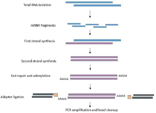
Figure 2.2 Basic flow chart of RNA sequencing preparation steps from sperm samples. ... 39
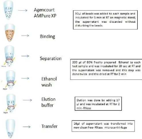
Figure 2.3 Brief description of beads clean-up step. ... 41
Figure 2.4 Brief description of second beads clean-up step ... 43
Figure 2.5 Brief description of third beads clean-up step ... 45
Figure 2.6 The Illumina reagent cartridge. All reagents required for sequencing and the library loading well number 10 of the cartridge. ... 48
Figure 2.7 The experimental design of RNA sequencing. ... 49
xii
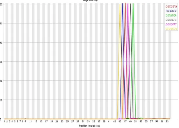
Figure 2.9 graph illustrating the Kmer content. ... 52
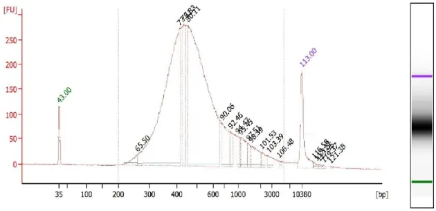
Figure 2.10 Schematic figure illustrating the distribution of reagents on the gel.68 Figure 2.11 Electropherogram of pooled purified cDNA libraries from the human sperm, ... 70
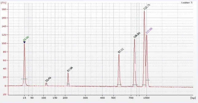
Figure 3.1 A representative bioanalyzer electropherogram of a human sperm mRNA library preparation. ... 73
Figure 3.2 The distribution of alignments yields, ... 77
Figure 3.3 Per sequence quality scores using Galaxy software for quality control. ...78
Figure 3.4 An insert length distribution boxplot, ... 79
Figure 3.5 MA plot of the differentially expressed genes ... 81
Figure 3.6 Original results obtained by DeSeq2 analysis of RNAseq (Illumina) sequencing data. ... 82
Figure 3.7 DESeq2 App 3D analysis ... 83
3Figure 3.8 A gene regulatory network of signaling pathways for transcripts ... 85
Figure 4.1 The videomicrography captured by in Sperminator® CASA system 99 Figure 4.2 The images illustrating the percentages of each motility grade ... 100
Figure 4.3 Volcano plot of miRNA differential expression in immotile sperm (D) and motile sperm (A), ... 106
Figure 4.4 Hierarchical clustering analysis diagram of the PCR array. ... 108
Figure 4.5 Illustration of different 20 related genes in sperm……….114
Figure 5.1 The progression of obesity trend in 1993, 2004 and 2015 ... 116
Figure 5.2 miR-21 conservation site ... …….125
Figure 5.3 Methylation of cytosine as part of the main key of epigenetic mechanism ... 127
xiii
Figure 5.5 Scatter plot of miRNA expression: ... 143 Figure 5.6 Hierarchical clustering heat map representation of the transcriptome analysis of miRNAs in the spermatozoa of mice. ... 144 Figure 5.7 Putative region of miRNA-21a structure and function, ... 146 Figure 5.8 Schematic structure of mouse sperm VmP1 transcript and miR-21a. ... 147 Figure 5.9 CpG site-specific methylation is represented for VmP1 in the sperm of HFD and AMC male mice. ... 149 Figure 5.10 miRNA pathways that affect spermatogenesis and sperm activity and related to hypertrophy of adipocytes ... 156
xiv
List of Abbreviation
5hmc 5-hydroxymethylcytosine AI Artificial insemination ART Assisted reproductive techniques ATP Adenosine Tri PhosphateBAM Binary Alignment Map
CASA Computer assisted semen analysis cDNA Complementary DNA
cmyc transcriptional regulator Myc-like CpG Cytosine-phosphate-guanosine motif
CRL Custom RNA Ladder
DGCR8 dsRNA binding protein DNA Deoxyribonucleic acid
dNTPs deoxynucleoside triphosphates dsDNA double strand DNA
eNOS endothelial nitric oxide synthase ERK5 extracellular signal regulated kinase5 FTH1 Ferritin heavy chain1
HML Ligation Buffer
hsa-miRNA Homo sapiens-miRNA HSP90A Heat shock protein 90A HRL High-Resolution Ladder HYA Hyaluronoglucosaminidase IVF in vitro fertilization
xv
KDM5D lysine demethylase 5D KLK2 Kallikrein-related peptidase2
miRNA MicroRNA
MMU Manchester Metropolitan University MiRNA Mus musculus- miRNA
mRNA messenger RNA
NaOAc Sodium acetate
NGS next generation sequencing NKX3-1 NK3 homeobox1
PAR masked Pseudo autosomal regions masked PCR Polymerase chain reaction
piRNA Piwi-interacting RNA
PML PCR mix
PAR masked Pseudoautosomal regions on chromosome Y PPARγ Peroxisome proliferator-activated receptor gamma
PrM Protamines
QC quality control
qRT-PCR Quantitative Real-time PCR
RA3’ RNA3’ Adaptor
RA5’ RNA5’ Adaptor
RIN RNA integrity number
RISC RNA-induced silencing complex RNA ribonucleic acid
RNAi RNA interference
xvi
RPI RNA PCR Index
RPL26 ribosomal protein L26 RPL5, L3 ribosomal protein L5, L3 RPL7 ribosomal protein L7
RPLP0 ribosomal protein lateral stalk subunit RPS27 ribosomal protein S24
RPS4X ribosomal protein S4, X-linked RPS4X ribosomal protein S4, X-linked rRNA ribosomal RNA
RT reverse transcription
RT room temperature
RTP RNA RT primer
RT-PCR Real-time PCR
SAM Sequence Alignment Map siRNA Small interference RNA SMCY Lysine demethylase 5D sncRNA small noncoding RNA SP17 Sperm protein 17 transcript SPM Sperm preparation medium ssDNA single strand DNA
STAR aligner Spliced Transcripts Alignment to a Reference aligner STP Stop solution
TBE Tris-buffered saline
TIMP3 metallopeptidase inhibitor3
xvii
tRNA transfer RNA
UCSC University of California, Santa Cruz UHR Universal Human Resource
UV Ultraviolet
VmP1 Vacuole membrane protein1 WHO World Health Organization ZFD Zinc finger domain
1
Abstract
Spermatozoa are known to be a carrier of genetic materials and serve no other function; However, it appears that it has a complex population of RNA including small noncoding RNA (sncRNA) that can deliver to the oocyte during fertilization. sncRNAs contribute to cellular gene regulation, in terms of their role in pre and post-fertilization genomic code, which in turn participate in the embryonic development process and any deviations in the gene expression pattern may lead to development retardation or early embryonic death. Moreover, phenotypic or environmental changes of parents can alter the phenotype of the next generation via specific miRNA expression regulation changes.
The present research investigated the profile of sperm mRNA and miRNA expression in both motile and immotile human sperm, in order to understand the role of sperm epigenetics in regulating genes that are involved in spermatogenesis and sperm function, in specific sperm motility, that has an impact on male fertility. Semen samples were collected from normo-spermic participants. RNA was extracted, and the mRNA profile in motile and non-motile sperm was investigated by Next Generation Sequencing. Results found that most transcripts expressed in motile sperm belonged to ribosomal mRNA.
Secondly, the thesis investigated and the miRNA expression changes in motile and immotile sperm. Preamplification of miRNAs with miscript PreAmp PCR kit before quantitative RT-PCR was performed using misprint PCR custom plate for 84 different sperm-specific miRNAs, in order to establish the miRNA profile in different motility grades.
2
The results suggested that miRNAs were differentially expressed in different sperm activity groups of the same sample and confirmed the role of miRNA in the physiological process of the spermatozoa. Data revealed that the miRNA expression profile in the sperm could serve as biomarkers for male fertility assessment.
To further, elucidate the relative expression and the epigenetic control of the sperm specific microRNAs in high-fat diet (HFD) mice and age-matched controls (AMC) by study the miRNA expression via qRT-PCR and DNA methylation of the most significant miRNA through finding its promoter region. The aim of this study was to explore the link between obesity and miRNA profile expression in a mouse model. We found that sperm-specific microRNAs from HFD mice were upregulated, with miR-21a-5p expression being highly significant and was regulated by methylation of the CpG islands on VMP1 promoter.
In conclusion, the current study demonstrated a differential expression of miRNA in sperm from motile and immotile populations. In addition, this study revealed links with obesity and altered expression of sperm miRNAs in a mouse model. There was a change in the methylation status and expression of miR-21a-5p, which may indicate the impact that paternal high fat diet has on sperm miRNA expression and DNA methylation.
3
Publications from this work
M. Khalid Abdulkareem Al-Gazi, S. Bradburn, S. Sugden, M. Ahmed, A. Greenstein, C. Murgatroyd, M. Carroll. Investigating the expression of sperm-specific microRNAs in a mouse model of obesity. ESHRE 34th annual meeting [2018].
MK. Al-Gazi, M. Carroll (2015). Sperm-Specific microRNAs - Their Role and Function. Journal of Human Genetics & Clinical Embryology. 1(1), pp.1-4 [review article].
M. Carroll, MK. Al-Gazi, C. Nevin. MicroRNA expression, and sperm motility. Association of Clinical Embryologist Annual Meeting [2015].
4
Chapter 1: General Introduction
1.1 Male Infertility
Infertility is a complex condition affecting around 15% of couples worldwide. It is well defined as an unsuccessful clinical pregnancy after 12 months or more of regular and unprotected intercourse (WHO, 2010; Hwang et al., 2011; Mascarenhas et al., 2012; HFEA, 2018). The main reasons for infertility in both male and female are varied due to endocrinopathies, infections, obesity, and genetic causes (for review see (O'Brien et al., 2010). Recent studies have reported a decline in male fertility and in particular sperm count (Levine et al, 2017).
Male infertility is due to sperm production or function defects. The main known causes could be a result of tubular obstruction, illness or injuries, cancer, mumps or inherited factors including Y chromosome microdeletions or Klinefelter (XXY) syndrome, and many other idiopathic aetiologies that cause genital tract disorders (Ferlin et al., 2006; Coward and Wells, 2013). Environmental and lifestyle factors also contribute to male infertility such as obesity that causes hormonal imbalance and sperm function defects (Vander et al., 2018). Low sperm count, poor sperm motility (asthenospermia), and high abnormal morphology percentage in the ejaculate are the most likely indicator for low fertilization rate, with an emphasis of the motility importance than other semen criteria (Donnelly et al., 1998; Shen et al., 2019).
Another cause of decreasing sperm quality is obesity, which can alter sperm production and quality through increases scrotal temperature and increased oxidative stress, resulting in reduced sperm motility and increased sperm DNA
5
damage (Katib, 2015). The prevalence of obesity has increased substantially globally with the increased burden of obesity-related complications. In men, obesity in addition to being a major risk factor for serious chronic diseases, there is a growing concern on the affects of ferritility (Rybar, R., et al., 2011) and more particularly, the long term health of the offspring. Obesity-related impaired spermatogenesis is associated with a decrease in microscopic and molecular sperm characteristics and pregnancy success. Much work is needed to unravel the link between obesity and the impact on sperm molecular andrology, including changes to the epigenome, which may translate to the offspring.
There has been a growing interest in the role of miRNAs and sperm function, in terms of sperm capacity to fertilize the oocyte. It is further suggested, that such sperm-specific miRNAs also drive the developing embryo and its differentiation (Ghorbian, 2012); although still relatively little is known about the mechanisms of action, as to how miRNAs exert their function. Regarding male infertility, several studies have found that some transcripts of miRNAs differ in expression between sperm from fertile and infertile men (Abu-Halima et al., 2013).
1.2 Spermatogenesis
Spermatogenesis is the process of sperm development within the seminiferous tubules in the testis after puberty. During embryogenesis, the activation of SRY gene (sex reversal Y) expression leads to the differentiation of seminiferous tubule from the genital cords in the gonadal medulla accompanied by differentiation of Sertoli cells from the epithelium layer of the tubules (Gilbert,
6
2000). The primordial germ cells then migrate from the genital ridge of the male embryo to enter the gonads under the control of TGF- beta 1, OCT 4, and alkaline phosphatase pathways and then give rise into sperm by spermatogenesis (Wilhelm et al., 2007).
The testis has a dual function that is regulated by the hypothalamus and pituitary glands: The first function is an endocrine function to produce the androgen (male sexual hormone) by the interstitial Leydig cells. The second function of the testis is the sperm production in the seminiferous tubules from the undifferentiated spermatogonial stem cells (SSCs), these cells are capable of self-renewal and can generate the same cells to produce the spermatogenic lineage via mitotic division, SSCs migrate to the adlumin for cytodifferentiation or spermiogenesis (the last stage of spermatogenesis).
Spermatogenesis requires 64 days and includes three phases:
1. Mitosis: the proliferation and differentiation of the spermatogonium (diploid germ cells, 2n) into type A1…A4 spermatogonium then divided into type B spermatogonium. B cells are the last precursor cells undergoing mitosis to form the primary spermatocytes.
2. Meiosis: the division of each primary spermatocytes to form two haploid secondary spermatocytes (n) in the first meiotic division, and the production of four spermatids in the second meiotic division.
3. Morphogenesis, in which the development and formation of the final elongated haploid spermatozoa with a condensed nucleus that is capable of fertilization within the testis.
7
Before the final stage, the spermatids are connected by cytoplasmic bridges that will be lost after moving toward the tubule’s lumen to form mature sperm. All developmental stages occur in close contact and support of Sertoli cells (Gilbert, 2000). The testis is capable to produce about 300-600 cell per second per gram of testis (Cooke and Saunders, 2002; Coward and Wells, 2013).
At the molecular level, genetic and epigenetic, transcription and post-transcription mechanisms are actively involved in the regulation of spermatogenesis. Perturbations in molecular control are known to be one of the critical causes of male infertility (Khalil and Wahlestedt, 2008; Zamudio et al., 2008; Bettegowda and Wilkinson, 2010).
Transcription in sperm is repressed due to chromatin remodeling and mRNA translation inhibition, the chromatin packaging changes of the round nuclear shape to an elongated one with a trace of cytoplasm content that accompanied by ribosome disappearance, as well as, 60% of histone is replaced first by intermediary transition proteins, then into protamines 1 and 2. In spite of chromatin loss and histone replacement, 15% of the chromatin stayed functional and provides a sperm transcriptional activity (McLay et al., 2003).
1.3 Sperm structure:
Sperm is a Greek word origin “sperma” (means ‘seed’). Sperm is a unique, highly specialized and differentiated cell, 60-70 μm in length with the principal oocyte fertilization purpose (Georgadaki et al., 2016). Sperm comprises of basic structures of the followings:
The sperm head, which is typically void of cytoplasm, contains a nucleus filled with the haploid paternal DNA securely twisted around protamine. The acrosome
8
cap, which lies at the front of the sperm head, contains proteolytic enzymes including acrosin, trypsin, and hyaluronidase that aids in oocyte penetration and fertilization. The sperm-oocyte interaction is facilitated by acrosin through the process of sperm capacitation and acrosomal reaction, which is essential to support the sperm-oocyte penetration reaction (Schill et al., 1988; Harper et al., 2008).
The midpiece holds the centriole with the fundamental purpose for mitosis and meiosis, while the mitochondria are required for sperm survival and the spermatozoa locomotion by oxidative phosphorylation of ATP and energy production.
The flagellum, or tail, that whips in motion to drive the sperm towards the oocyte. The tail functions through the central axoneme surrounded by two central singlet microtubules and nine doublet microtubules that are responsible for the motion by sliding past each other in the presence of ATP (Rauber, 2008; Chavarria et al., 1997), (Figure 1.1).
9
Figure 1.1 Structure of human sperm.
Schematic represents the human sperm head (containing the acrosome), midpiece and tail (principal piece). (B) Sperm morphology stain (Papanicolaou stain, 1000 X oil).
The sperm completes the first maturation process in the epididymis and acquires motility post emerging from the testis, with the aid of secondary glands secretion, which protects the sperm from the vaginal acidic environment. The second process called sperm capacitation in the female reproductive tract and acrosomal reaction activities when the two male and female pronuclei merged (Gervasi et al., 2018). The transport of a sperm-specific phospholipase C-ζ (PLC-ζ) to the female gamete during fertilization and initiation of the PLC-ζ signal and increase Ca++ influx which is essential for embryonic development (Saunders et al., 2002; Boerke et al., 2007).
The molecular changes of the mature haploid sperm cells are regulated by gene expression and accompanied by biochemical and physiological changes such as
10
the formation of disulfide bonds and the increased level of saturated fatty acids, as well as Ca++ signaling pathway that promotes sperm capacitation and oocyte fusion (Carvacho et al., 2018). Transcription in sperm is repressed due to the general concept that has been established previously of chromatin is an extremely compacted component and the protamines replace the histone content (chromatin remodeling process) and mRNA translation inhibition. The chromatin packaging changes of the round nuclear shape to an elongated one with a trace of cytoplasm content that accompanied by ribosome disappearance, as well as, 60% of histone is replaced first by intermediary transition proteins, then into protamines 1 and 2. In spite of chromatin loss and histone replacement, 15% of the chromatin stayed functional and provides a sperm transcriptional activity (McLay et al., 2003).
The protamine-DNA binding and compaction allow the DNA to fit into the relatively small spermatozoa head (Schagdarsurengin et al., 2012; Kanippayoor et al., 2013; Brunner et al., 2014). The interest of researchers in sperm biology has developed recently to decline the notion that sperm is the paternal genetic carrier only and serve no other function after fertilization, However, sperm molecular composition approved its importance post-fertilization and early embryonic development (Lin et al., 2013; Al-Gazi and Carroll 2015; Guo et al., 2017). A series of small non-coding RNAs (snc-RNAs) is One of the biological key regulator molecules for male fertility, which include the microRNAs (miRNAs) that are found in sperm and seminal plasma (Peña 2015).
11
1.4 Assessing male infertility
1.4.1 Semen Analysis
Semen analysis is an essential diagnostic assay in the evaluation of male reproductive health. There are a number of specific parameters important in evaluating male fertility according to WHO 2010 criteria:
Sperm concentration and sperm count: Normal sperm concentrations and counts range from 20 million/ml to over 200 million/ml. Concentrations lower than 15 million/ml are referred to as oligozoospermia and azoospermia refer to a lack of sperm in the ejaculate.
Sperm motility refers to the percentage of progressively motile sperm that is traveling in a straight-forward path or large circle with reasonable velocity. The immotile percentage describes sperm that shows no signs of movement; while non-progressive motility denotes an absence of progression, for example, swimming in small circles where the head is barely moving as the result of the flagellar movements (WHO, 2010), (Figure 1.2).
Normal levels of sperm motility are defined as levels higher than 40% of sperm in a sample showing progressive motility. The term asthenozoospermia is used to describe samples in which there is reduced sperm motility below 32 %. Earlier editions of the WHO manual classified sperm motility into 4 grades: Grade A (A): Rapid progressive motility (i.e. ≥25 µm/sec at 37°C, which is approximately equal to five heads length or half a tail length).
12
Grade C (C): Non-progressive motility (<5 µm/sec). Grade D (D): Immotile sperm that fails to move at all.
A high percentage of active sperm is obviously important to ensure the transport of sperm and achieve successful fertilization (Agarwal and Allamaneni, 2011; WHO, 2010). The sperm reaches to the female ovulation tract with the aid of specific molecules that act as chemotactic agents for the sperm, in the presence of Ca++ ions, to enhance sperm hyperactivation and penetrates the oocyte cumulus oophorous. The sperm that is aggressively motile can resist the female oviduct turbulences close to cilia and enter the lumen of fallopian tubes, hence stimulates the prostaglandins secretion and utero muscular contraction that are vital for fertilization (Kölle et al., 2015).
Figure 1.2 Sperm motility groups.
A – fast progressive sperm, moves linearly or in large circles quicker than 25 µm/sec. B – slow progressive sperm, moves linearly or in large circles slower than 25 µm/sec. C – nonprogressive sperm, moves in small circles or ‘twitches’. D – immotile sperm, no movement.
13
Sperm morphology: The presence of a wide range of morphological discrepancies between sperm in a population can make it difficult to judge the sample provided in vitro. Although the level of the clinical importance of morphology to fertility is debated, the normal morphology percentage that correlates with the success of fertilization in vivo or in vitro defined as over 4%. The term teratozoospermia describes the presence of a high percentage of abnormal spermatozoa in the semen sample, that were classified into three different categories depending on the abnormality location: head (acephalic, giant head, or multiple head), midpiece (presence of cytoplasmic droplet) and/or tail (short, long, or double tail). The main abnormality causes may be due to genetic, high-temperature exposure to the testes, and chemical or toxic substances contact, as well as lifestyle, and infections (Coutton et al., 2018).
Aspermia is another term that describes the lack of semen completely from the ejaculate as a result of the passing of semen into the bladder at ejaculation (WHO, 2010).
1.4.2 Sperm RNA
The concept that sperm carries only paternal DNA and the paternal RNA content in mature sperm were lost during spermatogenesis, and therefore served no function during fertilization has been challenged (Kramer and Krawetz, 1997). However, it is now established that spermatozoa contain a wide spectrum of RNA species including mRNAs, tRNAs and various non-coding RNAs (ncRNAs) such as miRNAs (Boerke et al., 2007). Studies have found that sperm RNA regulation provides a critical role in the process of fertilization when they are utilized to the
14
oocyte and participate in regulating the paternal genome and various biological events involved in early embryonic development (Miller et al., 2005; Lalancette et al., 2009; Kildemo, 2012). A cascade of paternal pathways may involve in regulating the events of postfertilization such as AKAP-4 and FOXG1B, and provide a level of control of the maternal genome transition into embryonic status (Ostermeier et al., 2005; Boeker et al., 2007).
Importantly, sperm does not contain ribosomal (r)RNA (18S and 28S rRNA), which are essential for protein synthesis, and they were depleted during spermiogenesis. Therefore, sperm cells are transcriptionally and translationally quiescent (Johnson et al., 2011).
mRNA and miRNA are RNAs that participate in gene regulation of sperm post-transcriptionally by controlling cellular fate and development and can be transferred through germ cells to the next generation (Krawetz, 2005; Ivey and Srivastava, 2010).
1.4.3 Sperm messenger RNA
Pessot 1989, was first who described the Messenger RNA (mRNA) in transcriptional inactive sperm (Pessot et al., 1989). mRNA is one kind of long RNA (comprises about 5% of total RNA) that is transcribed from DNA strand to form antisense strand that is used as a template for translation and the encoding proteins through genetic codes (Kukurba and Montgomery, 2015).
The translational ability of mRNA comes from the structure that contains methylated 5’cap end and 3’ polyA end units of non-coding sequence make it more stable and capable of translation into proteins. The coding region is the unit
15
of protein synthesis while the UTR region is for the expression regulation (Figure 1.3), (Kozak, 2005). During transcription, the mRNA contains exon regions only, while the intron regions are removed from the primary transcript during the silencing process of gene transcription (Keren et al., 2010).
Figure 1.3 mRNA regions. The figure is representing the five and three prime untranslated regions of mRNA transcript.
Mature sperm contains more than 3000 kinds of functionally viable mRNAs that play a critical role in gene expression during spermatogenesis, capacitation, acrosomal reaction, and onwards when has been delivered to the oocytes post fertilization (Ostermeier et al., 2004; Gur and Breitbart, 2006). Spermatozoal mRNA are not all located in the nucleus, some of them are present in the flagellar fibrous sheath like SP17 transcript (ChirivaInternati et al., 2009), or on the sperm surface like SMCY (also known as KDM5D, HYA or JARIDID), which have a major role after fertilization (Anderson, 2013). SMCY encodes a protein of ZFD, from this protein is a minor histocompatibility antigen for a graft rejection of sperm donor in an oocyte recipient (Dhanoa, Mukhopadhyay et al., 2016). SMCY is mainly functioned during sperm prophase stage of meiosis and in chromatin remodeling, any mutation in this transcript can lead to sperm maturation arrest and chromosome condensation during meiosis (De Jonge and Barratt, 2006). Sperm becomes incapable of transcription during the second meiosis of spermatogenesis and decreases mRNA transcription together with a decrease in a repertoire of mRNA content and 18S and 5S rRNA in the nucleus or present in
16
a negligible amount and is not enough to support cytoplasmic mRNA translation (Miller and Ostermeier, 2006). These events include histone hyperacetylation subsequent by replacement of histones by transition proteins mainly TNP1/2 then by protamines (PRM1/2) that condensed in the sperm nucleus that results in RNA content reduction and loss of transcription in spermatids which demand mRNA transcription in a high level but translationally delayed to another period of genesis (Cullinane, 2014). New proteins are needed for sperm morphological changes during maturation. rRNA is depleted in sperm during spermatogenesis, however, 95% of RNA in other cells are containing rRNA, which needs rRNA depletion step to get rid of this kind of RNA during the mRNA sequencing process.
1.5 Gene expression
Regulation of gene expression is imperative for the control of cell function and fate (Smorag, 2013). The processes of gene expression are comprising of transcription, RNA splicing, protein synthesis or translation, and post-translational modification of proteins. Gene expression may be modulated to alter the mRNA sequence level, which results in different protein structure, and can be studied to find out which genes are turned “on” to produce mRNA or “off” and enables to know the mRNA concentration can manipulate various cellular development phases. Different methods were used in studying the gene expression like Northern Blotting, SAGE (serial analysis of gene expression), microarrays, RT-PCR and RNA-Seq (Perdacher, 2011; Su et al., 2011).
This can explain how similar cells can behave differently and explains the phenotypic changes between species. Apparently, proteins have got an important
17
characterization in regulating gene expression, however, the discovery of ncRNA including miRNA has found that they are the most abundant RNA in gene regulation process post-transcriptionally by repressing mRNA (Bartel, 2004).
1.5.1 Sperm Epigenetics:
Epigenetics (“epi” from the Greek: means outside or over) or non-Mendelian para-mutation inheritance describes the alteration of gene expression without affecting the coding DNA sequence of the cell; therefore, it is relatively transient and potentially revised if the cause is corrected (Rassoulzadegan and Cuzin, 2015). Lifestyle and environment signals can promote diseases and health derivation via epigenetic mechanisms, such as histone modification, chromatin remodeling, DNA methylation, and non-coding RNA including miRNA expression (Lujambio and Esteller, 2009; Marczylo et al., 2012). There is growing evidence indicating that epigenetics could have a significant role in subfertility and the link between fertility and health (Dada et al., 2012), and any epigenetic modification during spermatogenesis can cause deleterious effects on sperm epigenome and affect its function and the subsequent progeny health (Schagdarsurengin et al., 2012; Marshall, 2015).
Small ncRNAs are epigenetic small molecules with a vast impact in regulating biological processes during mammalian development, coding refers to non-translated transcripts and most of them are cellular homeostasis regulators substances that are classified to be epigenetic regulators at a transcriptional and translational level in general. In sperm, miRNA, Piwi-interacting RNA (piRNA) and siRNAs are some sorts of short snRNAs (Santosh et al., 2015).
18
siRNAs (20-30 nt length) are endogenous molecules resulted from the cleavage of the dsRNAs by Dicer enzyme and regulating cell functions by RNA interference phenomenon or RNAi to initiate gene silencing by destabilisation of mRNAs, hence controlling cellular growth and development as well as the formation of heterochromatin (Dana et al., 2017). siRNAs have been implicated therapeutically for some diseases like cancer through its antiapoptotic and anti-proliferative action (Phillips, 2008).
piRNAs (24-31 nt length) have been counted as the largest class of small RNAs in mammalian tissues, it has been reported that they have an RNA silencing regulating function in germ cells especially spermatogenesis and sustain male fertility via their interaction with piwi protein (Tosar et al., 2018; Siomi et al., 2011; Capra et al., 2017).
Finally, miRNAs are considered as key regulators of all cellular functions. Regarding male gametes, a small amount of miRNA has been retained in sperm and are vital in the development of both sperm and embryos (Krawetz et al., 2011).
Another functional class called long noncoding RNA (lncRNA) which is known to have a function in chromatin remodeling, controlling the transcription and post-transcription events (Guttman et al., 2009).
1.5.2 miRNA
miRNAs are a novel class of endogenous ~17-26 nucleotide length, which control post-transcriptional gene expression by targeting 3’UTR mRNA resulting in its degradation or later protein expression inhibition, thus involved in all biological
19
process of living organisms. miRNA was first discovered in C.elegans in 1993 when lin-4 miRNA was detected with a conserved complementary site on the lin- 14 mRNA transcript (Lee et al., 1993; Wightman et al., 1993; Schickel et al., 2008). Later on, let-7 has been found to target the lin-41 and they have crucial effects on the developmental timing of C.elegans larvae (Reinhart et al., 2000). Lin4 and let-7 are highly conserved and have the same function in other species (Lagos-Quintana et al., 2001).
It has been well established that miRNAs have an impact on the pathogenicity of common diseases such as cardiovascular diseases, tumourigenesis, immune-inflammatory diseases, and metabolic disorders (Mogilyansky and Rigoutsos, 2013).
Studies on the relationship between miRNA and stress, which cause up or down-regulation of miRNA expression have revealed the mRNA targets effects and cellular response. However, the absence of miRNA function can cause impairment in the whole process of cellular development (Mendell and Olson, 2012).
Thousands of miRNA have been discovered, and an online database with analysis was established first by Ambros laboratory 2003 (Ambros et al., 2003). miRNA annotation and registry including all information about published miRNAs data can be explored via miRBase database system (http://www.mirbase.org/) by Sanger Institute. miRBase contains more than 25141 mature miRNAs in about 193 different organisms (Griffiths‐Jones 2004) or miRNA visa system database (www.cbrc.kaust.edu.sa/mirnavisa.org), (Kamanu et al., 2013). Development of deep sequencing techniques opened a new way of research of the miRNAs and the studying of the profile and function in normal and aberrant conditions as well
20
as prediction of new miRNAs which gave a hallmark of the miRNA importance (Kong et al., 2012).
1.5.2.1 miRNAs families and functions
miRNAs are situated in polycistronic (within one locus) miRNA “clusters” that have a similar functional role and are suitable for disease biomarkers such as cancer and cardiovascular disease (CVD). miRNAs families that have perfectly matching nucleotides in the seed region (2-7 nt) from 5’ end, as well as the complementary site for 3’UTR mRNA in this region, are highly conserved across species, therefore they are generated from a single primary transcript and regulating the same target genes (Guerra-Assunção and Enright, 2012). For example, miR 17~92 cluster, also known as oncomiR-1 because they are dysregulated in solid cancers, is located on chromosome 13 in human and include a range of different miRNAs that are important in normal development (Table 1.1), has been included a range of different miRNAs that are important in normal development. Also known as oncomiR-1 because they are dysregulated in solid cancers. miR-17-5p is highly expressed in some tumours while showing low expression pattern in the blood of non-small lung cancer patients. This family of miRNAs is also implicated in age-related conditions (Heegaard et al., 2012; Mogilyansky and Rigoutsos, 2013). A wide range of functions of miR-17 have been revealed such as enhancement of the prostate tumour invasion and growth through cellular proliferation exaggeration of tumour cells has approved by targeting TIMP3 (Concepcion et al., 2012). Furthermore, these miR-17 clusters have been involved in adipogenesis and found to have an inhibitory role and
21
adipogenesis promotion by targeting BMP2 mRNA along with miR-106a (Xu and Wong, 2008).
Table 1.1 miR-17 family.
The seed regions of 2-8 from 5’ end of miRNA sequences of miR-17 family are having the perfectly matching nucleotide (Hausser et al., 2013; Mogilyansky and Rigoutsos, 2013).
miRNAs miRNA sequences and the shaded seed region
hsa-miR-17-5p 5’CAAAGUGCUUAGUGCAGGUAGU 3’ hsa-miR-20A-5p 5’UAAAGUGCUUAUGUGCAGGUAG 3’ hsa-miR-20b-5p 5’CAAAGUGCUCAUAGUGCAGGUA 3’ hsa-miR-106b-5p 5’ UAAAGUGCUGACAGUGCAGAU 3’ hsa-miR-93-5p 5’CAAAGUGCUGUUCGUGCAGGUAG 3’ hsa-miR-106a-5p 5’CAAAGUGCUAACAGUGCAGGUA 3’
Another example, lethal-7 (let-7) miRNA family has similar sequences and only one nucleotide differs in order. To determine this difference, a letter at the end was added as let-7a, let-7b, let7c, etc. (Table 1.2). The most important function of let-7 family is to promote differentiation and timing of the development of organisms (Schulman et al., 2005; Roush and Slack, 2008). Let-7 dysregulation can cause cellular growth and development retardation and promotes diseases like cancer (Roush and Slack, 2008).
Table 1.2 Lists of isoform sequences of let-7 miRNAs family, the shaded area is
representing the miRNA seed region
miRNAs miRNA sequences and the seed shaded region
hsa-let-7a 5’ UGAGGUAGUAGGUUGUAUAGUU 3’
hsa-let-7b 5’ UGAGGUAGUAGGUUGUGUGGUU 3’
22
hsa-let-7e 5’ UGAGGUAGGAGGUUGUAUAGUU 3’
hsa-let-7f 5’ UGAGGUAGUAGAUUGUAUAGUU 3’
1.5.2.2 miRNA biogenesis: synthesis and functions
miRNAs are transcribed from specific genes by RNA polymerase II in the nucleus. They are first formed as primary transcripts (pri-miRNA) about 70nt, then folded into hairpin structures that are successively processed by several enzymes in the nucleus called Drosha and DGCR8 then transported to the cytoplasm by Expotin5 to be processed by Dicer enzyme (Almeida et al., 2011; Al-Gazi and Carroll, 2015), (Figure 1.4). A 22 nucleotide duplex, mature miRNA is then produced in which one strand assembles into a protein-RNA complex called RISC (RNA induced silencing complex) as a part of gene silencing process called interference RNA (RNAi) (Leung and Sharp, 2010). The small size of miRNA made them less prone to degradation and more stable than other types of long RNAs and mediates post-transcriptional gene suppression (MacRae et al., 2008; Zubakov et al., 2010). A single miRNA species can base pair to its target on multiple sites within a single mRNA transcript to cause their degradation. Additionally, one miRNA can regulate the expression of multiple gene targets and furthermore a single mRNA transcript can be targeted by more than one miRNAs (Smorag, 2013). miRNA and protein-coding genes expression regulation occur post-transcriptionally. Basically, DNA binding proteins like P53 and other transcription factors bind to the miRNA promoter region and regulate their expression (Boominathan, 2010). miRNA can reduce the protein output by inhibiting their translation without changes in the level of mRNA targets (Bagga et al., 2005; Curry et al., 2011).
23
Figure 1.4 Schematic representation of miRNA biogenesis.
a) miRNAs transcription is conducted via RNA polymerase II; b) A double-stranded hairpin called primary miRNA (pri-miRNA) is then is formed. c) pri-miRNA then cleaved by the help of Drosha, a member of superfamily RNase III endonuclease to form pre-miRNA molecule. d) exportin-5 is then assisted in pre-miRNA transport to the cytoplasm. e) Another enzyme called Dicer is then involved in processing pre-miRNA in the cytoplasm to produce short and double-stranded miRNA. f) The pre-miRNA act together with AGO (Argonaute) and other proteins to form the RISC component, and finally, the formation of a single-stranded mature miRNA. and g) mature miRNA targets the mRNA ( adapted from (Al-Gazi and Carroll, 2015).
miRNAs have a critical biological function upon binding at nucleotides 2-8 seed region to a complementary 3'UTR sequence of specific mRNA, leading to either mRNA degradation or protein translation arrest thus, influencing cell functions including gene expression mediation during development, differentiation, cell proliferation, cell fate decision, and stress response, apoptosis and death (Yerramilli et al., 2013). The half population of miRNA is encoded independently within noncoding gene transcripts while the others within intronic protein-coding genes (Guerra-Assunção and Enright, 2012). An individual miRNA could target
24
more than one protein coding mRNA post-transcriptionally through multiple pathways or act upon a single target to mediate a disease phenotype (Aurora et al., 2012). Nevertheless, until now little is known about their mechanism of action and how they exert their function. Regarding male infertility, the sperm contains an abundant profile of miRNA together with a set of mRNA targets also expressed in fertilized metaphase II oocyte, suggesting miRNA regulatory functions (Amanai et al., 2006). For instance, miR 143 (Esau et al., 2004), miR-27b (Karbiener et al., 2009), miR-375 (Ling et al., 2011), and miR-14 were involved in adipogenesis in mice. They act as a modulator for adipocytes differentiation, as well as miR-122, miR-370 (Iliopoulos et al., 2010), miR-335, miR-378/378*, and miR125a-5p all have a function in fatty acid and cholesterol metabolism regulation (Fernández-Hernando et al., 2011).
1.5.2.3 The relationship of miRNA and motility in male fertility:
Sperm motility is a critical factor in assessing male fertility. Several studies have reported differential miRNA expression between impaired and normal semen samples implying that miRNAs are key regulating players in spermatogenesis and production of a new, viable sperm in males (Wang et al., 2004; Reza et al., 2019). Some studies have established that some gene transcripts are regulated by miRNAs related to sperm structure, sperm morphology, motility, and metabolism. miRNAs studies were conducted on human sperm miRNAs with abnormal motility outcomes have found a positive relationship between the miRNAs expression level and semen quality which significantly affects reproduction patency (Ghorbian 2012; Abu-Halima et al., 2013). In addition, gene transcripts related to sperm motility are regulated by miRNAs. They have a role
25
during spermatogenesis, chromatin packaging, and early embryonic development, when delivered to the oocyte after fertilization, it can be passed down to the further generations (Wang et al., 2004; Jedrzejczak et al., 2007; Jodar et al., 2012; Kawano et al., 2012; Liu, Cheng et al., 2012; Abu-Halima et al., 2013).
miRNAs have different expression patterns between fertile and infertile men (Khazaie and Esfahani, 2014). Altered expression levels of sperm-specific miRNAs have been implicated with abnormal sperm parameters (Abu-Halima et al., 2013) and the propensity for transgenerational amplification of some conditions such as obesity and type-2 diabetes (Fullston et al., 2013). Marczylo 2012, reported that environmental changes targeted histone modification and miRNAs (for instance, altered the expression of hsa-mir-146b-5p, hsa-mir509-5p, hsa-mir-519d, and hsa-mir-652) profile in the sperm of infertile men as well as leading to alter next generation phenotype through impairment of male germ cells functions (Marczylo et al., 2012). Sperm function is influenced by miRNA expression changes. miR-122 is a specific sperm motility-related transcripts that have a role in male fertility via targeting TNP2, a testis-specific gene that involved in chromatin remodeling during spermatogenesis (Yu et al., 2005; Jodar et al., 2012; Lin et al., 2012). Hence, miR-122 inhibits the expression of proteins that have a significant impact on sperm development process (Liu et al., 2013). Sperm miRNAs are responsible for early embryonic development in mice (Liu et al., 2012). miR-34c was found in the sperm and it is important for cellular maturation, its inhibition causes detrimental development in mouse zygote (Choi et al., 2011) and expressed in human sperm by targeting DLL1 and NOTCH1 genes important for spermatogenesis (Krawetz et al., 2011). It is obvious that the lack of specific
26
miRNA can result in spermatogenesis impairment and infertility (Belleannée, 2015). miR-18 has a significant role in spermatogenesis by targeting Hsf2 gene (Björk et al., 2010).
miRNA function in spermatogenesis is still unclear. However, previous studies have compared normal fertile with impaired infertile cohorts (Figure 1.5), but to our understanding, there have not been any studies verifying miRNA expression in the same group of spermatozoa, i.e. actively motile and non-motile groups of spermatozoan according to the grading system of motility.
Figure 1.5 Historical miRNA discovery through a timeline since the first miRNA has been discovered in 1993.
Different studies of miRNAs in spermatozoa. 1 (Almeida et al., 2011), 2 (Wild and Roudebush, 2000), 3 (Garrido et al., 2004), 4 (Amanai et al., 2006), 5 (Yan et al., 2008), 6 (Bouhallier et al., 2010), and 7 (Abu-Halima et al., 2013).
27
1.5.2.4 miRNA and early embryonic development
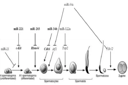
miRNAs are important for normal human and animal development. Changes or loss of the miRNA expression can cause embryonic death through the posttranscriptional mediation of pluripotent cells (Berardi et al., 2012). Different miRNAs can be specific targets of different pathways and transcripts during spermatogenesis and early embryonic development. miR-122 and miR-34c are spermatids specific miRNAs and maintain cell development by targeting c-Kit and Tnp2 genes (Figure 1.6) (Smorag, 2013).
Figure 1.6 miRNA and spermatogenesis.
miRNA targets different genes during spermatogenesis and after fertilization (Smorag, 2013).
Let-7 was the first miRNA that have found related to the development of C. elegans larvae by suppression of lin-28 and controls the transition of the larval stage into an adult (Reinhart et al., 2000). Lin (2013) reported that miRNA is highly expressed in the human embryos and some specific miRNAs are
28
upregulated at various stages of development as well as in tumorigenesis, which was confirmed using the microarray technique (Lin et al., 2013).
miR-17-192 cluster also was downregulated in week 6 of human embryogenesis and have oncogenic features at the same time by targeting CDKN1A (p21) and RUNX1 which is very important in controlling cell cycle and apoptosis. miR-290-295 cluster in mouse similar to 371-373 cluster in human and miR-302 clusters have a vital role during embryogenesis, also important in the cell cycle of embryonic stem cells and pluripotency phase by manipulating the expression of some cell cycle gene inhibitors like Rb1, RbI1 (Yuan et al., 2017), (Figure 1.6). they regulate embryonic stem cells by repressing mRNA embryonic stem cells regulators transcripts such as Nanog, Oct4, and Sox2 transcripts (Figure 1.7).
29
Figure 1.7 miRNAs and embryogenesis signaling pathway schematic diagram.
The miRNAs target genes involved in the up-regulation of core pluripotency factors that have an impact in embryogenesis and pluripotency.Let-7 repress differentiation transcripts (lin-28 and MYUC) and lin-28 conversely repress let-7 to maintain embryonic stem cell development. miRNAs on the right are suppressing other transcript and involved in the process of development also by repressing Casp3 leads to apoptosis. Pointed arrows representing activation while blunted arrows for repression. (adapted from (Berardi et al., 2012).
1.5.2.5 Methods of miRNA quantification
To study the cellular performance of miRNAs, mature miRNAs profile is the main target in normal and disease condition of different organs (Wark et al., 2008). Because of the miRNA short nucleotides length and limited expression level in a confined cell at a particular stage, as well as large sequence similarities make it a difficult molecule to isolate. Numerous quantification techniques were identified to quantify the miRNA expression with some limitations of advantages for each method (Baker, 2010).
1.5.2.5.1 Quantitative real-time PCR (qRT-PCR)
The polymerase chain reaction (PCR) is one of quantification method that allows a specific known region of DNA amplification using oligonucleotide primer complementary to the known sequence of DNA template. The DNA polymerase then used to make an extension of the primers on single strand DNA (ssDNA) in the presence of dNTPs under specific conditions, after that heat denaturation of double strand DNA (dsDNA) and cooling annealing (primer binding) to synthesize new DNA strands. The data output of quantification of gene expression using RT-PCR by cDNA amplification and transcription from RNA samples is expressed as a fold change or a fold difference of expression levels (Newton and Graham,
30
1994). qRT-PCR needs normalization of cDNA with the same RNA input to ensure the actual output obtaining using fluorescent reporter molecules emitted following each PCR cycle (Bustin et al., 2005).
1.5.2.5.2 Microarray
The microarray is a potent high-throughput implement to profile a large number of miRNA in parallel. Around 1000 miRNAs simultaneously can be detected in one run using a flexible probe and fluorescent dye, in order to label the specific gene of interest then dsDNA was formed by hybridization. Finally, the genes and their expression level were detected (Liu et al., 2008; Yang et al., 2008). However, it has several limitations like requisite of a previous understanding of the sequence being investigated, also the cross-hybridization of similar sequences, besides the inability to identify low or high expression level genes (Shendure, 2008).
1.5.2.5.3 Illumina High Through-put Next-generation sequencing
High Throughput Next-Generation Sequencing (NGS) is an advanced technology that allowed DNA and RNA sequencing directly by the synthesis in situ and dNTP can be detected simultaneously by Illumina sequencer system on a flow cell cartridge at millions of specific positions to provide an inclusive understanding of gene nature and function (Corney, 2013). RNA sequencing also known as RNA-Seq is a procedure of transcriptome analysis including mRNA analysis using various methods to study the presence and quantity of RNA in biological samples and to investigate gene expression profiling of the organisms. Simply, RNA samples are converted into cDNA library fragments to obtain short reads between 200-500 bp to be sequenced on Next-Seq 500 (an Illumina Genome analyzer
31
with a high throughput sequencing platform), which then aligned to a reference genome to generate a base-resolution expression profile (Datta and Nettleton, 2014).
1.5.2.6 miRNA therapy
miRNA are now well approved as biological pathways and functions regulators, and participating in the disease development. Various research has tried to modify miRNA to renovate therapeutics to be offered in the markets especially cancer therapeutic. With the unique miRNA conserved sequence among species that made it easy to synthesize to produce anti-miRs that have a high affinity to bind to miRNAs target and causing degradation of over-expressed miRNA (Christopher et al., 2016). Inactivating the pathological miRNAs and correcting the imbalance in genetic pathways caused by miRNA dysregulation can be achieved via introducing artificial miRNA, which will lead to increase the miRNA amounts and functional inhibition will be initiated (Negrini et al., 2007).
1.6 Summary
miRNA emerges to participate in many cellular function and biology and needs further investigations. This chapter of current research will emphasize the potential RNAs specific for progressively motile sperm, non-progressively motile sperm, and immotile sperm and may lead to identifying novel pathways and biomarkers associated with male infertility.
32
1.7 Hypothesis
Expresisons of mRNA and subpopulations of sperm micro RNAs responsible for sperm function and fertility is of increasing interst both in terms of sperm biology and potential fertility biomarkers.
The main aim of this research is to explore the relative epxresion of RNA in the sperm contains a myriad of RNA that expressed differentially in term of sperm activity.
The second aim is to explore the relative expression of mRNA and miRNA from different populations of isolated sperm cells from humans and to investigate the relative expression of sperm-specific miRNAs in an obese mouse model.
To achieve this, two main objectives of our research were:
1. Isolate and sequence humam sperm mRNA from motile and immotile sperm, establish optimal sperm RNA isolation [mRNA dan miRNA] and amplificaiton methodology from sperm samples – and sequence using the Ilumina platform.
2. To investigate the impact of highfat diet and obesity on the differtntial expression and epigenetic regulation of miRNA populations – an obese mouse model was utilised.
33
Chapter 2: General Materials and Methods
2.1 Exploring the relative mRNA expression in motile
and immotile human sperm
In order to explore the transcriptome content of motile and immotile human sperm, RNA sequencing (RNA-Seq) was performed using the Illumina Next-generation sequencing (NGS) platform (NextSeq 500 sequencer, Illumina, UK).
2.1.1 Procurement of semen
All sample participant consent and percurmment adhered to faculty ethics approval [SE111 229A Appendix 2]. Semen samples were obtained from recruited donors (aged 19-30 years) by masturbation after 2-5 days of abstinence. Donors were asked to fill a questionnaire detailing for health, lifestyle, and medications (Appendix 2). All participants have consented following faculty ethical approval at Manchester Metropolitan University (Appendix 2). Semen samples were produced on site in a designated, secure room were collected in sterile plastic containers (Sterilin, UK). Samples were divided into two portions for motile and immotile sperm from the same sample. Motility was assessed using the CASA system (Sperminator,® Procreative, UK).
34
2.1.2 Sperm preparation from human semen samples:
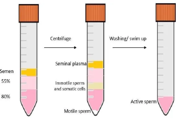
Isolating motile sperm from seminal plasma is a routine procedure in assisted reproduction technology (ART). These techniques are based on isolating viable, normal and motile sperm capable of fertilizing the oocyte. In order to separate different motility grades (Grade A - fast progressive motility, Grade B - slower progressive motility, Grade C- nonprogressive and Grade D immotile – referred to as A, B, C, and D henceforth). Conventional swim up methods was used to such isolation (Grunewald and Paasch 2012), (Figure 2.1). Samples of interest were assessed regarding sperm concentration, motility and the presence of round cells in semen sample according to WHO semen analysis criteria (2010) using CASA system software (Sperminator®, Procreative, UK). To select motile and immotile sperm from non-sperm and remove contaminants, a 55:80% discontinuous density gradient was performed.
Samples were allowed to liquefy for 30 min at 37oC before further analysis to
enable sperm to acquire swimming ability. Semen was analysed for volume, sperm concentration, and motility according to WHO guidelines (2010). Sperm concentration and grade motility were then assessed on a pre-warmed (37◦C) stage using CASA software (Sperminator®, Procreative, UK). Samples with more than 1x10⁶ round cells/ml were excluded from the study.
Sperm preparation was done using two swim-up purification methods (WHO, 2010). Firstly, sperm was separated by a discontinuous density gradient, a separation method based on cellular density separation. Briefly, 1 ml of semen samples were gently layered on top of 2 ml 55:80% Supra SpermTM media
35
(SPM) (Origio, Denmark), then centrifuged at 300 xg for 20 min at room temperature, then sperm pellets were washed twice in sperm preparation mediumTM (Origio, Denmark) at 300 xg for 10 min. The sperm was then
suspended in 1 ml of SPM for motile sperm portion and counted. While immotile sperm (D) was isolated from the intermediate layers of the gradient (Figure 2.1). The immotile portion washed twice with 1ml PBS at 300 xg for 10 min to ensure that sperm are free from seminal plasma and any other decapacitated contaminants. Finally, the washed portion was suspended with 0.5 ml PBS.
Figure 2.1 Schematic illustration of Discontinuous Density gradient method in vitro.
Secondly, the simple layer technique was done by gently placing 1 ml of semen sample underneath 2 ml of SPM (Origio, Denmark) at the bottom of a conical Falcon test tube, and placed in a 6.0% CO2 gassed incubator at 37°C at an