CODEN (USA)-IJPRUR,e-ISSN: 2348-6465
Original Article
Morphometry of Acromion Process: A Study of Indian
Scapulae
Shilpa Gosavi1, *, Surekha Jadhav2, Rajendra Garud1 1
Dept of Anatomy, BVDU medical college, Pune, Maharashtra, India- 411043. 2
Dept of Anatomy, PDVVPF’s Medical college, Ahmednagar, Maharashtra, India- 414111
A R T I C L E I N F O A B S T R A C T
_______________________________________________________________________________
1. INTRODUCTION
The acromion projects forwards almost at right angle,
from the lateral end of the spine of scapula. The lower
border of the crest of the spine becomes continuous
with the lateral border of the acromion at the acromial
angle which forms a subcutaneous bony landmark.
International Journal of Pharma Research and Health Sciences
Available online atwww.pharmahealthsciences.net
Received: 30 Aug 2015 Accepted: 28 Oct 2015
Objective: Anatomic details and variations of shoulder region are important for diagnosis and management of corrective surgeries in this area. Acromion morphology is believed to play a key role in impingement syndrome and the pathogenesis of rotator cuff diseases. The present study was carried out with the purpose to collect the morphological data of acromion process in Indian population and to compare it with similar studies in other population. Experimental approach:We studied 127 dry scapulae of unknown age and sex. Acromion processes were classified into Type I (flat), II (curved) and III (hooked). The length, breadth, anterior thickness, coraco-acromial distance, acromion-glenoid distance and the height of coraco-acromial arch was measured with the help of digital vernier caliper. The inclination of acromion was measured with the help of goniometer. Findings and Discussion: Type II acromion was observed in majority (81.88%), type I in 13.3% and type III in 4.7%. Mean coraco-acomial distance was 26.9 ± 5.6 mm while mean acromio-glenoid distance was 22.68 ± 3.3 mm. Mean height of coraco-acromial arch was 16.54 ± 2.8 mm. The inclination of acromion was less on the right side as compared to the left side (Rt. -33.460, Lt.- 43.40). Conclusion: Predominance of Type II acromion and bilateral symmetry in all other parameters was observed. When the results were compared with similar studies, differences were observed in most of the parameters. The results of the present study may be of help to the shoulder surgeons, Anthropologists and Anatomists.
Key words:Acromion, morphology, Scapula.
Corresponding author *
Dr. Shilpa Gosavi, Dept. of Anatomy,
The medial border of the acromion is short and is
marked anteriorly by a small oval facet directed
upwards and medially for articulation with the lateral
end of the clavicle. The tendon of supraspinatous
passes below the overhanging acromion and is
separated from it and from deltoid by the subacromial
bursa.1
The anatomy of the acromion and related structures in
the shoulder joint is of importance and useful to
successfully carry out interpretation of images and
surgical procedures in pathologies associated with this
joint.2Acromion morphology is believed to play a key
role in impingement syndrome and the pathogenesis of
rotator cuff diseases.3
The variation of the acromion form has been studied
by different authors.2-6 Bigliani et al.4 classified the
acromion in type I (plane), type II (curved) and type III
(hooked). The slope and length of the acromion and the
height of the arch are most closely associated with
degenerative changes.5
Anatomic details and variations of the region are
important for diagnosis and treatment. Furthermore, to
recognize that important parameters of the region may
change according to the race, gender and lateralization
will significantly increase the surgical achievement.6
The present study was carried out with the purpose to
collect the morphological data of acromion process in
Indian population and to compare it with similar
studies in other population.
2. MATERIAL AND METHOD
We studied one hundred and twenty-seven dry
scapulae (Right -52, Left- 75) available in the
department of anatomy, after taking approval from the
Ethical committee. The unbroken, intact adult scapulae
of unknown age and sex were included in the study.
Acromion processes were classified into Type I (flat),
II (curved) and III (hooked) as suggested by Bigliani et
al4.
Following morphological parameters were measured
using digital vernier caliper accurate up to 0.01 mm
and the angular measurements were taken with the help
of goniometer. (Fig.- 1)
Fig 1: Figure showing various measurements of acromion process. Blue line – Coraco-acromial distance, Yellow line – Height of coraco-acromial arch, Red dotted line –Acromio – glenoid distance, a = Inclination of acromian
The length of acromion process was measured as antero-posterior distance along the long axis
The width was measured as the maximum distance between lateral and medial borders.
Anterior thickness was measured at a point 1 cm posterior to anterior border and 1 cm medial to
lateral border.
The coraco-acromial ligament was simulated by a semi-rigid plastic strip and the shortest distance to
the supraglenoid tubercle was measured as height
of coraco-acromial arch (as described by Edelson
and Taitz5)
Distance between tips of coracoid and acromion processes was measured as C-A dist.
Acromio-glenoid distance (A-G dist) was measured as distance between supraglenoid
tubercle and inferior surface of acromion process.
Inclination of acromion was measured as the angle between the line tangential to the inferior surface
of acromion and horizontal plane.
The length and the width of the articular facet for the lateral end of clavicle were also noted.
The data was analyzed with the help of SPSS 17, mean,
3. RESULT
In a study of 127 dry scapulae of unknown sex and age
from Indian population, Type II (curved) acromions
were observed in majority (81.88%) of the scapulae.
Type I acromions were present in 13.3% and type III in
4.7%.
The mean length of acromion was 43.7 ± 6 mm and the
width was 22.78 ± 2.7 mm. Distance of the tip of
acromian measured from the tip of coracoid process
(C-A dist) was 26.9 ± 5.6 mm, while acromio - glenoid
distance (A-G dist) was 22.68 ± 3.3 mm. Mean height
of the coraco-acromial arch was 16.54 ± 2.8 mm. The
inclination of acromion was less on the right side as
compared to the left side (Rt. -33.460, Lt.- 43.40) but
the difference was not significant statistically (p>0.05).
Table 1: Classification of Acromion
Type Rt.(n=52) Lt( n=75) Total (n=127)
I 04 (7.69%) 13 (17.33%) 17 (13.38%)
II 46 (88.46%) 58 (77.33%) 104 (81.88%)
III 02 (3.84%) 04 (5.33%) 06 (4.72%)
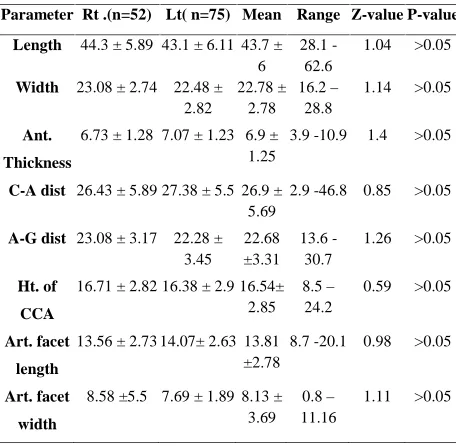
Table 2: Anatomical parameters for acromion process
Parameter Rt .(n=52) Lt( n=75) Mean Range Z-value P-value
Length 44.3 ± 5.89 43.1 ± 6.11 43.7 ± 6
28.1 -62.6
1.04 >0.05
Width 23.08 ± 2.74 22.48 ± 2.82 22.78 ± 2.78 16.2– 28.8 1.14 >0.05 Ant. Thickness
6.73 ± 1.28 7.07 ± 1.23 6.9 ± 1.25
3.9 -10.9 1.4 >0.05
C-A dist 26.43 ± 5.89 27.38 ± 5.5 26.9 ± 5.69
2.9 -46.8 0.85 >0.05
A-G dist 23.08 ± 3.17 22.28 ± 3.45 22.68 ±3.31 13.6 -30.7 1.26 >0.05 Ht. of CCA
16.71 ± 2.82 16.38 ± 2.9 16.54± 2.85 8.5– 24.2 0.59 >0.05 Art. facet length
13.56 ± 2.73 14.07± 2.63 13.81 ±2.78
8.7 -20.1 0.98 >0.05
Art. facet
width
8.58 ±5.5 7.69 ± 1.89 8.13 ± 3.69
0.8–
11.16
1.11 >0.05
Narrowest distance between coracoid and acromion
process in the present study was measured as 16.8 mm
whereas the narrowest distance between acromian and
glenoid was 13.6 mm.
The articular facet for the lateral end of the clavicle had
mean length of 13.81 ± 2.78 mm and mean width as
8.13 ± 3.69 mm.
4. DISCUSSION
During the evolution of the upper extremity, the
scapula, more than any other bone of the shoulder
girdle, reflects momentous alterations that have been
brought about by increased functional demands of a
prehensile limb. Changes in posture provided the
stimulus which initiated the numerous morphologic
changes. Gradual increase in the spine of the scapula
and the acromion process during development from the
pronograde to the orthograde. This change reflects the
increasing importance of the deltoid muscle.7
Collipol et al2 quoted that, the acromion morphology
according to Epstein et al. appears to have a prediction
value to determine the success of conservative medical
treatment in some cases and the need for surgery in
patients with joint impingement. Acromion of the hook
type (Type III) was observed with two times greater
frequency in patients with rotator cuff impingement
syndrome.2
High incidence of Type II followed by type I and very
low incidence of Type III scapulae was observed in the
present study. The findings were similar to
Sangiampong et al.8in Thai population, Musa et al.6in
Turkish population and Saha et al.9 in Indian
population. However high incidence of Type II,
followed by Type III and less number of Type I
acromion was observed by Coskun et al.3 in Turkish
population and Schetino et al.10in Brazilian population.
Table 3: Comparison of types of Acromion with some of the previous studies
Type (%)
Sangiampo ng et al8(Thai)
Coksun et al3 (Turkis
h)
Schetino et al10 (Brazilia n) Musa et al6 (Turkis h) Saha et al9 (India n)
Singh et al12 (India n)
Prese nt study
I 3.2 10 5.20 37 28 22.5 13.3
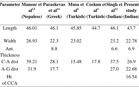
The length of the acromion was 43.7 ± 6 mm in the
present study; the findings were similar to most of the
earlier studies. (Table - 4) Edelson and Taitz5 had
observed that the thickness and width of Acromion
have no correlation with degenerative changes. The
width of the acromion in the present study did not
show much difference when compared with other
studies. The thickness of acromion observed by
Paraskevas et al.11was more (8.8 mm) as compared to
the present study (6.9 mm) and also another study in
Indian population by Singh et al.12(6.6 mm).
Table 4: Comparison of various parameters with some of the previous studies
Parameter Mansur et al13
(Nepalese)
Paraskevas et al11
(Greek)
Musa et al6
(Turkish)
Coskun et al3
(Turkish) Singh et
al12
(Indian) Present
study (Indian)
Length 46.01 46.1 45.85 44.7 46.1 43.7
Width 26.93 22.3 23.02 23.2 22.78
Ant. Thickness
8.8 6.6 6.9
C-A dist 39.21 28.1 15.48 17.8 37.5 26.9
A-G dist 31.9 17.7 27.0 22.68
Ht. of CCA
16.54
The predominant theory for the impingement syndrome
of the rotator cuff muscles classifies the contributing
factors as anatomical and functional. The anatomical
causes include the shape and the inclination of the
acromion.11 The Inclination of the acromion in the
present study was 33.460on the right side and 43.40on
the left side.
Sangiampong et al.8 quoted that, difference in the
development and morphology of acromion and the
presence of anterior acromial spur and inferior
acromioclavicular osteophytes decrease the volume of
subacromial space, leading to impingement and the
very close contact between the supraspinatous and the
anterior inferior part of the acromion, occurring at 900
abduction in internal rotation..In the present study we
found presence of acromial spur in three bones.
Some of the parameters of scapular morphometry vary
with sex and age, which also has a clinical
significance. Unfortunately, the data regarding age and
the sex of the bones studied was not available and that
we consider it as a weakness of this study
5. CONCLUSION
The results of the present study of 127 scapulae
revealed that Type II (Curved) acromion was
predominant. The bilateral symmetry was present in all
the parameters observed. The length and width of
acromion did not show much variation when compared
with other populations but coraco-acromial distance
and acromio-glenoid distance was variable. The results
of the present study may be of help to the shoulder
surgeons, Anthropologists and Anatomists.
6. ACKNOWLEDGEMENT
We acknowledge the suggestions and help by Dr. P.
Vatsalaswamy, Prof. Dept. of Anatomy, Dr. D. Y.
Patil medical college, Pimpri, Pune.
7. REFERENCES
1. Standring S. Gray’s Anatomy: the Anatomical
basis of clinical practice. Fortieth edition. Elsvier
ltd: London, UK. 2008.
2. Collipal E, Silva H, Ortega L, Espinoza E,
Martinez C. The acromion and its different forms.
Int J Morphol 2010; 28(4): 1189-1192.
3. Coskun N, Karaali K, Cevikol C, Demirel BM,
Sindel M. Anatomical basics and variations of the
scapula in Turkish adults. Saudi Med J 2006; Vol
27(9): 1320-1325.
4. Bigliani LU, Morrison DS, April EW. The
morphology of acromion and its relationship to
rotator cuff tears. Orthop. Trans 1986; 10:228.
5. Edelson JG, Taitz C. Anatomy of the
coraco-acromial arch. J Bone Joint Surg (Br) 1992; 74–B:
589-94.
6. Musa A, Tuba S, Mahinur U, Ismail Z, Serpil A,
analysis of the acromion with Multidetector
computerized tomography. Biomedical Research
2014; 25(3); 377-380.
7. DePalma AF, Brand RA. Origin and Comparative
Anatomy of the Pectoral Limb. Clin Orthop Relat
Res. v. 2008; 466(3).
8. Sangiampong A, Chompoopong S, Sangvichien S,
Thongtong P, Wongjittraporn S. The acromial
morphology of Thais in relation to gender and age:
Study in scapular dried bone. J Med Assoc Thai
2007; 90(3): 502-7.
9. Saha S, Vasudeva N, Paul S, Gautam VK. Study
of acromial morphology in Indian population. Rev
Arg de Anat Clin 2011; 3(2): 84-88.
10. Schetino LPL, Sousa Jr RR, Amâncio GPO,
Schetino MAA, Almeida-Leite CM, Silva JH.
Anatomical variations of acromions in Brazilian
adult’s scapulas. J Morphol Sci 2013; 30(2): 98
-102.
11. Paraskevas G, Traveas A, Papaziogas B, Kitsoulis
P, Spanidou S. Morphological parameters of the
acromion. Folia morphol 2008; 67(4): 255-260.
12. Singh J, Puhuja K, Agarwal R. Morphometric
parameters of the acromion process in adult human
scapulae. IJBAMR 2013; vol 2(8): 1165-1170.
13. Mansur DI, Khanal K, Haque MK, Sharma K.
Morphometry of acromion process of scapulae and
its clinical importance amongst Nepalese
population. Kathmandu Univ med J 2012; 38(2):
33-36
Conflict of Interest: None