Page 1 of 14
Open Access
Review
α
-Synuclein and neuronal cell death
Mark R Cookson
Address: Laboratory of Neurogenetics, National Institute on Aging, NIH. Building 35, Room 1A116, MSC 3707, 35 Convent Drive, Bethesda, MD 20982-3707, USA
Email: Mark R Cookson - [email protected]
Abstract
α-Synuclein is a small protein that has special relevance for understanding Parkinson disease and related disorders. Not only is α-synuclein found in Lewy bodies characteristic of Parkinson disease, but also mutations in the gene for α-synuclein can cause an inherited form of Parkinson disease and expression of normal α-synuclein can increase the risk of developing Parkinson disease in sporadic, or non-familial, cases. Both sporadic and familial Parkinson disease are characterized by substantial loss of several groups of neurons, including the dopaminergic cells of the substantia nigra that are the target of most current symptomatic therapies. Therefore, it is predicted that α-synuclein, especially in its mutant forms or under conditions where its expression levels are increased, is a toxic protein in the sense that it is associated with an increased rate of neuronal cell death. This review will discuss the experimental contexts in which α-synuclein has been demonstrated to be toxic. I will also outline what is known about the mechanisms by which α-synuclein triggers neuronal damage, and identify some of the current gaps in our knowledge about this subject. Finally, the therapeutic implications of toxicity of α-synuclein will be discussed.
All neurodegenerative diseases share the common phe-nomenon that neurons, usually relatively specific groups, are lost progressively as the disease develops. In some cases, we can provide partial relief for patients by treating some of their symptoms. However, because we don't understand the underling mechanisms of why neurons die, degeneration continues inexorably and old symp-toms often become unresponsive while new ones arrive. At the end of the disease process, we are left with only a few clues about what might have happened based on what we can glean from the pathology of the disease using post mortem samples. In general, the root cause of neurodegen-eration remains obscure although rare genetic variants are helpful in that we can be certain that an inherited muta-tion acts as the trigger for disease in that specific family.
Here, I will discuss cell loss related to Parkinson disease (PD) in the context of one protein, α-synuclein, that has several links to the disorder. In doing so, I will outline what we know about the ways in which a protein can lead to cell death. Before doing so, it is worth discussing what PD is, and what it isn't.
Cell death in PD
It is very commonly said that PD is the second most com-mon neurodegenerative disease and results from a loss of dopamine neurons. The first fact is dull and the second tells only part of the story. It is true that PD patients have a substantial loss of dopamine in the striatum resulting from a relatively selective loss of dopaminergic projection neurons in the substantia nigra pars compacta. Both
bio-Published: 4 February 2009
Molecular Neurodegeneration 2009, 4:9 doi:10.1186/1750-1326-4-9
Received: 17 December 2008 Accepted: 4 February 2009
This article is available from: http://www.molecularneurodegeneration.com/content/4/1/9
© 2009 Cookson; licensee BioMed Central Ltd.
chemical measures and imaging modalities suggest that at least a 70% decrease in striatal dopamine occurs before the onset of clinical parkinsonism and progresses over time [1]. These estimates of the extent of striatal dopamine depletion, combined with the observation that the majority of dopaminergic neurons are lost by the end of the disease process, imply that there is substantial cell death throughout the PD disease process. It is not possible to show this directly, but measurements of nigral cell numbers in neurologically normal people and in non-human primates reveal a slow progressive loss of dopamine neurons with age [2]. In this view, parkinson-ism is an accelerated, but still slow, cell death phenotype that would normally be seen with aging [3].
However, while there is relative vulnerability of dopamin-ergic neurons in the substantia nigra [4], not all dopamine cells are affected in PD. For example, although dopamin-ergic neurons in the ventral tegmental area that project to the nucleus accumbens do degenerate [5], compared to the dopaminergic neurons in the substantia nigra pars compacta these cells are relatively spared [6,7].
Furthermore, not all affected neurons in PD are dopamin-ergic. Non-motor symptoms are a serious problem for many PD patients and are often untreated by replacement therapy with L-DOPA (3,4-dihydroxy-L-phenylalanine) [8]. A good example of non-dopaminergic cells that degenerate in PD is the cholinergic neurons in the dorsal vagal nucleus [9]. It has been suggested that involvement of non-nigral regions underlies the complex clinical pic-ture in PD [10]. Therefore, although there is some specifi-city to cell death in PD, there is no absolute selectivity for any specific neurotransmitter group or anatomic region. It is also important to note that loss of nigral neurons occurs in diverse pathological situations [4] and that on its own, nigral cell loss defines the clinical term parkinsonism, not Parkinson disease.
This distinction is also important when discussing the other major pathological event in PD that appears along-side cell death, the formation of Lewy bodies and Lewy neurites. Lewy bodies are intracellular deposits of proteins and lipids [11] that were traditionally stained with eosin but now are more sensitively recognized by antibodies to specific marker proteins [12]. Using electron microscopy, Lewy bodies are fibrillar structures with a recognizable core and halo [13]. The range of Lewy pathology is now recognized as encompassing many regions of the diseased brain [14] including, for example, the olfactory bulb, raphe nucleus, locus coeruleus and the basal nucleus of Meynert. Additionally, some reports suggest that the nigra is not the first place where Lewy bodies form [15]. How this relates to the extent of cell loss in each region is not well defined. Lewy bodies are also seen in dementia with
Lewy bodies (DLB, also known as Diffuse Lewy body Dis-ease or DLBD), suggesting that PD and DLBD are related to one another by shared pathology and maybe by shared etiology.
Therefore, PD is a disease where substantial cell loss in the nigra occurs alongside the formation of Lewy bodies. Nei-ther cell loss nor Lewy bodies is absolutely specific for PD but both are required for a diagnosis of PD under current definitions [16]. This review will focus on cell death, but it is important to understand a little more about the most commonly used marker for Lewy bodies; α-synuclein.
α
-Synuclein is a marker of the PD process
The first member of the family of proteins for which α -synuclein is named was cloned from the neuromuscular junction of the electric eel [17]. Antibodies against that protein labeled both synapses and nuclei, leading to the naming of synuclein. A related protein was cloned from zebra finch as a protein upregulated during the process of song learning, a period of enormous synaptic plasticity [18]. In humans, there are three synuclein family mem-bers (α-,β-,γ-) and all synuclein genes are relatively well conserved both within and between species [19]. The synuclein genes are specific to the vertebrate lineage in that neither single cell organisms (including yeast) nor invertebrates (Drosophila melanogaster, Caenorhabditis ele-gans) have any apparent synuclein homologue. Addition-ally, primate α-synuclein sequences differ from other vertebrate synucleins by a substitution of Alanine for a Threonine at position 53 [20]. These two interesting facts about the evolutionary relationships in the synuclein fam-ily are important for understanding some of the experi-mental systems where synuclein has been explored.The normal function of α-synuclein is poorly understood. Although it is expressed at high levels in the brain, rela-tively specifically within neurons, it is also found in other tissues, e.g., hematopoietic cells [21,22]. α-Synuclein can bind to lipids [23] and, in neurons, is associated with pre-synaptic vesicles [24,25] and the plasma membrane, pos-sibly via lipid rafts [26]. The association of α-synuclein with vesicles is modulated by synaptic activity where the protein dissociates from vesicles after electrical stimula-tion of the neuron and only slowly re-associates [27]. However, α-synuclein knockout mice show only subtle abnormalities in neurotransmission [28-30], suggesting that α-synuclein plays a non-essential function at the syn-apse. There is some evidence that such a modulatory role may be more important under conditions where reactive oxygen species or nitric oxide are present [31,32], although the mechanism(s) are not yet fully defined.
Page 3 of 14 would be consistent with a predominantly synaptic
local-ization [25]. However, in PD brains, α-synuclein antibod-ies strongly stain Lewy bodantibod-ies [33] and Lewy neurites [34]. Because of this sensitivity, α-synuclein staining is now more commonly used than eosin or ubiquitin staining for these structures. Biochemical analyses have shown that α -synuclein is a major protein component of Lewy bodies and may be part of the fibrillar structure of these structures [35]. The deposited, pathological forms of α-synuclein are aggregated and show lower solubility than the normal protein [36] and may be modified post-translationally by truncation, nitration, ubiquitylation and phosphoryla-tion [37-40].
Therefore, α-synuclein protein deposition into Lewy bod-ies is a marker of the PD disease state. However, because we require Lewy bodies for a PD diagnosis this isn't an especially strong argument for involvement of α -synu-clein in the disease process. It is also important to note that, although we cannot determine if Lewy bodies previ-ously formed in the cells that eventually died, the individ-ual neurons where Lewy bodies are found are the ones that have survived the disease process (though they still may be dysfunctional). Very recently, it has been shown that Lewy bodies form in functional dopaminergic neu-rons grafted in to brains of people with PD as a therapeu-tic intervention [41,42], although this is not always seen [43]. These were embryonic cells that remained appar-ently healthy and were functional after grafting, which suggests that there is the environment of the PD brain pre-disposes even young cells to form Lewy bodies.
In summary, the available evidence identifies α-synuclein as a marker of the PD/DLB process but do not prove that it has a causal role. The evidence that it does comes from a variety of human genetic studies.
α
-Synuclein can cause PD
A key discovery in understanding PD was the report that an A53T mutation in the α-synuclein gene was causal for dominantly inherited disease [44]. This was the first clear report that a Mendelian gene could be a cause of PD, which to that point had been thought of as a non-genetic disease. It is interesting that the first mutation found was A53T, i.e. a reversion of the human Alanine to the ances-tral Threonine found in rodents and many other species. Since then, two other point mutations, A30P [45] and E46K [46], have been reported in different families. It is also important that while many cases are reported to have a phenotype of 'PD', in fact several patients in the A53T and E46K [46] families have a more diffuse involvement of synuclein deposition [47,48] and clinical features that presumably result from this degree of involvement of non-dopaminergic systems [49].
A second group of important cases have multiplications of the normal wild type allele of SNCA, the gene that
encodes for the α-synuclein protein. Cases with SNCA duplication have a brainstem-predominant PD pheno-type [50], while cases with a triplication have a Lewy body disease that again involves several brain regions [51,52]. Measurements of protein levels in triplication show the predicted doubling of α-synuclein in blood as well as increased levels and deposition of the protein in the cere-bral cortex where Lewy bodies are found [21]. Therefore, even without sequence variants, α-synuclein dosage can be causal for Lewy body disease.
A third piece of genetic evidence comes from the reports common variants around the α-synuclein gene are associ-ated with lifetime risk of sporadic PD. Both the promoter region, specifically the Rep1 polymorphic repeat [53], and polymorphisms towards the 3' end of the gene are associ-ated with PD [54]. Although it is not known specifically how these risk variants influence lifetime incidence of PD, it seems likely that they increase α-synuclein protein levels in the brain.
Collectively, the human genetic data strongly support a causal role for α-synuclein in PD/DLBD. Whether Lewy bodies are causal or consequential is less clear, but they do support the idea that α-synuclein represents an important link between sporadic and inherited PD. The various lines of evidence identify α-synuclein as a potentially toxic pro-tein, fulfilling the requirements of a causative agent in PD [55]. The question now is how, and in what context, is α -synuclein toxic, and can we do anything about it?
Where and when is
α
-synuclein toxic?
Given that cell loss is a major event in human PD, com-bined with the evidence that α-synuclein plays a causal role in disease, it is reasonable to infer that α-synuclein is toxic to human neurons. The time course is likely to be protracted, with the most likely explanation that there is asynchronous cell death that results in a slow depletion of the populations of relatively vulnerable neurons. How-ever, it is not possible to watch cells die in the human brain and so we have to turn to experimental models to confirm or refute the idea that α-synuclein is toxic.
organ-isms where the protein is not normally present. One read-ing of this data is that, at least in terms of toxicity occurring over days to weeks, the normal function of the protein is probably not relevant.
A situation where α-synuclein is normally present is in mammalian cell culture models. Two commonly used sys-tems are primary neurons, including dopaminergic cul-tures of the ventral midbrain, or neuroblastoma derived cell lines. Experiments showing the most substantial effects of α-synuclein include those where the protein is transiently expressed, e.g. from viral vectors [81-86], or expression is controlled from an inducible promoter sys-tem [87-89], although some authors have reported a lack of toxicity in similar circumstances [90]. In midbrain cul-tures, toxicity is higher for dopamine neurons than other cells [81], which may be relevant to the relative vulnera-bility of nigral neurons in PD. Some experiments show nicely that the difference between wild type and mutant protein is really a matter of dose and that at increasing expression levels, the normal protein becomes as toxic as the dominant mutants [89].
Although potentially useful in for understanding mecha-nisms, these cell-based models are taken out of their in vivo context and tend to show cell loss over a few days, compared to the predicted years of progress in the disease. A more intact approach is to express α-synuclein using transgenic technology in various parts of the mouse CNS. Some of these models show toxicity, particularly in the spinal cord, but nigral cell loss is absent or modest [91-97]. Several models do show accumulation and insolubil-ity of α-synuclein [e.g., [36,91,93,98]], although whether true Lewy bodies are formed is uncertain. Therefore, most mouse models reported to date are better for understand-ing α-synuclein deposition than frank cellular toxicity. Why this is the case is unclear, but it is interesting that crossing transgenic models with mouse α-synuclein knockouts exacerbates phenotypes [99-101], suggesting that the presence of the murine protein limits damage in some undefined way. The lack of an ideal PD mouse model that more completely captures the human pheno-type limits our current studies of α-synuclein toxicity. Though a goal worth pursuing, creation of such an ideal mouse model may be very challenging given the limita-tions of mouse lifespan and differences in physiology between mice and humans.
An alternate approach to traditional transgenics is to use viral vectors to deliver α-synuclein directly to the substan-tia nigra in mice [102], rats [103-106] or non-human pri-mates [107-109]. In these approaches, a significant cell loss is noted along with deposition of α-synuclein pro-tein. The extent of cell loss is less dramatic than in human PD and behavioral effects are similarly modest. However,
the critical observation here is that α-synuclein can induce toxicity in vivo using vertebrate organisms, with a time course of several weeks, allowing for some dissection of mechanism.
Taken together, all of this evidence suggests that α -synu-clein can induce toxicity in a variety of contexts, from sim-ple organisms to dopamine neurons in the primate substantia nigra. It is less clear whether all of these situa-tions are directly relevant to the human disease, where cell loss is probably more protracted, but as a practical matter such models at least afford an opportunity to examine mechanism(s) by which α-synuclein triggers neuronal death.
Why is
α
-synuclein toxic?
Some of the above model systems have been used to probe the mechanism(s) by which α-synuclein causes cell death. These can generally be sorted into aspects of the protein itself effects of the protein to the biological system (see figure 1). Appendix 1 highlights some of the key observations related to this critical question.
Aspects of protein chemistry of -synuclein and toxicity
α-Synuclein has a strong tendency to self-associate in vitro [110,111], and so a prime candidate for the underlying driving force for toxicity is the formation of aggregated species. One of the important questions about this idea is which species are present in cells/tissues. Oligomeric spe-cies can be isolated from cells [112-114] and from human [21] and mouse (both wild type and α-synuclein trans-genic) brain [115]. In both cells and brain, oligomers are particularly found in membrane-enriched fractions [112,115], suggesting a possible influence of the lipid environment on oligomer formation. Higher molecular weight forms have also been found in some models [116], especially after oxidative stress [117] or exposure to inflammatory triggers in mice [100]. Deposited α -synu-clein immunoreactivity has been seen in transgenic [91-97] or viral models [102-109]. However, the observation of aggregated α-synuclein by and of itself does not prove that aggregation is important; as discussed for Lewy bod-ies, all this proves is that deposition occurs, not that it is causal.
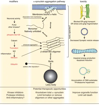
Page 5 of 14 Events in α-synuclein toxicity
Figure 1
Events in α-synuclein toxicity. The central panel shows the major pathway for protein aggregation. Monomeric α-synuclein is natively unfolded in solution but can also bind to membranes in an α-helical form. It seems likely that these two species exist in equilibrium within the cell, although this is unproven. From in vitro work, it is clear that unfolded monomer can aggregate first into small oligomeric species that can be stabilized by β-sheet-like interactions and then into higher molecular weight insoluble fibrils. In a cellular context, there is some evidence that the presence of lipids can promote oligomer formation: α -synuclein can also form annular, pore-like structures that interact with membranes. The deposition of α-synuclein into patho-logical structures such as Lewy bodies is probably a late event that occurs in some neurons. On the left hand side are some of the known modifiers of this process. Electrical activity in neurons changes the association of α-synuclein with vesicles and may also stimulate polo-like kinase 2 (PLK2), which has been shown to phosphorylate α-synuclein at Ser129. Other kinases have also been proposed to be involved. As well as phosphorylation, truncation through proteases such as calpains, and nitration, probably through nitric oxide (NO) or other reactive nitrogen species that are present during inflammation, all modify synu-clein such that it has a higher tendency to aggregate. The addition of ubiquitin (shown as a black spot) to Lewy bodies is prob-ably a secondary process to deposition. On the right are some of the proposed cellular targets for α-synuclein mediated toxicity, which include (from top to bottom) ER-golgi transport, synaptic vesicles, mitochondria and lysosomes and other pro-teolytic machinery. In each of these cases, it is proposed that α-synuclein has detrimental effects, listed below each arrow, although at this time it is not clear if any of these are either necessary or sufficient for toxicity in neurons.
toxicity
Impaired energy production Apoptosis induction Decreased Synaptic vesicle release
Accumulation of CMA substrates ?Proteasome impairment Blocked ER-golgi transport ER stress and golgi fragmentation
Monomer Natively unfolded
Membrane bound α-helix
β-sheet-like oligomer
fibrils
Lewy body
Neuronal activity
α-synuclein aggregation pathway
Ubiquitin
toxic species?
modifiers
PLK2 other kinases?
phospho-Ser129
Truncation
Calpain
dissociation from membranes
enhanced aggregation
Nitration
addition after deposition? Inflammation
NO
Potential therapeutic opportunities
Kinase inhibitors Protease inhibitors Anti-inflammatory?
Knockdown total α−synuclein Limit formation or remove oligomers or other aggregates
cells. The presence of fibrils in Lewy bodies would support this contention. However, α-synuclein can end up in the extracellular media [119] and it is possible that the condi-tions for aggregation might be more suitable in a milieu free of cells. The relevance of extracellular α-synuclein is an important question, raised also by the observation of Lewy bodies in grafted neurons [41,42] and the attendant hypothesis of 'host to graft transmission'.
Some studies have attempted to address whether intracel-lular aggregates of α-synuclein contribute to toxicity. For example, several imaging techniques shown that, in the context of a living cell, α-synuclein can form small oli-gomers, likely in an antiparallel configuration [114,120] and such oligomers can be associated with cell toxicity.
These approaches have been used to show that overex-pression of heat shock proteins (Hsps) can mitigate both oligomer formation and toxicity [114,120,121]. In vivo, Hsps can prevent toxic effects of α-synuclein in yeast [59] and in flies [67]. Whether these studies constitute formal proof that aggregation is required for toxicity is unclear as there are other theoretical interpretations of the data. For example, a formal possibility is that monomeric α -synu-clein is toxic and, thus, any protein binding the protein directly could limit toxicity. It should be stated that the mechanism(s) by which monomers of α-synuclein could be toxic are relatively unexplored but, equally, there is an absence of proof that aggregation is absolutely required for toxicity. Alternatively, Hsps could be limiting a detri-mental event downstream of the initial aggregation and thus may neither represent evidence for or against the role of aggregation in α-synuclein toxicity. Interestingly, Hsp expression in the fly model decreases neuronal toxicity without any change in the number of α-synuclein positive inclusions [67].
Overall, these considerations show that α-synuclein is capable of protein aggregation and can be deposited into inclusion bodies of various forms in vivo, but that there is insufficient evidence that aggregation or deposition is either necessary or sufficient for toxicity. In fact, several lines of evidence show that toxicity can be dissociated from deposition, including; the observation in cells of toxicity without deposition in some models [81]; differen-tial effects on toxicity and inclusions of various manipula-tions of α-synuclein in fly models [66,67]; and deposition of α-synuclein without clear toxic effects in some mouse models [e.g., [36]]. A key challenge for the field, therefore, is to understand whether protein aggregation is at all rele-vant for the toxic effects of α-synuclein. One way to poten-tially address this is to isolate various aggregated species of the protein and to express them within a neuron. This might be extraordinarily difficult from a technical stand-point and there is always possibility that the small
aggre-gates would seed larger ones may confound interpretation. Another potential approach would be to develop reagents that limit the biological availability of specific aggregated species and use these to probe which agents are toxic in intact cells. As an example, recom-binant single chain Fv antibody fragments against aggre-gated α-synuclein have been described [122,123] that might be helpful.
α-Synuclein has many additional properties as well as the tendency to aggregate. Some of the post-translational modifications that have been reported have also been explored as possible mediators of toxicity. For example, antibodies against α-synuclein phosphorylated at Ser129 are very good at identifying Lewy pathology in the human brain [38], suggesting either that Ser129 phosphorylation is a causal event for deposition or represents a common modification of the protein after it is deposited. Several groups have therefore made versions of α-synuclein that cannot be modified at this residue (S129A) or pseudo-phosphorylation mimics (S129D, S129E) and deter-mined the toxic effects of expression. In Drosophila mod-els, S129A is less toxic but has an increased tendency to form inclusion bodies compared to wild type protein [66]. The S129D phosphomimic has the opposite effect, i.e. increased toxicity but fewer inclusions. In contrast, similar experiments using viral overexpression in rats show the opposite result, namely that S129A greatly increases the toxic effects of expression [124]. In mamma-lian cell culture, S129A has a diminished tendency to form inclusion bodies [125].
At first glance, these results seem to suggest that the behav-ior of α-synuclein as it relates to toxicity is opposite in mammals compared to invertebrates where, it is impor-tant to note, the protein is not normally present. However, interpretation is complicated by several considerations. First, the expression levels of α-synuclein are critical for toxicity, which is shown by the human case where a differ-ence in protein levels is 2-fold in the triplication cases and 1.5-fold in the duplication cases. Second, recent data sug-gests that the phosphomimic S129D/E α-synuclein vari-ants have different biophysical properties compared to authentically phosphorylated wild type protein [126]. Overall, these considerations raise some important caveats about comparison of properties of α-synuclein in terms of concentration-dependent behaviors of the protein such as aggregation and toxicity.
Page 7 of 14 the polo-like kinase family, specifically PLK2, have been
shown to be active both in vitro and in vivo in generating pS129 α-synuclein [127]. What is interesting about PLK2 is that it is known to respond to neuronal activity [128], suggesting a possible link between neuronal phenotype and α-synuclein toxicity. However, it is not yet known in PLK2 inhibitors or gene knockout will limit the toxic effects of α-synuclein in vivo. Such experiments are feasi-ble in several species as PLK2 homologues are present in mice and flies, and there is at least one polo kinase in yeast.
There are a number of other modifications of α-synuclein that have been reported and some of these are found more often in pathological circumstances than under normal conditions, such as nitration or truncation. Truncation of α-synuclein is associated with a higher tendency for aggre-gation [129-131]. Transgenic mice expressing truncated α -synuclein have substantial cell loss [101] although in at least one line, this is a developmental and not degenera-tive phenotype [132]. Again, because the window for tox-icity is quite narrow, comparison between different lines is difficult. One question that arises for truncation is where such species are generated. α-Synuclein is predom-inantly degraded by lysosomal pathways [133,134], including chaperone-mediated autophagy [135], and the lysosomal cathepsins are important in proteolysis. There-fore, some truncated species are found in the lysosomes and it seems unlikely that they would cause damage to the cell. However, α-synuclein is also a substrate for cytoplas-mic calpains [136-139], which are therefore more likely to generate cytoplasmic toxic truncated species. Some detail is therefore needed to prove which truncated species mediate toxicity, if any of them in fact do.
Oxidative stress, including the neurotransmitter dopamine, has been linked to increased α-synuclein aggregation [89,140]. Dopamine itself may contribute to the toxic effects of α-synuclein in vitro [89], although such a mechanism cannot explain why non-dopaminergic neu-rons die early in the disease process. α-Synuclein expres-sion can enhance sensitivity to oxidative and nitrative stressors [141,142], although it can also be protective in some situations [143]. In most of these situations, the role of aggregation is unclear.
In summary, α-synuclein has properties, including the potential for aggregation and post-translational modifica-tions, which may influence its toxic effects. Whether these are required for toxicity is unclear, and some results still need to be resolved, for example for the work on S129 phosphorylation. However, there is a larger question, which is: what effects synuclein has on neurons that are responsible for its toxic effects?
Mediators of -synuclein toxicity in biological systems
Some of the relevant data from cellular systems has been reviewed previously [144] and will be discussed here in the context of examples across multiple models.
Presumably, α-synuclein might interact with other bio-molecules to mediate toxicity. Because α-synuclein can associate with lipids, membranes are one possible target. In vitro, α-synuclein can form pore-like structures [145,146], and annular rings of synuclein have been iso-lated from the brains of patients with multiple system atrophy, a synucleinopathy [147]. Cells expressing α -synuclein have increased cation permeability [148] and vesicles prepared from cultured cells or isolated from the adrenal medulla show leakage of catecholamines [149]. These events may be consistent with the formation of non-specific pores or similar structures at the plasma membrane or at a vesicle surface.
Because α-synuclein binds synaptic vesicles, it is possible that synaptic transmission would be directly or indirectly a target of synuclein toxicity. One example of this comes from work showing that A30P α-synuclein alters exocyto-sis of catecholamine containing vesicles in primary cells and in chromaffin cells [150]. The effect here is probably at a late stage of the exocytosis, before vesicle membrane fusion [150].
Further evidence for an effect of α-synuclein on vesicle function that may mediate toxicity comes from suppressor screens in yeast [63]. In the same organism, such defects can be localized to a block in endoplasmic reticulum (ER)-golgi vesicular trafficking [151]. Supporting this idea, there is evidence of ER stress [87] and golgi fragmen-tation [152] in mammalian cell systems.
Overexpression of Rab1, a GTPase that influences vesicle dynamics, was able to at least partially rescue the toxic effects of α-synuclein in yeast, worms and in mammalian cells [151]. Therefore, some of the toxic effects of α -synu-clein that are conserved across species involve damage to vesicular transport, which might express itself as damage to presynaptic vesicle release in a neuron.
[57]. The mechanism by which α-synuclein interacts with and causes damage to mitochondria is not fully resolved and, given the central role of mitochondria in apoptotic pathways, perhaps such effects are secondary to the induc-tion of apoptosis. Increased levels of α-synuclein are reported to trigger apoptosis in various cell types [159-161]. Several apoptotic markers are also seen in yeast models of synuclein toxicity [59]. α-Synuclein toxicity can be rescued by caspase inhibitors or knock down of cas-pase-12 [87]. Activation of caspase-3 has been reported in transgenic mice [162] caspase-9 has been reported in viral models in mice [102] and rats [106]. However, these stud-ies show only a few caspase positive cells, and so whether apoptosis is the only way in which cells expressing α -synuclein die remains unclear.
α-Synuclein can bind to the membranes of lysosomes [135] and inhibit lysosomal function [163] and chaper-one-mediated autophagy [135]. Recent results suggest that CMA is implicated in the regulation of the transcrip-tion factor MEF2D and that this can be disrupted by expression of α-synuclein, leading to neuronal death [164]. As another example of misregulated protein turno-ver, α-synuclein (and specifically α-synuclein oligomers) can also inhibit the proteasome [81,88,163,165-167], although it is not clear if the predicted altered turnover of proteasome substrates occurs in vivo [168].
The general principle is that multiple systems can be affected by α-synuclein expression and that if there is a common theme between them, it is likely to be that α -synuclein can binds lipids. Several lines of evidence sug-gest that lipid binding can promote the formation of oli-gomers [115,145,169]. Therefore, this interpretation links a primary protein abnormality to cellular targets of the protein. As discussed elsewhere [144], determining which events are truly primary and which are secondary remains a challenge. Although this distinction is an intellectual problem, it may also be relevant for deciding which aspects of cell death to target if we want to limit the dis-ease process in PD.
Potential therapeutic approaches related to
α
-synuclein toxicity
One of the key questions here is to decide whether to try and target the protein or the process that mediates cellular damage. Both are attractive for different reasons, although both are also difficult (see figure 1 for where these might be utilized and Appendix 2 for the critical next steps).
If there were a pathogenic aggregated form of α-synuclein, then one tactic would be to target that species. If we pro-pose that insoluble fibrils are toxic, then a 'fibril-buster' would be the way forward [reviewed in [111]], but if sol-uble oligomers damage cells then we would want to pre-vent their formation or encourage their turnover. As
discussed above, both fibrils and oligomers can be found in different models and either alone, or both, could be toxic. For oligomers, the situation is more complicated if different oligomeric forms have different toxic properties [118], suggesting that we may need to be careful about which oligomers we target.
Alternatively, we could be agnostic about which species are important and try and decrease all α-synuclein expres-sion. There are reports that increasing autophagy can help clear aggregation-prone proteins, including α-synuclein [170]. Antisense approaches might also be helpful, and have been reported to work in the rat [171] and mouse [172] brain. This approach is predicated on the idea that α-synuclein really is dispensable for CNS function in humans, as it appears to be in the mouse [28,30], but per-haps even a modest decrease in protein levels would be enough to decrease PD progression.
We might also try to change the modifications of α -synu-clein, especially if these are specific for pathogenic forms. For example, example of PLK2 as a kinase for Ser129 [127] may provide a way to test the idea that phosphorylation at this residue is key for pathogenesis, if sufficiently specific kinase inhibitors can be developed. Again, assuming spe-cificity can be achieved, it might be interesting to block other modifications such as truncation or nitrosylation – the latter might be part of the general rubric of anti-inflammatory approaches. However, such approaches would only be helpful if the modification is truly specific for the pathogenic form and makes an active contribution to cellular toxicity, ie is not a bystander in the process.
Finally, we may target one or more of the cellular effects of α-synuclein that are associated with toxicity. This might have the advantage of leaving the protein alone, which may be useful if it turns out that α-synuclein has a specific function in the human brain. The difficulty, of course, is in understanding why the protein is toxic, although the work with Rab1 [151,173] suggests that this is a tractable problem, at least in principle.
Conclusion
Page 9 of 14 well as at developing models that have a stronger cell
death signal, to more accurately represent the substantive loss of neurons seen in PD.
Abbreviations
DLB/DLBD: Dementia with Lewy bodies/Diffuse Lewy Body Disease; ER: endoplasmic reticulum; L-DOPA: 3,4-dihydroxy-L-phenylalanine; PD: Parkinson disease.
Competing interests
The author declares that they have no competing interests.
Appendix 1: key observations
The role of α-synuclein in PD and related disease is high-lighted by the convergence of pathological and genetic data. Because part of the pathological phenotype of PD involves cell death of neurons, particularly but not exclu-sively dopamine neurons in the substantia nigra pars compacta, this suggests that α-synuclein may be a toxic protein. The following key observations have been made in various experimental systems to support this conten-tion:
- In pure in vitro assays, α-synuclein shows a lack of con-formational restraint that tends to promote inappropriate aggregation. This can be enhanced by mutation, increas-ing concentration or any of several protein modifications associated with pathological deposition of the protein in vivo. α-Synuclein can also bind lipids and membranes in vitro
- In a variety of species, expression of α-synuclein can pro-mote toxic events. These include organisms such as yeast, worms and flies, where no α-synuclein homologue is present, suggesting that irrespective of its normal func-tion, the protein can be toxic.
- Data in mammalian cell culture also supports a toxic effect of α-synuclein, particularly to dopaminergic cells. Results in intact in vivo systems are mixed, with toxicity limited to the spinal cord in some transgenic mouse mod-els and modest toxic effects to dopaminergic neurons using viral mediated overexpression in rodents and non-human primates.
- The mechanism(s) involved are currently unclear, but binding to several cellular membranes may contribute to toxic events.
Appendix 2: critical next steps
The following critical issues need to be addressed before our understanding of α-synuclein pathobiology can be applied to therapeutic development:
- We need to better understand normal function of α -synuclein, such that we can assess both what role it might play in toxicity in the mammalian CNS and so we can highlight potential detrimental effects of limiting expres-sion or function of the protein.
- We need to clearly identify which cellular pathways con-tribute to the pathological effects of the protein. Some great work has been performed in yeast models that high-light interruption of vesicle transport, but it is important now to establish what the analogous process is in neurons and whether this is sufficient to explain α-synuclein toxic-ity in this system.
- We need to develop models where there is a lesion that better approximates the severity of cell loss seen in human PD. This will allow for a more rigorous test of pathways involved in toxicity as the disease progresses. An acceler-ated time course would be helpful, and may be necessary, but the pathology should be similar to human PD in that nigral neurons should be affected at some point in the model but not necessarily first or exclusively.
Acknowledgements
This research was supported by the Intramural Research Program of the NIH, National Institute on Aging.
References
1. Brooks DJ: The early diagnosis of Parkinson's disease. Ann Neu-rol 1998, 44:S10-18.
2. Chu Y, Kordower JH: Age-associated increases of alpha-synu-clein in monkeys and humans are associated with nigrostri-atal dopamine depletion: Is this the target for Parkinson's disease? Neurobiol Dis 2007, 25:134-149.
3. Thal DR, Del Tredici K, Braak H: Neurodegeneration in normal brain aging and disease. Sci Aging Knowledge Environ 2004, 2004:pe26.
4. Dickson DW: Linking selective vulnerability to cell death mechanisms in Parkinson's disease. Am J Pathol 2007, 170:16-19.
5. Hirsch EC: Biochemistry of Parkinson's disease with special reference to the dopaminergic systems. Mol Neurobiol 1994, 9:135-142.
6. Chung CY, Seo H, Sonntag KC, Brooks A, Lin L, Isacson O: Cell type-specific gene expression of midbrain dopaminergic neu-rons reveals molecules involved in their vulnerability and protection. Hum Mol Genet 2005, 14:1709-1725.
7. Surmeier DJ: Calcium, ageing, and neuronal vulnerability in Parkinson's disease. Lancet Neurol 2007, 6:933-938.
8. Langston JW: The Parkinson's complex: parkinsonism is just the tip of the iceberg. Ann Neurol 2006, 59:591-596.
9. Gai WP, Blumbergs PC, Geffen LB, Blessing WW: Age-related loss of dorsal vagal neurons in Parkinson's disease. Neurology 1992, 42:2106-2111.
10. Jellinger KA: Post mortem studies in Parkinson's disease–is it possible to detect brain areas for specific symptoms? J Neural Transm Suppl 1999, 56:1-29.
11. Gai WP, Yuan HX, Li XQ, Power JT, Blumbergs PC, Jensen PH: In situ and in vitro study of colocalization and segregation of alpha-synuclein, ubiquitin, and lipids in Lewy bodies. Exp Neu-rol 2000, 166:324-333.
13. Gibb WR, Scott T, Lees AJ: Neuronal inclusions of Parkinson's disease. Mov Disord 1991, 6:2-11.
14. Braak H, Del Tredici K, Rub U, de Vos RA, Jansen Steur EN, Braak E: Staging of brain pathology related to sporadic Parkinson's disease. Neurobiol Aging 2003, 24:197-211.
15. Del Tredici K, Rub U, De Vos RA, Bohl JR, Braak H: Where does parkinson disease pathology begin in the brain? J Neuropathol Exp Neurol 2002, 61:413-426.
16. Hughes AJ, Daniel SE, Kilford L, Lees AJ: Accuracy of clinical diag-nosis of idiopathic Parkinson's disease: a clinico-pathological study of 100 cases. J Neurol Neurosurg Psychiatry 1992, 55:181-184. 17. Maroteaux L, Campanelli JT, Scheller RH: Synuclein: a neuron-specific protein localized to the nucleus and presynaptic nerve terminal. J Neurosci 1988, 8:2804-2815.
18. George JM, Jin H, Woods WS, Clayton DF: Characterization of a novel protein regulated during the critical period for song learning in the zebra finch. Neuron 1995, 15:361-372.
19. George JM: The synucleins. Genome Biol 2002, 3:REVIEWS3002. 20. Hamilton BA: alpha-Synuclein A53T substitution associated
with Parkinson disease also marks the divergence of Old World and New World primates. Genomics 2004, 83:739-742. 21. Miller DW, Hague SM, Clarimon J, Baptista M, Gwinn-Hardy K,
Cook-son MR, Singleton AB: Alpha-synuclein in blood and brain from familial Parkinson disease with SNCA locus triplication. Neu-rology 2004, 62:1835-1838.
22. Scherzer CR, Grass JA, Liao Z, Pepivani I, Zheng B, Eklund AC, Ney PA, Ng J, McGoldrick M, Mollenhauer B, Bresnick EH, Schlossmacher MG: GATA transcription factors directly regulate the Par-kinson's disease-linked gene alpha-synuclein. Proc Natl Acad Sci USA 2008, 105:10907-10912.
23. Jo E, McLaurin J, Yip CM, St George-Hyslop P, Fraser PE: alpha-Synuclein membrane interactions and lipid specificity. J Biol Chem 2000, 275:34328-34334.
24. Withers GS, George JM, Banker GA, Clayton DF: Delayed localiza-tion of synelfin (synuclein, NACP) to presynaptic terminals in cultured rat hippocampal neurons. Brain Res Dev Brain Res
1997, 99:87-94.
25. Irizarry MC, Kim TW, McNamara M, Tanzi RE, George JM, Clayton DF, Hyman BT: Characterization of the precursor protein of the non-A beta component of senile plaques (NACP) in the human central nervous system. J Neuropathol Exp Neurol 1996, 55:889-895.
26. Fortin DL, Troyer MD, Nakamura K, Kubo S, Anthony MD, Edwards RH: Lipid rafts mediate the synaptic localization of alpha-synuclein. J Neurosci 2004, 24:6715-6723.
27. Fortin DL, Nemani VM, Voglmaier SM, Anthony MD, Ryan TA, Edwards RH: Neural activity controls the synaptic accumula-tion of alpha-synuclein. J Neurosci 2005, 25:10913-10921. 28. Abeliovich A, Schmitz Y, Farinas I, Choi-Lundberg D, Ho WH, Castillo
PE, Shinsky N, Verdugo JM, Armanini M, Ryan A, Hynes M, Phillips H, Sulzer D, Rosenthal A: Mice lacking alpha-synuclein display functional deficits in the nigrostriatal dopamine system. Neu-ron 2000, 25:239-252.
29. Steidl JV, Gomez-Isla T, Mariash A, Ashe KH, Boland LM: Altered short-term hippocampal synaptic plasticity in mutant alpha-synuclein transgenic mice. Neuroreport 2003, 14:219-223. 30. Cabin DE, Shimazu K, Murphy D, Cole NB, Gottschalk W, McIlwain
KL, Orrison B, Chen A, Ellis CE, Paylor R, Lu B, Nussbaum RL: Syn-aptic vesicle depletion correlates with attenuated synSyn-aptic responses to prolonged repetitive stimulation in mice lack-ing alpha-synuclein. J Neurosci 2002, 22:8797-8807.
31. Martin ED, Gonzalez-Garcia C, Milan M, Farinas I, Cena V: Stressor-related impairment of synaptic transmission in hippocampal slices from alpha-synuclein knockout mice. Eur J Neurosci 2004, 20:3085-3091.
32. Liu S, Ninan I, Antonova I, Battaglia F, Trinchese F, Narasanna A, Kolodilov N, Dauer W, Hawkins RD, Arancio O: alpha-Synuclein produces a long-lasting increase in neurotransmitter release. Embo J 2004, 23:4506-4516.
33. Spillantini MG, Schmidt ML, Lee VM, Trojanowski JQ, Jakes R, Goed-ert M: Alpha-synuclein in Lewy bodies. Nature 1997, 388:839-840.
34. Takeda A, Mallory M, Sundsmo M, Honer W, Hansen L, Masliah E: Abnormal accumulation of NACP/alpha-synuclein in neuro-degenerative disorders. Am J Pathol 1998, 152:367-372.
35. Crowther RA, Daniel SE, Goedert M: Characterisation of iso-lated alpha-synuclein filaments from substantia nigra of Par-kinson's disease brain. Neurosci Lett 2000, 292:128-130. 36. Kahle PJ, Neumann M, Ozmen L, Muller V, Odoy S, Okamoto N,
Jacobsen H, Iwatsubo T, Trojanowski JQ, Takahashi H, Wakabayashi K, Bogdanovic N, Riederer P, Kretzschmar HA, Haass C: Selective insolubility of alpha-synuclein in human Lewy body diseases is recapitulated in a transgenic mouse model. Am J Pathol 2001, 159:2215-2225.
37. Anderson JP, Walker DE, Goldstein JM, de Laat R, Banducci K, Cac-cavello RJ, Barbour R, Huang J, Kling K, Lee M, Diep L, Keim PS, Shen X, Chataway T, Schlossmacher MG, Seubert P, Schenk D, Sinha S, Gai WP, Chilcote TJ: Phosphorylation of Ser-129 is the dominant pathological modification of alpha-synuclein in familial and sporadic Lewy body disease. J Biol Chem 2006, 281:29739-29752. 38. Fujiwara H, Hasegawa M, Dohmae N, Kawashima A, Masliah E, Gold-berg MS, Shen J, Takio K, Iwatsubo T: alpha-Synuclein is phospho-rylated in synucleinopathy lesions. Nat Cell Biol 2002, 4:160-164. 39. Giasson BI, Duda JE, Murray IV, Chen Q, Souza JM, Hurtig HI, Ischi-ropoulos H, Trojanowski JQ, Lee VM: Oxidative damage linked to neurodegeneration by selective alpha-synuclein nitration in synucleinopathy lesions. Science 2000, 290:985-989. 40. Tofaris GK, Razzaq A, Ghetti B, Lilley KS, Spillantini MG:
Ubiquiti-nation of alpha-synuclein in Lewy bodies is a pathological event not associated with impairment of proteasome func-tion. J Biol Chem 2003, 278:44405-44411.
41. Kordower JH, Chu Y, Hauser RA, Freeman TB, Olanow CW: Lewy body-like pathology in long-term embryonic nigral trans-plants in Parkinson's disease. Nat Med 2008, 14:504-506. 42. Li JY, Englund E, Holton JL, Soulet D, Hagell P, Lees AJ, Lashley T,
Quinn NP, Rehncrona S, Bjorklund A, Widner H, Revesz T, Lindvall O, Brundin P: Lewy bodies in grafted neurons in subjects with Parkinson's disease suggest host-to-graft disease propaga-tion. Nat Med 2008, 14:501-503.
43. Mendez I, Vinuela A, Astradsson A, Mukhida K, Hallett P, Robertson H, Tierney T, Holness R, Dagher A, Trojanowski JQ, Isacson O: Dopamine neurons implanted into people with Parkinson's disease survive without pathology for 14 years. Nat Med 2008, 14:507-509.
44. Polymeropoulos MH, Lavedan C, Leroy E, Ide SE, Dehejia A, Dutra A, Pike B, Root H, Rubenstein J, Boyer R, Stenroos ES, Chandrasekhar-appa S, Athanassiadou A, Papapetropoulos T, Johnson WG, Lazzarini AM, Duvoisin RC, Di Iorio G, Golbe LI, Nussbaum RL: Mutation in the alpha-synuclein gene identified in families with Parkin-son's disease. Science 1997, 276:2045-2047.
45. Kruger R, Kuhn W, Muller T, Woitalla D, Graeber M, Kosel S, Przuntek H, Epplen JT, Schols L, Riess O: Ala30Pro mutation in the gene encoding alpha-synuclein in Parkinson's disease.
Nat Genet 1998, 18:106-108.
46. Zarranz JJ, Alegre J, Gomez-Esteban JC, Lezcano E, Ros R, Ampuero I, Vidal L, Hoenicka J, Rodriguez O, Atares B, Llorens V, Gomez Tor-tosa E, del Ser T, Munoz DG, de Yebenes JG: The new mutation, E46K, of alpha-synuclein causes Parkinson and Lewy body dementia. Ann Neurol 2004, 55:164-173.
47. Duda JE, Giasson BI, Mabon ME, Miller DC, Golbe LI, Lee VM, Tro-janowski JQ: Concurrence of alpha-synuclein and tau brain pathology in the Contursi kindred. Acta Neuropathol 2002, 104:7-11.
48. Yamaguchi K, Cochran EJ, Murrell JR, Polymeropoulos MH, Shannon KM, Crowther RA, Goedert M, Ghetti B: Abundant neuritic inclu-sions and microvacuolar changes in a case of diffuse Lewy body disease with the A53T mutation in the alpha-synuclein gene. Acta Neuropathol 2005, 110:298-305.
49. Markopoulou K, Dickson DW, McComb RD, Wszolek ZK, Katecha-lidou L, Avery L, Stansbury MS, Chase BA: Clinical, neuropatho-logical and genotypic variability in SNCA A53T familial Parkinson's disease. Variability in familial Parkinson's dis-ease. Acta Neuropathol 2008, 116:25-35.
50. Chartier-Harlin MC, Kachergus J, Roumier C, Mouroux V, Douay X, Lincoln S, Levecque C, Larvor L, Andrieux J, Hulihan M, Waucquier N, Defebvre L, Amouyel P, Farrer M, Destee A: Alpha-synuclein locus duplication as a cause of familial Parkinson's disease.
Lancet 2004, 364:1167-1169.
Page 11 of 14
Baptista M, Miller D, Blancato J, Hardy J, Gwinn-Hardy K: alpha-Synuclein locus triplication causes Parkinson's disease. Sci-ence 2003, 302:841.
52. Fuchs J, Nilsson C, Kachergus J, Munz M, Larsson EM, Schule B, Lang-ston JW, Middleton FA, Ross OA, Hulihan M, Gasser T, Farrer MJ: Phenotypic variation in a large Swedish pedigree due to SNCA duplication and triplication. Neurology 2007, 68:916-922. 53. Maraganore DM, de Andrade M, Elbaz A, Farrer MJ, Ioannidis JP, Kruger R, Rocca WA, Schneider NK, Lesnick TG, Lincoln SJ, Hulihan MM, Aasly JO, Ashizawa T, Chartier-Harlin MC, Checkoway H, Fer-rarese C, Hadjigeorgiou G, Hattori N, Kawakami H, Lambert JC, Lynch T, Mellick GD, Papapetropoulos S, Parsian A, Quattrone A, Riess O, Tan EK, Van Broeckhoven C: Collaborative analysis of alpha-synuclein gene promoter variability and Parkinson dis-ease. JAMA 2006, 296:661-670.
54. Mueller JC, Fuchs J, Hofer A, Zimprich A, Lichtner P, Illig T, Berg D, Wullner U, Meitinger T, Gasser T: Multiple regions of alpha-synu-clein are associated with Parkinson's disease. Ann Neurol 2005, 57:535-541.
55. Cookson MR: The biochemistry of Parkinson's disease. Annu Rev Biochem 2005, 74:29-52.
56. Brandis KA, Holmes IF, England SJ, Sharma N, Kukreja L, DebBurman SK: alpha-Synuclein fission yeast model: concentration-dependent aggregation without plasma membrane localiza-tion or toxicity. J Mol Neurosci 2006, 28:179-191.
57. Buttner S, Bitto A, Ring J, Augsten M, Zabrocki P, Eisenberg T, Jung-wirth H, Hutter S, Carmona-Gutierrez D, Kroemer G, Winderickx J, Madeo F: Functional mitochondria are required for alpha-synuclein toxicity in aging yeast. J Biol Chem 2008, 283:7554-7560.
58. Dixon C, Mathias N, Zweig RM, Davis DA, Gross DS: Alpha-synu-clein targets the plasma membrane via the secretory path-way and induces toxicity in yeast. Genetics 2005, 170:47-59. 59. Flower TR, Chesnokova LS, Froelich CA, Dixon C, Witt SN: Heat
shock prevents alpha-synuclein-induced apoptosis in a yeast model of Parkinson's disease. J Mol Biol 2005, 351:1081-1100. 60. Liang J, Clark-Dixon C, Wang S, Flower TR, Williams-Hart T, Zweig
R, Robinson LC, Tatchell K, Witt SN: Novel suppressors of alpha-synuclein toxicity identified using yeast. Hum Mol Genet 2008, 17:3784-3795.
61. Outeiro TF, Lindquist S: Yeast cells provide insight into
alpha-synuclein biology and pathobiology. Science 2003,
302:1772-1775.
62. Sharma N, Brandis KA, Herrera SK, Johnson BE, Vaidya T, Shrestha R, Debburman SK: alpha-Synuclein budding yeast model: toxic-ity enhanced by impaired proteasome and oxidative stress. J Mol Neurosci 2006, 28:161-178.
63. Willingham S, Outeiro TF, DeVit MJ, Lindquist SL, Muchowski PJ: Yeast genes that enhance the toxicity of a mutant huntingtin fragment or alpha-synuclein. Science 2003, 302:1769-1772. 64. Zabrocki P, Bastiaens I, Delay C, Bammens T, Ghillebert R, Pellens K,
De Virgilio C, Van Leuven F, Winderickx J: Phosphorylation, lipid raft interaction and traffic of alpha-synuclein in a yeast model for Parkinson. Biochim Biophys Acta 2008, 1783:1767-1780. 65. Zabrocki P, Pellens K, Vanhelmont T, Vandebroek T, Griffioen G, Wera S, Van Leuven F, Winderickx J: Characterization of alpha-synuclein aggregation and synergistic toxicity with protein tau in yeast. FEBS J 2005, 272:1386-1400.
66. Chen L, Feany MB: Alpha-synuclein phosphorylation controls neurotoxicity and inclusion formation in a Drosophila model of Parkinson disease. Nat Neurosci 2005, 8:657-663.
67. Auluck PK, Chan HY, Trojanowski JQ, Lee VM, Bonini NM: Chaper-one suppression of alpha-synuclein toxicity in a Drosophila model for Parkinson's disease. Science 2002, 295:865-868. 68. Park SS, Lee D: Selective loss of dopaminergic neurons and
for-mation of Lewy body-like aggregations in alpha-synuclein transgenic fly neuronal cultures. Eur J Neurosci 2006, 23:2908-2914.
69. Haywood AF, Staveley BE: Mutant alpha-synuclein-induced degeneration is reduced by parkin in a fly model of Parkin-son's disease. Genome 2006, 49:505-510.
70. Periquet M, Fulga T, Myllykangas L, Schlossmacher MG, Feany MB: Aggregated alpha-synuclein mediates dopaminergic neuro-toxicity in vivo. J Neurosci 2007, 27:3338-3346.
71. Kontopoulos E, Parvin JD, Feany MB: Alpha-synuclein acts in the nucleus to inhibit histone acetylation and promote neuro-toxicity. Hum Mol Genet 2006, 15:3012-3023.
72. Auluck PK, Bonini NM: Pharmacological prevention of Parkin-son disease in Drosophila. Nat Med 2002, 8:1185-1186. 73. Feany MB, Bender WW: A Drosophila model of Parkinson's
dis-ease. Nature 2000, 404:394-398.
74. Pesah Y, Burgess H, Middlebrooks B, Ronningen K, Prosser J, Tiruna-garu V, Zysk J, Mardon G: Whole-mount analysis reveals normal numbers of dopaminergic neurons following misexpression of alpha-Synuclein in Drosophila. Genesis 2005, 41:154-159. 75. Kuwahara T, Koyama A, Koyama S, Yoshina S, Ren CH, Kato T, Mitani
S, Iwatsubo T: A systematic RNAi screen reveals involvement of endocytic pathway in neuronal dysfunction in alpha-synu-clein transgenic C. elegans. Hum Mol Genet 2008, 17:2997-3009. 76. van Ham TJ, Thijssen KL, Breitling R, Hofstra RM, Plasterk RH, Nollen EA: C. elegans model identifies genetic modifiers of alpha-synuclein inclusion formation during aging. PLoS Genet 2008, 4:e1000027.
77. Kuwahara T, Koyama A, Gengyo-Ando K, Masuda M, Kowa H, Tsun-oda M, Mitani S, Iwatsubo T: Familial Parkinson mutant alpha-synuclein causes dopamine neuron dysfunction in transgenic Caenorhabditis elegans. J Biol Chem 2006, 281:334-340. 78. Ved R, Saha S, Westlund B, Perier C, Burnam L, Sluder A, Hoener M,
Rodrigues CM, Alfonso A, Steer C, Liu L, Przedborski S, Wolozin B: Similar patterns of mitochondrial vulnerability and rescue induced by genetic modification of alpha-synuclein, parkin, and DJ-1 in Caenorhabditis elegans. J Biol Chem 2005, 280:42655-42668.
79. Springer W, Hoppe T, Schmidt E, Baumeister R: A Caenorhabditis elegans Parkin mutant with altered solubility couples alpha-synuclein aggregation to proteotoxic stress. Hum Mol Genet
2005, 14:3407-3423.
80. Lakso M, Vartiainen S, Moilanen AM, Sirvio J, Thomas JH, Nass R, Blakely RD, Wong G: Dopaminergic neuronal loss and motor deficits in Caenorhabditis elegans overexpressing human alpha-synuclein. J Neurochem 2003, 86:165-172.
81. Petrucelli L, O'Farrell C, Lockhart PJ, Baptista M, Kehoe K, Vink L, Choi P, Wolozin B, Farrer M, Hardy J, Cookson MR: Parkin pro-tects against the toxicity associated with mutant alpha-synu-clein: proteasome dysfunction selectively affects catecholaminergic neurons. Neuron 2002, 36:1007-1019. 82. Zach S, Bueler H, Hengerer B, Gillardon F: Predominant neuritic
pathology induced by viral overexpression of alpha-synuclein in cell culture. Cell Mol Neurobiol 2007, 27:505-515.
83. Zhou W, Hurlbert MS, Schaack J, Prasad KN, Freed CR: Overex-pression of human alpha-synuclein causes dopamine neuron death in rat primary culture and immortalized mesen-cephalon-derived cells. Brain Res 2000, 866:33-43.
84. Zhou W, Schaack J, Zawada WM, Freed CR: Overexpression of human alpha-synuclein causes dopamine neuron death in primary human mesencephalic culture. Brain Res 2002, 926:42-50.
85. Zhou W, Freed CR: DJ-1 up-regulates glutathione synthesis during oxidative stress and inhibits A53T alpha-synuclein toxicity. J Biol Chem 2005, 280:43150-43158.
86. Liu F, Hindupur J, Nguyen JL, Ruf KJ, Zhu J, Schieler JL, Bonham CC, Wood KV, Davisson VJ, Rochet JC: Methionine sulfoxide reduct-ase A protects dopaminergic cells from Parkinson's disereduct-ase- disease-related insults. Free Radic Biol Med 2008, 45:242-255.
87. Smith WW, Jiang H, Pei Z, Tanaka Y, Morita H, Sawa A, Dawson VL, Dawson TM, Ross CA: Endoplasmic reticulum stress and mito-chondrial cell death pathways mediate A53T mutant alpha-synuclein-induced toxicity. Hum Mol Genet 2005, 14:3801-3811. 88. Tanaka Y, Engelender S, Igarashi S, Rao RK, Wanner T, Tanzi RE, Sawa A, V LD, Dawson TM, Ross CA: Inducible expression of mutant alpha-synuclein decreases proteasome activity and increases sensitivity to mitochondria-dependent apoptosis. Hum Mol Genet 2001, 10:919-926.
89. Xu J, Kao SY, Lee FJ, Song W, Jin LW, Yankner BA: Dopamine-dependent neurotoxicity of alpha-synuclein: a mechanism for selective neurodegeneration in Parkinson disease. Nat Med 2002, 8:600-606.
are not toxic to human neuronal cells. J Neuropathol Exp Neurol
2008, 67:1084-1096.
91. Giasson BI, Duda JE, Quinn SM, Zhang B, Trojanowski JQ, Lee VM: Neuronal alpha-synucleinopathy with severe movement dis-order in mice expressing A53T human alpha-synuclein. Neu-ron 2002, 34:521-533.
92. Kahle PJ, Neumann M, Ozmen L, Muller V, Jacobsen H, Schindzielorz A, Okochi M, Leimer U, Putten H van Der, Probst A, Kremmer E, Kretzschmar HA, Haass C: Subcellular localization of wild-type and Parkinson's disease-associated mutant alpha -synuclein in human and transgenic mouse brain. J Neurosci 2000, 20:6365-6373.
93. Lee MK, Stirling W, Xu Y, Xu X, Qui D, Mandir AS, Dawson TM, Copeland NG, Jenkins NA, Price DL: Human alpha-synuclein-harboring familial Parkinson's disease-linked Ala-53 --> Thr mutation causes neurodegenerative disease with alpha-synuclein aggregation in transgenic mice. Proc Natl Acad Sci USA
2002, 99:8968-8973.
94. Masliah E, Rockenstein E, Veinbergs I, Mallory M, Hashimoto M, Takeda A, Sagara Y, Sisk A, Mucke L: Dopaminergic loss and inclusion body formation in alpha-synuclein mice: implica-tions for neurodegenerative disorders. Science 2000, 287:1265-1269.
95. Matsuoka Y, Vila M, Lincoln S, McCormack A, Picciano M, LaFrancois J, Yu X, Dickson D, Langston WJ, McGowan E, Farrer M, Hardy J, Duff K, Przedborski S, Di Monte DA: Lack of nigral pathology in trans-genic mice expressing human alpha-synuclein driven by the tyrosine hydroxylase promoter. Neurobiol Dis 2001, 8:535-539. 96. Richfield EK, Thiruchelvam MJ, Cory-Slechta DA, Wuertzer C,
Gai-netdinov RR, Caron MG, Di Monte DA, Federoff HJ: Behavioral and neurochemical effects of wild-type and mutated human alpha-synuclein in transgenic mice. Exp Neurol 2002, 175:35-48. 97. Putten H van der, Wiederhold KH, Probst A, Barbieri S, Mistl C, Dan-ner S, Kauffmann S, Hofele K, Spooren WP, Ruegg MA, Lin S, Caroni P, Sommer B, Tolnay M, Bilbe G: Neuropathology in mice expressing human alpha-synuclein. J Neurosci 2000, 20:6021-6029.
98. Kahle PJ, Neumann M, Ozmen L, Muller V, Jacobsen H, Spooren W, Fuss B, Mallon B, Macklin WB, Fujiwara H, Hasegawa M, Iwatsubo T, Kretzschmar HA, Haass C: Hyperphosphorylation and insolubil-ity of alpha-synuclein in transgenic mouse oligodendrocytes.
EMBO Rep 2002, 3:583-588.
99. Cabin DE, Gispert-Sanchez S, Murphy D, Auburger G, Myers RR, Nussbaum RL: Exacerbated synucleinopathy in mice express-ing A53T SNCA on a Snca null background. Neurobiol Aging
2005, 26:25-35.
100. Gao HM, Kotzbauer PT, Uryu K, Leight S, Trojanowski JQ, Lee VM: Neuroinflammation and oxidation/nitration of alpha-synu-clein linked to dopaminergic neurodegeneration. J Neurosci
2008, 28:7687-7698.
101. Tofaris GK, Garcia Reitbock P, Humby T, Lambourne SL, O'Connell M, Ghetti B, Gossage H, Emson PC, Wilkinson LS, Goedert M, Spill-antini MG: Pathological changes in dopaminergic nerve cells of the substantia nigra and olfactory bulb in mice transgenic for truncated human alpha-synuclein(1–120): implications for Lewy body disorders. J Neurosci 2006, 26:3942-3950. 102. St Martin JL, Klucken J, Outeiro TF, Nguyen P, Keller-McGandy C,
Cantuti-Castelvetri I, Grammatopoulos TN, Standaert DG, Hyman BT, McLean PJ: Dopaminergic neuron loss and up-regulation of chaperone protein mRNA induced by targeted over-expres-sion of alpha-synuclein in mouse substantia nigra. J Neurochem
2007, 100:1449-1457.
103. Kirik D, Rosenblad C, Burger C, Lundberg C, Johansen TE, Muzyczka N, Mandel RJ, Bjorklund A: Parkinson-like neurodegeneration induced by targeted overexpression of alpha-synuclein in the nigrostriatal system. J Neurosci 2002, 22:2780-2791.
104. Lauwers E, Debyser Z, Van Dorpe J, De Strooper B, Nuttin B, Baeke-landt V: Neuropathology and neurodegeneration in rodent brain induced by lentiviral vector-mediated overexpression of alpha-synuclein. Brain Pathol 2003, 13:364-372.
105. Lo Bianco C, Ridet JL, Schneider BL, Deglon N, Aebischer P: alpha -Synucleinopathy and selective dopaminergic neuron loss in a rat lentiviral-based model of Parkinson's disease. Proc Natl Acad Sci USA 2002, 99:10813-10818.
106. Yamada M, Iwatsubo T, Mizuno Y, Mochizuki H: Overexpression of alpha-synuclein in rat substantia nigra results in loss of
dopaminergic neurons, phosphorylation of alpha-synuclein and activation of caspase-9: resemblance to pathogenetic changes in Parkinson's disease. J Neurochem 2004, 91:451-461. 107. Eslamboli A, Romero-Ramos M, Burger C, Bjorklund T, Muzyczka N,
Mandel RJ, Baker H, Ridley RM, Kirik D: Long-term consequences of human alpha-synuclein overexpression in the primate ventral midbrain. Brain 2007, 130:799-815.
108. Kirik D, Annett LE, Burger C, Muzyczka N, Mandel RJ, Bjorklund A: Nigrostriatal alpha-synucleinopathy induced by viral vector-mediated overexpression of human alpha-synuclein: a new primate model of Parkinson's disease. Proc Natl Acad Sci USA
2003, 100:2884-2889.
109. Yasuda T, Miyachi S, Kitagawa R, Wada K, Nihira T, Ren YR, Hirai Y, Ageyama N, Terao K, Shimada T, Takada M, Mizuno Y, Mochizuki H: Neuronal specificity of alpha-synuclein toxicity and effect of Parkin co-expression in primates. Neuroscience 2007, 144:743-753.
110. Uversky VN: A protein-chameleon: conformational plasticity of alpha-synuclein, a disordered protein involved in neurode-generative disorders. J Biomol Struct Dyn 2003, 21:211-234. 111. Uversky VN: Neuropathology, biochemistry, and biophysics of
alpha-synuclein aggregation. J Neurochem 2007, 103:17-37. 112. Lee HJ, Choi C, Lee SJ: Membrane-bound alpha-synuclein has a
high aggregation propensity and the ability to seed the aggregation of the cytosolic form. J Biol Chem 2002, 277:671-678.
113. Esteves AR, Arduino DM, Swerdlow RH, Oliveira CR, Cardoso SM: Oxidative Stress involvement in alpha-synuclein oligomeri-zation in Parkinsons disease cybrids. Antioxid Redox Signal 2008. 114. Outeiro TF, Putcha P, Tetzlaff JE, Spoelgen R, Koker M, Carvalho F, Hyman BT, McLean PJ: Formation of toxic oligomeric alpha-synuclein species in living cells. PLoS ONE 2008, 3:e1867. 115. Sharon R, Bar-Joseph I, Frosch MP, Walsh DM, Hamilton JA, Selkoe
DJ: The formation of highly soluble oligomers of alpha-synu-clein is regulated by fatty acids and enhanced in Parkinson's disease. Neuron 2003, 37:583-595.
116. Lee HJ, Lee SJ: Characterization of cytoplasmic alpha-synu-clein aggregates. Fibril formation is tightly linked to the inclusion-forming process in cells. J Biol Chem 2002, 277:48976-48983.
117. Ostrerova-Golts N, Petrucelli L, Hardy J, Lee JM, Farer M, Wolozin B: The A53T alpha-synuclein mutation increases iron-dependent aggregation and toxicity. J Neurosci 2000, 20:6048-6054.
118. Danzer KM, Haasen D, Karow AR, Moussaud S, Habeck M, Giese A, Kretzschmar H, Hengerer B, Kostka M: Different species of alpha-synuclein oligomers induce calcium influx and seeding. J Neu-rosci 2007, 27:9220-9232.
119. Lee HJ, Patel S, Lee SJ: Intravesicular localization and exocytosis of alpha-synuclein and its aggregates. J Neurosci 2005, 25:6016-6024.
120. Klucken J, Outeiro TF, Nguyen P, McLean PJ, Hyman BT: Detection of novel intracellular alpha-synuclein oligomeric species by fluorescence lifetime imaging. Faseb J 2006, 20:2050-2057. 121. Yu F, Xu H, Zhuo M, Sun L, Dong A, Liu X: Impairment of redox
state and dopamine level induced by alpha-synuclein aggre-gation and the prevention effect of hsp70. Biochem Biophys Res Commun 2005, 331:278-284.
122. Emadi S, Barkhordarian H, Wang MS, Schulz P, Sierks MR: Isolation of a human single chain antibody fragment against oligo-meric alpha-synuclein that inhibits aggregation and prevents
alpha-synuclein-induced toxicity. J Mol Biol 2007,
368:1132-1144.
123. Emadi S, Liu R, Yuan B, Schulz P, McAllister C, Lyubchenko Y, Messer A, Sierks MR: Inhibiting aggregation of alpha-synuclein with human single chain antibody fragments. Biochemistry 2004, 43:2871-2878.
124. Gorbatyuk OS, Li S, Sullivan LF, Chen W, Kondrikova G, Manfredsson FP, Mandel RJ, Muzyczka N: The phosphorylation state of Ser-129 in human alpha-synuclein determines neurodegenera-tion in a rat model of Parkinson disease. Proc Natl Acad Sci USA
2008, 105:763-768.