S. Prathy usha et al., Jour. of Sci. Res. in Phar. 2012, 1(2), 43-54
J
ournal of
S
cientific
R
esearch in
P
harmacy
Research Article
Available online thr oug h
ISSN: 2277-9469
www.jsrponline.com
Formulation and Evaluation of a Polyherbal drug for Anti-Diabetic Activity
S. Prathyusha1*, B. Rama Rao2, Stellaa Robertson1
1SRM College of Pharmacy, SRM University, Kattankulathur – 603203. Kancheepuram Dist. Tamilnadu, India. 2Indian Ins titute Of Chemical Technology, Uppal Road, Hyderabad-500007
Received on: 15-05-2012; Revised on: 16-05-2012; Accepted on: 22-05-2012
ABS TRACT
The herbal formulation, AMT/11, elicits hypoglycemic or anti-diabetic effects in both normal and experimentally induced hyperglycemic
(streptozotocininduced ) rats. The AMT/11 also elicited a significant antioxidant effects in streptozotocininduced diabetic rats as reflected by its
ability to inhibit lipid peroxidation and to elevate the enzymatic antioxidants in pancreatic tissue. The histopathological s tudies during the long term
treatment have shown to ameliorate the streptozotocininduced histological damage of islets of langerhans. The Pharmacological evaluation of normal
fasted rats shows onset of hypoglycaemic a ctivity of AMT/11 at 125, 250 and 500 mg/kg was evident between 1 -2 hr, the peak was found to be at 4 hr. The rats receiving 500 mg/kg of AMT/11 showed the onset of effect a t 1 hr with a peak effect at 4 hr. The hypoglycaemic effect of AMT/11 at 500 mg/kg (13.8% fall) was found to be nearly comparable to that of glibenclamide (5 mg/kg) (20.3% fall). The inh ibitory effects on biochemical and histological
parameters induced by herbal formulation at a dose of 500 mg kg were almost comparable (p<0.01) to that of standard drug, Glibenclamide (5mg/Kg).
It is possible that the herbal formulation may a ct through both, pancreatic and extra-pancreatic me chanism(s).The present study demonstrates that herbal formulation exhibits promising anti-diabetic activity and helps to maintain good glycemic and metabolic control.
Key words: Antidiabetic activity, streptozotocin , Herbal formulation, Antioxidant enzymes.
INTRODUCTION
The word diabetes was coined by the Greek physician
Aeretaeus in the first century A.D [1].Diabetes is a clinical syndrome
characterised by inappropriate hyperglycaemia caused by a relative or absolute deficiency of insulin or by a re sistance to the action of insulin at the cellular level. Diabetes mellitus has been known since ages and the sweetness of diabetic urine has been mentioned in Ayurveda by Sushruta. In the 17th century, Willis observed that the urine of diabetics as wonderfully sweet as if imbued with honey or sugar.
The presence of sugar in the urine of diabetics was demonstrated by Dobson in 1755. Diabetes is a chronic disease affecting around 2-3% of the population worldwide. Unfortunately, after the introduction of sulfonylurea and metformin about 50 years back no major lead has been obtained in this direction of finding a proper drug for diabetes. Plant materials which are being used as traditional medicine for the treatment of diabetes are considered one of the good sources for a new drug or a lead to make a new drug.
Diabetes mellitus is wide spread disorder, which has long been in the history of medicine. Before the advent of insulin and oral hypoglycaemic drugs the major form of treatment involved the use of the plants. But now from the last two decades there has been a new trend in the preparation and marketing of herbal drugs.
Further it has been estimated that in the U.S. 25% of all prescription dispensed from community pharmacies contain plant extracts.
AMT/11, a poly herbal anti diabetic formulation containing three ingredients of herbal origin that is used in traditional medicine to treat type-1 and type-2 diabetes.
Aegle marmelos ,Momordica charantia ,Trigonella foenum-graecum, are proven antidiabetic drugs that alone or in combination act to control the diabetes.
Aegle marmelos leaf extract is being used in Ayurveda as a medicine for diabetes. It effectively reduced the oxidative stress induced by streptozotocin and produced a reduction in blood sugar [1].
Momordica charantia has tremendous potential values not only in controlling glucose in diabetic patients and also it inhibits the uptake of glucose from the gut and stimulates the glucose uptake by the skeletal muscle.
*Corresponding author:
S. Prathyusha
SRM College of Pharmacy, SRM University,
Kattankulathur–603203. Kancheepuram Dist. Tamilnadu, India.
*E-Mail: [email protected]
Trigonella foenum-graecum is currently available commercially in encapsulated forms and is being prescribed as dietary supplements for the control of hypercholesterolemia and diabetes by practitioners of complementary and alternative medicine [2].
MATERIALS AND METHODS
Plant Profile: [3]
1. Biological Name : Aegle marmelos L.corr., (leaves)
Family : Ruta ceae
Common Name : Bengal quince, golden apple, stone apple
Vernacular Names: Telugu - Maredu
Tamil - kuvalum
Sanskrit - Bilwa
Hindi - Bael
Bengali - Bel
Gujrati - Bil
Kannada - Bilpatra, kumbala, malura
Fig.1: Leaves of Aegle marmelos
Fig.2: Whole plant of Aegle marmelos
Chemical Constituents: Yield an essential oil, 4 alkaloids besides
aegeline, aegelinine; also condensed tannins, phlobotannins, flavon-3-ols, leucoanthocyanins, anthocyanins, flavanoids, glycosides, skimianine, β -sitosterol, rutin and marmesinin.
Uses: Anti-diabetic, anti-oxidant, astringent, digestive and Stomachic,
Peptic ulcer.
2. Biological Name : Momordica charantia Linn (Fruit) [4]
Family : Cucurbitaceae
Common Name : Bitter gourd
Vernacular Names : Telugu - Kaakarakaaya
Tamil - Paharkai
Sanskrit - Karavella
Hindi - Karelas
Kannada - Hagalakai
Malayalam - Kaippa
Marathi - Karla
Fig. 3: Fruit of Momordica charantia
Chemical Constituents: It contains an array of biologically active plant
chemicals including triterpenes, proteins, and acylglucosyl sterols, lipids, glucoside and ascorbigen. These chemicals that lower blood sugar include a mixture of steroidal saponins known as charantins, insulin -like peptides, and alkaloids.
Uses: Hypoglycaemic, bacterial, cancerous, fertility,
Anti-viral.
3. Biological Name : Trigonella foenum-graecum (seed) [5]
Family : Fabaceae
Common Name : Fenegreek , Trigonella
Vernacular Names : Telugu - Mentikura, Mentulu; Menthiakulu
Tamil - Me ti, Vendayam, Vetani; Vendayakirai
Sanskrit - Methika
Hindi - Kasurimethi, Methi, Sagmethi
Kannada - Mente, Mentya
Fig. 4: Whole plant of Trigonella foenum-graecum
Fig. 5: Seeds of Trigonella foenum-graecum
Chemical Constituents: Fenugreek seeds are a rich source of the
polysaccharidegalactomannan. They are also a source of saponins such as diosgenin, yamogenin, gitogenin, tigogenin, and neotigogens.
Uses: Anti-diabetic, Fenugreek is frequently used by lactating women to
increase milk supply
Preparation of Ethanolic Extract: [6]
Materials required: Test drugs extra ct, ethanol, Soxhlet apparatus
Procedure: Aegle marmelos (leaves), Momordica charantia (fruit),
Trigonella foenum graceum (seeds) has to bewashed, dried and make into powder form separately through 10 mesh sieve. The separated plant powder will be then packed into different soxhlet apparatus and subjected to hot continuous percolation using ethanol (95%v/v) as solvent. The extracted solution received from ethanolic extra ctions will be filtered hot through muslin cloth. The filtrate has to be concentra ted
in a vacuum evaporator at 600 C and dried in an oven set at 400 C
separately. The dried flakes are pulverised to 80 -100 mesh and packed under hygienic condition.
Preliminary Phytochemical Screening: [7-10]
All the three plants extracts are subjected to qualitative tests for the identification of various active constituents viz. carbohydrate, glycosides, alkaloids, amino acids, flavonoids, fixed oil, tannins, gum and mucilage, phytosterols etc. according to standard procedures.
Formulation of Herbal Tablet Amt/11: [11]
Table No. 1: Composition of Herbal Formulation (AMT/11)
Herbs Quantity (g/100g)
Aegle marmelos (Leaves) 25
Momordica charantia (Fruits) 40
Trigonella foenum graceum (Seeds) 35
Preparation of Tablets: The preparation of tablets is carried by direct
compression, 7 mm round flat- faced punch of rotary ta blet machine, single station. Compression force will be kept constant for all formulations. Each tablet weight has to be 250 mg.
Evaluation of Tablet Characteristics: [12]
1. Weight variation, friability, h ardness and thickness: Friability
tester (Roche friabilator). Vernier caliper and Monsanto hardness tester respectively.
2. Disintegration Test: Disintegration test was performed in distilled
water using USP disintegration apparatus. The mean ± SD of 6 tablets were calculated.
In Vitro Anti-Oxidant Activity:
Nitric oxide free radical scavenging activity:
Materials required: Sodium nitroprusside (10 mM),Disodium
hydrogen phosphate, Sulphanilic acid reagent.NED (0.1% w/v), Ascorbic acid.
Procedure: The method of Garrat (1964) was used to determine the
nitric oxide radical scavenging activity of AMT/11. A volume of 2ml of 10
mM sodium nitroprusside in 0.5ml phosphate buffer saline (pH 7.4) was
mixed with 0.5 ml extract or standard solutions of different concentrations (1000, 500, 250, 125, 62.5, 31.25 µg/ml in ethanol).The
mixture was incubated under light at 250 C for 150 min. After incubation,
The activity given as nitric oxide radical scavenging was calculated according to the following equation:
Control Absorbance – Extract Absorbance
% No radical scavenging = X 100
Control Absorbance
Pharmacological Evaluation of AMT/11: [13-21]
Animals: Male albino Wister rats (50-120 g body weight) and female
albino mice (15-25 g) procured from Indian Institute Of Chemical Technology, Hyderabad, were used for the study. The work was got approved from the Institutional Animal Ethical Committee (IAEC).
Institution registration number:97/1999/CPCSEA, dated 28.04.1999.
Preparation of Animals: Animals were kept for one week to acclimatize
to laboratory conditions before starting the experiment, they were allowed to free access of tap water and standard rat feed.
Acute Toxicity Studies: Organization for Economic co-operation and
Development (OECD) regulates guideline for oral acute toxicity study. It is an International Organization which work with the aim of reducing both the number of animals and the level of pain associated with acute toxicity testing.
Following are the main type of guideline followed by OECD Guideline 420, Fixed dose procedure. (5 animals used) Guideline 423, Acute toxic class. (3 animals used) Guideline 425, Up and. Down method. (1 animal used)
Methods for acute toxicity study:
The adult female albino mice 15-25 g were randomly distributed to 5 different groups with 3 animals in each group. The animals was fasted overnight and the Formulation AMT/11 was administered orally (10 ml/kg BW) of various doses levels (100, 200, 500, 1000 and 2000 mg/kg body weight) dissolved in 1% Carboxy Methyl Cellulose, (CMC).
The animals was observed continuously for two hour and then occasionally for further four hours and finally any mortality. Behaviour (gross behaviour, general motor a ctivity, writhing, convulsion, response to tail pinching, pupil size, faecal output, water intake, feeding
behaviour, sedation etc.) of the animals and any other toxic symptoms
also observed for 72 hours and the animals were kept under observati on up to 14 days. The maximum dose was found to be 2000mg/kg. The
Effective Dose (ED50) of formulation AMT/11 was decided 1/10 of
maximum dose.
Hypoglycemic activity Screening in Normal Rats: Experimental Design:
Group 1: Treated with 1%CMC solution (1ml/kg)
Group 2: Treated orally with AMT/11, 125mg/kg
Group 3: Treated orally with AMT/11, 250mg/kg
Group 4: Treated orally with AMT/11, 500mg/kg
Group 5: Treated orally with Glibenclamide 5mg/kg
Blood samples were collected from tail vein prior and 1, 2, 4 and 6 hour after treatment. Fasting blood glucose (FBG) was determined
by the glucose oxidase method using CONTOURTMTS Blood Glucose
Meter with same test strips. The percentage (%) fall in blood glucose level was also calculated at peak hour of effect.
Antidiabetic activity in Experimentally Induced Diabetic Rats: Induction of experimental diabetes:
Overnight fasted albino rats were made diabetic by injecting streptozotocin (in the ice cold normal saline) intraperitonially at a dose of 150 mg/kg body weight. Diabetes was confirmed in streptozotocin injected rats by measuring the fasting blood glucose concentration, 72 hr
after the streptozotocination. Rats with blood glucose level above 250 mg/dl were considered to be diabetic and were used in this study. The diabetic rats were divided into 5 groups of 6 rats each.
Experimental design:
Group 1: Normal control and received vehicle i.e. 1% CMC Solution (1ml/kg, po)
Group 2: Diabetic control and received 1% CMC solution (1ml/kg/BW)
Group 3: Treated orally with AMT/11, 125mg/kg/BW
Group 4: Treated orally with AMT/11, 250mg/kg/BW
Group 5: Treated orally with AMT/11, 500mg/kg/BW
Group 6: Received glibenclamide 5mg/kg/BW, orally on 3rd day after
streptozotocination (i.e. 1st day of treatment)
In single-dose, short term study:
Fasting Blood Glucose was estimated from the tail vein prior and 1, 3 and 6 hr after administration of test drugs and vehicle.
In multi dose long term study:
The same animals were continued with the same dose of vehicle, AMT/11and glibenclamide once daily for 15 days.
Fasting Blood Glucose in the blood was collected at and measured 24 hr
after the previous dose on 3, 6, 9, 12 and 16th day.
Biochemical parameters determinations [22]
After 15 days of treatment, overnight fasted rats were sacrificed and blood was collected. The serum was separated and analysed for lysosomal enzymes such as transaminases (Serum Glutamate Oxaloacetate Transaminase, SGOT and Serum Glutamate Pyruvate Transaminase, SGPT), and Alkaline Phosphatase (ALP), by colorimetri c method.
The liver and pancreas were dissected out and washed with ice-cold saline immediately. A portion of pancreatic tissue was homogenized and the extract was used for the estima tion of enzymatic antioxidants( catalase, CAT and Glutathione Peroxidase, GPX ) activities including also Lipid Peroxidation process to see the effect of 15 days treatment with AMT/11.
Determination of Blood Glucose: The test provides a quantitative
measurement of glucose in blood from 10 to 600 mg/dl as described in the manual of manufacurer Bayer polychem (India) Limited
(CONTOURTMTS Blood Glucose Meter with same Test Strips) as follows:
Determination of Serum glutamate oxaloacetate transaminase
(SGOT): Me thod using SGOT kit.
Principle:
SGOT catalyses the following reaction
SGO T
α -Keto glutarate + L-asparate L -glutamate + Oxalacetate pH 7.4
Alkaline
Oxaloacetate + 2,4 DNPH 2,4-dinitrophenyl hydrazone
Medium (Brown coloured)
Oxaloacetate formed in the rea ction is spontaneously converted to pyruvic acid. Rate of reaction is then determined by the estimation of pyruvic acid using dinitrophenyl hydrazine. Dinitrophenyl hydrazine formed was estimated at 505 nm. The unreacted α-keto glutarate also gives coloured product with color reagent but the intensity was much less than that of pyruvate and hence it was negligible.
Reagents:
Reagent 1: Buffered alanine α-KG substrate, pH 7.4 Reagent 2: DNPH colour reagent
Reagent 3: Sodium hydroxide 4 N
Reagent 4: Working pyruvate standard, 2mM
Table No. 2: Determination of SGOT
Tube No. 1 2 3 4 5
Enzyme activity (units/ml) 0 24 61 114 190
Reagent1: Buffered alanine, pH 7.4 (ml) 0.5 0.45 0.4 0.35 0.3
Reagent4: Working pyruvate standard, 2mM (ml) - 0.05 0.1 0.15 0.2
Purifiedwater (ml) 0.1 0.1 0.1 0.1 0.1
Reagent 2: DNPH colour reagent (ml) 0.5 0.5 0.5 0.5 0.5
Mix well and allow to stand at room temperature for 20 min.
Solution I (ml) 5.0 5.0 5.0 5.0 5.0
Table No. 3: Test procedure of SGOT
Reagents Blank Test
Reagent 1: Buffered alanine, pH 7.4 0.5ml 0.5 ml
Incubate at 37˚C for 5 min.
Serum 0.1 ml
Mix well and incubate at 37˚C for 60 min.
Reagent 2: DNPH colour reagent 0.5ml 0.5 ml
Mix well and allow to stand at room temp. for 20 min.
Distilled water 0.1ml 0.1 ml
Working sodium hydroxide 5.0ml 5.0 ml
Mix well and allow to stand at room temperature for 10 min. Estimated with the help of spectrophotome ter at 505nm and expressed as U/l.
Determination of Serum glutamic pyruvic transaminase (SGPT):
Method using SGPT kit.
Principle:
SGPT (ALT) catalyses the following reaction
SGPT
α -Keto glutarate + L-alanine L - glutamate + pyruvate pH 7.4
Alkaline
Pyurate + 2,4 DNPH 2,4-dinitrophenyl hydrazone
Medium (Brown coloured)
Pyruvate was coupled with 2,4-dinitrophenyl hydrazine (2,4-DNPH) to give the corresponding hydrazone, which gives the brown color in alkaline medium and this can be measured colorimetrically.
Reagents:
Reagent 1: Buffered alanine α-KG substrate, pH 7.4 Reagent 2: DNPH colour reagent
Reagent 3: Sodium hydroxide 4 N
Reagent 4: Working pyruvate standard, 2Mms
Preparation of working solutions:
Solution I: Dilute 1 ml of reagent 3 to 10 ml with purified water.
Table No. 4: Determination of SGPT
Tube No. 1 2 3 4 5
Enzyme activity (units/ml) 0 28 57 97 100
Reagent1: Buffered alanine, pH 7.4 (ml) 0.5 0.45 0.4 0.35 0.3
Reagent4: Working pyruvate standard, 2mM (ml) - 0.05 0.1 0.15 0.2
Purifiedwater (ml) 0.1 0.1 0.1 0.1 0.1
Reagent 2: DNPH colour reagent (ml) 0.5 0.5 0.5 0.5 0.5
Mix well and allow to stand at room temperature for 20 min.
Solution I (ml) 5.0 5.0 5.0 5.0 5.0
Mix well by inversion. Allow to stand at room temperature for 20 min. and measure the O.D . of all the five tubes against purified water on a colorimeter using a green filter.
Table No. 5: Test procedure of SGPT
Reagents Blank Test
Reagent 1: Buffered alanine, pH 7.4 0.5ml 0.5 ml
Incubate at 37˚C for 5 min.
Serum 0.1 ml
Mix well and incubate at 37˚C for 60 min.
Reagent 2: DNPH colour reagent 0.5ml 0.5 ml
Mix well and allow to stand at room temp. for 20 min.
Distilled water 0.1ml 0.1 ml
Working sodium hydroxide 5.0ml 5.0 ml
Mix well and allow to stand at room temperature for 10 min. Estima ted with the help of spectrophotometer at 505nm and expressed as U/L
Determination of serum alkaline phosphatase (SALP): The alkaline
phosphates level was estimated by p-Nitrophenyl phosphate (PNPP) method.
Principle:
The determination of the activity of alkaline phosphatase in serum based on the hydrolysis of p- nitro phenyl phosphate (PNPP) by the enzyme with the formation of free p- nitro phenol. This compound was yellow in alkaline solution. The formation of yellow colour can be
spectro-photometrically readapt 405 nm, whichwas directly
proportional to the enzymatic activity of alkaline phosphatase in serum / plasma.
Alkaline Phosphatase
PNPP + H2O P- nitrophenol + Phosphate The method has been recommended by the German Socie ty of clinical chemistry and by the committee on enzyme of the Scandinavian Society of Clinical Chemistry and Clinical Physiology.
Reagents:
Reagents 1: Substrate Reagents 2: Buffer
Preparation of working solution: Dissolve each vial content (Reagent
1) of dry substance with 3.0 ml of buffer (Reagent 2). Mix to dissolve by slow stirring to ensure uniform mixing.
Table No. 6: Determination of SALP
Test(T) Blank(B)
Working reagent 1.0 ml Distilled water
Sample 20 µl Distilled water
Mix well and read the absorbance at 60, 90, 120 and 150 sec at 405 nm. Determine the A /min from the linear part of the assay.
Calculation: IU /L of Alkaline phosphatase = A /min × 2713
Where F=2713was calculated on the basis of molar extincti on coefficient for p- nitrophenol and total assay volume to sa mple volume.
Measurement of Lipid Peroxidation (LPO):
The concentration of thiobarbituric acid rea ctive substances (TBARS) was measured (lipid peroxidation product maondialdehyde
(MDA) was estimated) in liver using the method of Okhawa et al., (1979).
1 ml of the sample was mixed with 0.2 ml 4 % (w/v) sodium dodecyl sulfate, 1.5 ml 20% acetic acid in 0.27 M hydrochloric acid (pH 3.5) and 15 ml of 0.8% thiobarbituric acid (TBA, pH 7.4). The mixture was heated in a hot water bath at 85˚C for 1 h. The intensity of the pink colour developed was read against a reagent blank at 532 nm following centrifugation at 1200 g for 10 min. The concentration was expressed as
n moles of MDA per mg of protein using 1,1,3,3,-tetra-ethoxypropane as
the standard.
Determination of Glutathione- Peroxidase activity:
The reaction mixture contained 0.1 M reduced glutathione, 10 U/ml of glutathione reductase, 2 mM nicotinamide adenine dinucleotide phosphate reduced (NADPH), 0.05 M phosphate buffer (pH 7.0) and 7 Mm t-butyl hydroperoxide. Decrease in absorbance of NADPH was measured as GPx activity at 340 nm. One unit of GPx is equal to the
number of nano moles of NADPH oxidized/utilized per min at 25˚C.6
Measurement of Catalase (CAT):
In animals, catalasewas present in all major body organs, especially being concentrated in liver and erythrocyte. During β -oxidation of fatty acids by flavoprotien dehydrogenase, hydrogen peroxidewas generated, whichwas accepted upon by catalase present in peroxisomes. (Nichollas and Schonbaum, 1963).
The catalase activity was assayed by the method of catalase catalyses the rapid decomposition of hydrogen peroxide to water.
2H2O2 2H2O + O2
The decomposition of hydrogen peroxide by catalase proceeds at one of the highest rates known for enzymatic reactions.
Reagents:
Dichromate-acetic acid reagent: Five % potassium dichromate was prepared with acetic acid (1:3 v/v in distilled water). Phosphate buffer - 0.01M, pH 7.0: 173 mg of disodium hydrogen phosphate and122 mg of sodium dihydrogen phosphate were dissolved in 61 ml and 39 ml of distilled water respectively and made upto 200 ml with distilled water.
Hydrogen peroxide – 0.2M: 2.27 ml h hydrogen peroxide was made upto 100 ml with distilled water.
Procedure:
0.1 ml of liver homogenate was taken, to which 1.0 ml of ea ch
phosphate buffer and hydrogen peroxide were added and a timer started. The reaction was arrested by the addition of 0.2 ml dichromate acetic acid reagent. Standard hydrogen peroxide in the range of 4 to 20 µm were taken and treated similarly. The tubes were heated in a boiling water bath for 10 minutes. The green color developed was read at 570 nm in a Double beam UV-VIS spectrometer (Perkin Elmer), Germany. Catalase activity was expressed as IU/ L.
Histopathological Studies:
Pancreas were isolated and preserved in 10% formalin.
Section of the pancreas, tissues were made, stained with Haematoxylin and Eosin reagent and observed under low and high power objective for histopathological changes. The alteration and changes in the histology of pancreas were shown in vide plate and the results with phutomicrograph were given in the result section.
RESULTS
Aegle marmelos:
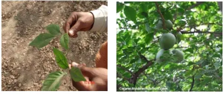
Macroscopical Characters Of Leaf: Organoleptic evaluations of leavas
of Aegle marmelos L. were reported.
Fig. 6: Morphological features of Aegle marmelos L.
Leaves:
Shape - Ovate to lanceolate Colour - Pale green Venation - Reticulate pinnate
Surface - S mooth
Margin - Five foliate with crenate margin Length - 5.7 cm
Width - 2.8 cm Petiole - Long Odour - Peculiar Taste - Bi tter
Microscopical Characters of Leaf:
Fig. 6.1: T.S of leaf through Midrib
Midrib: The leaf has thick and prominent midrib which is broadly
conical on the adaxial side and more or less flat on the abaxial side (Fig 6.1). The midrib is 600 µm thick and 400 µm wide. The epidermis is thin and the cells are smaller and circular. The abaxial part of the midrib consists of two or three layers of collenchyma; rest of the tissue is parenchymatous with circular compact cells. The adaxial conical portion has trancurrent palisade tissue and narrow zone of parenchymatous cells. The vascular system of the midrib is double stranded. There is an
adaxial strand and an abaxial strand, both occurring juxtaposed. (Fig.
6.1).
Fig. 6.2: Midrib Vascular strand – A portion enlarged
(ABS- abaxial side; ABVB- abaxial Vascular Bundle ; ADH- Adaxial Hump; ADVB- adaxial Vascular Bundle; EP: Epidermis ; GT : Ground Tisssue ; La: Lamina; Ph: Phloem; X: Xylem; Sc: sclerenchyma). The adaxial strand is smaller than the abaxial strand, both are collateral. The vascular strand consists short, parallel lines of xylem elements and wide leaves of phloem elements. These are wide, elliptical cells running in between the xylem and phloem lines. Xylem elements are angular,
wide and thick walled (Fig 6.2). Beneath phloem zone occurs thick band
of sclerenchyma cells.
Lamina:
Fig. 6.3: T.S of lamina
of the lateral veins and veinislets are located in the middle region of the
lamina. The vascular strands are circular and collateral (Fig.6.3).
Epidermal Cells and Stomata:
Fig. 6.4: Paradermal section of the ep idermis.
(ABE: Abaxial Epidermis; ADE: Adaxial Epidermis; E.C: Epidermal cells; L.V: Lateral Vein; S.C: Subsidary Cells; St: Stomata; P.M: Palisade Mesophyll; S.M: Spongy Mesophyll).
Two paradermal section , the epidermal cells appear polygonal with the straight anticlinical walls. No cuticular markings are evident. Stomata occur only on the lower epidermis (hypostomatic). The stoma ta are cytocyti c type. Each stoma is circulated by three circles of subsidiary cells; in each circle occur three to six subsidiary cells which
are rectangular in shape (Fig.6.4).
Petiole:
Fig. 6.5: Microscopy of petiole
(A. whole view, B. edge view, C. vascular region)
(col. collenchyma, fi. fibers; epi. epidermis, mes. mesophyll, par. parenchyma, ph. phloem, pi. pith, sd. secretory cannal, stm. stoma, t. trichome, xy. Xylem)
Powder Microscopic Characters:
The microscopic examination of powdered leaf material was performed to detect and established various identifying microscopic characters which will be help full in differentiation of the substi tute of the drug supplied in the form of dried powder. The photomicrographs of the identifying features of the plant material are shown in fig.The covering trichomes and stomata were present in the sample. The covering trichomes were multicelluler, uniseriate and the stomata were paracytic type. It was found that the powdered leaf showed groups of fibres with calcium oxalate crystals. Calcium oxalate crystals were numerous and mainly of cluster crystal type. Some xylem vessels (pitted vessels) were also visible which were lignified and cells of palisade and spongy parenchyma were also observed
Fig. 6.6: Epidermis showing paracytic stomata Fig. 6.7: Calcium oxalate crystal
Momordica charantia:
Macroscopical Characters of the Fruit: Organoleptic evaluations of
fruit of. Momordica charantiaL. were reported.
Fruit: (Fig. 3)
Colour - Bright orange-col oured
Shape - Pendulous, fusiform, usually pointed, ribbed and bearing numerous
Length - 5-15 cm long, Odour - Bitter Taste - Bitter
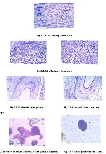
Microscopical Characters of the Fruit:
The fruit elongated and cylindrical, dark green with several fleshy thick outer growths on the surface. Two cross section, the outer line of the fruit shows large, fleshy bodies of varying shape and size. The fruit has the epidermal layer and homogenous parenchymatous tissue of
the fleshy bodies of outer growth (Fig 7.1).
(EP: Epidermis; FO: Fleshy out growth; GT: Growth tissue; PC: Pericarp; VS: Vascular strand)The fleshy outer growths receive well
developed vascular strands from the me socarp (Fig. 7.3).
The pericarp of the fruit is differentiated in to outer zone of ground tissue which include, loosely arranged lobed parenchyma cells
with scattered air chambers (Fig 7.4).
The inner pericarp has circular compact parenchymatous
cells with many sca tterd vascular strands (Fig. 7 .5). The vascular
strands have less prominent xylem and phloem.
(EP: Epidermis; PC: Pericarp; SC:seed coat). Seeds are much elongated and have thick seed coat and cellular endosperm. The seed coat has outer wide epidermal cells and inner parenchymatous tissue
(Fig. 7.6, 7.7).
(Cr: Crystals; SG: Starch Grains). The powder of the fruit exhibits large concratic starch grains. The starch grains are not abundant. Cells possessing dense content of crystals are more common
in the powder (Fig. 7.8, 7.9).
(EP: Epidermis; IMC: Inner Mesocarp; OMC: Outer Mesocarp; Ph: Phloem; VS: Vascular strand; X: Xylem).
Fig. 7.4: T.S of Pericarp- Outer zone
Fig. 7.5: T.S of Pericarp- Inner zone
Fig. 7.6: LS of seed – Upper portion Fig. 7.7: LS of seed – Lower portion
Powder Microscopy:
Fig. 7.8: Masses of parenchyma tissue with glandular crystals Fig. 7 .9: A starch grain stained with IKI.
Trigonella foenum-graecum:
Macroscopical Characters of Seeds: Organoleptic evaluations of fruit of. Momordica charantiaL. were reported.
Fig. 8: Macroscopy of seeds of Trigonella foenum-graecum
Seeds:
Colour - Light greyish, brown, olive green or cinnamon coloured,
Shape - Re ctangular to rounded in outline with a deep groove between the radical and cotyledons. Length - 3.5– 6mm
width - 2.5– 4mm and
Microscopical Characters of Seeds:
Fig. 8.1: L.S View Fig.8.2: Seed coat (Testa) Fig. 8.3: Sclerotesta
Powder Microscopy:
Fig. 8.4: Starch Grains Fig. 8.5: Sarcotesta (linear) Fig. 8.6: scleroids Fig. 8.7: Th ick bundles of scleroids
Physicochemical Parameters:
Table No. 7: Ash valu e
S. No. Plants Total ash (% w/w) Acid insoluble ash (% w/w) Water soluble ash (%w/w)
1 Aeglemarmelos 6.30 2.55 1.27
2 Momordicacharantia 8.80 0.60 6.60
3 Trigonellafoenumgreacum 3.92 0.44 3.48
Table No. 8: Extractive value
S. No. Plants Water soluble (% w/w) Alcohol soluble (% w/w)
1 Aeglemarmelos 9.28 14.21
2 Momordicacharantia 24.00 8.10
3 Trigonellafoenumgreacum 35.00 14.50
The yield of the alcoholic extract of dried powdered of Aeglemarmelos ,Momordica- charantia and Trigonellafoenumgreacum were 12.5%
w/w, 15.18% w/w, 9.10% w/w respectively.
Table No. 9: Preliminary phyto-chemical screening
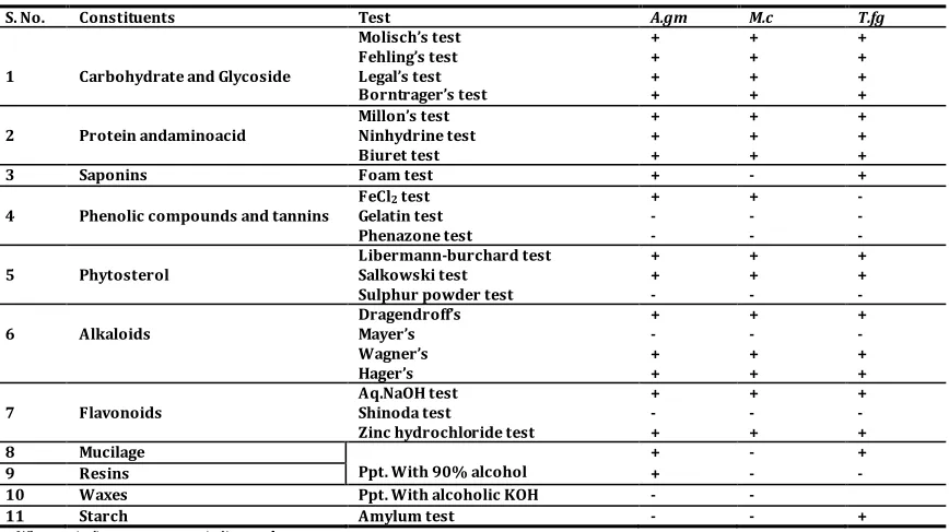
S. No. Constituents Test A.gm M.c T.fg
1 Carbohydrate and Glycoside
Molisch’s test + + +
Fehling’s test + + +
Legal’s test + + +
Borntrager’s test + + +
2 Protein andaminoacid
Millon’s test + + +
Ninhydrine test + + +
Biuret test + + +
3 Saponins Foam test + - +
4 Phenolic compounds and tannins
FeCl2 test + + -
Gelatin test - - -
Phenazone test - - -
5 Phytosterol
Libermann-burchard test + + +
Salkowski test + + +
Sulphur powder test - - -
6 Alkaloids
Dragendroff’s + + +
Mayer’s - - -
Wagner’s + + +
Hager’s + + +
7 Flavonoids
Aq.NaOH test + + +
Shinoda test - - -
Zinc hydrochloride test + + +
8 Mucilage
Ppt. With 90% alcohol
+ - +
9 Resins + - -
10 Waxes Ppt. With alcoholic KOH - -
11 Starch Amylum test - - +
Table No. 10: Evaluation of herbal tablet AMT/11
Drug Weight (mg) Thickness (mm) Hardness (Kg/cm2) Disintegration (minutes) Friability (%)
AMT/11 200.13±0.98 3.20±0.00 3.5±0.497 8.05±0.6194 0.8
All values are Mean ± SD (n=6)
Table No. 11: LD50 value of formulation AMT/11
Groups No. of animals Dose / mg/kg Results
1 3 100 No Death
2 3 200 No Death
3 3 500 No Death
4 3 1000 No Death
5 3 2000 No Death
Table No. 12: Nitric oxide free radical scavenging activity
S. No. Conc. (g/ml)
Standard (Ascorbic acid) AMT/11
%Inhibition %Inhibition
1 1000 93.75% 67.5%
2 500 89.5% 62.5%
3 250 83.5% 52.5%
4 125 79.75% 42.5%
5 62.5 72.5% 32.5%
6 31.25 66.5% 27.5%
Table No. 13: Effect of AMT/11 on blood glucose level in normal fasted rats
Group Treatment (dose mg/kg, po)
Blood Glucose level (mg/dl)
0 hr 1 hr 2 hr 4 hr 6 hr
Normal control 63.78±1.54 62.5±1.66 61.85±0.97 60.88±0.71 59.23±1.51
AMT/11(125) 65.08±1.51 64.3±0.65 (1.19) 63.5±1.17 (2.42) 61.4±1.59 (5.65) 59.63±0.99a (8.37)
AMT/11(250) 67.1±2.85 65.0±1.78 (3.1) 63.5±2.66 a (5.3) 60.3±2.19 b (10.1) 62.0±1.47b (7.6)
AMT/11 (500) 69.03±1.75 67.2±0.75a (2.6) 65.25±1.74b (5.4) 59.5±0.89b (13.8) 63.6±2.34b (7.8)
Glibenclamide (5) 59.25±0.78 54.5±1.24 (8.01) 50.45±1.41b (14.9) 47.23±0.73b (20.3) 49.56±1.44b (16.4)
(Values are mean ± SD from 6 animals in each group.Figure in parenthesis indicates; % fall in BGL as compared to 0 hr)P value:a<0.05,b P<0.01
Table No. 14: Effect of AMT/11on blood glucose level in streptozotocininduced diabetic rats (Single-dose short term study)
Group Treatment (dose mg/kg, p.o) Blood Glucose Level (mg/dl)
0 hr 1 hr 3 hr 6 hr
Normal control 64.3±1.61 62.5±1.66 64.1±1.35 63.9±1.05
Diabetic control 292.35±1.15a 304.8±2.09a (-4.2) 296.08±1.90a (-1.2) 297.6±2.30a (-1.8)
AMT/11(125) 30.8±1.27 270.3±1.93b (11.6) 209.21±1.98b (31.5) 207±2.11b (32.3)
AMT/11 (250) 303.5±1.09 235.6±2.06b (22.3) 203.9±1.88b (32.8) 199.6±1.71b(34.2)
AMT/11 (500) 301.7±1.71 211.3±1.01b (29.9) 136.5±2.17b (54.7) 153.2±1.86b (49.2)
Glibenclamide (5) 296.08±1.89 207±1.91b (30.08) 117.1±1.60b (60.4) 134.6±1.50b (54.5)
(Values are mean ± SD from 6 animals in each group.Figure in parenthesis indicates % fall in BGL as compared to 0 hr); Pvalue: <0.01; compared to a normal
group, b diabetic group
Table No. 15: Effect of multidose administration of AMT/11on blood glucose level in streptozotocin induced diabetic rats (long term study of 15 days daily once)
Group treatment dose mg/kg; po Blood glucose level (mg/d l)
Day 3 Day 6 Day 9 Day 12 Day 16
Normal control 59.1±1.46 60.0±1.18 60.1±1.05 62.5±1.36 59.8±1.37
Diabetic control 312.7±1.26 a 307.5±0.94a (1.66) 291.5±1.09a (6.7) 280.0±0.90a (10.45) 275.0±1.23 a (12.05)
AMT/11(125) 293.5±0.74 b 280.6±1.09c (4.3) 260.2±1.32c (11.3) 231.9±1.09c (20.9) 195.2±0.81 c(33.4)
AMT/11(250) 290.5±0.77 c 271.3±0.80c (6.5) 229.4±1.19c (21.0) 186.5±0.72c (35.7) 173.8±1.05 c(40.1)
AMT/11(500) 284.8±1.20 c 254.0±0.82c (10.7) 219.2±1.16c (23.0) 181.05±0.95c (36.4) 165.3±0.88c(41.9)
Glibenclamide(5) 279.6±0.67 c 249.2±1.43c (10.8) 218.3±1.36c (21.9) 178.5±1.36c (36.1) 159.9±0.93 c (42.8)
(Values are mean ± SD from 6 animals in each group. Figure in parenthesis indicates % fall in BGL as compared to Day 3); P values: <0.01, as compared to a normal
group; c diabetic control group b<0.05 compared to diabetic group.
Table No. 16: Effect of formulation AMT/11on body weight in Normal and streptozotocin induced diabetic rats.
S. No Groups Initial Body weight (g) Final Body weight (g) %increased/ decreased of body weight
1 Normal control 88.00±0.74 104.30±1.47 + 18.52
2 Diabetic control 101.11±1.09 56.55±1.53 - 44.07
3 AMT/11 (125) 80.00±1.02 50.25±1.19 - 37.19
4 AMT/11 (250) 87.06±1.05 63.66±0.88 - 26.94
5 AMT/11 (500) 91.90±1.12 74.1±1.04 - 19.30
6 Glibenclamide (5mg/kg) 91.80±12.4 75.50±0.73 - 17.70
0 20 40 60 80 100
3 1 .2 5 6 2 . 5 1 2 5 2 5 0 5 0 0 1 0 0 0
66.5 72.5
79.75 83.5 89.5 93.5
27.5 32.5 42.5 52.5 62.5 67.5 % In h ib it io n Concentration (µg/ml) Antioxidant activity of AMT/11
Standard Test 0 20 40 60 80
0 hr 1 hr 2 hr 4 hr 6 hr
B lo o d g lu co se l ev el s (m g/ d l)
Effect of AMT/11 on blood glucose level in normal fasted rats
Normal control AMT/11(125)
Fig. 9: Antioxidant activity of AMT/11 Fig. 10: Effect of AMT/11 on blood glucose level in normal fasted rats
0
200
400
0 hr 1 hr 3 hr 6 hr
B lo o d g lu co se l ev el s (m g /d l )
Fig: Effect of AMT/11 on blood glucose level in streptozotocin induced diabetic rats (single dose short term study) Normal control Diabetic control AMT/11(125) AMT/11(250) AMT/11(500)
Fig. 11: Effect of AMT/11 on blood glucose level in streptozotocin induced diabetic rats (single dose short term study)
0 50 100 150 200 250 300 350
Day 3 Day 6 Day 6 Day 12 Day 16
B lo o d g lu co se l ev el s( m g/ d l)
Effect of multidose administration of AMT/11 on blood glucose level in streptozotocin diabetic rats(long term study of 15 days daily once)
Normal control Diabetic control AMT/11(125) AMT/11(250) AMT/11(500) Glibenclamide(5)
Fig. 12: Effect of multi dose administration of AMT/11 on blood glucose level instreptozotocin induced diabetic rats
(long term study of 15 days daily once)
Table No. 17: Effect of formulation AMT/11 on biochemical parameters in streptozotocin induced diabetic rats
Group treatment (dose mg/kg, po
SGOT (IU/L) SGPT (IU/L) Alkaline phosphatise
(IU/L)
%Lipid Peroxi-dation
CAT (U/mg) GPX (U/mg)
Normal control 70.25±0.078 40.10±0.363 136.92±0.251 - 3.160±0.140 2.58±0.107
Diabetic control 138.20±0.178a 100.16±0.544a 254.23±0.232a 100±1.059 1.988±0.169a 1.936±0.175a
AMT/11(125) 115.8±0.126b 75.16±0.058b 216.30±0.429b 80.56±0.304b 2.20±0.817b 2.10±0.090b
AMT/11(250) 105.23±0.096b 64.66±0.360b 194.20±0.143b 74.36±0.165b 2.46±0.069b 2.26±0.044b
AMT/11(500) 90.36±0.066b 55.34±0.045b 169.20±0.127b 65.32±0.058b 2.76±0.031b 2.688±0.147b
Glibenclamide (5 mg/kg) 83.33±0.198b 48.30±0.224 b 155.30±0.040b 64.93±0.336b 2.86±0.233b 2.58±0.135b
Values were expressed as Mean ± SD of 6 rats in each group; P value: <0.01; compared to anormal group b diabetic
Histopathological Studies:
Microscopically examine pancreas section show the following features:
Pancreas section of rat of normal group (Fig. 13) showed that
normal architecture of pancreas with acini of serous epithelial cells along with nest of endocrine cells separated by fi brocollaoenous, stroma into lobules. No fibrosis or inflammation was found.
Pancreas section of rat of diabetic control group (Fig. 14)
showed that normal architecture of pancreas with acini of serous epithelial cells along with nest of endocrine cells separated by fibrocollagenous, stroma into lobules. No fibrosis or inflammation was found.
Pancreas section of rat treated with AMT/11(125). (Fig. 15)
showed that normal architecture of pancreas with acini of serous epithelial cells along with nest of endocrine cells separated by fibrocollagenous,stroma into lobules. No fibrosis or inflammation was
found.
Pancreas section of rat treated with AMT/11(250). (Fig. 16)
showed that normal architecture of pancreas with acini of serous epithelial cells along with nest of endocrine cells separated by fibrocollagenous, stroma into lobules. No fibrosis or inflammation was found.
Pancreas section of rat treated with AMT/11(500). (Fig. 17)
showed that normal architecture of pancreas with acini of serous epithelial cells along with nest of endocrine cells separated by fibrocollagenous, stroma into Mules. No fibrosis or inflammation was found.
Pancreas section of rat treated with 5 mg/kg Glibenclamide
(Fig. 18) showed that normal architecture of pancreas with acini of
Fig. 13: Normal Control Fig. 14: Diabetic Control Fig. 15: AMT/11 (125)
Fig. 16: AMT/11(250) Fig. 17: AMT/11 (500) Fig. 18: Glib enclamide (5mg/k g)
DISCUSSION
It is essential to meet the quality and efficacy of the herbal drugs, hence standardization of herbal drug as mandatory which may help one to understand, some of the controversies in identification, quality and therapeutic action of the drug. The drug materials such as
Aegle marmelos (Leaves), Momordica charantia (Fruit), Trigonellum foenum graceum (Seeds) chosen for this work was standardized based on the parameters with reference to WHO guidelines for Pharmacognostical standardization of a herbal drug.
The physico chemical evaluation of the leaf powder of Aegle
marmelos was found to 6.30 %w/w total ash, 2.55%w/w acid in soluble ash, 1.27 %w/w water soluble ash, 9.28 %w/w water soluble extractive value, 14.21%w/w alcohol soluble extractive.
The fruit powder of Momordica charantia was found to
8.80%w/w total ash, 0.60%w/w acid insoluble ash, 6.60%w/w water soluble ash, 24.00%w/w water soluble extractive value, 8.10%w/w alcohol soluble extractive.
The seedpowder of Trigonella foenum-graecum was found to
3.92%w/w total ash, 0.44%w/w acid insoluble ash, 3.48%w/w water soluble ash, 35.00%w/w water soluble extractive value, 14.50%w/w alcohol soluble extractive.
The yield of the alcoholic extract of dried powdered of Aegle
marmelos,Momordica charantia and Trigonella foenum greacum were
12.5% w/w, 15-18% w/w, 9-10% w/w respectively.
The preliminary phytochemical screening of the drug has been revealed the presence of flavonoids, phytosterols, tannins, carbohydrates, glycosides, alkaloids.
In the acute toxicity study, AMT/11 up to the dose level of 2000 mg/kg of body weight did not exhibit any lethality or toxic symptoms. Further dosing to estimate the LD50 of the drug was not performed. According to OECD guidelines for acute oral toxicity, an LD50 dose of 2000mg/kg and above is categ orized as unclassified and hence the drug is found to be safe.
The In vitro anti oxidant studies of AMT/11 were carried out using Nitric Oxide Radical Scavenging Method, shows significant results.
The Pharmacological evaluation of normal fasted rats shows onset of hypoglycaemic a ctivity of AMT/11 at 125, 250 and 500 mg/kg was evident between 1-2 hr, the peak was found to be at 4 hr. The rats receiving 500 mg/kg of AMT/11 showed the onset of effect at 1 hr with a peak effect at 4 hr. The hypoglycaemic effect of AMT/11 at 500 mg/kg (13.8% fall) was found to be nearly comparable to that of glibenclamide (5 mg/kg) (20.3% fall) (Table. 13).
Streptozotocin induced diabetic rats shows a single-dose
administration of AMT/11 (125, 250 and 500 mg/kg, p o) on 3rd day
after induced streptozotocin, showed a significant (p<0.01) reduction in
blood glucose level (BGL) after 1 and 3 hr interval. Maximum reduction in BGL to 136.5±2.17 mg/dl was seen at 3 hr after administration of 500 mg/kg of AMT/11 Glibenclamide (5 mg/kg, p o) also showed maximum reduction to 117.1±1.60 mg/dl at 3 hr. At 6 hr the BGL slightly increased as compared to 3 hr values (Table 14).
On repeated administration of either vehicle, AMT/11 or
glibenclamide for 15 days, a sustained and significant (P<0.01) decrease
in the blood glucose of the diabetic rats was observed at a dose of 250 (39.9 % fall) and 500 mg/kg (35.7% fall), in a dose dependent manner as compared to the vehicle treated group. Glibenclamide also showed a
significant (P<0.01) decrease in blood glucose (42.9% fall) at a dose of
5mg/kg, as compared with vehicle treated group (Table 15).
Type 2 diabetic rats gained relatively less weight during the course of development as compared to normal rats. Decrease in uptake of glucose, free fatty acids from circulation and accelerated beta-
oxidation in adipose tissue leads to weight loss in diabetes.6 it was
observed that as dose of formulation AMT/11 increases from 125 to 500 mg/kg , the loss of body weight significantly decreases from 35.87 % to 18.59 %, where as Standard drug Glibenclamide (5mg/kg) have 16.04 % value.
The biochemical parameter involves the Serum SGOT and
SGPT levels were elevated significantly (P<0.01) in streptozotocin
induced diabetic rats as compared to normal rats. In streptozotocin diabetic rats when treated with the AMT/11 glibenclamide there was a
significant (P<0.01) reduction in the elevated levels of SGOT and SGPT.
Similarly, elevated ALP level in serum during streptozotocin induced
diabetes were found to be significantly (P<0.01) lowered by AMT/11 and
glibenclamide treatment.
Superoxide anion radical (O2.-), hydrogen peroxide (H2O2)
and hydroxyl radical (OH*) are the major reactive oxygen species generated during oxidative stress. Free radical decrease the insulin receptor substrate ( IRS) tyrosine phosphorylation and in turn the activity of phosphatidyl inositol (PI) 3- kinase. Altered insulin signalling pathway exerts insulin resistance, a state of type 2 diabetes. Aerobic cells are endowed with extensive anti-oxidant defence mechanism including both low molecular weight scavengers such as reduced glutathione, ascorbic acid, vitamin E and enzyme system namely SOD, CAT and GPx.
Decrease in CAT activity could be possi bly due to less availability of NADPH or gradual decrease in erythrocyte CAT concentration by excessive generation of O2** that inactivates the enzyme. Since the activity of an enzyme depends upon its substrate, depletion of Glutathione may be the reason for decreased glutathione peroxidise activity. In other words levels of both enzymatic anti-oxidants (GPx and CAT) decreased and lysosomal enzymes increased in diabetic rats.
It is important to know in AMT/11 treated rats there were increased levels of the antioxidant enzymes (GPx and CAT). AMT/11 (500) increased 38.8% of both CAT and GPx with respect to Diabetic control where as Glibenclamide (5mg/kg) increased 43.8% of CAT and 33.2% of GPx. This shows that AMT/11 can reverse all these abnormalities either by pancreatic or hepatic mechanism.
Lipid Peroxidation is considered to be a primary mechanism of cell membrane destruction by free radicals. The extent of lipid peroxidation is analyzed by the formation of MDA (Marker). MDA conjugate with amino group of protein to form intra and inter molecular cross-links. These cross-links inactivate the membrane bound enzymes
and receptors.6 Type 2 diabetic rats showed elevated plasma MDA due to
peroxidation of lipids. Decrease in MDA by AMT/11 showed the ability of drug to prevent oxidative damage. The % Lipid peroxidation value decreased with AMT/11 (125), (250), (500) and Glibenclamide (5) were 19.4%, 25.6%, 34.68% and 35.07% respectively.
Histopathological findings of pancreas of the diabetic rats showed necrosis, atrophy and fibrotic changes. But, the pancreas of the rats treated with AMT/11 and glibenclamide showed minimal necrosis and mild to moderate atrophy and fibrotic changes.
CONCLUSION
The present study demonstrates that herbal formulation exhibits promising antidiabetic activity and help to maintain good glycemic and metabolic control. The herbal formulation, AMT/11, elicit hypoglycaemic/antidiabetic effects in both normal and experimentally induced hyperlycemic (Streptozotocin) rats. The herbal formulation under acute toxicity studies by OECD guideline shows it is non -toxic up to 2000mg/kg BW, so i t can be recommended for human conception after a safe clinical trial. It is possible that the herbal formulation may a ct through both, pancreatic and extra-pancreatic mechanism(s). The AMT/11 also elicited a significant antioxidant effect in Streptozotocin diabetic rats as reflected by its ability to inhibit lipid peroxidation and to elevate the enzymatic antioxidants in pancreatic tissue. The histopathological studies during the long term treatment have shown to ameliorate the Streptozotocin induced histological damage of islets of Langerhans. The inhibitory effects on biochemical and histological parameters induced by herbal formulation at a dose of 500 mg/kg were almost comparable to that of standard drug, glibenclamide (5mg/kg).
REFERENCE:
1. Nahar N. Traditional medicine. Oxford, OBH Publishing Co. Pvt.
Ltd., New Delhi. 18th Ed. 1993; 205-209.
2. Sharma VN. Some observati ons on hypoglycaemic activity of
Momordica charantia. Indian J. Med. Res.1960: 48(4); 471-47.
3. Ponnachan PTC, Paulose CS. Panikkar KR.Effect of leaf extract of
Aegle marmelos in diabetic rats. Indian J. Exp. Biology.1993: 31; 345-347.
4. Tiangda C, Mekmanee R, Praphapraditchote K, Ungsurungsie M,
Paovalo C. The hypoglycaemic activity of Momordica charantia
Linn. in normal and alloxan- induced diabetic rabbits. Journal of
the National Research Council. 1987: 19; 1-11.
5. Shani. J, Goldschmid, A. Ahronson, Z. Sulman, FG. Hypoglycaemic
effect of Trigonella foenum-graecum and Lupinus termis seeds
and their major alkaloids in alloxan diabetic and normal rats.
Arch. Int. Pharmacodyn. Ther. 1974: 210; 27-36.
6. Abdel-Barry J. A, Abdel-Hassan I. A, Al-Hakiem M. H.
Hypoglycemic and anti hyperglycaemic effects of Trigonella
foenum-graecum leaf in normal and alloxan induced diabetic
rats. J. ethnopharmacol. 1997: 58; 149-155.
7. Resmi CR. Anti diabetic effects of herbal drug in Alloxan -
Diabetic Rats, Indian Drugs. 2001: 38(6); 319.
8. Kokate CK, Purohit P, Gokhale SB. Pharmacognosy, Nirali
Prakashan, Pune. 27th Ed. 2001; 124.
9. Hakim ZS. Potential Anti diabetic Agents from Plant Sources;
Pharmacological Aspects, Indian J. Natural Product. 1995: 11(1);
3.
10. Mchael JB. Ethnobotany and the identification of Therapeutic
agents from plants. John Wiley, Chichester. 1990; 22-39.
11. Wendell DW, YS Huo, DL Yao. Inhibition of the progression of
type 2 Diabetes in the C57Bl/6J Mouse Model by an Anti-diabetes Herbal Formula. Phytotherapy
12. Chandra shekhar J, Ekambaram Sanmuga Priya, Subramanian
Venkataraman. Acute and Subacute Toxicity Studies on the Polyherbal Antidiabetic formulation diakyur in Experimental
Animal Models. Journal of Health science.2007: 53(2);245-249.
13. Kamalkkannan N, Prince PS. Hypoglycemic effect of water
extract of Aegle marmelos fruits in streptozotocin diabetic rats. J.
Ethnopharmacol. 2003: 87; 207-210.
14. Trease GE, Evan WC. A Text book of Pharmacognosy. London:
Bailliere Tinall Ltd; 13th Ed. 1989.
15. Srivastava, Sudhir KY, Venkatakrishna BH, Verma Y, Prem AS.
Retardation of retinopathy by Momordica charantia Linn
(Bitter gourd) fruit extract in alloxan diabetic rats. Ind. Exp.
Biology. 1987: 25; 571-572.
16. Lotlikar MM, Rajarama RMR. Pharmacology of a hypoglycemic
principle isolated from the fruit of Momordica charantia Linn.
Ind. J. Pharmacy. 1996: 28; 129-133.
17. Leung L. Anti – diabetic and hypoglycaemic effects of Momordica
charantia ( bitter melon): a mini review. Br. J. Nutr. 2009: 102(12); 1703-8.
18. Teoh. A histological study of the structural changes in the liver of
streptozotocin – induced diabetic rats treated with or without
Momordica charantia (bitter gourd). Clin. Ter. 2009: 160(4); 283-6.
19. Nahas R. Complementary and alternative medicine for the
treatment of type 2 diabetes.Can. Fa m. Physician. 2009: 55(6);
591-6.
20. Inayat-ur-Rahman,Serum sialic acit changes in
non-insulin-dependant diabetesmellitus (NIDDM) patients following bitter
melon (Momordica charantia) and rosiglitazone (Avandia)
treatment. Phytomedicine. 2009: 16(5); 401-5.
21. Nivitabishekam S. Pharmacodynamic interaction of Momordica
charantia with rosiglitazone in rats. Chem. Biol. Interact. 2009: 177(3); 247-53.
22. Okinaka S,Kumagia H, Ebashi S, Sugita H, Momoi H, Toyokura Y,
et al. Serum creatine phosphokinase activity in progressive muscular dystrophy and neuromuscular diseases. Arch. Neurol. 1961: 4; 520-5.