R E S E A R C H A R T I C L E
Open Access
Multiple genetically engineered humanized
microenvironments in a single mouse
Jungwoo Lee
1,4*, Dirk Heckl
2and Biju Parekkadan
1,3*Abstract
Background:Immunodeficient mouse models that accept human cell and tissue grafts can contribute greater
knowledge to human stem cell research. In this technical report, we used biomaterial implants seeded with genetically engineered stromal cells to create several unique microenvironments in a single mouse. The scope of study was focused on human CD34 hematopoietic stem/progenitor cell (HSPC) engraftment and differentiation within the engineered microenvironment.
Results:A mouse model system was created using subdermal implant sites that overexpressed a specific human
cytokines (Vascular Endothelial Growth Factor A (hVEGFa), Stromal Derived Factor 1 Alpha (hSDF1a), or Tumor Necrosis Factor Alpha (hTNFa)) by stromal cells in a three-dimensional biomaterial matrix. The systemic exposure of locally overexpressed cytokines was minimized by controlling the growth of stromal cells, which led to autonomous local, concentrated sites in a single mouse for study. This biomaterial implant approach allowed for the local analysis of each cytokine on hematopoietic stem cell recruitment, engraftment and differentiation in four different tissue microenvironments in the same host. The engineered factors were validated to have bioactive effects on human CD34+ hematopoietic progenitor cell differentiation.
Conclusions:This model system can serve as a new platform for the study of multiple human proteins and their
local effects on hematopoietic cell biology for in vivo validation studies.
Keywords:Genetically engineered stroma, Chimeric mouse model, Human hematopoietic stem and progenitor
cells, Implantable microenvironments, Scaffolds, Bone marrow stromal cells
Background
Immunodeficient mice lack immune cell activity and do not acutely reject xenogenic cells and tissues [1, 2]. These mouse strains have been an enabling tool for understanding human cell growth and functions in the context of living systems [3–6]. In particular, these mouse models have greatly advanced functional characterization of human hematopoietic stem and progenitor cells (HSPCs) [7, 8]. Human HSPCs can be intravenously injected and engrafted into a host immunodeficient mouse to form a chimeric hematopoietic system. These experiments have helped to identify and validate factors that control human HSPCs migration, engraftment, self-renewal, and differentiation in
mice for mechanistic and therapeutic purposes [9]. Yet, there still remain critical areas to improve these immuno-deficient mouse models to better model human HSPCs [10]. For example, human HSPCs do not engraft into a human stromal cell bed which may be important for long-term support of human stem cell function by direct phys-ical contact or the local secretion of stromal soluble factors. Several approaches have been tested in order to improve the functional support of human HSPCs in immunodeficient mice. First, human cytokines have been directly injected to a host mouse before and after human HSPCs injection that promoted engraftment and differ-entiation of human cells when compared to the control [11, 12]. Since single injection of human cytokines only produces a transient effect, repeated injections are typ-ically required, which is cost-prohibitive and burden-some. Alternatively, human gene encoded lentiviral vectors [13] or plasmid deoxyribonucleic acids (DNAs) [14] have been applied to directly transfect host cells * Correspondence:[email protected];biju_parekkadan@hms.
harvard.edu
1Department of Surgery, Center for Engineering in Medicine, Massachusetts General Hospital & Harvard Medical School and Shriners Hospital for Children, Boston, MA, USA
Full list of author information is available at the end of the article
and synthesize human cytokines. Second, using gene knock-in methods, immunodeficient mouse strains have been generated to express human major histocom-patibility complex (MHC) while suppressing mouse MHC molecules that improved human immune system development [2, 15]. Transgenic expression of human cytokines and growth factors has been also demon-strated to modulate human HSPCs responses [14]. For instance, human stem cell factor, granulocyte-macrophage colony stimulating factor and interleukin-3 genes inserted NOD-scid IL2rγnull (NSG) mice models demonstrated enhanced hematopoietic reconstitution of human HSPCs [16]. Although this approach is promising, it is labor/ time-intensive, requires specialized facilities and equip-ment, and is limited in throughput of genes expressed in a single mouse. Moreover, ubiquitous expression of human molecules could be problematic to interpret experimental outcomes. Lastly, human cells and tissues can be directly introduced into the murine host. For example, human bone marrow stromal cells (hBMSCs) were introduced into a mouse bone marrow cavity by intra-femoral injec-tion. The presence of human stromal cells in a mouse bone marrow increased engraftment of subsequently injected human HSPCs [17]. However, this invasive pro-cedure is compatible with only a few bones which limits the throughput of this approach in supporting human HSPCs function. Human fetal thymic and fetal liver tissues, known sites of early hematopoiesis, have been implanted in an immunodeficient mouse prior to human HSPCs injection. A mouse carrying primary human fetal tissue organoids demonstrated complete hematopoietic reconstitution of human HSPCs [18]. Although this study demonstrated the possibility to recreate a full spectrum human hematopoietic cells in mouse models, human fetal thymus and liver tissues are difficult to widely source mak-ing broad adoption unattainable.
In this report, we introduce an integrated bioengin-eering approach that can increase the throughput of model systems research for human HSPCs by using genetically engineered scaffold microenvironments. Engineered stromal cells, stably synthesizing a human cytokine or growth factor, were embedded in three-dimensional (3D) porous hydrogel scaffolds and implanted into immunodeficient mice in different subcutaneous locations. Each implant created a unique hematopoietic-supportive tissue microenvironment over time. These implants exhibited a locally concen-trated environment of human cells and soluble factors with the opportunity to make multiple, autonomous scaffolds implanted into the same mouse. We demon-strated the engineering of stromal cells with individual human cytokines, the control of local and systemic exposure of engineered cytokines, and functional characterization of the implanted microenvironments
with human HSPCs. The presented method is versatile and can be readily applied to other human cells and cytokines with minor modifications for better under-standing human biology.
Methods
All chemicals and supplies were purchased from Sigma Al-drich or Fisher Scientific unless otherwise stated. All mouse and primary human cell experiments were reviewed and approved by an internal review board of Massachusetts General Hospital.
Stromal cell culture
Primary hBMSCs were isolated from healthy donor’s fresh bone marrow (Lonza) following a previously reported protocol [19]. Briefly, hBMSCs were cultured with medium composed of 15 % fetal bovine serum, 100 U/mL penicillin, 100 μg/mL streptomycin, 20 mg/L gentamicin, 1 ng/L fibroblast growth factor, and 3 g/L sodium bicarbonate in alpha-minimum essential medium (a-MEM). Human um-bilical vein endothelial cells (HUVECs) were purchased from Invitrogen and cultured with medium composed of 10 % low serum growth supplement, 100 U/mL penicillin and 100 μg/mL streptomycin. Mouse bone marrow stro-mal cells (mBMSCs) were purchased from Invitrogen and cultured with medium composed of 15 % fetal bovine serum, 100 U/mL penicillin, 100 μg/mL streptomycin, 20 mg/L gentamicin, 1 ng/L fibroblast growth factor, and 3 g/L sodium bicarbonate in a-MEM. All cell cultures were maintained at 37 °C, 5 % CO2and 100 % humidity. Human
peripheral blood derived CD34 cells were purchased from StemCell Technologies and after thawing immediately used for in vivo experiments without culture.
Generation of lentiviral particles encoded human VEGFa, SDF1a and TNFa genes
Generation of genetically engineered mouse bone marrow stromal cells
mBMSCs were plated in a 12-well plate. Once the cul-ture reached about 50 % confluence, the medium was replaced with 0.5 ml of new medium containing 1:10 diluted lentiviral particles and 2 μg/ml Polybrene (Hexadimethrine bromide), and incubated for 8 h. Afterwards the cell surface was washed with PBS three times and 1 ml of normal medium was added in each well. After 48 h, mBMSCs expressing green cent protein (GFP) were sorted out using a fluores-cence activated cell sorting (FACS) machine (BD FACSAria II cell sorter). Expression of GFP was con-firmed under a fluorescent microscope (Zeiss Axio 200) and secretion of hVEGFa, hSDF1a, and hTNFa was determined by enzyme-linked immunosorbent assay (ELISA) kits (R&D systems).
Fabrication of inverted colloidal crystal hydrogel scaffolds and in vitro 3D stromal cell culture
Polyacrylamide hydrogel based inverted colloidal crystal scaffolds were prepared following the previously reported methods [20]. Final hydrogel scaffolds were consisted of regularly arranged spherical pores (D = 250 ± 30 μm) of which surface was coated with type I collagen utilizing amine reactive heterbifunctional cross-linker (Sulfo-SANPH). Dimensions of cylindrical shape hydrogel scaf-folds were about 1.5 mm in thickness and 6.5 mm in diameter. Prior to cell seeding, hydrogel scaffolds were dehydrated in a laminar hood for 30 min and then a dense genetically engineered mBMSCs or hBMSCs (0.5 × 10⁶ cells in 40μl) was dropped on the hydrogel scaffold. Rehy-dration process promoted effective cell distribution into a complex 3D porous geometry and seeded stromal cells formed stable adhesion on collagen fiber coated pore sur-face in 4–6 h. After 3 days culture, the level of hVEGFa, hSDF1a, and hTNFa molecules in the medium was deter-mined using ELISA kits. Total cell mass in each scaffold was quantified using a MTT 3-(4,5-dimethylthiazol-2-yl)-2,5-diphenyltetrazolium bromide (MTT) reagent (ATCC) and used for normalization of cytokine secretion.
In vivo microenvironments formation with genetically engineered stromal cells
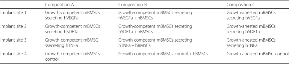
Three different stromal compositions were used for creating subcutaneous microenvironments in single host mouse (Table 1); (i) Growth-competent genetically engi-neered mBMSCs (0.5 × 10⁶ cells in 40 μl); (ii) 1:1 mixture of growth-competent genetically engineered mBMSCs (0.25 × 10⁶ in 20 μl) and hBMSCs (0.25 × 10⁶ in 20 μl); (iii) Growth-arrested genetically engineered mBMSCs (0.5 × 10⁶ in 40 μl). Growth-arrested engi-neered stromal cells were prepared by treating growth
competent stromal cells with 10 μg/mL mitomycine C containing medium for 3 h. Post 3-day in vitro culture, stromal cell-scaffolds were subdermally implanted on the dorsal side of a 8 week-old NOD/SCID/IL2νnull (NSG) male mouse (Jackson Laboratory) following the previously reported method [20]. Four types of cell-scaffolds were implanted beneath the skin of immunode-ficient mice. A distance between implanted scaffolds was maintained at least 2 cm in order to prevent their contact. After 6 weeks, mice were scarified and periph-eral blood (0.5–1 ml) and implanted scaffolds samples were retrieved. Bloods were allowed to clot at room temperature for 1 h, centrifuged at 6000 rpm for 10 min, and serum was collected. The level of hVEGFa, hSDF1a, and hTNFa molecules in serum was determined using ELISA kits. A low level of hSDF1a was detected in control NSG mice without carrying scaffolds due to anti-body cross-reactivity with mouse SDF1a. This back-ground signal was subtracted from the final data. Explanted scaffolds were embedded in optical cutting temperature compound, snap frozen with dry ice-chilled 2-methylbutane, and saved for histological analysis.
Systemic migration of human CD34 cells to the implanted microenvironments
Direct implantation of human CD34 cells with genetically engineered stromal cell-scaffolds and ex vivo methylcellulose assay
A 2:1 ratio mixture of growth-arrested genetically engi-neered mBMSCs (0.5 × 10⁶ in 40 μl) and hBMSCs (0.25 × 10⁶in 20μl) was introduced into a scaffold. Post 3 days culture, cell-scaffold plates were placed in a refrigerator for 20 min and then removed medium both from a well and a hydrogel scaffold using a pipette. Next ice-cold, moderately dehydrated cell-scaffolds were trans-ferred to a 96-well plate and 50 μl of ice-cold matrigel containing the mixture of 2 × 10⁵ HUVECs and 2 × 10⁵ hCD34 cells was infiltrated into the hydrogel scaffold pores via centrifugation at 4 °C, 1500 rpm for 15 min. The matrigel-cell filled scaffolds were incubated for 20 min at 37 °C incubator to solidify the gel and then 1 ml of culture medium was placed in each well.
After 3 weeks in vivo implantation, hematopoietic cells were retrieved from explanted scaffolds following the above procedure. Hematopoietic cells were dispersed in methylcellulose medium containing recombinant cyto-kines and erythropoieting for human cells (MethoCult H4034, Stem Cell Technologies). Final concentration was 1 × 10⁵hematopoietic cells in 1.1 ml methylcellulose medium that was plated in a 35 mm dish following a protocol provided by the vendor. After 2 weeks incuba-tion, colony forming units of (i) erythroid, (ii) granulo-cyte and macrophage, (iii) granulogranulo-cyte, erythrogranulo-cyte, monocyte and megakaryocyte, and (iv) burst forming unit of erythroid were distinguished and counted under an inverted light microscope.
Histological analysis
Frozen scaffold blocks were cut into 10–30μm thickness using a cryostat (Leica Biosystems) and stored at−80 °C until use. For hematoxylin and eosin, and Masson’s Trichrome staining, frozen tissue sections were fixed with 10 % buffered formalin solution and stained following the vendor’s protocol (American Master Tech). For im-munofluorescence staining, frozen tissue sections were fixed with ice-cold acetone, blocked with 10 % normal goat serum and 1 % bovine serum albumin diluted in phosphate buffered saline (PBS). Slides were incubated
with rat anti-mouse CD31 (BD Pharmingen) and rabbit anti-human CD34 antibody (Abcam) for overnight. Subse-quently the slides were stained by goat anti-rat immuno-globulin G (IgG) conjugated with alexa fluor 488 and goat anti-rabbit IgG conjugated with alexa fluor 568 (Invitro-gen) for 1 h. Finally, VectaShield mounting medium with DAPI was applied and slides were imaged under a fluores-cence (Zeiss 200) microscope. Open source image analysis software, ImageJ, was used to process and quantify areas of collagen fibers and vasculatures in Masson’s Trichrome and mouse CD31 stained slides, respectively.
Imaging
Explanted scaffolds were fixed in 2 % glutaladehyde solu-tion for 6 h and then serially dehydrated with 20, 50, 70, 90 and 100 % ethanol solution. The scaffolds were further dried using a lyophilzer overnight. A thin platinum/gold coating was made on the samples using a sputter coating machine (208HR,Cressington) and imaged under FESEM Ultra55 (Zeiss).
Statistics
Statistical comparisons of data were per- formed using SPSS version 17 software. Nonparametric tests, i.e., Kruskal-Wallis and Mann-Whitney tests, were applied for ELISA and LSK cell analysis, respectively. Compari-sons of human cell engraftment in long-term engraft-ment studies were performed using an unpaired Student ttest on GraphPad PRISM version 5.
Results
Genetically engineered mouse stromal cell lines secreting human VEGFa, SDF1a, or TNFa
In order to create a specific human soluble factor enriched microenvironment, we first designed lentiviral vectors that encoded human vascular endothelial growth factor a (hVEGFa), human stromal cell derived factor-1 alpha (hSDF1a), and human tumor necrosis factor alpha (hTNFa) genes along with enhanced green fluorescent protein (eGFP) (Fig. 1a). A lentiviral control was also ap-plied expressing eGFP but not a specific cytokine. mBMSCs were infected with lentiviral particles and sorted by FACS to purify eGFP cells. Mouse cells were
Table 1Three stromal cell-seeding compositions for creating 4 distinct microenvironments in a mouse
Composition A Composition B Composition C
Implant site 1 Growth-competent mBMSCs secreting hVEGFa
Growth-competent mBMSCs secreting hVEGFa + hBMSCs
Growth-arrested mBMSCs secreting hVEGFa
Implant site 2 Growth-competent mBMSCs secreting hSDF1a
Growth-competent mBMSCs secreting hSDF1a + hBMSCs
Growth-arrested mBMSCs secreting hSDF1a
Implant site 3 Growth-competent mBMSC ssecreting hTNFa
Growth-competent mBMSCs secreting hTNFa + hBMSCs
Growth-arrested mBMSCs secreting hTNFa
Implant site 4 Growth-competent mBMSCs control
used for these studies to ensure long-term survival of engineered stromal cells because even severely immun-compromised mice still retain immune compartments that can detect human cells. The purified cells were culture-expanded to establish 3 genetically engineered mBMSC-lines i.e. mBMSC-hVEGFa, mBMSC-hSDF1a, and mBMSC-hTNFa (Additional file 1: Figure S1).
Genetically engineered stromal cells were then seeded into the 3D hydrogel scaffolds following the previously reported methods [20]. These hydrogel scaffolds con-sisted of regularly arranged spherical cavities, whereby the cavity surfaces were coated with type I collagen. This coating method promoted homogenous stromal cell seeding and subsequent adhesion (Fig. 1b). The charac-terized rate of soluble factor secretion of genetically engineered stromal cells in the scaffolds was 4.42 ± 0.24 μg/mL for hVEGFa, 0.87 ± 0.16μg/mL for hSDF1a, and 2.7 ± 0.02 μg/mL for hTNFa over 3 days. When compared to primary hBMSCs growing in the scaffolds, normalized hVEGFa and hSDF1a secretion were about 4.8 and 3.7 folds higher, respectively (Fig. 1c-e). hBMSCs do not naturally secrete hTNFa. These stable cell lines were advanced for further in vivo testing.
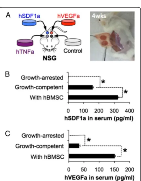
Control of systemic and local exposure of engineered factors after in vivo implantation
We subcutaneously implanted genetically engineered growth-competent stromal cell seeded scaffolds into immunodeficient NOD-scid IL2rγnull (NSG) mice and determined whether these engineered factors could be detected in vivo. Four different types of engineered stro-mal cell-seeded scaffolds were implanted into a NSG mouse (Fig. 2a). Peripheral blood samples were collected at 6 weeks post implantation and the level of human cytokines in serum was measured using ELISA. Detectable levels of hVEGFa (33.93 ± 3.88 pg/ml) and hSDF1a (238.97 ± 8.01 pg/ml) were found in peripheral blood while there was no hTNFa. We next examined whether systemic exposure of secreted molecules can be controlled
Fig. 1Creating genetically engineered stromal cell-coated implantable microenvironments.aDesign of lentiviral vectors encoding hVEGFa, hSDF1a, and hTNFa genes for genetically engineered mBMSC-line generation. b Microfabricated hydrogel scaffold that represents a standardized and fully interconnected porous microstructure (top) and a fluorescent image of genetically engineered mBMSC residing in a 3D scaffold (bottom).c-dNormalized secretion of (c) hVEGFa, (d) hSDF1a, and (e) hTNFa from genetically engineered stromal cells for 3 days. The secretion rates were compared with hBMSC growing in the same hydrogel scaffolds
by manipulating the growth of genetically engineered stro-mal cells. In our previous studies, hBMSCs accelerated and augmented inter-scaffold angiogenic process via secreting pro-angiogenic and immunomodulatory mole-cules [20, 21]. To enhance the survival and systemic distri-bution of secreted molecules, we co-seeded a 1:1 ratio hBMSCs and engineered stromal cells into the scaffolds. Peripheral blood analysis 6 weeks after implantation re-vealed significantly increased level of hVEGFa and hSDF1a, but again no hTNFa was detected. We then hy-pothesized that systemic exposure of cytokines secreted from the engineered stromal cells could be reduced by limiting stromal cell proliferation. To test this hypothesis, we treated genetically engineered stromal cells with mito-mycine C that bound to microtubules and blocked cellular division. Growth-arrested stromal cells remained viable and maintained comparable levels of human cytokine secretion during 3 weeks of in vitro culture (Additional file 1: Figure S2). Growth arrested stromal cell-seeded scaffolds were subdermally implanted to NSG mice and 6 weeks after peripheral blood levels of human cytokines were measured. We confirmed the absence of hVEGFa and hTNFa, and a lower level of hSDF1a (Fig. 2b, c). The residual hSDF1a level was caused by cross-reactivity of the antibody with mouse SDF1a (Additional file 1: Figure S3). Collectively these results indicate that genetically engi-neered stromal cells remained bioactive after in vivo implantation. The secretion levels were controllable by manipulating their survival and growth.
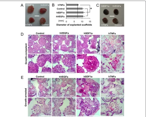
Local tissue formation in scaffolds is altered by stromal cell proliferation
The next aspect of our study was to understand local tissue development in scaffolds that were seeded with growth-arrested stromal cells compared to growth-competent stro-mal cells. Gross images of explanted scaffolds seeded with growth-competent engineered stromal cells showed exces-sive tissue development that completely engulfed the implanted scaffolds except the hTNFa scaffolds (Fig. 3a, b). On the other hand, growth-arrested stromal cell scaffolds maintained their original dimension and showed dark red color due to locally recruited red blood cells (Fig. 3c). Histopathological analysis of growth-competent stromal cell scaffolds revealed a densely populated stromal cells and deposited extracellular matrix (ECM) with few hematopoietic cells, while hTNFa scaffolds were mostly filled with hematopoietic cells that mimics tissue inflam-mation (Fig. 3d). In case of growth-arrested stromal cell scaffolds, developed tissues were hypocellular and accom-modating both stromal and hematopoietic components in an organized manner. Hematopoietic cells were primarily found at the pore surface and stromal cells were located in the pore center. Control and hVEGFa scaffolds showed comparable microenvironments, while hSDF1a scaffolds
exhibited significantly increased hematopoietic cells. Again hTNFa scaffolds showed partially acellular areas with an in-flammatory leukocyte reaction (Fig. 3e). Further SEM ana-lysis distinguished densely filled fibroblastic tissue and ECM in growth-competent stromal cell-scaffolds, and co-existence of hematopoietic and stromal cells in growth-arrested stromal cell-scaffolds (Additional file 1: Figure S4).
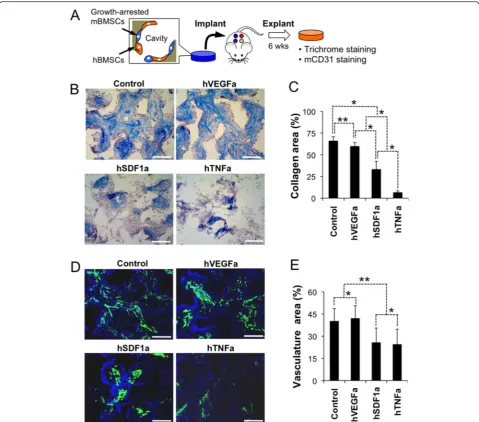
Implanted microenvironments support systemic migration and retention of human CD34 cells
Having multiple, autonomous microenvironments in a single mouse provided an opportunity to study human HSPCs function in unique ways. In our next set of ex-periments, we exploited growth-arrested stromal cell scaffolds that form locally enriched human soluble mi-croenvironments while providing room for growth of human HSPCs. Prior to seeding growth-arrested stromal cells were mixed with hBMSCs that improved survival of stromal cells after in vivo implantation in the above study (Fig. 4a). We first characterized soluble factor-dependent tissue microenvironments focusing on inter-scaffold ECM deposition and angiogenesis. Trichrome staining and semi-quantitative image analysis revealed significantly enhanced collagen deposition in control and hVEGFa scaffolds, whereas only sparse collagen fibers were deposited in hTNFa scaffolds reminiscence to a necrotic tissue (Fig. 4b, c and Additional file 1: Figures S5 and S6). Immunohistostaining of mouse CD31 (mCD31) endothelial cells showed considerably more blood vessels in control and hVEGFa scaffolds than hSDF1a and hTNFa scaffolds (Fig. 4d and e). Collectively these results indicate that growth arrested stromal cells created a soluble factor enriched local micro-environments that induced distinct tissue formation.
between 24 and 72 h characterization. On the other hand, hCD34 cell number in the bone marrow and spleen were reduced after 72 h (Fig. 5c). In general, an implanted microenvironment recruited circulating hCD34 cells and retained them, though differential mi-gration to a preferential scaffold was not observed in this model system.
Skewed in vivo differentiation of hCD34 cells in engineered microenvironments
The soluble factors that were selected for overexpression in stromal cells all have known effects on HSPCs migra-tion, differentiamigra-tion, and maintenance [22–24]. In this subset study, we wanted to verify the bioactivity of these molecules on differentiation in our model system.
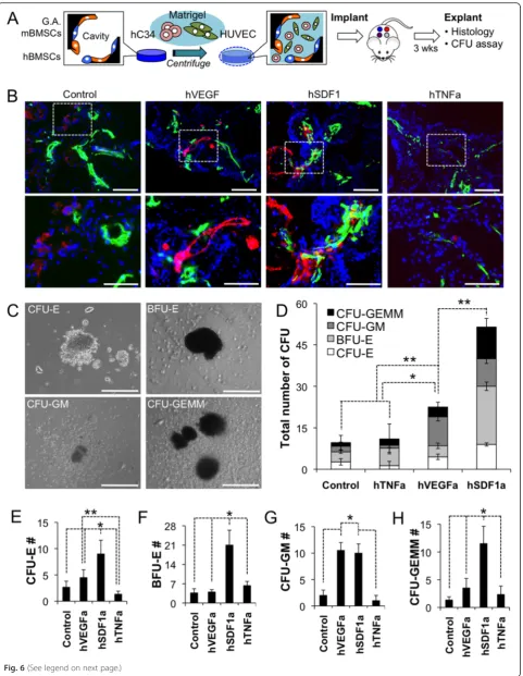
Scaffolds pre-seeded with growth-arrested genetically engineered stromal cells and hBMSCs were cultured for 3 days. On the day implantation, hCD34 cells and HUVECs dispersed in matrigel were infiltrated into the cavities of hydrogel scaffolds by centrifugation and then solidified resulting in a complex 3D tissue structure that was durable throughout the implantation procedure (Fig. 6a). HUVECs were used to induce rapid angiogen-esis in the implanted microenvironments for promoting the survival of hCD34 cells since they were directly im-planted into a non-vascularized scaffold in this study. This tissue construct was implanted into mice at 4 dif-ferent locations representing the one difdif-ferent engi-neered soluble factor that was overexpressed. After 3 weeks of in vivo growth, the scaffolds were explanted
and stained for hCD34 and mCD31 expressing cells. The results reveled that hCD34 cells were still present in the scaffold at this time point and located in close proximity with vasculatures in all scaffolds, except hTNFa scaffolds (Fig. 6b). Separately, scaffold cells were retrieved and put in a colony forming unit (CFU) assay to assess the number of progenitor cells and their skewed differentiation poten-tial. Isolated cells from each environment showed visual signs of erthyroid, granulocyte, monocyte, macrophage, and megakaryocyte by 14 days of differentiation culture in methylcellulose with supplemented human growth factors and cytokines. Isolated cells from each environment
showed visual signs of erthyroid, granulocyte, monocyte, macrophage, and megakaryocyte by 14 days of differenti-ation culture in methylcellulose with supplemented human growth factors and cytokines (Fig. 6c). Total CFU counting result distinguished that hSDF1a scaffolds retain substantially higher number of hematopoietic progeni-tor cells followed by hVEGFa scaffolds. Control and hTNFa scaffolds accommodate comparable level, low number of hematopoietic progenitor cells (Fig. 6d). Further enumeration of specific colonies that were grown from each stromal cell type, revealed a very potent effect of hSDF1a on the maintenance of a
progenitor pool in all lineages (Fig. 6e-h). Other major notable findings were the reduction of erythroid pro-genitors by hTNFa (Fig. 6e) and the positive effects of hVEGFa overexpression on the number of granulocyte/ macrophage progenitor populations (Fig. 6g). The re-sults show that this overexpression system produces bioactive human molecules that have differentiation effects on the human hematopoietic system as corrobo-rated by several previous studies [25–28].
Discussion
The goal of humanized mouse models is to reconsti-tute specific cellular and tissue dynamics to study hu-man cell biology in vivo and potentially become a testbed to predict human responses for precision medicine. Studies using immunodeficient mice that accept human cells/tissue have generally shown that by adding complimentary human tissues, a more human-ized response can be seen [10]. In this study, we wanted to create a testing platform where molecules of interest to the human hematopoietic system can be studied in a locally enriched environment with easy ac-cess and analytical capability. Our hypothesis was that a single mouse can harbor multiple independent
microenvironments that have site-specific bioactivity by implanting genetically engineered stromal cell-laden scaffolds. Mouse cells are generally more difficult to infect with lentivirus than human cells, though they are more proliferative after infection and also could survive longer after in vivo implantation than human cells [29]. Our infection efficiency of mouse stromal cells was ~10–30 %. The transfected cells maintained proliferative capacity after purification by flow sorting and expressed properly folded hVEGFa, hSDF1a, and hTNFa. The constitutive translation of these proteins by engineered cells was 4–5 fold more in concentra-tion than normal hBMSCs with respect to hVEGFa and hSDF1a. hTNFa is not naturally expressed by hBMSCs and this construct was included to mirror an inflam-matory reaction site. This mouse cell expression was robust and took only a few weeks as opposed to months/years for transgenic mice, which can further benefit from improvement in infection efficiency while exploring new promoter systems and unknown mole-cules of interest to microenvironment research.
When compared to in vivo gene delivery methods that non-specifically infect host cells to synthesize desired human cytokines, an engineered ectopic tissue analogue
is an alternate approach with multiple advantages. First, the porous hydrogel scaffolds were a very useful tool for standardizing and analyzing engineered tissue microen-vironments. The fully interconnected microscale cavities made with a synthetic hydrogel matrix promoted angio-genesis and, in turn, improved the survival and function of genetically engineered stromal cells after implant-ation. These features allowed for direct comparison of the effects of the overexpressed gene on local tissue de-velopment and human HSPC fate. Second, these engi-neered tissue analogues could be studied for local or systemic effects by simply controlling the growth poten-tial of the engineered stromal cells. This also enables an increase in the throughput of studies by implanting several unique microenvironments into a single host mouse. Finally, implanted microenvironments can ac-commodate human HSPCs and be directly accessed for various imaging and analytical tools [20, 30]. Thus, im-plantable microenvironments that harbor genetically engineered cells can broaden the study of human cells in immunodeficient mouse models.
In our in vivo studies with engineered stromal cells, we observed systemic levels of expressed cytokine and a mild sarcoma-like overgrowth of these stromal cell lines that were not under growth arrest. A typical fibroblastic compartment is quiescent, unless the tissue bed is acti-vated at which time the cells have been observed to pro-liferate. The tissue cavity was not considered conducive to support primary functions of hematopoiesis. In-stead, a growth-arrested stromal cell population could still express soluble, bioactive factors in an enriched environment while allowing for additional tissue ele-ments (e.g. hBMSCs, HUVECs, hCD34 cells) to be included and maintain viability within the space as seen in Fig. 5. Using growth-arrested stromal cells eliminated systemic levels of expressed cytokines as well. This stromal population was put on test in a competitive transplantation assay and we did not ob-serve site-specific homing of hCD34 cells, even to a potent chemokine hSDF1a. The lack of a systemic che-mokine gradient may be a plausible explanation for non-specific homing results and can be an area of improvement with a temporal and stronger burst of cytokine from a non-proliferative cell mass.
The human cytokines overexpressed in our model sys-tem, namely hVEGFa, hSDF1a, and hTNFa, were selected based on known biological responses to hematopoietic ac-tivity to benchmark our bioacac-tivity studies. hVEGFa is a potent stimulator of angiogenesis and has direct effect on promoting the survival of HSPCs [31, 32]. hSDF1a is a well-known chemokine molecule that actively recruits circulating HSPCs [27, 33, 34] and maintains their activ-ities [35]. hTNFa restricts HSPCs activity functioning as a key precursor for inflammatory response [36, 37]. Al-though these cytokines are designed for human specific, evolutionary conservation of key structures and functions in these molecules exhibited cross-reactivity with murine tissue/cells that in turn promote distinct tissue micro-environment formation depending on types of engineered stromal cells. We observed sustained vascular area in hVEGFa constructs, more cellularity and progenitor cell activity in hSDF1a constructs, and more inflammatory cell recruitment and collagen remodeling in hTNFa con-structs. Our model system further reflected key biological responses in vivo interacting with hCD34 HSPCs. This model system can be deemed suitable to understand short-term effects of engineered human proteins on HSPCs function for future discovery and validation studies. There may also be cross-reactivity of mouse molecules on human HSPCs and vice versa, though we maintained the same cellular composition of our scaf-folds to control for this potential effect. A long-term evaluation of these microenvironments is necessary to establish true stem cell functions of serial transplant-ation, reconstitution, and tri-lineage differentiation after months in vivo.
Conclusion
In conclusion, we presented a bioengineering strategy to improve humanized mouse models by creating implantable microenvironments that release human specific cytokines and growth factors in a controlled manner. Genetically engineered stromal cells stably synthesize encoded human molecules after in vivo implantation. Porous hydrogel scaf-folds facilitated the usage of engineered stromal cells as forming tissue-engineered depots that continuously release engineered human molecules. Presented approach is sim-ple and versatile that can be easily applied to other soluble (See figure on previous page.)
and insoluble molecules. Combination of engineered stro-mal cells and implantable microenvironments is expected to be a valuable tool for generating humanized mouse models and utilizing them for basic and translational hu-man HSPCs as well as cancer research.
Additional file
Additional file 1: Figure S1.Selection and conformation of lentivial transfected mouse stromal cells. (A) Flow cytometric analysis of GFP mBMSC, (B) Culture-expanded genetically engineered mBMSCs. (Scale bar, 200μm). Figure S2.Characterized secretion of human cytokines from genetically engineered stromal cells in 1 and 3 weeks in vitro culture.Figure S3.hSDF1a ELISA in mouse blood serum. Control mice without scaffold implantation showed a background level of SDF1a signal due to cross-reactivity. This level was used as a baseline and was also observed in growth arrested, which was concluded, as undetectable. The other groups showed measurable levels above background and were concluded to be true hSDF-1a detection. Figure S4.SEM images of growth-competent genetically engineered stromal cell-seeded scaffolds. (A) Cross-sectional images of human soluble factor secreting engineered stromal cell-seeded scaffolds after 6 weeks subcutaneous implantation. Except hTNFa, entire pores were completely filled with tissue cells with no hematopoietic components. (B) Closed-up image of growing engineered stromal cell-seeded scaffolds. Figure S5.Examples of semi-quantitative image analysis using ImageJ. (A) Collagen fiber area estimation from a Masson’s Trichrome staining image, (B) Vasculature area estimation from an immunohistostaining mCD31 and DAPI image.Figure S6.Long-term maintenance of inflammation-mimicking tissue microenvironment indirectly indicates survival and function of growth-arrested hTNFa secreting engineered stromal cells in the implanted scaffolds. (DOCX 2962 kb)
Abbreviations
a-MEM, alpha-minimum essential medium; CFU, colony forming unit; ECM, extracellular matrix; eGFP, enhanced green fluorescent protein; FACS, fluorescence activated cell sorting; hBMSCs, human bone marrow stromal cells; hSDF1a, stromal derived factor 1 alpha; HSPCs, hematopoietic stem and progenitor cells; hTNFa, tumor necrosis factor alpha; HUVECs, human umbilical vein endothelial cells; hVEGFa, vascular endothelial growth factor a; mBMSCs, mouse bone marrow stromal cells; MHC, major histocompatibility complex; MTT, MTT 3-(4,5-dimethylthiazol-2-yl)-2,5-diphenyltetrazolium bromide; NSG, NOD-scid IL2rγnull; SEM, scanning electron microscopy
Acknowledgements
We give thanks to Drs. Rebekka Schneider-Kramann and Benjamin Ebert for providing reagents for the study and helpful discussions. We also acknowledge the efforts of Jessica Elman and Ryan Murray for assisting lentiviral gene transduction and FACS cell sorting experiments, and tail vein injection of hCD34 cells, respectively.
Funding
This work was supported by NIH Grants R01EB012521 (B.P.), K01DK087770 (B.P.), R00CA163671 (J.L.) and the Shriners Hospitals for Children (B.P., J.L.).
Availability of data and materials
Additional file 1: Figure S1-S6 are available on the web.
Authors’contributions
JL and BP conceived the idea, designed experiments, analyzed data and wrote the manuscript. DH prepared Lentivirus particles. All authors read and approved the final manuscript.
Competing interests
The authors declare that they have no competing interests.
Consent for publication Authors consent for publication.
Ethics approval and consent to participate
All mouse and primary human cell experiments were reviewed and approved by an internal review board of Massachusetts General Hospital.
Author details
1Department of Surgery, Center for Engineering in Medicine, Massachusetts General Hospital & Harvard Medical School and Shriners Hospital for Children, Boston, MA, USA.2Department of Medicine, Brigham and Women’s Hospital, Boston, MA, USA.3Harvard Stem Cell Institute, Cambridge, MA, USA. 4Department of Chemical Engineering, Institute for Applied Life Sciences, University of Massachusetts, Amherst, MA, USA.
Received: 10 February 2016 Accepted: 13 June 2016
References
1. Bosma GC, Custer RP, Bosma MJ. A severe combined immunodeficiency mutation in the mouse. Nature. 1983;301(5900):527–30.
2. Shultz LD, Ishikawa F, Greiner DL. Humanized mice in translational biomedical research. Nat Rev Immunol. 2007;7(2):118–30.
3. Ito M et al. NOD/SCID/gamma(c)(null) mouse: an excellent recipient mouse model for engraftment of human cells. Blood. 2002;100(9):3175–82. 4. Legrand N et al. Humanized mice for modeling human infectious disease:
challenges, progress, and outlook. Cell Host Microbe. 2009;6(1):5–9. 5. Morton CL, Houghton PJ. Establishment of human tumor xenografts in
immunodeficient mice. Nat Protoc. 2007;2(2):247–50.
6. Quintana E et al. Efficient tumour formation by single human melanoma cells. Nature. 2008;456(7222):593–8.
7. McCune JM et al. The SCID-hu mouse: murine model for the analysis of human hematolymphoid differentiation and function. Science. 1988; 241(4873):1632–9.
8. Traggiai E et al. Development of a human adaptive immune system in cord blood cell-transplanted mice. Science. 2004;304(5667):104–7.
9. Doulatov S et al. Hematopoiesis: a human perspective. Cell Stem Cell. 2012; 10(2):120–36.
10. Shultz LD et al. Humanized mice for immune system investigation: progress, promise and challenges. Nat Rev Immunol. 2012;12(11):786–98.
11. Dao MA, Pepper KA, Nolta JA. Long-term cytokine production from engineered primary human stromal cells influences human hematopoiesis in an in vivo xenograft model. Stem Cells. 1997;15(6):443–54.
12. Lapidot T et al. Cytokine stimulation of multilineage hematopoiesis from immature human cells engrafted in SCID mice. Science. 1992;255(5048): 1137–41.
13. O’Connell RM et al. Lentiviral vector delivery of human interleukin-7 (hIL-7) to human immune system (HIS) mice expands T lymphocyte populations. PLoS One. 2010;5(8):e12009.
14. Chen Q, Khoury M, Chen J. Expression of human cytokines dramatically improves reconstitution of specific human-blood lineage cells in humanized mice. Proc Natl Acad Sci U S A. 2009;106(51):21783–8.
15. Covassin L et al. Human peripheral blood CD4 T cell-engrafted non-obese diabetic-scid IL2rgamma(null) H2-Ab1 (tm1Gru) Tg (human leucocyte antigen D-related 4) mice: a mouse model of human allogeneic graft-versus-host disease. Clin Exp Immunol. 2011;166(2):269–80.
16. Billerbeck E et al. Development of human CD4+FoxP3+ regulatory T cells in human stem cell factor-, granulocyte-macrophage colony-stimulating factor-, and interleukin-3-expressing NOD-SCID IL2Rgamma(null) humanized mice. Blood. 2011;117(11):3076–86.
17. Muguruma Y et al. Reconstitution of the functional human hematopoietic microenvironment derived from human mesenchymal stem cells in the murine bone marrow compartment. Blood. 2006;107(5):1878–87. 18. Melkus MW et al. Humanized mice mount specific adaptive and innate
immune responses to EBV and TSST-1. Nat Med. 2006;12(11):1316–22. 19. Parekkadan B et al. Mesenchymal stem cell-derived molecules reverse fulminant
hepatic failure. PLoS One. 2007;2(9):e941.
20. Lee J et al. Implantable microenvironments to attract hematopoietic stem/ cancer cells. Proc Natl Acad Sci U S A. 2012;109(48):19638–43.
21. Bersani F et al. Bioengineered implantable scaffolds as a tool to study stromal-derived factors in metastatic cancer models. Cancer Res. 2014;74(24):7229–38. 22. Adams GB, Scadden DT. The hematopoietic stem cell in its place. Nat Immunol.
23. Wilson A, Trumpp A. Bone-marrow haematopoietic-stem-cell niches. Nat Rev Immunol. 2006;6(2):93–106.
24. Zhang CC, Lodish HF. Cytokines regulating hematopoietic stem cell function. Curr Opin Hematol. 2008;15(4):307–11.
25. Lapidot T, Kollet O. The essential roles of the chemokine SDF-1 and its receptor CXCR4 in human stem cell homing and repopulation of transplanted immune-deficient NOD/SCID and NOD/SCID/B2m(null) mice. Leukemia. 2002;16(10): 1992–2003.
26. Lataillade JJ et al. Stromal cell-derived factor 1 regulates primitive hematopoiesis by suppressing apoptosis and by promoting G(0)/G(1) transition in CD34(+) cells: evidence for an autocrine/paracrine mechanism. Blood. 2002;99(4):1117–29. 27. Peled A et al. Dependence of human stem cell engraftment and repopulation
of NOD/SCID mice on CXCR4. Science. 1999;283(5403):845–8.
28. Stellos K et al. Platelet-derived stromal cell-derived factor-1 regulates adhesion and promotes differentiation of human CD34+ cells to endothelial progenitor cells. Circulation. 2008;117(2):206–15.
29. Ricks DM et al. Optimized lentiviral transduction of mouse bone marrow-derived mesenchymal stem cells. Stem Cells Dev. 2008;17(3):441–50. 30. Zhang YS et al. Optical-resolution photoacoustic microscopy for volumetric
and spectral analysis of histological and immunochemical samples. Angew Chem. 2014;53(31):8099–103.
31. Gerber HP et al. VEGF regulates haematopoietic stem cell survival by an internal autocrine loop mechanism. Nature. 2002;417(6892):954–8. 32. Ziegler BL et al. KDR receptor: a key marker defining hematopoietic stem
cells. Science. 1999;285(5433):1553–8.
33. Dar A, Kollet O, Lapidot T. Mutual, reciprocal SDF-1/CXCR4 interactions between hematopoietic and bone marrow stromal cells regulate human stem cell migration and development in NOD/SCID chimeric mice. Exp Hematol. 2006;34(8):967–75.
34. Hattori K, Heissig B, Rafii S. The regulation of hematopoietic stem cell and progenitor mobilization by chemokine SDF-1. Leuk Lymphoma. 2003;44(4): 575–82.
35. Greenbaum A et al. CXCL12 in early mesenchymal progenitors is required for haematopoietic stem-cell maintenance. Nature. 2013;495(7440):227–30. 36. Lu L et al. Effects of recombinant human tumor necrosis factor alpha,
recombinant human gamma-interferon, and prostaglandin E on colony formation of human hematopoietic progenitor cells stimulated by natural human pluripotent colony-stimulating factor, pluripoietin alpha, and recombinant erythropoietin in serum-free cultures. Cancer Res. 1986;46(9):4357–61. 37. Pronk CJ et al. Tumor necrosis factor restricts hematopoietic stem cell activity in
mice: involvement of two distinct receptors. J Exp Med. 2011;208(8):1563–70.
• We accept pre-submission inquiries
• Our selector tool helps you to find the most relevant journal
• We provide round the clock customer support
• Convenient online submission
• Thorough peer review
• Inclusion in PubMed and all major indexing services
• Maximum visibility for your research
Submit your manuscript at www.biomedcentral.com/submit