Iran J Public Health, Vol. 49, No.1, Jan 2020, pp.30-36
Original Article
Correlation between Deep Venous Thrombosis and
Inflamma-tion in Patients after ImplantaInflamma-tion of Permanent Pacemaker
Jianxin MA
1, Lian CUI
1, Wenjin HUO
1, Guanghui WANG
1, Xin QUAN
2, *Jinping
ZHANG
31. Department of Health Care, 305 Hospital of Chinese PLA, Beijing 100017, P.R. China
2. Department of Echocardiography, Fuwai Hospital, Peking Union Medical College, Beijing 100017, P.R., China 3. Medical Department, 305 Hospital of Chinese PLA, Beijing 100017, P.R. China
*Corresponding Author: Email: [email protected]
(Received 17 Mar 2019; accepted 26 Jun 2019)
Introduction
Clinically, the application of permanent pace-maker implantation is increasingly common, es-pecially in patients with arrhythmia (1), but clini-cians cannot overcome a series of complications occurring after the permanent pacemaker implan-tation, such as postoperative infection, implant electrode shedding, deep venous thrombosis
(DVT), which affect prognosis and even lead to death (2). Since the permanent pacemaker im-plantation was put into clinical use, it has been applied from indications of arrhythmia to long QT syndrome, heart failure, vagal dizziness (3). Regarding complications, infection and DVT are the most serious. For patients with DVT, pulmo-Abstract
Background: The correlation between postoperative deep venous thrombosis (DVT) and inflammation in patients with permanent cardiac pacemaker implantation was analyzed.
Methods: A total of 130 cases undergoing permanent pacemaker implantation in the 305 Hospital of Chinese PLA and Fuwai Hospital from May 2014 to February 2017 were selected. Of the 130 cases, 60 patients compli-cated with DVT were selected as the observation group, and the remaining 70 cases without complications of DVT were selected as the control group. The relationship and influence of various factors were explored. Results: The number of patients smoking and the number of subjects with DVT history in the observation group were higher than those in the control group. In the observation group, plasminogen activator inhibitor (PAI)-1: Ag, PAI-1: Ac, thrombin-activated fibrinolysis inhibitor (TAFI): Ag, and TAFI: Ac levels were higher than those in control group (P<0.05). The levels of inflammatory factors of the peripheral blood of the obser-vation group were significantly higher than those of the control group (P<0.05). In the correlation analysis of serum inflammatory factors and coagulation factors, CRP, IL-6, IL-10 were positively correlated with PAI-1: Ag level. Age, BMI, smoking history, number of implanted electrodes, DVT history, duration of immobiliza-tion and inflammatory factor levels had independent predictive value on postoperative complicated DVT. Conclusion: The serum inflammatory factors are closely associated with postoperative DVT in patients im-planted with permanent cardiac pacemaker, and the serum inflammatory factors are a good reference for the evaluation of DVT.
nary embolism may also occur, increasing the mortality rate (4). In most cases, at the initial stage of DVT, no obvious or only slight clinical manifestations have been observed, which have often not been taken seriously or treated prompt-ly (5). Among fibrinoprompt-lytic systems, plasminogen activator inhibitor (PAI)‑1 and throm-bin‑activated fibrinolysis inhibitor (TAFI) are the major and most important components (6). The PAI-1 and TAFI content increase leads to re-duced fibrinolytic activity, and an important fac-tor in DVT is the reduced fibrinolytic activity (7). Overreaction of the inflammatory system in-creases the risk of DVT because a damage to the venous wall is a key factor in the development of DVT and thrombosis may aggravate the damage to the vascular wall, especially in the veins, and elevated levels of inflammatory cytokines in pe-ripheral blood are a risk factor for the develop-ment of DVT (8,9).
Therefore, analyzing the relationship between inflammation and DVT is helpful to reduce the risk of developing DVT in patients with perma-nent pacemaker implantation, or to achieve early treatment and improve prognosis.
Patients and Methods
General information
A total of 130 cases undergoing permanent pacemaker implantation in 305 Hospital of Chi-nese PLA (Beijing, China) and Fuwai Hospital (Beijing, China) from May 2014 to February 2017 were selected. Among them, 60 cases with DVT were selected as the observation group, and the other 70 patients without DVT as control group, including 83 males and 47 females, aged 68-78 years, with an average age of 73.73±10.71 years. All patients were selected after permanent pace-maker implantation.
Patients with DVT were diagnosed according to ultrasound and clinical manifestations, by apply-ing pressure on the limbs usapply-ing a probe and comparing with normal limbs. DVT showed ob-vious expansion of the lumen, no echo or low echo in the lumen. The lumen cannot be
com-pressed or thrombus can be seen moving in the blood vessels. Exclusion criteria: long-term pre-operative immobilization; systemic infection; the use of estrogen or anticoagulant, thrombolytic drugs; malignant tumor; autoimmune diseases; recent surgery or severe trauma; severe liver and kidney dysfunction; mental illness; clinical data mismatched or refused to sign informed consent. The study was approved by the Ethics Commit-tee of 305 Hospital of Chinese PLA. Informed consents were signed by the patients or guardi-ans.
Methods
Clinical data from all patients were retrospective-ly anaretrospective-lyzed, including age, sex, BMI, smoking his-tory, the number of electrodes implanted, and history of DVT. Fasting peripheral blood was drawn 10h after fasting for 10 days from all en-rolled cases. Biochemical indexes were deter-mined by detecting the upper serum. The data of total cholesterol (TC), triglyceride (TG), LDL-C and HDL-C were measured by an automatic bio-chemical analyzer provided by Hitachi, Ltd. (To-kyo, Japan). The levels of inflammatory cytokines such as CRP, IL-10 and IL-6 were measured by an immunoturbidimetric assay. PAI-1: Ac, TAFI: Ag, TAFI: Ac levels were measured by enzyme-linked immunosorbent assay (ELISA).
Statistical methods
SPSS 19.0 statistical software (IBM Corp., Ar-monk, NY, USA) was used to process the data. The data were expressed as x±s. Pearson correla-tion analysis was used for the calculacorrela-tion of the r-values between serum inflammatory and clotting factors. Logistic regression analysis was used to examine risk factors for DVT. P<0.05 was con-sidered statistically significant.
Results
Comparison of general information between observation and control group
but the age, BMI, number of patients smoking, number of electrodes implanted, number of pa-tients with DVT history in the observation group were higher than that of the control group; and
the immobilization time of the observation group was also longer than that of the control group. The differences were statistically significant (P<0.05) (Table 1).
Table 1: Comparison of general information between observation and control group
General information
Observa-tion(n=60) Control(n=70) P-value
Age (yr) 77.63±5.38 69.21±7.16 0.043
Sex (f/m) 38/22 45/25 0.793
BMI (kg/m²) 30.06±2.82 27.74±2.88 0.039
Smoking history (yes/no) 48/12 30/40 0.028
No. of electrodes implanted 4.52±1.80 1.40±0.39 0.003
DVT history (yes/no) 45/15 23/47 0.001
Postoperative immobilization time (h) 75.17±4.80 48.39±4.65 0.001 DVT, deep venous thrombosis.
Comparison of coagulation factor levels The levels of PAI-1: Ag, PAI-1: Ac, TAFI: Ag, TAFI: Ac in the observation group were higher
than those in the control group and the differ-ences were statistically significant (P<0.05) (Table 2).
Table 2: Comparison of clotting factor levels in observation and control group
Index Observation
(n=60) Control (n=70) P-value
PAI-1:Ag (ng/ml) 44.39±4.68 27.67±2.52 0.002
PAI-1:Ac (AU/ml) 0.75±0.29 0.49±0.28 0.017
TAFI:Ag (%) 108.53±22.41 93.73±25.31 0.001
TAFI:Ac (µg/ml) 33.93±7.37 15.51±5.42 0.001
PAI, plasminogen activator inhibitor; TAFI, thrombin‑activated fibrinolysis inhibitor
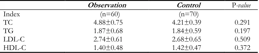
Comparison of biochemical indicators be-tween the observation and control group Compared between the two groups, the levels of TC, TG and LDL-C in the observation group
were higher than those in the control group, but the differences were not statistically significant (Table 3).
Table 3: Biochemical indicators of the observation and control group (µmol/l)
Observation Control P-value
Index (n=60) (n=70)
TC 4.88±0.75 4.21±0.39 0.291
TG 1.87±0.68 1.84±0.59 0.197
LDL-C 2.74±0.61 2.68±0.65 0.509
HDL-C 1.40±0.48 1.42±0.47 0.372
Serum inflammatory factors in observation and control group
The inflammatory factors (CRP, IL‑6, IL‑10) in peripheral blood serum of patients in the
obser-vation group were significantly higher than those in the control group and the differences were sta-tistically significant (P<0.05) (Table 4).
Table 4: Comparison of serum levels of inflammatory factors in observation and control group
Index Observation
(n=60) Control (n=70) P-value
CRP (mg/l) 28.39±2.23 3.32±1.49 0.001
IL-6 (pg/ml) 13.56±5.37 3.02±1.70 0.001
IL-10 (pg/ml) 2.96±1.42 0.91±0.56 0.019
Correlation analysis of serum inflammatory and clotting factors
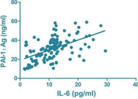
CRP (r=0.796, P<0.001), IL-6 (r=0.664,
P<0.001) and IL-10 (r=0.731, P<0.001) were all
positively correlated with PAI-1: Ag level (Figs. 1-3).
Fig. 1: Correlation of CRP with PAI-1: Ag. PAI, plasminogen activator inhibitor
Fig. 3: Correlation of IL-10 with PAI-1: Ag. PAI, plasminogen activator inhibitor
Logistic regression analysis of risk factors for DVT after permanent pacemaker implanta-tion.
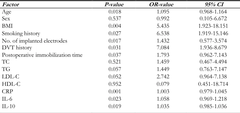
Age, BMI, smoking history, the number of im-planted electrodes, the history of DVT, the
post-operative immobilization time and the levels of inflammatory factors had independent predictive value on postoperative complicated DVT (Table 5).
Table 5: Logistic regression analysis of risk factors for DVT after permanent pacemaker implantation
Factor P-value OR-value 95% CI
Age 0.018 1.095 0.968-1.164
Sex 0.537 0.992 0.105-6.672
BMI 0.004 5.435 1.923-18.151
Smoking history 0.027 6.538 1.919-15.146
No. of implanted electrodes 0.017 1.432 0.577-3.574
DVT history 0.031 7.084 1.936-8.679
Postoperative immobilization time 0.037 1.793 0.962-7.143
TC 0.521 1.459 0.467-4.494
TG 0.057 1.449 0.763-7.147
LDL-C 0.052 2.742 0.964-7.138
HDL-C 0.952 0.079 0.451-18.714
CRP 0.001 1.003 0.979-1.045
IL-6 0.023 1.058 0.969-1.218
IL-10 0.019 1.035 0.985-1.036
DVT, deep venous thrombosis; TC, total cholesterol; TG, triglyceride.
Discussion
Clinically, permanent pacemakers are widely used in patients with cardiac arrhythmias by generating and releasing a relative amount of magnetic im-pulses that interconnect with the patient's cardiac tissue cells (10). After receiving the pulse wave, the myocardial cells can affect the surrounding
myocardial tissue cells and reconduct the pulse wave so that all heart cells can receive a certain degree of stimulation, resulting in normal heart diastolic and systolic exercise, which is the main mechanism of treating patients by permanent pacemaker (11).
ac-tivity, they can mainly affect cardiomyocytes through stimulations for therapeutic purposes (12). In patients with arrhythmia, permanent pacemaker implantation is prone to lead to some common complications, such as infection, DVT; these complications are critical and will even lead to death (13). After operation, the release of co-agulation factors is disturbed and further for-mation of thrombus is promoted because the implanted electrodes damage the blood vessel wall tissue cells to varying degrees (14). Further-more, the contact between the electrode and the original lesion in the thrombus also increases the probability of DVT (15). After a permanent pacemaker implantation, DVT tends to occur because the patient has an excessive BMI and prolonged postoperative immobilization due to the patient's blood flow slowing down when the patient is immobilized for an extended period of time, DVT is likely to occur (16). The age and BMI of the observation group was greater than that in the control group, the number of elec-trodes implanted and the number of patients with DVT history were more than those in the control group, and postoperative immobilization time was also longer than that in the control group (P<0.05), which was consistent with the above conclusion.
In the development and progression of DVT, activators such as plasminogens are one of the most dangerous factors and are associated with an overreaction with the inflammatory system (17). In the blood system, the most important reason for stimulating fibrinolytic time is throm-bin, and PAI‑1, TAFI‑1 are closely related with fibrinolysis time and they are major markers of fibrinolysis (18). PAI‑1, TAFI‑1, which are main-ly expressed as PAI-1: Ac, TAFI-1: Ac after acti-vation, they are the main force of fiber dissolu-tion (19). The PAI-1: Ag, PAI-1: Ac, TAFI: Ag, TAFI: Ac levels in patients with DVT after per-manent pacemaker implantation were significant-ly higher than those patients without DVT, and the difference was statistically significant (P<0.05).
The disorder of the inflammatory system can lead to or aggravate the formation of DVT, and DVT can further promote the inflammatory reaction of the blood vessel wall, resulting in a vicious circle (8). The activation and continual regulation of tissue factor affect the inflammatory system, and the increased level of inflammatory factors in pe-ripheral blood is a high-risk factor of DVT (9). Large amounts of data have shown that CRP, IL-6 and IL-10 levels and other inflammatory cyto-kine levels in the peripheral blood serum of pa-tients with DVT are increased up to nearly eight times, and it has been suggested that DVT itself is accompanied by overreaction of the inflamma-tory system (20). The present study found that the levels of peripheral inflammatory factors in the observation group were higher than those in the control group, and the correlation analysis between serum inflammatory factors and blood coagulation factors showed that the levels of CRP, IL-6, IL-10 and PAI-1:Ag level were posi-tively correlated, indicating that the higher the level of inflammatory cytokines in serum, the greater the risk of DVT, which is consistent with most of the findings.
Conclusion
To explore the relationship between DVT and inflammation in patients after permanent pace-maker implantation can reduce the incidence of DVT, or provide early intervention in the early stage of disease to improve the prognosis and reduce mortality.
Ethical considerations
Ethical issues (Including plagiarism, informed consent, misconduct, data fabrication and/or fal-sification, double publication and/or submission, redundancy, etc.) have been completely observed by the authors.
Acknowledgements
References
1. Bianchi G, Marzocchi R, Lorusso C, Ridolfi V, Marchesini G (2008). Nutritional treatment of chronic liver failure. Hepatol Res, 38 (Suppl 1): S93-S101.
2. Squizzato A, Ageno W (2005). D-dimer testing in ischemic stroke and cerebral sinus and ve-nous thrombosis. Semin Vasc Med, 5: 379-386. 3. Manly DA, Boles J, Mackman N (2011). Role of
tissue factor in venous thrombosis. Annu Rev Physiol, 73: 515-525.
4. Sideris S, Archontakis S, Dilaveris P, et al (2017). Leadless Cardiac Pacemakers: Current status of a modern approach in pacing. Hellenic J Cardiol, 58: 403-410.
5. Yoo MC, Cho YJ, Ghanem E, Ramteke A, kim kI (2009). Deep vein thrombosis after total hip arthroplasty in korean patients and D-dimer as a screening tool. Arch Orthop Trauma Surg, 129: 887-894.
6. Snow V, Qaseem A, Barry P, et al (2007). Joint American College of Physicians/american Academy of Family Physicians Panel on Deep Venous Thrombosis/pulmonary Em-bolism: Management of venous thromboem-bolism: A clinical practice guideline from the American College of Physicians and the American Academy of Family Physicians. Ann Fam Med, 5: 74-80.
7. Al Fagih A, Al Najashi K, Dagriri K, et al (2010). Feasibility of cardiac resynchronization thera-py in a patient with complex congenital heart disease and dextrocardia, facilitated bycardiac computed tomography and coronary sinus venography. Hellenic J Cardiol, 51: 178-182. 8. Yoshikane H, Yamamoto T, Ozaki M,
Matsuza-ki M (2007). Clinical significance of high‑sensitivity C‑reactive protein in lifestyle-related disease and metabolic syndrome. J Cardiol, 50: 175-182 (In Japanese).
9. Sidelmann JJ, Sjøland JA, Gram J, et al (2007). Lupus anticoagulant is significantly associated with inflammatory reactions in patients with suspected deep vein thrombosis. Scand J Clin Lab Invest, 67: 270-279.
10. Vormittag R, Vukovich T, Schönauer V, et al (2005). Basal high-sensitivity-C-reactive pro-tein levels in patients with spontaneous
ve-nous thromboembolism. Thromb Haemost, 93: 488-493.
11. Eren E, Yilmaz N, Pence S, Kocoglu H, Göksu S, Kocabaş R, Kadayifci S (2002). Diagnostic value of C-reactive protein in patients with angiographically documented coronary heart disease. Acta Medica (Hradec kralove), 45: 155-160.
12. Rozmus G, Daubert JP, Huang DT, Rosero S, Hall B, Francis C (2005). Venous thrombosis and stenosis after implantation of pacemakers and defibrillators. J Interv Card Electrophysiol, 13: 9-19.
13. Peng WH, Chen GL, Zhou Y, Fan SY, Fu DL and Wang Y (2017). Study of expression of circulating inflammatory factors in ACS rats with low dose of Tripterygium Wilfordii. Hel-lenic J Cardiol, 59: 46-47.
14. Kearon C (2001). Natural history of venous thromboembolism. Semin Vasc Med, 1: 27-37. 15. Syed FF, Beeching NJ (2005). Lower-limb
deep-vein thrombosis in a general hospital: Risk factors, outcomes and the contribution of in-travenous drug use. QJM, 98: 139-145. 16. Whisenant B, kar S, Bunch TJ (2014). Left atrial
appendage occlusion addresses the tremen-dous unmet needs of stroke prevention in atrial fibrillation that persist despite recent ad-vances in anticoagulation therapy. Circulation, 130: 1516-1523.
17. Sabzi F, Faraji R (2013). Right atrial thrombosis associated with sagittal sinus thrombosis caused by protein C-S deficiency. JNMA J Nepal Med Assoc, 52: 378-383.
18. Karatzis EN (2005). The role of inflammatory agents in endothelial function and their con-tribution to atherosclerosis. Hellenic J Cardiol, 46: 232-239.
19. Lim kE, Hsu WC, Hsu YY, Chu PH, Ng CJ (2004). Deep venous thrombosis: Compari-son of indirect multidetector CT venography and sonography of lower extremities in 26 pa-tients. Clin Imaging, 28: 439-444.