Whiplash evokes descending muscle recruitment
and sympathetic responses characteristic of
startle
Daniel WH Mang,
BSc, MSc, PhD(c)1Gunter P Siegmund,
PhD, P.Eng1,2Jean-Sébastien Blouin,
DC, MSc, PhD1,3,41 School of Kinesiology, UBC 2 MEA Forensic Engineers & Scientists 3 Brain Research Centre
4 Institute for Computing, Information and Cognitive Systems (ICICS)
Corresponding Author: Jean-Sébastien Blouin School of Kinesiology, University of British Columbia 210 – 6081 University Boulevard.
Vancouver, BC V6T1Z1 Email: [email protected]
This study was funded by the Michael Smith Foundation for Health Research (MSFHR) and infrastructure support was obtained from Canada Foundation for Innovation (CFI) – BC Knowledge Development Fund – and MSFHR. JSB received salary support from the MSFHR and Canadian Chiropractic Research Foundation. GPS received support from Natural Sciences and Engineering Research Council of Canada (NSERC). DWHM received support from the Canadian Institutes of Health Research (CIHR) – Auto 21.
©JCCA 2014
Whiplash injuries are the most common injuries following rear-end collisions. During a rear-end collision, the human muscle response consists of both a postural and a startle response that may exacerbate injury. However, most previous studies only assessed the presence of startle using data collected from the neck muscles and head/neck kinematics. The startle response also evokes a descending pattern of muscle recruitment and changes in autonomic activity. Here we examined the recruitment of axial and appendicular muscles along with autonomic responses to confirm whether these other features of a startle response were present during the first exposure to a whiplash perturbation. Ten subjects experienced a single whiplash perturbation while recording electromyography, electrocardiogram,
Introduction
Whiplash injuries are the most common injury caused by motor vehicle collisions, and rear-end collisions pose the greatest risk of whiplash injury.1 Although the exact aetiology of whiplash injuries remains unclear, a startle response elicited by a multisensory stimulus (somatosen-sory, acoustic and vestibular) is part of the neuromuscular response to a rear-end collision.2-5 The startle response in-creases neck muscle activity and increased activity in the posterior neck muscles, in particular the cervical multi-fidus muscle, may exacerbate strains in posterior neck structures (i.e. cervical facet joint) while these neck struc-tures are already strained by the collision-induced inter-vertebral motion.4,6 Thus, it is important to understand how the startle response contributes to the neuromuscular response during rear-end collisions.
Previous whiplash experiments have focused primarily on neck muscle responses and head/neck kinematics as indicators of the presence of a startle response.3,6-8 How-ever, the startle response, which is found in all mammals9, elicits a descending pattern of involuntary, axial and ap-pendicular muscle recruitment originating from the cau-dal brainstem10. The startle response also influences auto-nomic physiological responses: it activates sympathetic post-ganglionic neurons that innervate organs such as the heart, blood vessels and sweat glands.11-17 As a result, startle-induced sympathetic responses include an increase in heart rate and electrodermal activity (EDA), which is a technique used to infer sympathetic drive from meas-urable changes in skin conductance at the surface of the skin.15,18
Here we attempt to confirm that a startle response
forms part of the neuromuscular response evoked during a rear-end collision. Specifically, we investigate the re-cruitment of axial and appendicular muscle responses and changes in autonomic responses as additional indicators of the presence of a startle response during a rear-end col-lision. If a startle forms part of the response to a whiplash collision, we expect a whiplash-like perturbation to evoke a descending recruitment of muscles and an increase in heart rate and electrodermal activity characteristic of a startle response.
Methods
Subjects
Ten subjects with no history of neurological disorders par-ticipated in this experiment (5M/5F, 27±8 years, 169±11 cm tall, 70±14 kg). All subjects provided written informed consent and were paid a nominal fee for participating. The research protocol was approved by the UBC Clinical Ethics Review Board (H07-01281) and conformed to the Declaration of Helsinki.
Instrumentation
Surface electromyography (EMG) electrodes (Ambu Blue Sensors: N type, Ballerup, Denmark) were placed unilaterally on muscles on the left side of the body: stern-ocleidomastoid (SCM), cervical paraspinal (PARA) at the C4 level, triceps brachii (TRI), first dorsal interosseous (FDI), erector spinae (ES) at the L4 level and rectus femoris (RF). Due to the multi-layered architecture of the posterior neck muscles, we use the term paraspin-als muscles (PARA) to describe the total muscle activity
and electrodermal responses. All subjects exhibited a descending pattern of muscle recruitment, and increasing heart rate and electrodermal responses following the collision. Our results provide further support that the startle response is a component of the response to whiplash collisions.
(JCCA 2014;58(2):109-118)
k e y w o r d s: whiplash, startle, perturbation,
chiropractic
les sujets ont présenté un recrutement musculaire descendant, une augmentation du rythme cardiaque et des réponses électrodermales suivant la collision. Nos résultats soutiennent l’idée selon laquelle la réaction de sursaut est une composante de la réponse aux collisions avec coup de fouet.
(JCCA 2014;58(2):109-118)
m o t s c l é s : coup de fouet, sursaut, perturbation,
recorded at these electrodes. Reference electrodes were placed bilaterally on the acromion to satisfy the internal grounding requirements of the EMG recording system. EMG recording sites were shaved, cleaned with alcohol and lightly abraded with NuPrep gel (D.O. Weaver and Co., Aurora, CO, USA). All EMG signals were ampli-fied using a Neurolog system (Digitimer, Welwyn Garden City, Hertfordshire, England, UK) at subject dependent gains (ranging from ×1000-5000) and analogue band-pass filtered from 10 to 1000 Hz.
Electrocardiography (ECG) was measured in a bipolar recording configuration with a pre-amplification device (Grass Technologies P55 A.C. Pre-Amp, West Warwick, RI, USA). Disposable surface electrodes (Ambu Blue Sensors: M type, Ballerup, Denmark) were placed on the right side of the chest just below the clavicle med-ial to the deltoid muscle, on the left side of the chest at the level of the 5th intercostal space on the mid-clavicular line, and on the right side of the chest at the level of the 5th intercostal space adjacent to the mid-axillary line to act as reference. The ECG signals were amplified ×1000 and analogue band-pass filtered between 0.3 Hz – 100 Hz. Electrodermal activity (EDA) was recorded using a skin conductance module (Cambridge Electronic Design (CED) 2502, Cambridge, England, UK) and disposable surface electrodes were placed at the thenar and
hypoth-enar eminences of the right hand. The EDA signals were passed through a second order low-pass analogue filter with a 10 Hz cut-off frequency to remove any high fre-quency noise in the recordings.
Kinematics of the head, torso and trunk were recorded with transducers to document the occupant responses and seat interaction. Head acceleration was measured using a nine accelerometer array (8 Kistler 8302B20S1; ±20g, Amherst, NY, USA. and 1 Silicon Design 2220-010; ±10g, Issaquah, WA, USA) arranged in a 3-2-2-2 configuration19 and securely fastened to the subject’s head. Upper torso acceleration was measured using a tri-axial linear accel-erometer (Summit 34103A; ±7.5 g, Akron, OH) mounted to an aluminum plate that was securely fastened to the chest immediately below the sternal notch. Lower lumbar acceleration was measured using a uniaxial linear accel-erometer (Silicon Design 2220-020; ±20g, Issaquah, WA, USA) fastened to the skin between the L5 and S1 spinal levels with the sensing axis orthogonal to the seatback/ back interface. A motion capture system (Optotrak Cer-tus, Northern Digital, Waterloo, ON, Canada) was used to measure head, torso and sled displacements. Twelve infrared (IRED) markers were affixed in groups of four to the head accelerometer array, torso chest plate, and car seat/sled platform. The location of the accelerometers and IRED markers were digitized relative to anatomical landmarks using Optotrak so that the kinematics could be transformed to anatomically relevant locations (i.e. atlanto-occipital joint and head centre of mass).2,3,7 Sled acceleration was measured with a uni-axial accelerometer (Silicon Design Inc. 2220-100; ±100g. Issaquah, WA, USA). All accelerometer data were digitally low-pass fil-tered using a 4th order, dual-pass Butterworth filter with a cut-off frequency of 100 Hz.
EMG, ECG, EDA and accelerometer signals were sam-pled simultaneously at 2000 Hz using a National Instru-ment Data Acquisition (DAQ) PXI system and a custom Labview program, (National Instruments Corporation, Austin, Texas, USA). Optotrak data were acquired at 200 Hz per marker and the capture of each frame was trig-gered by the DAQ system to ensure synchronized data. For all trials, data were recorded for a total of 40 s: 10 s before and 30 s after the onset of sled acceleration.
Test Procedures
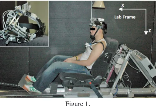
Subjects were seated on a feedback-controlled linear sled Figure 1.
Photographs of the experimental showing the location of the head and torso accelerometers, horn speaker and
laboratory reference frame (X, Z). Inset: Close-up view of the nine accelerometer array on the headgear device.
fitted with the driver’s seat of a 2005 Honda Accord (Fig-ure 1). Subjects were instructed to sit comfortably facing forward, rest their forearms on their lap, and relax their head and neck muscles. The head restraint was removed from the top of the seat back to prevent head-to-head-re-straint interaction that could affect the head/neck kinemat-ics or generate additional sensory inputs. The sled gener-ated no audible or mechanical pre-perturbation signals that could be used to predict the onset of a perturbation. The ambient background noise level in the lab was 64 dB. Each subject experienced a single forward horizontal translation with an average speed change of 75.00±0.03 cm/s, a peak acceleration of 19.5±0.2 m/s2, and a duration of 53.20±0.05 ms. To closely replicate a real automotive collision, the onset of the acceleration matched the onset of a vehicle-to-vehicle collision with a speed change of 8 km/h (2.22 m/s;20) and was presented simultaneously with the sound recorded from of an actual 8-km/h vehicle-to-barrier crash (peak 109 dB, time-to-peak 34ms). To re-main naïve to the experiment, subjects received neither practice nor demonstration trials of the perturbation.
Data Analysis
After data collection, all EMG data were digitally high-pass filtered using a 4th order, dual-pass Butterworth fil-ter with a 30 Hz cut-off frequency to further remove any motion artifact. To determine the recruitment order of axial and appendicular muscle responses, we compared the onsets of activity in the different muscles recorded. EMG onset was defined as the time when the root-mean-squared (RMS) amplitude (20 ms window) reached 10% of its maximum value8, and was then confirmed visually. Instantaneous heart rate (IHR) was obtained from the R-R intervals on the ECG signals to detect changes in the beat-to-beat intervals during and following the whiplash perturbation. Baseline IHR and EDA were defined as the average value over 5 s immediately preceding each per-turbation. Peak amplitude and time-to-peak for both IHR and EDA responses were determined as the first peak to occur within the 10 s period following the onset of the perturbation. The timeframe for IHR and EDA responses to return to baseline values were defined as the first in-stance IHR and EDA responses returned to their respect-ive baseline values following the perturbation.
The head acceleration data were transformed from the head accelerometer array to the atlanto-occipital
joint (AOJ) location and reported in the global reference frame (x-forward, y-right, z-down; for detailed proced-ures, see6). The AOJ was estimated to be 24 mm poster-ior and 37 mm inferposter-ior to the head’s center of mass21 and the head’s center of mass was estimated to lie in the mid-sagittal plane, rostral to the inter-aural axis by 17% of the distance between the interaural axis and the vertex22. All head and trunk accelerometers were corrected for the earth’s gravitational field. The onsets of head (x- and z-axis), chest (x-z-axis), lower lumbar (x-z-axis), and sled ac-celerations (x-axis) were determined directly from the transformed accelerometer data using a finite difference algorithm (5 ms moving window at a threshold value of 2 times the maximum pre-perturbation baseline value)8 and then confirmed visually. All data analyses were per-formed using MATLAB (The Mathworks, Natick, MA).
Statistical Analysis
Non-parametric statistics were used to determine the sig-nificance differences between muscle response onsets by ranking the recruitment order of axial and appendicular muscle responses. A Friedman rank sum test was first used to determine whether the recruitment of EMG responses was different between muscles. A paired Wilcoxon rank sum test was then used to determine individual differen-ces between each pair of muscles. Similar non-parametric statistics were performed to determine the significant dif-ferences within the order of acceleration onsets. A Fried-man rank sum test was used to determine whether the ac-celeration onsets were different between accelerometer locations and a paired Wilcoxon rank sum test further determined individual differences between each pair of accelerometer locations. Autonomic responses were ana-lyzed with a parametric paired-sample Student’s T-Tests to compare pperturbation baseline IHR and EDA re-sponses to the respective peak response occurring within the first 10 seconds following sled perturbation. All sta-tistical analyses were performed using MATLAB at a sig-nificance level of p = 0.05.
Results
Figure 2.
A sample of kinematic, muscular and autonomic responses from a single subject during the first exposure to a whiplash perturbation. Due to the different timing of responses, kinematic and muscular data have been grouped in panel a., and autonomic responses in panel b. Hollow circles
and dotted lines represent the onsets of accelerations and muscle responses to illustrate the propagation order of accelerations and the recruitment order of axial and appendicular muscles,
respectively. The vertical scale bars are aligned with the onset of the sled perturbation and are consistent between trials. Kinematic data: subscript x and z refers to the x- and z-directions,
respectively, for sled, lumbar, trunk and head accelerations. Electromyographic data: sternocleidomastoid (SCM), cervical paraspinal (PARA). triceps brachii (TRI), erector spinae at the level of L4 (ES), first dorsal interosseous (FDI) and rectus femoris (RF). Autonomic data:
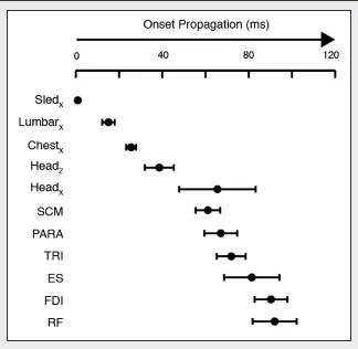
ms) and then head (z-axis: 38.9±6.9 ms, x-axis: 65.5±17.7 ms) (χ2(3)=30.00, p<0.0001) (Figure 2a & Figure 3). Wilcoxon rank sum tests indicated a significantly earlier onset of sled acceleration than onsets of lumbar spine, upper torso and head (z-axis) accelerations (multiple p values<0.0001), earlier onset of lumbar spine accelera-tions than onsets of upper torso and head (z-axis) accel-erations (multiple p values<0.0002), and earlier onset of upper torso accelerations than onset of head (z-axis) ac-celerations (p<0.0002), to establish an upward propaga-tion of accelerapropaga-tions (Sled Pelvis Upper Torso
Head). In contrast to the upward propagation of ac-celeration onsets, we observed a downward recruitment of muscle onsets from the neck muscles to the appendicu-lar muscles (χ2(5)=43.08, p<0.0001). Neck muscles were activated first (SCM: 61.1±5.7 ms & PARA: 67.1±7.6 ms) followed by TRI (71.9±6.7 ms), ES (81.4±12.8 ms), FDI (90.3±7.6 ms) and RF (92.0±10.2 ms). Wilcoxon rank sum tests indicated no difference between SCM and
PARA onsets (Z =-1.7047, p=0.0883), but did show that SCM was active before TRI, ES, FDI, and RF (multiple p values<0.0028) and that PARA was active before ES, FDI, and RF (multiple p values<0.0058) though not TRI (Z=-0.9085, p=0.3636). Furthermore, TRI was active be-fore FDI (Z=-3.5920, p=0.0003) and ES was active bebe-fore RF (Z =-2.1560, p=0.0311). Thus, two descending muscle recruitment schemes were observed: 1.) SCM TRI
FDI and 2.) SCM/PARA ES RF.
Concurrent sympathetic responses (IHR and EDA) were observed in all subjects following the simulated col-lision (Figure 2b). Baseline IHR ranged from 54 to 91 beats per minute (bpm) with an average IHR of 70±12 bpm. IHR increased by 14.3±5.7 bpm (p<0.0001) at 4.7±1.6 s after the onset of perturbation to 84±11 bpm. IHR returned to baseline levels in all subjects within 30 seconds following the collision. Baseline EDA values of -3.460±1.1 µmho increased by 2.08±1.1 µmho (p=0.0002) at 6.7±2.0 s after the onset of perturbation to an average
Figure 3.
value of -1.405±1.7 µmho. In comparison to IHR, EDA did not return to baseline within the recording duration of the experimental trial (30s).
Discussion
The goal of this study was to confirm the presence of a startle response within the neuromuscular response to a rear-end collision using two indirect measures of the star-tle response: recruitment order of muscle responses and autonomic physiological responses. A single whiplash-like perturbation evoked a descending recruitment pattern of axial and appendicular muscles and increased sym-pathetic responses (IHR and EDA). These observations were consistent with responses evoked independently by an acoustic startling stimulus (muscle responses10 and autonomic responses11-17) and provide further support that startle contributes to the overall response evoked during a rear-end collision.
Descending recruitment of muscle responses indicative of startle
A rear-end car collision is a complex, multi-sensory perturbation that stimulates the visual, vestibular, som-atosensory, and auditory systems. Recent human vol-unteers studies involving seated transient perturbations have suggested that the startle reflex forms part of the neuromuscular response to a rear-end collision.2,3,6,23 The startle response elicits a descending pattern of involun-tary axial and appendicular muscle activity such as facial grimacing, abduction of the upper arms and bending of the knees.10,24 From our study, we observed axial and ap-pendicular muscle responses with a descending recruit-ment of muscle activations from neck muscles (SCM and PARA) to more distal axial muscles (ES) to appendicular muscles (FDI and RF). These results were similar to those elicited by the acoustic startle response and further sup-port the presence of the startle responses.10
Alternatively, Forssberg and Hirschfeld (1994) pro-posed that somatosensory afferents derived from the backwards rotation and translation of the pelvis were responsible for triggering postural responses during sit-ting.25 Somatosensory receptors located in both the trunk and the pelvis are the first detectors of the physical onset of a whiplash perturbation as we observed an ascending propagation of accelerations from the seat to the head (lumbarx: 15ms, torsox: 26ms, and headz: 39ms). If the
trunk and pelvis were indeed responsible for the triggering of the postural responses, one may expect segmental re-flexes from the lumbar (ES muscle) to occur first through fast conducting monosynaptic stretch reflexes to maintain posture. These segmental reflex loops would then evoke an ascending recruitment of muscle activity along with the ascending propagation of accelerations. However, the current study observed two descending recruitment patterns of axial and appendicular muscles (SCM TRI FDI & SCM/PARA ES RF) despite an ascending propagation of accelerations. The observed downward recruitment of muscles responses further sup-port the idea that startle reflex forms part of the neuro-muscular responses to a rear-end collision.
Sympathetic responses indicative of startle
Sympathetic neural activity mediates the human body’s fight-or-flight responses to maintain homeostasis follow-ing situations perceived as startlfollow-ing or dangerous.26 Chan-ges in instantaneous heart rate (IHR) and electrodermal activity (EDA) can be used to infer the body’s regulation of this sympathetic drive during threatening situations. Following an unexpected rear-end collision, we observed an increase in sympathetic drive resulting in IHR and EDA increases of 14.1 bpm and 2.1 µmho, respectively. Similar increases in IHR and EDA were observed in volunteers who were driving on public roads and encountered a start-ling scenario involving an unexpected pedestrian crossing the road or a potential collision with another vehicle.16 Moreover, a startling auditory (110 dB) stimulus has been shown to evoke an average IHR increase of 11 bpm in human volunteers lying in a supine position.12 Thus, the sympathetic responses (within the first 10s) observed here support the presence of a startle response during a rear-end collision.
Implication for whiplash injury prevention
observed here provides further support for investigating methods of reducing the startle response following low-speed rear-end collisions. If the startle component of the posterior neck muscle responses can be decreased, then the strain applied to posterior neck structures and the risk of whiplash injury may be reduced. We have pre-viously shown that a loud (105 dB) pre-stimulus tone, presented 250 ms before the onset of impact, inhibits the startle component of the neuromuscular response evoked during a whiplash collision.7 The pre-stimulus tone decreased the kinematics of the head (horizontal acceleration and angular acceleration in extension by 23%) and neck muscle responses (SCM by 16% and PARA by 29%). Thus, we suggest that startle responses should be addressed in the development of future anti-whiplash safety devices to reduce, and possibly prevent, the risk of whiplash injuries.
Our observations that a whiplash-evoked startle re-sponse elicits muscle activity throughout the body may have several clinical implications for the management of whiplash injuries. Although whiplash injuries remain pri-marily associated with neck pain (80%-100%), patients have also reported localized pain in the lumbar region (30%-60%) and extremities (12%-35%)32-38. In follow-up reports two years after the motor vehicle collisions, patients reported chronic pain in the lumbar region (6%-25%) and in the extremities (8%-17%).32,34,35,37 The aeti-ology of the lumbar symptoms remains unclear, but the present findings imply that increased axial muscle activity can potentially lead to chronic low-back pain symptoms reported by patients with whiplash-associated disorders. It may be that increased activation of lower back muscles increases internal loads on lumbar structures by altering the kinematic and kinetic responses of the lumbar spine despite being supported by the car seat throughout the whiplash collision. Future in-vivo studies are needed to confirm this hypothesis and to characterize the kinematic and kinetic responses of the lumbar spine during whiplash collisions. Understanding the neuromechanics of whip-lash injuries will ultimately lead to injury prevention, bet-ter management and improve the life quality of patients with whiplash-associated injuries.
The whiplash perturbation used in this study is less severe than many real-life whiplash injury-inducing col-lisions39 and volunteer studies (higher speed changes: 4 to 16 km/h and peak accelerations: up to 6.0 g)40-44.
How-ever, startle responses have been shown to increase with stimulus intensity and rise time.45 If the startle response is present in the neuromuscular response to the acceleration pulse used in this study, the startle response should in-crease as stimulus intensity inin-creases. Nevertheless, fur-ther work is needed to confirm that our results are relevant at higher collision severities. Investigation into specific neurophysiological pathways responsible for triggering and modulating muscular and autonomic responses was outside the scope of this study. Thus, the exact nature of the sensory afferents triggering the startle reflex during rear-end collisions remains unanswered.
Conclusion:
This study provided further support that the startle re-sponse contributes to the neuromuscular rere-sponse evoked during a rear-end collision. We observed a descending re-cruitment pattern of axial and appendicular muscles and increased sympathetic responses indicative of a startle response. Increasing our understanding of how the star-tle response contributes to the neuromuscular response during rear-end collisions will lead to the development of more effective anti-whiplash safety devices to reduce, and possibly prevent, the risk of whiplash injuries. References
1. Jakobsson L, Lundell B, Norin H, Isaksson-Hellman I. WHIPS – Volvo’s Whiplash Protection Study. Accid Anal Prev. 2000;32(2):307-19.
2. Blouin JS, Inglis JT, Siegmund GP. Auditory startle alters the response of human subjects exposed to a single whiplash-like perturbation. Spine. 2006;31(2):146-54. 3. Blouin JS, Inglis JT, Siegmund GP. Startle responses
elicited by whiplash perturbations. J Physiol. 2006;573(3):857-67.
4. Siegmund GP, Blouin JS, Carpenter MG, Brault JR, Inglis JT. Are cervical multifidus muscles active during whiplash and startle? An initial experimental study. BMC Musculoskeletal Disorders. 2008;9(80). doi:10.1186/1471-2474-9-80
5. Siegmund GP, Blouin JS, Inglis JT. Does startle explain the exaggerated first response to a transient perturbation? Exerc Sport Sci Rev. 2008;36(2):76-82.
6. Blouin JS, Siegmund GP, Timothy Inglis J. Interaction between acoustic startle and habituated neck
postural responses in seated subjects. J Appl Physiol. 2007;102(4):1574-86.
8. Siegmund GP, Sanderson DJ, Myers BS, Inglis JT. Rapid neck muscle adaptation alters the head kinematics of aware and unaware subjects undergoing multiple whiplash-like perturbations. J Biomech. 2003;36(4):473-82.
9. Davis M. The mammalian startle response. In: Eaton RC, editor. Neural Mechanisms of Startle Behavior. New York, NY: Plenum Press; 1984. p. 287-351.
10. Brown P, Rothwell JC, Thompson PD, Britton TC, Day BL, Marsden CD. New observations on the normal auditory startle reflex in man. Brain. 1991;114 ( Pt 4):1891-902.
11. Dimberg U. Facial electromyographic reactions and autonomic activity to auditory stimuli. Biol Psychol. 1990;31(2):137-47.
12. Holand S, Girard A, Laude D, Meyer-Bisch C, Elghozi JL. Effects of an auditory startle stimulus on blood pressure and heart rate in humans. J Hypertens. 1999;17(12 Pt 2):1893-7.
13. Valls-Sole J, Kumru H, Kofler M. Interaction between startle and voluntary reactions in humans. Exp Brain Res. 2008;187(4):497-507.
14. Yeomans JS, Li L, Scott BW, Frankland PW. Tactile, acoustic and vestibular systems sum to elicit the startle reflex. Neurosci Biobehav Rev. 2002;26(1):1-11. 15. Eder DN, Elam M, Wallin BG. Sympathetic nerve and
cardiovascular responses to auditory startle and prepulse inhibition. Int J Psychophysiol. 2009;71(2):149-55. 16. Yoshino K, Edamatsu M, Yoshida M, Matsuoka K. An
algorithm for detecting startle state based on physiological signals. Accid Anal Prev. 2007;39(2):308-12.
17. Vossel G, Zimmer H. Stimulus rise time, intensity and the elicitation of unconditioned cardiac and electrodermal responses. Int J Psychophysiol. 1992;12(1):41-51. 18. Sibley KM, Mochizuki G, Esposito JG, Camilleri JM,
McIlroy WE. Phasic electrodermal responses associated with whole-body instability: presence and influence of expectation. Brain Res. 2008;1216:38-45.
19. Padgaonkar AJ, Krieger KW, King AI, editors. Measurement of angular acceleration of a rigid body using linear accelerometers. Applied Mechanics Summer Conference, Symposium on Biomechanics; 1975; Troy, N.Y.: The American Society of Mechanical Engineers. 20. Siegmund GP, King DJ, Lawrence JM, Wheeler JB,
Brault JR, Smith TA, editors. Head/neck kinematic response of human subjects in low-speed rear-end collisions (973341). Proceedings of the 41st Stapp Car Crash Conference; 1997; Warrendale, PA: Society of Automotive Engineers.
21. Siegmund GP, Blouin JS, Brault JR, Hedenstierna S, Inglis JT. Electromyography of superficial and deep neck muscles during isometric, voluntary, and reflex contractions. J Biomech Eng. 2007;129(1):66-77. 22. NASA. Anthropometric Source Book, NASA Reference
Publication 1024. Hanover, MD, USA: National
Aeronautics and Space Administration, Scientific and Technical Information Office; 1978.
23. Grosse P, Brown P. Acoustic startle evokes bilaterally synchronous oscillatory EMG activity in the healthy human. J Neurophysiol. 2003;90(3):1654-61.
24. Landis C, Hunt WA. The Startle Pattern. New York, NY: Farrar and Rinehart; 1939.
25. Forssberg H, Hirschfeld H. Postural adjustments in sitting humans following external perturbations: muscle activity and kinematics. Exp Brain Res. 1994;97(3):515-27. 26. Tortora GJ, Derrickson B. Principles of anatomy and
physiology. 11th ed. Hoboken, NJ: J. Wiley; 2006. 1 v. (various pagings) p.
27. Barnsley L, Lord S, Bogduk N. Whiplash injury. Pain. 1994;58(3):283-307.
28. Bogduk N, Yoganandan N. Biomechanics of the cervical spine Part 3: minor injuries. Clin Biomech (Bristol, Avon). 2001;16(4):267-75.
29. Pearson AM, Ivancic PC, Ito S, Panjabi MM. Facet joint kinematics and injury mechanisms during simulated whiplash. Spine. 2004;29(4):390-7.
30. Anderson JS, Hsu AW, Vasavada AN. Morphology, architecture, and biomechanics of human cervical multifidus. Spine. 2005;30(4):E86-91.
31. Winkelstein BA, Nightingale RW, Richardson WJ, Myers BS. The cervical facet capsule and its role in whiplash injury: a biomechanical investigation. Spine. 2000;25(10):1238-46.
32. Hildingsson C, Toolanen G. Outcome after soft-tissue injury of the cervical spine. A prospective study of 93 car-accident victims. Acta Orthop Scand. 1990;61(4):357-9.
33. Hincapié CA, Cassidy JD, Côté P, Carroll LJ, Guzmán J. Whiplash injury is more than neck pain: a population-based study of pain localization after traffic injury. J Occup Environ Med. 2010;52:434-40.
34. Hohl M. Soft-tissue injuries of the neck in automobile accidents. Factors influencing prognosis. J Bone Joint Surg Am. 1974;56(8):1675-82.
35. Maimaris C, Barnes MR, Allen MJ. ‘Whiplash injuries’ of the neck: a retrospective study. Injury. 1988;19(6):393-6. 36. Norris SH, Watt I. The prognosis of neck injuries resulting
from rear-end vehicle collisions. J Bone Joint Surg Br. 1983;65(5):608-11.
37. Radanov BP, Sturzenegger M, Di Stefano G. Long-term outcome after whiplash injury. A 2-year follow-up considering features of injury mechanism and somatic, radiologic, and psychosocial findings. Medicine (Baltimore). 1995;74(5):281-97.
38. Sturzenegger M, DiStefano G, Radanov BP, Schnidrig A. Presenting symptoms and signs after whiplash injury: the influence of accident mechanisms. Neurology. 1994;44(4):688-93.
consequences? Vehicle and human factors. Traffic Inj Prev. 2002;3:89-97.
40. Brault JR, Siegmund GP, Wheeler JB. Cervical muscle response during whiplash: evidence of a lengthening muscle contraction. Clin Biomech. 2000;15(6):426-35. 41. Brault JR, Wheeler JB, Siegmund GP, Brault EJ. Clinical
response of human subjects to rear-end automobile collisions. Arch Phys Med Rehabil. 1998;79(1):72-80. 42. Matsushita T, Sato TB, Hirabayashi K, Fujimara A,
Asazuma T, Takatori T, editors. X-ray study of the human neck due to head inertia loading (942208). Proceedings of the 38th Stapp Car Crash Conference; 1994; Warrendale, PA: Society of Automotive Engineers.
43. McConnell WE, Howard RP, van Poppel J, Krause R,
Guzman HM, Bomar JB, et al., editors. Human head and neck kinematic after low velocity rear-end impacts – Understanding “whiplash” (952724). Proceedings of the 39th Stapp Car Crash Conference; 1995; Coronado, CA: Society of Automotive Engineers.
44. Szabo TJ, Welcher JB, Anderson RD, Rice MM, Ward JA, Paulo LR, et al. Human occupant kinematic response to low speed rear-end impacts (940532). Occupant containment and methods of assessing occupant protection in crash environment; Warrendale, PA: Society of
Automotive Engineers; 1994. p. pp. 23-35.