Corresponding Author E-mail: [email protected] 1505 | Page http://www.ijabbr.com
online at Available
International journal of Advanced Biological and Biomedical Research
Volume 1, Issue 11, 2013: 1505-1511
Study of Photoisomerization in Cis-Retinal as a Natural Photo switch in Vision Using
Density Functional Theory
Seyed Mahdi Seyed Hosseini*1, Mohammad. Agha Mohammadi2, Mohsen Mousavi1
1
Department of Chemistry, Saveh Branch, Islamic azad University, Saveh, Iran 2
Department of Chemistry, Bourojerd Branch, Islamic Azad University, Bourojerd, Iran
ABSTRACT
In the present study, theoretical chemical reactivates Photo isomerization in Cis-Retinal as a Natural Photo switch in Vision. DFT hybrid functional, B3LYP and, post-HF method, were the theoretical methods applied utilizing G09 software. 6-31G+ (d,p) basis set employed for structural optimizations, and single point computations performed using B3LYP/6-31G+(d,p). The isomers cis molecule retinal from 0 to 180 ° in steps of 10 degrees to the trans isomers convert and we calculate the energy in each step, we determined that the most appropriate angle for the conversion of isomers of cis to trans angle = 90.11 the energy barrier less be.It identified that transmission S0 S1 is the most likely transmission.
Key words: Photoisomerization, Retinal, Photo switch, DFT, B3LYP
INTRODUCTION
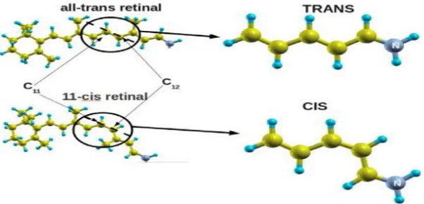
The mechanism of vision is present in most living creatures (Hargrave et al., 1983). In animals this mechanism is supported by a complex apparatus, but the ability to transform external optical perturbations in chemical signals is also present in the most primitive formsof life such as bacteria (Meyer, 1987). In all cases the first and most important step of the mechanism is the photoisomerization of the rhodopsin chromophore. Upon the absorption of a photon, its geometrical con-formation changes from 11-cisto all-trans by a rotation along theaxis connecting two carbon atoms of the polyenic chain (C11 andC12, see Figure 1).
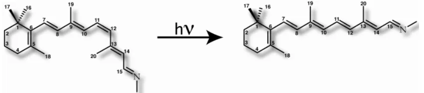
1506 | Page Indicate the rotation axis during the isomerization process Despite the importance of this process andseveral experimental (Kukura., 2005) and theoretical studies (Valsson, 2010). On this topic, the photoisomerizationmechanism is not yet fully understood (Nielsen, 2006). A theoretical description and evaluation of the efficiency andvelocity of the photoisomerization of the 11-cisretinal is possible only by a very precise calculation ofthe excited-state energies along all the phases of the isomerization. This study attempts to investigate the exact reaction pathway and to answer the question of howmany excited electronic states are involved in the internal conversion process as well as to calculate the energetics of the initial step of cis–transtrans formation for the photoisomerization of the retinal chromophore.Vision is a complex process in which light is converted into a nervous impulse through a chain of biochemical reactions taking place along various specialized cells and tissues, from the retina of the eye to the brain. In the eye, specialized cells of the retina called rod cells contain a protein acting as a photoreceptor, rhodopsin and possess the unique ability to convert and generate the neuronal signal. In order to become activated and induce the enzymatic cascade leading to vision (Albert, 2002) rhodopsin relies on its chromophore, retinal (Hofmann KP. 2000). Retinal is an organicmolecule (Figure 2) covalently-bound to rhodopsin in anticipation of its photo-activationThe 11-cis-retinal isomer is a key photosensitive component of rhodopsin. It is a molecular trigger capable of converting the electromagnetic energy carried by a photon into mechanical energy to be transferred to the protein in order to induce slower conformational changes, eventually leading to the initialization of the chain of biochemical reactions leading and finally to neural signal (Sakmar,1998). To perform its function, 11-cis-retinal undergoes a fast photoisomerization leading to all-trans-retinal,a process which triggers the conformational changes of the receptor to its active form (metarhodopsin II) (Yoshizawa T 1963).
Figure 2.Chemical structure of 11-cis-retinal and all-trans-retinal.
Computational methods
1507 | Page correspond respectively to a rotation around the main molecular axis of about (0,10,20,…,180), (table 1, table 2).For the potential energy surface of both the ground and excited state, the rotation about the C11 –C12 bond, characterized by the twist angle (C10 –C11 –C12 –C13 ), is considered.
Table1. Energy Specifications of the ground state Energy(ev)
Change angle
-23035.96 0
-23035.96 10
-23035.98 20
-23035.99 30
-23035.91 40
-23035.72 50
-23035.74 60
-23034.97 70
-23034.47 80
-23034.00 90
-23034.50 100
-23035.00 110
-23035.44 120
-23035.85 130
-23036.21 140
-23036.49 150
-23036.71 160
-23036.82 170
1508 | Page Table2. Energy Specifications and wave length thefirstexcitation
Singlet Excitation wavelength(nm) (ev)Energy Change angle 411.49 3.01 0 414.06 2.99 10 420.85 2.94 20 433.07 2.86 30 454.12 2.73 40 490.80 2.52 50 558.11 2.22 60 693.86 1.78 70 1041.52 1.19 80 1079.53 0.11 90 1018.14 1.21 100 681.58 1.81 110 547.83 2.26 120 481.38 2.57 130 445.69 2.78 140 425.61 2.91 150 414.24 2.99 160 408.44 3.03 170 406.85 3.04 180
Results and discussion
1509 | Page As is thecase for time-dependent DFT calculations, excitation energies are underestimated. Rhodopsin and bacteriorhodopsin have beenextensively investigated. Many calculations usingforce-field, (Humphrey 1994)semi empirical(Warshel, 1976)and ab initio methods, (Vreven 1997) and alsocombined classical/quantum mechanical simulations, applied to more or less complexmodels, have been performed; the references citedhere represent a selection from the huge literatureon the subject. However, there are still many openquestions in understanding the photoreaction details.
Figure3. Energydiagram ofthe changemodesdepending onthe degree
1510 | Page Figure.4. C11-C12 bond length as a function of the torsional angle for the protonated Schiff base of retinal without (filled symbols) and with the countering group and water molecule (empty symbols) in the ground (squares) and excited (circles) states.
REFERENCES
Albert AD, Yeagle PL. (2002).Structural studies on rhodopsin.BiochimBiophysActa 1565: 183 Warshel, A. (1976), Bicycle-pedal model for the first step in the vision process Nature 260 679.
Hofmann KP. (2000). In Handbook ofBiological Physics: MolecularMechanisms in Visual Transduction,ed. DG Stavenga, WJ DeGrip, ENPugh, pp. 91–142. New-York:Elsevier/North-Holland
Nielsen, L I.B. Lammich, L.H. Andersen, (2006), S1 and S2 Excited States of Gas-Phase Schiff-Base Retinal Chromophores, Phys. Rev. Lett, 96, 018304.
Parrinello M., (1998) maximally localized Wannier functions for disordered systems: application to amorphous silicon Solid State Commun. 102 107
Valsson, C. Filippi, (2010), Photoisomerization of model retinal chromophores: Insight from quantum Monte Carlo and multiconfigurational perturbation theory, J. Chem. Theory Comput, 64, 1275.
Kukura, D.W. McCamant, S. Yoon, D.B. Wandschneider, R.A. Mathies, (2005), Structural observation of the primary isomerization in vision with femtosecond-stimulated Raman, Science, 310, 1006
1511 | Page Singh, B.M. Deb,(1999) Time-Dependent Density-Functional Theory: Concepts and Applications Phys. Rep. 311 47.
Sakmar TP. (1998). Rhodopsin: aprototypical G protein-coupledreceptor. Prog Nucleic Acid Res MolBiol 59: 1
Meyer, E. Yakali, M.A. Cusanovich, G. Tollin, (1987), A water-soluble yellow protein from E. halophila was previously shown to be photoactive, Biochemistry, 262, 418.
Vreven, F. Bernardi, M. Garavelli, M. Olivucci, M.A. Robb, H.B. Schlegel, (1997) Conical intersection mechanism for photochemical ring opening in benzospiropyran compounds J. Am. Chem. Soc. 119 12687.
Humphrey, I. Logunov, K. Schulten, M. Sheves, (1994), Molecular dynamics study of bacteriorhodopsin and artificial pigments ,Biochemistry 33 ,3668.