Structural Correlates of Preterm Birth in the
Adolescent Brain
WHAT’S KNOWN ON THIS SUBJECT: A large number of studies identified preterm birth as a risk factor for long-term cognitive and structural brain development. However, it is known that developmental outcomes can differ on the basis of neonatal treatment and care.
WHAT THIS STUDY ADDS: This is one of the largest neuroimaging cohort studies investigating the long-term effects of preterm birth. Previous investigators did not combine structural brain images with DTI data. Furthermore, our findings are milder than those reported for similar cohorts.
abstract
OBJECTIVE:The Stockholm Neonatal Project involves a prospective, cross-sectional, population-based, cohort monitored for 12 to 17 years after birth; it was started with the aim of investigating the long-term structural correlates of preterm birth and comparing findings with reports on similar cohorts.
METHODS:High-resolution anatomic and diffusion tensor imaging data measuring diffusion in 30 directions were collected by using a 1.5-T MRI scanner. A total of 143 adolescents (12.18 –17.7 years of age) participated in the study, including 74 formerly preterm infants with birth weights ofⱕ1500 g (range: 645–1486 g) and 69 term control subjects. The 2 groups were well matched with respect to demographic and socioeconomic data. The anatomic MRI data were used for calcu-lation of total brain volumes and voxelwise comparison of gray matter (GM) volumes. The diffusion tensor imaging data were used for voxel-wise comparison of white matter (WM) microstructural integrity.
RESULTS:The formerly preterm individuals possessed 8.8% smaller GM volume and 9.4% smaller WM volume. The GM and WM volumes of individuals depended on gestational age and birth weight. The reduc-tion in GM could be attributed bilaterally to the temporal lobes, central, prefrontal, orbitofrontal, and parietal cortices, caudate nuclei, hip-pocampi, and thalami. Lower fractional anisotropy was observed in the posterior corpus callosum, fornix, and external capsules.
CONCLUSIONS:Although preterm birth was found to be a risk factor regarding long-term structural brain development, the outcome was milder than in previous reports. This may be attributable to differences in social structure and neonatal care practices.Pediatrics2009;124: e964–e972
AUTHORS:Zoltan Nagy, MSc,a,bJohn Ashburner, PhD,b
Jesper Andersson, PhD,cSaad Jbabdi, PhD,dBogdan
Draganski, MD,bStefan Skare, PhD,cBirgitta Bo¨hm, PhD,e
Ann-Charlotte Smedler, PhD,fHans Forssberg, MD, PhD,e
and Hugo Lagercrantz, MD, PhDa
aNeonatal andeNeuropediatric Units, Department of Woman and Child Health, andcMagnetic Resonance Center, Department of Clinical Neuroscience, Karolinska Institute, Stockholm, Sweden; bWellcome Trust Center for Neuroimaging, Institute of Neurology, University College London, London, England;dCentre for Functional Magnetic Resonance Imaging of the Brain, Department of Clinical Neurology, University of Oxford, Oxford, England; andfDepartment of Psychology, University of Stockholm, Stockholm, Sweden
KEY WORDS
preterm, magnetic resonance imaging, brain, diffusion tensor imaging, follow-up evaluation
ABBREVIATIONS
BW— birth weight
DTI— diffusion tensor imaging WM—white matter
GM— gray matter FA—fractional anisotropy GA— gestational age GMV— grey matter volume SGA—small for gestational age TBV—total brain volume WMV—white matter volume
www.pediatrics.org/cgi/doi/10.1542/peds.2008-3801 doi:10.1542/peds.2008-3801
Accepted for publication Jun 4, 2009
Address correspondence to Zoltan Nagy, MSc, Department of Woman and Child Health, Karolinska Institute, Astrid Lindgren Children’s Hospital, Q2:07, 171 76 Stockholm, Sweden. E-mail: [email protected]
PEDIATRICS (ISSN Numbers: Print, 0031-4005; Online, 1098-4275). Copyright © 2009 by the American Academy of Pediatrics
Many investigators have found that preterm birth affects structural devel-opment unfavorably. Children born preterm tend to achieve smaller brain volumes1,2 and have greater tenden-cies for structural brain lesions, intra-ventricular hemorrhage, intra-ventricular dilation,3,4and impaired growth of the corpus callosum.5,6They also possess a vulnerable white matter (WM) micro-structure at term-equivalent age.7,8
However, it is now apparent that differ-ences exist in neonatal care methods,9 which might have significant effects on developmental outcomes.10,11 The Stockholm Neonatal Project12 was started to investigate how the long-term outcomes of individuals born preterm fit into the internationally re-ported spectrum. Therefore, it was de-signed as a prospective study of chil-dren born preterm during a 4.5-year period between 1988 and 1993. At 5.5 years of age, the formerly preterm children and their matched control subjects underwent neuropsychologi-cal assessments.13,14
In reports of similar adolescent co-horts, the investigators used morpho-metric analysis of either T1-weighted MRI structural images15–17or diffusion tensor imaging (DTI) data.18–20 Interest-ingly, these 2 methods have not been combined before.
We expected that we would observe an association between preterm birth and structural brain development and that the gestational age (GA) and the birth weight (BW) would be inversely related to the severity of structural ab-normality. Another aim of this study was to compare the outcomes with those of other cohorts cared for in other centers of the world.
METHODS
Subjects
Between September 1988 and March 1993, infants born at the Karolinska
Hospital or transferred to the Karolin-ska Hospital from Lo¨wenstro¨mKarolin-ska Hospital in Stockholm were included if their BWs wereⱕ1500 g and their GAs were ⱕ36 weeks. In addition, all in-fants born in the entire county of Stockholm with BWs ofⱕ1000 g who received neonatal intensive care were invited to participate in the study.
Of the 291 infants who were included at birth, 55 died in the neonatal period or in early infancy, and 54 either had moved out of the Stockholm area or declined participation. At 5.5 years of age, 182 were available for follow-up evaluations.13 At that time, a control group of 125 children with GAs of ⱖ37 weeks was recruited from a population-based register, according to birth date and birth hospital.14
All parts of this study were performed with the approval of the local ethics committee, and both the participant and an accompanying adult signed written consent forms. The 182 avail-able case subjects and 125 control subjects were invited in random order, and 74 case subjects and 69 control subjects complied. The rest of the sub-jects either failed to respond to the in-vitation or declined. The participants did not differ from the original avail-able group with respect to GA, BW, gen-der distribution, mother’s age at birth, mother’s level of education, or general cognitive development (verbal, perfor-mance, and full-scale IQ scores) at 5.5
years of age.14There was no statisti-cally significant difference between the 2 groups with respect to weight and height at the time of scanning, gender distribution, mother’s age at birth, or mother’s level of education (Table 1).
MRI Data Collection
All participants underwent a cranial MRI examination at the Karolinska Hospital between April 2005 and Feb-ruary 2006, with a 1.5-T GE Signa Ex-cite Twin-speed scanner (Waukesha, WI). All except 2 of the examinations were performed by Mr Nagy. The protocol (Table 2) included T1/T2-weighted MRI images and a DTI data set with 30 diffusion-weighted MRIs (b⫽1000 seconds/mm2) distributed evenly21and 4 reference images (b⫽ 0 seconds/mm2). The DTI data set was collected twice, with alternate-phase encoding directions to remove susceptibility-induced artifacts.22 Al-though the T1-weighted MRI images were of good quality for all 143 sub-jects, the DTI images for 11 subjects (4 control subjects) were excluded from further analysis because of exces-sive movement artifacts.
Preprocessing of T1-Weighted MRI Scans
By using SPM 5 (Wellcome Trust Centre for Neuroimaging, London, England), a freely available software package, the
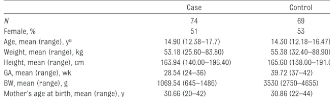
TABLE 1 Description of the 2 Groups in This Study
Case Control
N 74 69
Female, % 51 53
Age, mean (range), ya 14.90 (12.38–17.7) 14.30 (12.18–16.47)
Weight, mean (range), kg 53.18 (25.60–83.80) 55.38 (32.40–88.90) Height, mean (range), cm 163.94 (140.00–196.40) 165.60 (138.00–191.00) GA, mean (range), wk 28.54 (24–36) 39.72 (37–42) BW, mean (range), g 1069.54 (645–1486) 3530 (2750–4655) Mother’s age at birth, mean (range), y 30.66 (20–42) 30.86 (22–44) The mothers’ level of education is not shown because the scoring is on an ordinal scale and therefore mean values are not representative.
T1-weighted MRI images were seg-mented into gray matter (GM), WM, and cerebrospinal fluid.23The GM and WM segments were warped to the space of the average of all of the sub-jects by using DARTEL.24The output im-ages were modulated by the Jacobian determinants to preserve total volume irrespective of warping, resampled to 1.5-mm isotropic voxels, and smoothed with a 6-mm (full width at half-maximum), isotropic, Gaussian kernel before voxelwise statistical analysis. The total GM volume (GMV) and total WM volume (WMV) were estimated by integrating the GM and WM segment images, respectively. GMV and WMV were added to yield the total brain vol-ume (TBV).
Statistical Analysis of T1-Weighted MRI Images
The GMV and WMV were compared be-tween the groups by using 2-sample, 2-tailedttests and assuming unequal variances and also were correlated with age at the time of scanning, BW, and GA. For voxel-based morphometry (VBM),25the first analysis was a simple comparison of the 2 groups by using voxelwisettests. The second analysis included age, gender, and TBV as co-variates. Because 16 of the formerly preterm subjects were born small for GA (SGA),26 a separate analysis was performed with only the 58 indi-viduals who were born with an ap-propriate BW.
Seventeen formerly preterm subjects (4 of them also SGA) and 4 control sub-jects had radiologic structural abnor-malities (data not shown).27 A final comparison was performed with only
the 65 control subjects and 57 for-merly preterm adolescents who had negative MRI findings. Within the group of formerly preterm adolescents, pos-itive and negative correlations were made in a voxelwise manner by using GA or BW as a covariate.
In each of the analyses, correction was made for the large number of multiple comparisons. After the correction, P
values ofⱕ.05 were considered statis-tically significant.
Preprocessing of DTI Data
The data sets for the 67 formerly pre-term subjects and 65 control subjects with good-quality DTI data were cor-rected individually for eddy current ar-tifacts and realigned.28The mean of the 4 reference images was used to esti-mate the extent of susceptibility-induced distortion.22 The 2 data sets were then combined into a single un-distorted series (Fig 1), from which the diffusion tensor and fractional anisot-ropy (FA) images29were calculated.
Statistical Analysis of FA Images
By using tools from the freely available FSL software package (Functional MRI of the Brain Centre, University of Ox-ford, OxOx-ford, England), the tract-based, spatial, statistic pipeline was fol-lowed.30Nonbrain tissue was stripped from the mean reference image (brain extraction tool)31 and was used to mask the FA image of the same subject. The FA images were aligned into a com-mon space and resampled to 1-mm isotropic voxels (FMRIB’s Non-linear Image Registration Tool). The mean FA image was thinned to create a mean FA skeleton, representing the centers of
major WM tracks common to all of the subjects in the 2 groups. Finally, each of the FA images was projected onto this mean skeleton.
The statistical analyses were per-formed only with the set of voxels that belonged to the mean FA skeleton, with a minimal FA value of 0.2. Be-cause the null distribution was not known and the images had not been smoothed, permutation methods (ran-domize tool) were used32on the cluster level.33Student’sttests were used to compare the groups, and correlations between variables of interest and the
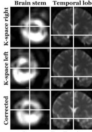
FIGURE 1
Illustration of the need for removal of susceptibility-induced distortions. The top 2 rows illustrate the original data sets, with the phase-encoding direction being left to right (ie, k-space is collected to the right) for the top row and right to left for the middle row. The bottom row is the corrected images that were used for the analyses. Left images are axial images at the level of the brainstem; right, coronal brain slices.
TABLE 2 Acquisition Parameters for T1-Weighted and DTI Data
Image Type Mode Sequence Echo
Time, ms
Repetition Time, ms
Flip Angle, °
Voxel Size, mm3
voxelwise values were also performed. Although the skeleton is only 1 voxel thick, the displayed results are en-larged slightly to aid visualization.
Separate analyses were performed af-ter exclusion of adolescents who were born SGA or demonstrated positive ra-diologic findings. P values of ⱕ.01 were considered statistically signifi-cant, after corrections for multiple comparisons had been made.
Statistical Analysis of Nonimage Data
For comparisons of the groups with re-spect to age, height, weight, and moth-er’s age at birth, 2-tailedttests were used, assuming unequal variances. A 2-sample test for binomial proportions was used to compare the gender dis-tributions of the 2 groups. The moth-er’s level of education was categori-cally compiled, with 0⫽no schooling, 1⫽6 years of schooling, 2⫽9 years of schooling, 3⫽ ⬍3 years of high school education, 4 ⫽ ⱖ3 years of high school education, 5⫽ ⬍3 years of uni-versity education, 6⫽ⱖ3 years of uni-versity education, and 7⫽doctoral de-gree. For these data, the Wilcoxon rank-sum test (a nonparametric ana-log of a 2-samplettest) was used. In all cases,Pvalues of⬍.05 were consid-ered statistically significant.
RESULTS
Total GMV and WMV
The GMV (Fig 2A) and WMV (Fig 3A) were smaller in the group of adoles-cents born preterm, by 8.8% and 9.4%, respectively (P⬍.001). The GMV/WMV ratio did not differ statistically signifi-cantly between the 2 groups.
A statistically significant linear rela-tionship emerged in which older sub-jects had smaller GMVs (Fig 2B); how-ever, the slopes were not different between the groups. Correspondingly, WMV increased with the age of the
sub-ject, but this linear relationship was not statistically significant (Fig 3B).
Both GMV (Fig 2 C and D) and WMV (Fig 3 C and D) depended directly on BW and GA (more pronounced for WMV).
These dependences were nonlinear and were strongest for adolescents who had been born most preterm and with the lowest BW. The GMV was independent of BW above ⬃1000 g,
FIGURE 2
Plots of GMV versus preterm birth, age, GA, and BW. Red circles indicate adolescents born preterm (open circles, SGA); blue circles, term. A, Comparison of GMVs between the groups. Horizontal bars indicate means and SDs. B, GMV plotted against age at the time of scanning. The slope of the reduction in GMV versus age was statistically significant (control:P⫽.016; formerly preterm:P⫽.039), but there was no statistically significant difference between the slopes (P⫽.596). C, GMV plotted against BW for the formerly preterm adolescents. D, GMV plotted against GA for the group of formerly preterm adolescents.
FIGURE 3
whereas the WMV even started to de-crease in the range of 1000 to 1500 g. Both the GMV and WMV seemed to be independent of GA at⬎30 weeks, but there was a bias toward the children being SGA because of the inclusion cri-terion based on BW alone.
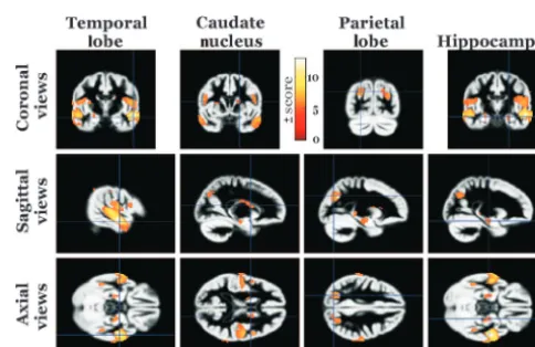
Voxelwise Comparison of GM Images
In comparisons of all of the 74 formerly preterm individuals with the 69 control subjects, the VBM analysis indicated a reduced grey matter volume in spe-cific areas within all 4 lobes of the brain, the caudate nuclei, hippocampi, thalami all bilaterally. Interestingly, the cerebella were almost entirely un-affected (Table 3 and Figs 4 and 5). The converse analysis did not indicate any regions in which the control group had smaller GMV.
After correction for the effects of gen-der, age at the time of scanning, and TBV, the results became more focal, with involvement of only the temporal and parietal lobes (data not shown). Again, the converse analysis did not yield any regions in which the control group had smaller GMV.
Exclusion of the individuals who were born SGA did not have a noteworthy effect on the results. The major
find-ings did not change significantly after exclusion of all individuals with abnor-mal radiologic findings.
Only 2 focal regions of insignificant vol-ume resulted from the analysis that correlated BW or GA with GMV in a vox-elwise manner. The clusters were lo-cated in the left prefrontal and parietal cortices and consisted of 32 and 20 voxels, respectively.
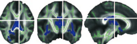
Voxelwise Comparison of FA Data
Figure 6 displays the regions within WM tracts in which FA was higher in the control population than in group of formerly preterm adolescents. The af-fected areas included the external cap-sule bilaterally (potentially involving the uncinate fasciculus or the inferior frontooccipital fasciculus), the poste-rior body, isthmus, and anteposte-rior parts of the splenium of the corpus callo-sum, the forceps major (bilaterally), and the fornix (bilaterally). There was
FIGURE 4
Results of the voxel-based morphometric analysis. The highlighted regions represent voxels where the GMV was on average higher for the group of control subjects than for the group of adolescents born preterm (P⬍.05, after correction for multiple comparisons). Most of the results show a striking bilaterality.
FIGURE 5
Surface-rendered display of the results of the voxel-based morphometric analysis. The results from Fig 4 (red surface) are overlaid on a volume-rendered depiction of the GM/WM boundary (white surface). Both hemispheres are shown to demonstrate the bilaterality of the findings (P⬍.05, after correction for multiple comparisons).
TABLE 3 MNI Coordinates for Voxels With PeakzandtScores
Region MNI Coordinates P zScore t
x y z
no significant cluster when the oppo-site contrast was tested.
FA correlated with BW and GA re-gardless of whether subjects were born preterm. Again, the external/ extreme capsule, corpus callosum, and fornix were involved; however, when only the control population was considered, these covariations were not significant.
The results did not change signifi-cantly when all subject born SGA were excluded from the analysis. Exclusion of those with abnormal radiologic find-ings restricted the differences be-tween the groups to only the external capsules.
DISCUSSION
By using state-of-the-art MRI acquisi-tion and image-processing methods, we examined a large cohort and found that the total GMVs and WMVs were smaller in formerly preterm adoles-cents, compared with matched control subjects. Voxel-based morphometric measurement of the GM revealed that this difference was localized to the me-dial and superior temporal cortex, so-matosensory cortex, parietal cortex, and subcortical GM structures. Track-based spatial statistics revealed cor-respondingly lower FA in the corpus callosum, external capsule, and fornix for formerly preterm adolescents. Fur-thermore, the GMV and WMV attained depended on both BW and GA, whereas voxelwise analyses revealed
statisti-cally significant correlations only be-tween FA and BW/GA.
We found that formerly preterm ado-lescents had smaller GMV (8.8%) and WMV (9.4%), on average (Figs 2A and 3A). This finding is not unprecedent-ed1,2,34 and, given the common struc-tural abnormalities in this group,3,4,35it was expected. With respect to the GMV, it is possible that, in response to the distressing extrauterine environment, endogenous corticosteroid levels were increased, which is known to inhibit growth.11 However, the large overlap between the groups is notable (Figs 2A and 3A). Although the results were sta-tistically significant, the biological or clinical significance may not be as alarming. For example, in a similar study, Nosarti et al1found a similar ef-fect size (11.8%) in comparisons of GMV but no statistically significant dif-ference in WMV.
In line with previous reports, the GMV decreased monotonically36,37while the WMV increased correspondingly34,36in both groups between 12 and 17 years of age, although the increase in WM was not statistically significant in this study. In our cohort, the GMV/WMV ra-tio did not differ between the 2 groups, which indicated that the brain as a whole, not only the GM or WM, devel-oped differently.
The voxelwise comparison between the groups, without covariates, yielded extended regions in which the
adoles-cents who were born preterm had smaller GMVs (Figs 5 and 6). These findings are in excellent agreement with the report by Peterson et al2and have commonalities with that by Nosarti et al.15
It is debatable whether the variability attributable to TBV should be included as a covariate. Although it is not always included,15most investigators remove the effects of TBV before presenting their results.1,2,34When such an exten-sive set of regions are involved, it is not surprising that regional differences covary with TBV. When we included TBV as a covariate, it explained most of the difference between the 2 groups and only the temporal and parietal lobe re-gions remained statistically signifi-cant, to a much reduced spatial extent, which was much less severe than that reported previously.1,2,15,34
Simple voxelwise comparison of the 2 groups indicated that FA was lower in the group of formerly preterm adoles-cents in the posterior part of the cor-pus callosum, the fornix, and the exter-nal capsules bilaterally. These findings are not unprecedented. Both Consta-ble at al20and Vangberg et al18found similar results, at 12 and 15 years of age, respectively. It should be noted, however, that the extent of WM regions in which we could find statistically sig-nificant differences was much less.
The regions in which lower FA was de-tected generally connect cortical ar-eas in which lower GMV was found. The body of the corpus callosum connects the bilateral motor and sensory corti-ces, the isthmus contains fibers con-necting the parietal and temporal cor-tices as well as the parahippocampal gyrus, and axons in the splenium run mainly between the occipital corti-ces.38,39The external capsule contains fibers that run to the striatum, fibers in the fornix pass the hippocampi,40 and the orbitofrontal and anterior
FIGURE 6
temporal areas are connected by the uncinate fasciculus.41
Although there is evidence that injured WM impairs the migration and proper development of neurons,42,43 it is also known that electrical activity induced by neuronal activity is necessary for proper WM development.44 With its observational design, the present stu-dy cannot unquestionably distinguish whether inferior WM connectivity led to the lower GMV or whether injury to the GM caused the reduction of FA in the corresponding WM fiber tracts.
Whether being born SGA has detrimen-tal effects on brain and cognitive devel-opment is still debated. There are reports supporting both possibili-ties.45–47The differences in opinion may be a result of differences in the co-horts. This is not yet conclusive, how-ever, because brain volumes alone may not always accurately reflect cog-nitive ability.1,6It should also be noted that, at 5.5 years of age, the IQ was sig-nificantly lower for the eldest GA group (32–36 weeks), of whom 63% were SGA. Our negative findings may be ex-plained by catch-up development or may be a result of the small number of individuals (n⫽16) in this subgroup.
In a separate analysis, we also ex-cluded all individuals with positive radiologic diagnoses, to investigate whether the reductions in GMV or FA were apparent only in individuals with structural brain injuries. How-ever, this affected only the FA analy-sis. This is probably because most of the identified injuries affected the WM, to a larger extent than injuries to the GM.
Overall, the differences between for-merly preterm adolescents and appro-priate control subjects were subtler than had been reported previously. This is in line with previous examina-tions and current radiologic findings for the same cohort. A large propor-tion of the infants were supported only with supplemental oxygen (25%) or na-sal continuous positive airway pres-sure therapy (35%). Less than 25% of the infants were supported with me-chanical ventilation only.12The neona-tal morneona-tality rate was low (17.5%), and corticosteroid prophylaxis and surfac-tant therapy were not yet routine (⬍10% of infants). A comprehensive neuropsychological assessment at 5.5 years of age revealed that the preterm group scored lower than the control subjects on the Wechsler Preschool and Primary Scale of Intelligence-Revised performance scale, which in-dicated deficits in visuospatial cogni-tive functioning, whereas no deficits were observed in the verbal domain.14 Also, at 5.5 years of age, the preterm chil-dren tended to score lower on tests of executive functioning.13 However, these observed deficits were minor and sub-clinical in nature. Structural abnormali-ties in this cohort were less frequent and less severe27than reported elsewhere.47 Furthermore, in a recent report from our center,48structural brain abnormal-ities of neonates born preterm were less frequent than reported from other cen-ters. We hypothesize that a combination of social structure and minimally inva-sive neonatal care may underlie this fa-vorable outcome.
All of the case subjects (N⫽182) and control subjects (N⫽125) who
partic-ipated in the follow-up evaluation at 5.5 years of age are presently (2007–2010) invited to participate in a psychologi-cal assessment at 18 years of age. The possible relationships between the morphologic findings in this study and the results of this psychological as-sessment will be investigated. Future plans also include tractography based on the DTI data, to elucidate whether the regions with lower FA values in for-merly preterm individuals are parts of the fiber tracks connecting the areas where the cortex was found to be smaller.
ACKNOWLEDGMENTS
Funding support was provided by Sa¨ll-skapet Barnavård (Sweden), the Swed-ish Research Council, the Bank of Swe-den Tercentenary Foundation, and the Wellcome Trust (United Kingdom).
Many thanks go to Jessica Schiott for contacting and booking the subjects. Yords O¨sterman and Marie Lundberg are experienced MRI radiographers who trained Mr Nagy extensively in data collection. Patient comfort and data quality were improved because of their involvement. Either Dr Anna-Karin Edstedt Bonamy (pediatrician) or Ms Jonna Karlen (medical student) at-tended each of the scans, to provide support in case of medical emergency and to assist with the scans. Their hard work and dedication were invaluable in the completion of this study. The late Dr David Freedman of the Department of Statistics, University of California, Berkeley, provided patient advice. Fi-nally, the manuscript was much im-proved after Drs Geraint Rees and Chloe Hutton offered their assistance with the preparation.
REFERENCES
1. Nosarti C, Al Asady MH, Frangou S, Stewart AL, Rifkin L, Murray RM. Adolescents who were born very preterm have decreased brain volumes.
Brain.2002;125(7):1616–1623
2. Peterson BS, Vohr B, Staib LH, et al. Regional brain volume abnormalities and long-term
cognitive outcome in preterm infants.
JAMA.2000;284(15):1939 –1947
3. Dyet LE, Kennea N, Counsell SJ, et al. Natural history of brain lesions in extremely pre-term infants studied with serial magnetic resonance imaging from birth and
neuro-developmental assessment.Pediatrics.
2006;118(2):536 –548
4. Rutherford MA.MRI of the Neonatal Brain. London, England: Saunders; 2002 5. Anderson NG, Laurent I, Woodward LJ, Inder
cor-pus callosum in premature infants. Pediat-rics.2006;118(3):951–960
6. Stewart AL, Rifkin L, Amess PN, et al. Brain structure and neurocognitive and behav-ioural function in adolescents who were born very preterm.Lancet.1999;353(9165): 1653–1657
7. Hu¨ppi PS, Maier SE, Peled S, et al. Micro-structural development of human newborn cerebral white matter assessed in vivo by diffusion tensor magnetic resonance imag-ing.Pediatr Res.1998;44(4):584 –590 8. Anjari M, Srinivasan L, Allsop JM, et al.
Dif-fusion tensor imaging with tract-based spatial statistics reveals local white matter abnormalities in preterm infants. Neuro-image.2007;35(3):1021–1027
9. Van Reempts P, Gortner L, Milligan D, et al. Characteristics of neonatal units that care for very preterm infants in Europe: results from the MOSAIC study. Pediatrics.2007; 120(4). Available at: www.pediatrics.org/cgi/ content/full/120/4/e815
10. Gressens P, Rogido M, Paindaveine B, Sola A. The impact of neonatal intensive care prac-tices on the developing brain.J Pediatr.
2002;140(6):646 – 653
11. Murphy BP, Inder TE, Hu¨ppi PS, et al. Im-paired cerebral cortical gray matter growth after treatment with dexametha-sone for neonatal chronic lung disease. Pe-diatrics.2001;107(2):217–221
12. Katz-Salamon M, Forssberg H, Lagercrantz H. The Stockholm Neonatal Project: very low birthweight infants in the late 20th century in Stockholm.Acta Paediatr.1997;86(suppl 419):1– 43
13. Bo¨hm B, Smedler AC, Forssberg H. Im-pulse control, working memory and other executive functions in preterm children when starting school.Acta Paediatr.2004; 93(10):1363–1371
14. Bo¨hm B, Katz-Salamon M, Smedler AC, La-gercrantz H, Forssberg H. Developmental risks and protective factors for influencing cognitive outcome at 5 1/2 years of age in very-low-birthweight children. Dev Med Child Neurol.2002;44(8):508 –516 15. Nosarti C, Giouroukou E, Healy E, et al. Grey
and white matter distribution in very pre-term adolescents mediates neurode-velopmental outcome. Brain.2008;131(1): 205–217
16. Allin M, Henderson M, Suckling J, et al. Ef-fects of very low birthweight on brain struc-ture in adulthood.Dev Med Child Neurol.
2004;46(1):46 –53
17. Martinussen M, Fischl B, Larsson HB, et al. Cerebral cortex thickness in 15-year-old
adolescents with low birth weight mea-sured by an automated MRI-based method.
Brain.2005;128(11):2588 –2596
18. Vangberg TR, Skranes J, Dale AM, Martinus-sen M, Brubakk AM, Haraldseth O. Changes in white matter diffusion anisotropy in ado-lescents born prematurely. Neuroimage.
2006;32(4):1538 –1548
19. Skranes J, Vangberg TR, Kulseng S, et al. Clinical findings and white matter abnor-malities seen on diffusion tensor imaging in adolescents with very low birth weight.
Brain.2007;130(3):654 – 666
20. Constable RT, Ment LR, Vohr BR, et al. Pre-maturely born children demonstrate white matter microstructural differences at 12 years of age, relative to term control subjects: an investigation of group and gender effects.
Pediatrics.2008;121(2):306 –316
21. Jones DK, Horsfield MA, Simmons A. Optimal strategies for measuring diffusion in aniso-tropic systems by magnetic resonance im-aging.Magn Reson Med.1999;42(3):515–525 22. Andersson JL, Skare S, Ashburner J. How to
correct susceptibility distortions in spin-echo spin-echo-planar images: application to dif-fusion tensor imaging.Neuroimage.2003; 20(2):870 – 888
23. Ashburner J, Friston KJ. Unified segmenta-tion.Neuroimage.2005;26(3):839 – 851 24. Ashburner J. A fast diffeomorphic image
registration algorithm.Neuroimage.2007; 38(1):95–113
25. Ashburner J, Friston KJ. Voxel-based mor-phometry: the methods.Neuroimage.2000; 11(6):805– 821
26. Fenton TR. A new growth chart for preterm babies: Babson and Benda’s chart updated with recent data and a new format.BMC Pediatr.2003;3:13
27. Nagy Z, Jonsson B. Cerebral MRI findings in a cohort of ex-preterm and control adoles-cents.Acta Paediatr.2009;98(6):996 –1001 28. Andersson JL, Skare S. A model-based
method for retrospective correction of geo-metric distortions in diffusion-weighted EPI.
Neuroimage.2002;16(1):177–199
29. Basser PJ, Pierpaoli C. Microstructural and physiological features of tissues elucidated by quantitative-diffusion-tensor MRI.J Magn Reson B.1996;111(3):209 –219
30. Smith SM, Jenkinson M, Johansen-Berg H, et al. Tract-based spatial statistics: voxel-wise analysis of multi-subject diffusion data.Neuroimage.2006;31(4):1487–1505 31. Smith SM. Fast robust automated brain
extraction. Hum Brain Mapp.2002;17(3): 143–155
32. Nichols TE, Holmes AP. Nonparametric per-mutation tests for functional neuroimaging:
a primer with examples.Hum Brain Mapp.
2002;15(1):1–25
33. Hayasaka S, Nichols TE. Validating cluster size inference: random field and permuta-tion methods. Neuroimage. 2003;20(4): 2343–2356
34. Reiss AL, Abrams MT, Singer HS, Ross JL, Denckla MB. Brain development, gender and IQ in children: a volumetric imaging study.Brain.1996;119(5):1763–1774 35. Skranes J, Evensen KI, Løhaugen GC, et al.
Abnormal cerebral MRI findings and neuro-impairments in very low birth weight (VLBW) adolescents.Eur J Paediatr Neurol. 2008; 12(4):273–283
36. Giedd JN, Blumenthal J, Jeffries NO, et al. Brain development during childhood and adolescence: a longitudinal MRI study.Nat Neurosci.1999;2(10):861– 863
37. Sowell ER, Peterson BS, Thompson PM, Wel-come SE, Henkenius AL, Toga AW. Mapping cortical change across the human life span.
Nat Neurosci.2003;6(3):309 –315
38. Sunderland S. The distribution of commis-sural fibres in the corpus callosum in the macaque monkey.J Neurol Psychiatry.1940; 3(1):9 –18
39. Moses P, Courchesne E, Stiles J, Trauner D, Egaas B, Edwards E. Regional size reduction in the human corpus callosum following pre- and perinatal brain injury.Cereb Cor-tex.2000;10(12):1200 –1210
40. Poletti CE, Creswell G. Fornix system effer-ent projections in the squirrel monkey: an experimental degeneration study.J Comp Neurol.1977;175(1):101–128
41. Petrides M, Pandya DN. Association fiber pathways to the frontal cortex from the su-perior temporal region in the rhesus mon-key.J Comp Neurol.1988;273(1):52– 66 42. Inder TE, Hu¨ppi PS, Warfield S, et al.
Periven-tricular white matter injury in the prema-ture infant is followed by reduced cerebral cortical gray matter volume at term. Ann Neurol.1999;46(5):755–760
43. Leviton A, Gressens P. Neuronal damage ac-companies perinatal white-matter damage.
Trends Neurosci.2007;30(9):473– 478 44. Demerens C, Stankoff B, Logak M, et al.
In-duction of myelination in the central ner-vous system by electrical activity.Proc Natl Acad Sci U S A.1996;93(18):9887–9892 45. Tolsa CB, Zimine S, Warfield SK, et al. Early
alteration of structural and functional brain development in premature infants born with intrauterine growth restriction. Pedi-atr Res.2004;56(1):132–138
Growth and development are similar in VLBW children born appropriate and small for gestational age: an interim report on 97 preschool children.J Pediatr Endocrinol Metab.2007;20(9):1017–1026
47. Skranes JS, Martinussen M, Smevik O, et al. Cerebral MRI findings in very-low-birth-weight and small-for-gestational-age chil-dren at 15 years of age. Pediatr Radiol.
2005;35(8):758 –765
DOI: 10.1542/peds.2008-3801 originally published online October 26, 2009;
2009;124;e964
Pediatrics
Lagercrantz
Stefan Skare, Birgitta Böhm, Ann-Charlotte Smedler, Hans Forssberg and Hugo
Services
Updated Information &
http://pediatrics.aappublications.org/content/124/5/e964
including high resolution figures, can be found at:
References
http://pediatrics.aappublications.org/content/124/5/e964#BIBL
This article cites 45 articles, 5 of which you can access for free at:
Subspecialty Collections
http://www.aappublications.org/cgi/collection/neurology_sub
Neurology
following collection(s):
This article, along with others on similar topics, appears in the
Permissions & Licensing
http://www.aappublications.org/site/misc/Permissions.xhtml
in its entirety can be found online at:
Information about reproducing this article in parts (figures, tables) or
Reprints
http://www.aappublications.org/site/misc/reprints.xhtml
DOI: 10.1542/peds.2008-3801 originally published online October 26, 2009;
2009;124;e964
Pediatrics
Lagercrantz
Stefan Skare, Birgitta Böhm, Ann-Charlotte Smedler, Hans Forssberg and Hugo
Zoltan Nagy, John Ashburner, Jesper Andersson, Saad Jbabdi, Bogdan Draganski,
Structural Correlates of Preterm Birth in the Adolescent Brain
http://pediatrics.aappublications.org/content/124/5/e964
located on the World Wide Web at:
The online version of this article, along with updated information and services, is
by the American Academy of Pediatrics. All rights reserved. Print ISSN: 1073-0397.