HEPATOCYTE GROWTH FACTOR/SCATTER^
FACTOR—
AND
RENAL MORPHOGENESIS
By
Maria Kolatsi
a Thesis submitted for the degree of
Doctor of Philosophy
in the University of London
August 1996
Institute of Child Health
Developmental Biology Unit
30 Guilford Street
London
ProQuest Number: 10105163
All rights reserved
INFORMATION TO ALL USERS
The quality of this reproduction is dependent upon the quality of the copy submitted. In the unlikely event that the author did not send a complete manuscript and there are missing pages, these will be noted. Also, if material had to be removed,
a note will indicate the deletion.
uest.
ProQuest 10105163
Published by ProQuest LLC(2016). Copyright of the Dissertation is held by the Author. All rights reserved.
This work is protected against unauthorized copying under Title 17, United States Code. Microform Edition © ProQuest LLC.
ProQuest LLC
789 East Eisenhower Parkway P.O. Box 1346
Abstract.
Hepatocyte growth factor/scatter factor (HGF/SF) is a growth factor which has
a variety of effects on many target cells. Its effects have been reported to be
mitogenic, motogenic and morphogenic. HGF/SF and its receptor MET, were
studied in the developing and adult mouse kidney and in the developing human
fetus. From investigating the early development of the mouse metanephros,
HGF/SF was found to be expressed by renal mesenchymal cells while MET
was expressed by both the ureteric bud and the mesenchyme. Conditionally-
immortal cell lines derived from renal mesenchyme expressed either MET or
both the ligand and receptor. Nephron formation and branching
morphogenesis were disrupted when metanephric rudiments were grown in
serum-free organ culture with neutralising anti-HGF/SF antibodies. Hence
HGF/SF may have an autocrine and/or paracrine role in nephrogenesis.
By studying the metanephros in more detail, I found that the vascular stalk of
the glomerulus expressed the MET protein. Therefore I studied the role of
HGF/SF and MET in individual mesangial cells derived from the adult kidney.
Both HGF/SF and MET mRNA and protein were expressed by these
mesangial cells and HGF/SF phosphorylated the MET receptor and caused the
cells to aojj/iW an elongated shape. Hence, HGF/SF may have an autocrine
From screening a number of human fetal organs taken from the first trimester
of pregnancy, HGF/SF and MET were detected in organ systems which
originate from mesenchymal-epithelial conversions and include the kidney,
liver, lung and gut. HGF/SF and MET were also expressed in the chorionic
villi of the placenta. Taken together these data imply that HGF/SF and MET
may not only be important in mouse nephrogenesis but also in early human
Acknowledgements.
Firstly, I would like to express my thanks and appreciation to Dr Adrian Woolf
for giving me the opportunity to carry out the work in the first place and for
supporting me throughout. Dr W oolfs enthusiasm for science and medicine
knows no bounds and he is a constant source of ideas, advice and help. I
would like to give credit to Dr Woolf who derived the metanephric clonal cell
lines and mesangial cell lines which provided me with the basis of my work. I
would like to express how grateful I am to Dr Patricia Hardman for her expert
training in microdissection of embryonic kidneys, and for all her hard work on
double staining and handling of the confocal microscope.
I would like to thank Professor Peter Thorogood for giving me permission to
use tissues from the human embryo bank and thanks must also go to Rachel
Moore and Jo Chan for collecting the tissues. My appreciation and thanks
goes to all the people who have taught me various techniques over the last
three years especially to Dr Debbie Henderson. Dr Paul Hunt helped with
computing, and Dr Paul Winyard advised me on immunohistochemical
techniques.
I would like to acknowledge and give thanks to our collaborators Dr Ermanno
Gherardi and his group. Dr Gherardi and his group from Cambridge have
goes to Dr Elisabetta Andemarcher who performed the in situ hybridisation. I
would like to express my gratitude to Dr Stan White from Sheffield University
for taking the time to isolate glomeruli fi’om adult mice.
I would like to say thank you to all my colleagues and all the team leaders in
the Developmental Biology and Neural Development Units at the Institute of
Child Health for making it a very happy and pleasant place to work and I have
enjoyed and learnt very much from my time spent with them. On a more
personal note, I would like to say a big thank you to my family for supporting
and encouraging me throughout my Ph D and for making sure I fed and
Abbreviations.
ABC avidin-biotin peroxidase complex
ATP adenosine triphosphate
BMP bone morphogenetic protein
bp base pair
BSA bovine serum albumin
cAMP cyclic adenosine monophosphate
cDNA complementary DNA
CRE cAMP response element
dATP deoxyadenosine triphosphate
DAB diaminobenzadine tetrahydrochlorodihydrate
dCTP deoxycytidine triphosphate
DBPC diethyl pyrocarbonate
dGTP deoxyguanosine triphosphate
DNA deoxyribonucleic acid
DNAse deoxyribonuclease
dNTP ’ s deoxyiibonucleotriphosphates
dXTP deoxythymidine triphosphate
DTT dithiothreitol
EDTA ethylene-diamine tetra-acetic acid
EGF epidermal growth factor
aFGF acidic fibroblast growth factor
bFGF basic fibroblast growth factor
FITC fluoroscein isothiocyanate
HEPES N-2-hydroxyethylpiperazine-N’ -2-ethanesulphonic acid
HGF/SF hepatocyte growth factor/scatter factor
HLP hepatocyte growth factor-like protein
HOX homeobox
IGF insulin-like growth factor
IGFbp insulin-like growth factor binding protein
IL interleukin
kb kilobase
kDa kilodaltons
LIF leukaemia inhibitory factor
mag. X magnification times
mRNA messenger ribonucleic acid
MSP macrophage stimulating protein
NGF nerve growth factor
PAX pairedbox
PCR polymerase chain reaction
PDGF platelet-derived growth factor
RNA ribonucleic acid
RNAse ribonuclease
RT-PCR reverse transcriptase polymerase chain reaction
SDS sodium lauryl sulphate
SSC sodium chloride/sodium citrate buffer
TAE tris acetate EDTA electrophoresis buffer
TBE tris borate EDTA electrophoresis buffer
TE tiis-EDTA
TEMED N,N,N’ ,N’tetramethyl ethyl enediamine
TESPA 3 -aminopropyltriethoxy sil ane
TRITC texas red isothiocyanate
TGF a and P transforming growth factor alpha and beta
TIE TGFp inhibitory element
UTP uridine triphosphate
TNFa tumour necrosis factor alpha
UV ultra violet
VEGF vascular endothelial growth factor
X-gal 5-bromo-4-chloro-3 -indolyl-p -D-galactosidase
Contents.
Title page 1
Dedication 2
Abstract 3
Acknowledgements 5
Abbreviations 7
Contents 10
List of Figures 18
List of Tables 20
Chapter I. Introduction
1.1 Anatomy of nephrogenesis 21
1.2 The pronephros 22
1.3 The mesonephros 23
1.4 The metanephros 24
1.4.1 The glomerulus 31
1.4.2 Mesangial cells 31
1.4.3 Proximal tubules 35
1.4.4 The loop of Henle 35
1.5 Fetal function of the mammalian kidney 36
2. Basic mechanisms of nephrogenesis 37
2.1 Cell proliferation 37
2.2 Cell death 38
2.3 Induction 38
2.4 Morphogenesis 39
2.5 Migration 39
2.6 Differentiation 40
3.1 Descriptive studies 41
3.2 Functional studies 41
3.2.1 Whole organ culture 41
3.2.2 Induction of renal mesenchyme 42
3.2.3 Nephrogenic cell lines 45
3.2.4 Transgenic studies 46
3.2.5 Overexpression 47
3.2.6 Gene ablation 47
4. Growth factors and kidney development 51
4.1 Growth factors which enhance nephrogenesis 52
4.1.1 Epidermal growth factor and Transforming growth factor a 52
4.1.2 Fibroblast growth factors 52
4.1.3 Insulin like growth factors 53
4.1.4 Platelet derived growth factor 54
4.1.5 Vascular endothelial derived growth factor 54
4.1.6 GDNF and RET 55
4.1.7 ROS 55
4.1.9 Nerve growth factors 56
4.2 Growth factors which inhibit nephrogenesis 57
4.2.1 Transforming growth factor p 57
4.2.2 Leukaemia inhibition factor 58
4.2.3 Tumour necrosis factor a 58
5. Survival factors 58
6. Transcription factors 60
7. Ceil adhesion molecules and extracellular matrix 62
8. Hepatocyte growth factor/scatter factor and its receptor
tyrosine kinase MET 65
8.1 Discovery and isolation of HGF/SF 65
8.3 Regulation of HGF/SF expression and activity 69
8.3.1 Regulatory elements 69
8.3.2 Secretion 70
8.3.3 Proteolytic activation 70
8.4 Structure of MET 71
8.5 HGF/SF is a mitogen in vitro 73
8.6 HGF/SF is a motogen in vitro 74
8.7 HGF/SF and branching morphogenesis in vitro 75
8.8 HGF/SF and the cytoskeleton 78
8.9 HGF/SF and angiogenesis 79
8.10 HGF/SF and neoplasias 80
9. HGF/SF and MET expression during development 81
9.1 Human 81
9.2 Mouse 83
9.3 Rat 84
9.4 Chicken 85
9.5 Xenopus 85
10. HGF/SF and regeneration of organs in vivo 86
11. HGF/SF and MET transgenic mice 88
12. Other members of the HGF/SF and MET receptor tyrosine
kinase families 90
12.1 Hepatocyte growth factor-like protein 90
Chapter H. Experimental Strategy
1. HGF/SF and MET in the early development of the mouse
metanephros 96
1.1 Expression of HGF/SF and MET 96
1.2 Metanephric cell lines 96
1.3 Metanephric organ culture 97
2. HGF/SF and MET in glomerular mesangial cells 97
2.1 Mesangial shape change 99
2.2 Expression of HGF/SF 99
3. Expression of HGF/SF and MET in human organs 100
3.1 Expression of HGF/SF and MET 100
Chapter III. Materials
1. General reagents 102
2. Sources of human fetuses 102
3. Tissue culture equipment 103
4. Sources of mouse tissues and cells 103
5. Enzymes 104
6. Photography, autoradiography and blotting 104
7. Radioisotopes 104
8. Gel electrophoresis 104
9. Bacterial strains 104
10. Molecular size markers and cloning vectors 105
11. Oligonucleotides 105
12. Western blotting equipment 106
13. Immunohistochemistry and in situ hybridisation equipment 107
14. Antibodies 107
1. Expression studies 109
1.1 DNA protocols 109
1.1.1 Subcloning DNA fragments into pGEM-T vector 109
1.1.2 Recovery of the PCR product from the agarose gel 109
1.1.3 Ligation reaction 110
1.1.4 Transformation of foreign DNA into bacterial cells 111
1.1.5 Large scale isolation of plasmid DNA 112
1.1.6 Small scale isolation of plasmid DNA 114
1.2 RNA protocols 116
1.2.1 Preparation of solutions, glassware and plasticware 116
1.2.2 Extraction of total RNA from mouse and human tissues 117
1.2.3 Extraction of total RNA from cells grown in monolayer 118
1.2.4 Reverse transcription and the polymerase chain reaction 119
1.2.5 Reverse transcription (RT) 119
1.2.6 Polymerase chain reaction (PCR) 120
1.2.7 Analysis of the PCR products 122
1.2.8 Double stranded sequencing 122
1.2.9 Denaturing polyacrylamide gel electrophoresis 124
1.3 In situ hybridisation of mouse metanephroi 125
1.3.1 Preparation of slides 126
1.3.2 Preparation of template DNA 126
1.3.3 Riboprobe synthesis 127
1.3.4 Removal of unincorporated nucleotides 128
1.3.5 Alkaline digestion of the riboprobe 129
1.3.6 Fixation, embedding and sectioning 130
1.3.7 Pretreatments of sections 131
1.3.8 Hybridisation 132
1.4 Autoradiography 134
1.4.1 Developing 135
1.4.2 Photography of slides 135
1.5 Protein analysis 136
1.5.1 Immunoprécipitation 136
1.5.2 Westem blotting 137
1.5.3 Immunological detection 141
1.5.4 Western blotting for HGF/SF 142
1.6 Histological analysis 143
1.6.1 Fixation, embedding and sectioning of tissues 143
1.6.2 Hematoxylin and eosin (H&E) staining 144
1.6.3 Immunohistochemistry 144
1.6.4 Preparation of slides 146
1.6.5 Fixation, embedding and sectioning of tissues 146
1.6.6 Immunohistochemistry using avidin-biotin peroxidase (ABC) 146
1.6.7 Immunocytochemistry of cells and frozen sections 148
1.6.8 Transgenic metanephric cells 148
1.6.9 Mesangial cells 149
1.7 Analysis of slides 150
2. Functional studies 151
2.1 Derivation of conditionally-immortal cell lines 151
2.2 Derivation of renal mesenchymal lines 151
2.3 Derivation of mesangial cell lines 152
2.4 Bioassay or “scatter assay” 154
2.5 Organ culture 156
Chapter V. Results
1. Roles of HGF/SF and MET in the early development of the
mouse metanephros 160
1.1 HGF/SF and MET expression in early nephrogenesis 160
1.2 mRNA expression as assessed by RT-PCR 160
1.3 mRNA localisation by in situ hybridisation 161
1.4 Immunohistochemistry for HGF/SF and MET 161
1.5 Metanephric mesenchymal cell lines 162
1.6 Gross effects of anti-HGF/SF antibody in metanephric
organ culture 164
1.7 Confocal microscopy of metanephric organ culture 166
2. Summary 167
Chapter VI. Results
1. HGF/SF and MET in renal mesangial cells 189
1.1 Isolation of conditionally-immortal mesangial cells 189
1.2 Mesangial cells express MET 190
1.3 HGF/SF changes mesangial cell shape 191
1.4 Cultured renal mesangial cells express HGF/SF 192
2. Summary 193
Chapter VII. Results
1. The expression of HGF/SF and MET in human embryonic
and fetal organs 215
1.1 mRNA expression as assessed by RT-PCR 215
1.2 HGF/SF and MET protein expression 216
1.3 Metanephros 217
1.5 Placenta 218
1.6 Other sites of MET protein expression 219
2. Summary 220
Chapter V m . Discussion
8.1 Involvement of HGF/SF and the MET receptor in the
early development of the metanephros 232
8.1.1 Expression of MET in E ll mesenchyme 235
8.1.2 Effects of anti-HGF/SF in metanephric organ culture 237
8.1.3 Putative roles of HGF/SF in kidney development 239
8.2 Met transduces a morphogenetic signal in renal glomerular
flhromuscular mesangial cells 244
8.2.1 Fibromuscular mesangial cells express MET 244
8.2.2 MET transduces a morphogenetic signal in mesangial cells 246
8.2.3 Mesangial cells express HGF/SF 247
8.2.4 Possible roles of the mesangial HGF/SF-MET axis in health
and disease 249
8.3 Expression of HGF/SF and MET in human tissues 253
8.3.1 Expression of HGF/SF and MET in human fetal tissues 254
8.3.2 HGF/SF and MET in human development 256
9.
Summary
25910. Further work
26iReferences
265List of Figures
1. Early development of the metanephros 26
2. Cell lineages in nephrogenesis 30
3. Diagram of a longitudinal section through a glomerulus 33
4. Diagram of a cross section of a glomerular capillary and its
relationship to the mesangium 34
5. Primary structure of the murine HGF/SF precursor 67
6. RT-PCR of HGF/SF and MET during nephrogenesis
and in renal mesenchymal cell lines 168
7. In situ hybridisation for metanephric HGF/SF and MET 170
8. Immunohistochemistry for metanephric HGF/SF and MET 172
9. Derivation of metanephric cell lines 174
10. Proliferation of conditionally-immortal renal mesenchymal
cell lines 176
11. Phenotype of two metanephric mesenchymal cell lines 178
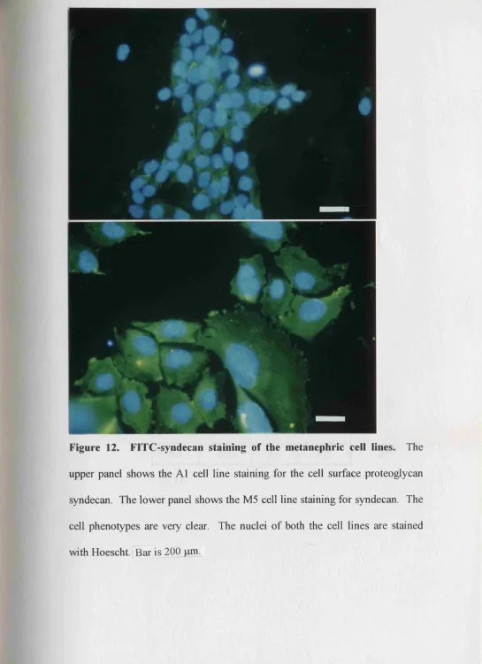
12. Syndecan staining of the metanephric cell lines 179
13. Western blotting for HGF/SF 180
14. Effects of growth factors on proliferation of renal mesenchyme
cell lines 182
15. Blockade of HGF/SF in metanephric organ culture 184
16. Tissue effects of anti-HGF/SF antibody 186
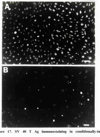
17. SV40 T Ag immunostaining in conditionally-immortal
mesangial cells 194
18. Characterisation of mesangial cell lines 195
19. Mesangial lines are conditionally-immortal 197
20. Expression of HGF/SF and MET mRNA by mesangial cells 199
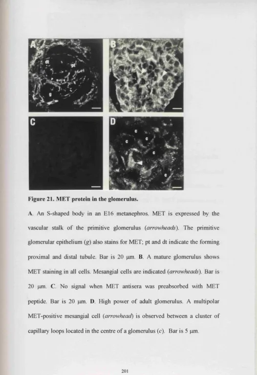
21. MET protein in the glomerulus 201
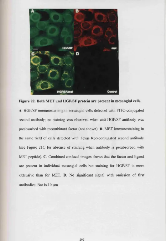
23. Mesangial MET phosphorylation by recombinant HGF/SF 203
24. Exogenous HGF/SF induces mesangial shape changes 205
25. Effects of HGF/SF on mesangial F-actin 207
26. Ultrastructural effects of HGF/SF 209
27. HGF/SF reduces mesangial fîbronectin 211
28. Anti-HGF/SF antibody and Suramin alters the shape of
mesangial cells 213
29. RT-PCR of HGF/SF and MET in a 10 week human embryo 221
30. MET immunohistochemistry in a 9 week gestation human
metanephros 224
31. Western blots for HGF/SF and MET in 10 week human
fetal liver 226
32. HGF/SF and MET protein expression by human trophoblast 228
33. MET immunohistochemistry in human fetal lung, intestine
and spinal nerve root 230
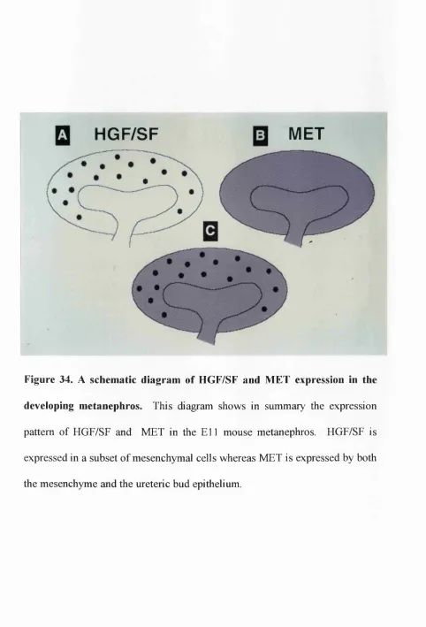
34. A schematic diagram of HGF/SF and MET expression in
the developing metanephros 234
35. Schematic diagram representing a mesangial cell in a normal
List of Tables
1. Events in human and mouse kidney development 28
2. Transgenic mice generated with kidney malformations 49
3. Effects of anti-HGF/SF antibodies on apoptosis in E l 1
metanephros cultured for 48 hours 188
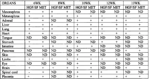
4. HGF/SF and MET mRNA expression as assessed by
Chapter I. Introduction
1.1 Anatomy of Nephrogenesis.
The mammalian kidneys are essential for life of the post-natal organism. They
are responsible for filtering nitrogen-containing waste out of the blood stream,
they regulate the amount of water passed out of the body and maintain the
correct balance of electrolytes in the body. For example, if potassium ions and
protons are allowed to accumulate they are harmful to the physiology and
metabolism of the cell. In addition, sodium determines the extracellular fluid
volume because it is the main cation within this compartment and the
excretion of this ion is also determined by the kidney. For adult kidneys to be
able to function correctly they must have first undergone a complex program
of development called nephrogenesis.
The mature mammalian kidney is derived from 2 major embryonic cell types.
These are the nephrogenic mesenchyme and ureteric bud epithelium, both of
which are found at the inception of the metanephros. The former forms the
nephrons while the bud cells form the collecting ducts. One molecule which is
implicated in both morphogenetic events is hepatocyte growth factor/scatter
factor (HGF/SF). This molecule is the subject of this Thesis.
There are three sets of kidneys in the mammalian embryo, the pro-, meso-, and
Human and mouse kidney development involves similar morphogenetic events
but the timings differ. It is in the mouse in which the process of nephrogenesis
has been most studied.
1.2 The Pronephros.
The first signs of a urinary excretory system are at day 22 after fertilization in
humans and embryonic day 9 in mice. The pronephric duct develops from the
intermediate mesoderm which lies lateral to the notocord (Gilbert, 1991). As
this duct elongates caudally it is referred to as the mesonephric, or Wolffian
duct. The mechanism involved in duct elongation in amniotes has not as yet
been reported although much work on this subject has been done in other
animals; such as axolotl, Xenopus, and chick. Apparently, cues for
pronephric duct migration come from the overlying epidermis in the axolotl
(Drawbridge et ah, 1995). The duct elongates by both active migration and
recruitment of cells from the intermediate mesoderm in Xenopus (Cornish et
ah, 1993). Polysialic acid, a component of neural cell adhesion molecule is
required for chick duct elongation ( Bellairs et ah, 1995).
The anterior of the pronephric duct induces the adjacent intermediate
mesoderm to form simple tubules which fuse with the duct (Shostak, 1991;
Kaufinan 1992). This process of mesenchymal-epithelial conversion is
reiterated in the meso- and metanephros. From experiments carried out on
exclusive to the pronephric duct, because neural tissue could induce the
intermediate mesoderm to differentiate into nephron tubules (Gruenwald,
1943). This suggests that both the duct and the neural tissue could share an
inducing factor but this has yet to be identified. In humans and mice
pronephric tubules are transient structures which probably have no excretory
fimction.
1.3 The Mesonephros.
By day 25 after fertilization in humans (Gilbert, 1991), the pronephros
degenerates in an anterior-posterior direction but the mesonephric duct induces
mesonephric tubules to form in the adjacent mesoderm. The ends of the
tubules open into the mesonephric duct, as it proceeds down to the cloaca,
which will eventually form the bladder. In humans there are about 40
mesonephric tubules at day 25 but they regress in a cranio-caudal fashion
leaving only about 20 pairs by day 40 (Kaufinan, 1992, Larsen 1993).
The intermediate end of each mesonephric tubule becomes invaginated in a
cup-shaped epithelial membrane which is comparable to the Bowman capsule
in the metanephros. This structure encases a knot of capillaries and is known
as the glomerulus. These structures are probably ftmctional (Moore, 1988). In
contrast to humans, well differentiated glomeruli are absent in murine
mesonephroi. It is possible that these murine mesonephric tubules could
Recent evidence suggests that mesonephroi could be involved in the paracrine
stimulation of other organs such as the forelimb; factors involved are IGF I
and FGF8 (Geduspan et a l, 1992; 1993; Crossley et a l, 1996). The
mesonephric region has also been implicated in haemopoiesis (Medvinsky et
al, 1993). In humans and mice the mesonephros involutes in mid-late
gestation but in males the duct is retained as the vas deferens (Moore, 1988).
1.4 The Metanephros.
The development of the metanephros can be divided into two parts; i)
development of the ureteric bud, and ii) differentiation of the nephrons.
Grobsteins’ metanephric organ culture work (Grobstein, 1953b, 1955, 1956),
discussed later in the Thesis, provides the generally accepted view that the
ureter, renal pelvis, calyces and collecting tubules differentiate from the
ureteric bud, but the nephrons differentiate from the metanephric mesenchyme.
However, from a recent study by Qiao et al, (1995) it appears that retrovirally-
tagged mesenchyme or bud can contribute to a minor extent to the collecting
system and nephrons respectively. This suggests that renal cell lineages may
be more plastic than previously considered.
The development of the permanent kidney (metanephros) begins at day 28
after fertilization in the human. It begins with the branching of the ureteric
bud from the mesonephric duct. By day 32 after fertilization in humans, the
mesenchyme, the intermediate mesoderm which has condensed around the
bud. In humans the first glomeruli have formed by 8-9 weeks after gestation
and nephrogenesis continues until the 34th week.
In mice, the ureteric bud enters the metanephric mesenchyme by embryonic
day 11.5 (El 1.5) and initiates metanephrogenesis (Fig. 1). The first glomeruli
have formed by E l4 and nephrogenesis continues for two weeks post-natally.
Table 1 summarizes the timings at which nephrogenic events take place
between humans and mice and figure 1 shows the major events in early mouse
E l l
F 16.1
B
E
E11
G
E13
E1 2
Figure 1. Early development of the metanephros. Stereomicroscopic images
of the mouse metanephros (A-D) and cross-sectional diagrams of the
metanephros (E-H) on embryonic day 11 (A and E), embryonic day 12 (B and
F), embryonic day 13 (C and G), and embryonic day 14 (Dand H). Note that
the most primitive structures are located in the periphery of the embryonic day
14 metanephric rudiment. Arrowheads in B indicate the first branch tips of the
ureteric bud. u, ureteric bud; m, mesenchyme; me, mesenchymal condensate; c
and s, comma and S-shaped bodies, respectively; g, glomerulus; and w,
Wolffian duct. Bar, 50pm in A-F. E-H are not drawn to scale. This figure is
Table 1: Nephrogenic events in human and mouse development.
Times when first
detected
Appearance of Mouse Human
structures
Length of gestation 20 days 40 weeks
Pronephros 9 days 22 days
Mesonephros 10 days 24 days
Metanephros 11.5 days 32 days
Renal pelvis 12.5 days 33 days
Collecting tubules 13 days 44 days
and nephrons
Glomeruh 14 days 8 weeks
The initial ureteric bud ampulla will give rise to the renal pelvis and collecting
ducts, the stalk of the ureteric bud becomes the ureter. This process of
branching morphogenesis results in a complex tree-like structure. As the
mesenchyme induces the ureteric bud to form these structures, the ureteric bud
induces the mesenchyme to form the nephrons. At each branch tip, loose
mesenchymal cells form condensates which form an epithelium with a lumen,
the vesicle. This elongates to form a comma shape which folds back on itself
to become an S-shaped body. This epithelial tubule continues to elongate and
differentiate to give rise to the proximal convoluted tubule, the descending and
ascending limbs of the loop of Henle, and the distal convoluted tubule. The
distal end fuses with the collecting ducts to form a continuous functional unit.
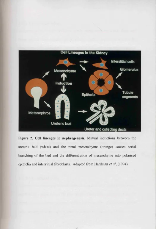
Figure 2 represents a simple diagram of the structures formed once the ureteric
Cell Lineages In the Kidney
Interstitial cells
Mesenchyme
c
♦
^ induction
I *
Metanephros
Epithella
Ureteric bud
Ureter and collecting ducts
Figure 2. Cell lineages in nephrogenesis. Mutual inductions between the
ureteric bud (white) and the renal mesenchyme (orange) causes serial
branching of the bud and the differentiation of mesenchyme into polarised
1.4.1 The glomerulus.
The proximal end of the S-shaped tubule invaginates into a cup shape and
forms the Bowman capsule, which encases a tuft of capillaries to form the
glomerulus. The glomerulus consists of four main cell types when matured:
the visceral epithelia (podocytes), parietal epithelia (Bowman’s capsule),
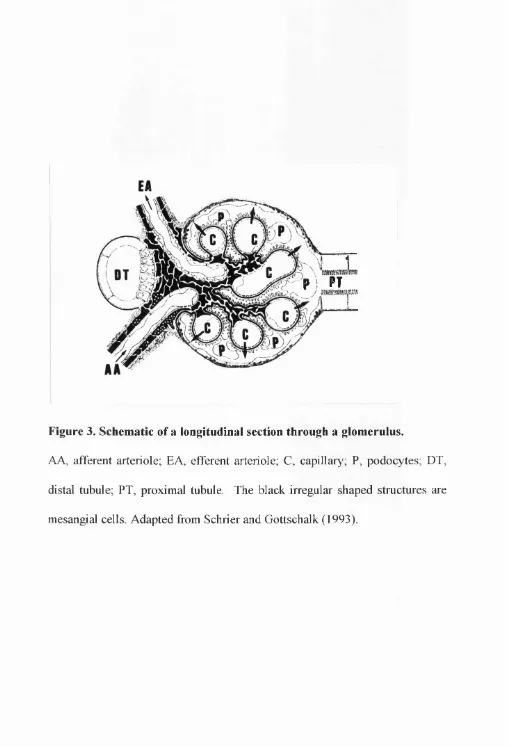
mesangial cells and endothelium (Fig. 3). The renal capillaries are thought to
arise from direct ingrowths of vessel branches originally derived from the
dorsal aorta (Larsen, 1993).
The glomerular basement membrane envelopes the capillaries itI_order to
separate the blood from the glomerular filtrate. The podocyte foot processes
support the basement membrane and capillaries. Between the foot processes
are slit pores which facilitate the filtration process. The glomerulus is
responsible for the production of an ultrafiltrate of plasma. Ultrafiltration
occurs due to the hydrostatic pressure in the capillaries. Molecules of a
relative molecular mass less than 6 8 , 0 0 0 pass through the glomerular basement
membrane to form the glomerular filtrate (Hebert and Kriz, 1993).
1.4.2 Mesangial cells
Fibromuscular mesangial cells are embedded in their own extracellular matrix
and form the central core of the glomerulus. These cells can change their cell
morphology in response to growth factors and hormones. Like smooth muscle
1992), and exhibit contractile properties, (Kreisberg et a l, 1985). Their role is
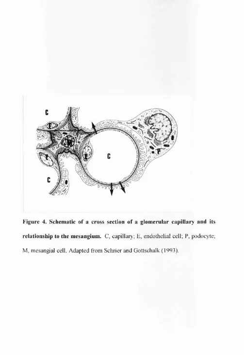
to physically support glomerular capillary loops (Fig. 4) and modulate plasma
ultrafiltration (Kriz et ah, 1990; Arendshorst and Navar, 1993). Another role
for mesangial cells is suggested by their ability to degrade and clear immune
complexes from the glomerulus. Finally mesangial cell proliferation and
matrix production have been implicated in a number of glomerular diseases
P
PT
;
Figure 3. Schematic of a longitudinal section through a glomerulus.
AA, afferent arteriole; EA, efferent arteriole; C, capillary; P, podocytes; DT,
distal tubule; FT, proximal tubule. The black irregular shaped structures are
Figure 4. Schematic of a cross section of a glomerular capillary and its
relationship to the mesangium. C, capillary; E, endothelial cell; P, podocyte;
1.4.3 Proximal tubules.
The proximal tubule continues directly after the glomerulus and constitutes the
middle part of the embryonic S-shaped body. It consists of an initial
convoluted portion, the pars convoluta, (a continuation of the parietal
epithelium of Bowman capsule), and a straight portion, the pars recta. The
epithelial cells have well developed brush-borders of microvilli which
increases the surface area for reabsorption. The main role of the proximal
convoluted tubule is to reabsorb ions (Na^, HCO3 , Cl’, K^, Ca^, P0 4 ^’) water
and organic solutes such as glucose and amino acids. Approximately 2/3 of
the ultrafiltrate is reabsorbed in the proximal tubule.
1.4.4 The loop of Henle.
The loop of Henle is composed of the straight portion of the proximal tubule
(pars recta), the thin limb segments, and the straight portion of the distal tubule
(thick ascending limb). The limbs of the loop of Henle generate an osmolar
gradient which permits the reabsorption of water by the collecting ducts. The
maturation of the loops occur post-natally.
Collecting ducts contain three ftmctionally distinct cell types. Principle cells
reabsorb Na^ and secrete by means of basolateral Na/K-ATPase, amiloride
sensitive Na^ channels and Ba^^-sensitive channels in the apical membrane.
secrete HCO 3. These cell types demonstrate plasticity of their phenotypes in
vitro (Fejes-Toth et a l, 1992) and therefore might have a common progenitor
in the ureteric bud branches.
1.5
Fetal function of the mammalian kidney.
The placenta eliminates the metabolic waste from the fetal blood. The role of
the fetal kidney is to regulate the production of the amniotic fluid. Too small a
volume of amniotic fluid results in a condition called oligohydramnios which
causes lung hypoplasia and limb distortion as well as dry, wrinkly skin.
2. Basic Mechanislms of Nephrogenesis.
A balance of key cellular events are crucial to the formation of a kidney.
These are: cell proliferation, programmed cell death (apoptosis), induction,
motility, differentiation and morphogenesis. If the balance is disturbed, so is
the normal development of the kidney. Such aberrations form the basis of
disease such as kidney malformations, cystic diseases and tumours (Bard and
Woolf, 1992).
2.1 Cell Proliferation.
Stem cells are cells which replicate themselves and which generate another
type of daughter cell which differentiates. They are located in the
undifferentiated renal mesenchyme (Burrow and Wilson, 1993). Stem cells
and their immediate progeny are called precursors. These cells secrete a
number of growth factors which could act in a paracrine or autocrine fashion
to stimulate their growth as discussed later. This Thesis shows that one such
factor is HGF/SF. Although stem cells probably do not exist in the normal
mature kidney, it is possible for dedifferentiation and regeneration to occur in
tubular epithelia (Kawaida et ah, 1994). Thus, the study of nephrogenesis is
2.2
Cell Death.
Approximately 50% of embryonic kidney cells die during nephrogenesis
(Coles et al, 1993). This programmed cell death occurs by apoptosis (Koseki
et a l, 1992; Coles et a l, 1993). Apoptosis is characterized by nuclear
condensation and DNA fragmentation into 180-200 base pairs by a calcium-
dependeut endonuclease. Such cells are phagocytosed by neighbouring
nephrogenic cells or macrophages (Camp and Martin, 1996). Genetic
experiments involving the generation of BCL 2 null mutant mice, suggest that
apoptosis plays an important role in determining the final number of nephrons
(Veis et al, 1993; Sorenson et a l, 1995). It has been suggested that clonal
cells derived from the metanephrogenic mesenchyme expressing BCL 2 are
destined to undergo differentiation, whereas those cells which do not express
BCL 2 die by apoptosis (Winyard et a/.,1996a,b). Work presented in this
Thesis suggests that HGF/SF maybe a factor which enhances cell survival.
2.3 Induction.
When the differentiation fate of a group of embryonic cells is altered an
inductive event is said to have occurred. Induction plays a major part in
organogenesis, for instance when renal mesenchyme is induced to convert to
nephrons. Two types of induction exists: i) permissive, and ii) instructive
(Bard, 1990a). The induced metanephric mesenchyme already contains the
ureteric bud is needed to start the process of nephrogenesis (Shostak, 1991).
This inductive process is called permissive. In contrast when a lung bud is
cultured with stomach mesoderm, gastric epithelial glands develop, while
hepatic cords develop when lung buds are cultured with liver mesoderm
(Shostak, 1991). Thus the lung buds can not only form bronchial epithelia but
they can be induced to form diverse epithelia by the action of heterologous
mesenchymes. This type of induction is called instructive.
2.4 Morphogenesis.
Morphogenesis is the acquisition of shape. In order for morphogenesis to
occur the cells must be able to communicate with each other and their
surrounding environment, be able to move and change their cell shape. There
are intrinsic and extrinsic factors which influence morphogenesis, a) cell-cell
contact, b) cell-matrix contact, c) cell cytoskeleton, d) traction, and e)
diffusion of soluble factors (Bard, 1990a). These processes are important in
epithelial-mesenchymal interactions, especially in i) branching morphogenesis
which occurs in the developing kidneys, salivary glands, pancreas, teeth,
mammary glands, liver and lungs and ii) the formation of nephrons from
undifferentiated mesenchyme (Gilbert, 1991; Hardman et al., 1994)
2.5 Migration.
In nephrogenesis this process is important during caudal migration of the
a/., 1995; Bellairs et al., 1995). Within the metanephros itself, migration does
not appear to play a central role in development.
2.6 Differentiation.
When cells within the developing kidney acquire a specialised phenotype they
are said to have undergone differentiation, a classic example is the
mesenchyme to epithelial transition in which renal mesenchyme differentiates
into nephrons. It is thought that the metanephric mesenchyme contains
multipotent stem cells that can give rise to epithelia of the glomerulus,
3. Studying Nephrogenesis
in vitro
and
in vivo.
3.1 Descriptive Studies.
There are two basic ways with which to studying nephrogenesis, the first is by
a descriptive approach, and the second is by a functional approach.
Descriptive studies document the anatomy of the kidney and correlate
morphology with the sequential expression of genes in terms of mRNA and
protein. These results can suggest that specific patterns of genes are
associated with the events in nephrogenesis, but functional studies are required
to prove that a particular molecule has a developmental role. The results of
experiments of these kinds has prompted the establishment of a kidney
development database (Internet address
httm://www. ana. ed. ac.uk/anatomy/kidbase). This allows descriptive
information on kidney development to date to be obtained easily. At the time
of writing, over 2 0 0 genes are known to be expressed during nephrogenesis
and there are many potential functional overlaps which could lead to
redundant pathways.
3.2 Functional Studies.
3.2.1 Whole Organ Culture.
The metanephros has the capacity to develop in vitro. Organ culture has been
Intact mouse metanephric rudiments are isolated by dissection, placed on
membranes and can be cultured for up to 7 days during which time nephrons,
avascular glomeruli and tubules are formed (Grobstein.,1955; Avner et ah,
1985; Saxene^a/., 1987; Hardman., 1990).
By using this technique it is possible to culture kidneys in the presence of
growth factors, blocking antibodies to growth factors or cell adhesion
molecules and antisense oligonucleotides to perturb expression transcription
factors and examine their effects on nephrogenesis. These types of studies
suggest that 5 classes of molecules are important for nephrogenesis: i) growth
factors, ii) transcription factors, iii) survival factors iv) cell-cell adhesion
molecules, and v) cell-matrix adhesion molecules.
Cell lineage in organ culture has been examined using the retroviral mediated
gene transfer technique, which allows cell fates of the metanephrogenic
mesenchyme and ureteric bud to be followed in vitro (Herzlinger et ah, 1992;
Qiao et ah, 1995).
3.2.2 Induction of renal mesenchyme.
The isolated ureteric bud epithelium and metanephric mesenchyme can be
studied in isolation in order to try and identify the underlying mechanisms of
the induction process. In the 1950’s, Clifford Grobstein performed
from the ureteric bud and recombined them on a glass-clot interface with
another source of inductive stimulus on the other side of the filter. He
concluded that: a) the mesenchyme and ureteric bud cannot survive in
isolation, b) the mesenchyme and epithelial ureteric bud need to interact with
each other to undergo growth, differentiation and morphogenesis, c) tubule
production can be induced in metanephrogenic mesenchyme by salivary
mesenchyme and by dorsal embryonic spinal cord (Grobstein, 1955 and 1956).
At the time Grobstein was performing the reconstitution experiments, the
inducing factor in the spinal cord was unknown. It seems that the signal works
over a short range through secreted growth factors, cell-cell, or cell-matrix
interactions (Lehtonen, 1975). The work carried out in this Thesis suggests
that HGF/SF is also involved in nephron formation from renal mesenchyme.
WNT genes are candidates for an induction molecule. The WNT gene family
has been shown to be involved in developmental regulation. They encode
secretory glycoproteins which binds matrix and acts via autocrine and
paracrine routes (Nusse and Varmus, 1992). WNT 1 mRNA is expressed in
the embryonic spinal cord at the time the spinal cord is known to induce
nephron formation. WNT 1- transfected fibroblasts can induce nephrogenesis
in isolated mesenchyme (Herzlinger et al, 1994). Although WNT 1 is
expressed by the ureteric bud, other members of the WNT gene family are also
WNT 4 have developmentally retarded kidneys (Stark et al 1994), WNT 4 is
expressed during the mesenchymal-epithelial conversion and is not likely to be
an inductive factor secreted by the ureteric bud in vivo.
Grobstein performed transfilter experiments with isolated metanephric
mesenchyme on one side of the filter and spinal cord on the other and found
that nephrons formed (Grobstein, 1953). Sariola and co-workers found that
neurons had penetrated the filter pores and made contact with the metanephric
mesenchyme on the other side. Upon destruction of the neural tissue within
the spinal cord, induction was abolished (Sariola et ah, 1989). When
antibodies against ganglioside D3, (a molecule common to neural tissue) were
added to metanephroi in organ culture, ureteric bud branching was inhibited
(Sariola et ah, 1988b). Since neurons are associated with the ureteric bud and
neural tissue is a potent inducer of nephrogenesis in vitro, this supports the
theory that neurons are involved in induction in vivo.
Perantoni et al, (1991a) reported that pituitary extract, epidermal growth factor
(EGF) and type IV collagen promote tubulogenesis in isolated rat
metanephrogenic mesenchyme. Recently Perantoni and collègues report that
basic fibroblast growth factor (FGF2) can promote uninduced
metanephrogenic mesenchyme to form condensates, the first morphogenetic
step in the mesenchymal-epithelial conversion (Perantoni et a/., 1995). Finally,
kidney tubule differentiation in isolated renal mesenchyme (Davies and
Garrod, 1995). The physiological significance of this observation is not
known.
The ureteric bud has proved even more challenging to culture in isolation
although Perantoni and coworkers found that isolated rat ureteric buds branch
when suspended in collagen type I gels (Perantoni et a l, 1991b). However,
branching is very slow compared to in vivo.
3.2.3 Nephrogenic Cell Lines.
Since the kidney consists of several cell lineages, the generation of
nephrogenic cell lines allows the study of purified populations of cells(Burrow
et a l, 1993; Karp et a l, 1994; Woolf et a l, 1995). Each cell type exhibits a
specific phenotype in vitro which is characterised by their morphology,
membrane constituents, intracellular proteins, synthesis and release of specific
products such as growth factors, and extracellular matrix proteins. Cell
phenotypes can be influenced by culture conditions; so it is essential to define
the culture conditions which will allow the cell to behave as normal, and have
the same phenotype in vitro as in vivo.
Cultunng cell lines is useful in testing effects of selected substances on cell
proliferation, adhesion, migration, differentiation, and contraction.
through receptors. Culture conditions can be changed to mimic diseased states
and so the molecular and cellular processes which are involved can be
analysed. I have used cell lines from renal mesenchyme to study the
expression of HGF/SF (Chapter IV).
3.2.4 Transgenic Studies.
Transgenic animals provide a means by which to study the consequences of
overexpression of specific genes, and the consequences of gene ablation in
vivo. Transgenes can be introduced into the host cells by i) microinjecting
recombinant DNA into the pronuclei of fertilized eggs, ii) retrovirally infecting
preimplantation embryos, iii) reconstitution of early embryos with genetically
engineered embryonal stem (ES) cells (Schuchardt et ah, 1994; Bladt et ah,
1995; Schmidt et ah, 1995; Uehara et ah, 1995; Dudley et ah, 1995). ES cells
are isolated from the inner cell mass of explanted embryos and they can then
be grown and genetically manipulated in tissue culture.
Genes can be targeted by homologous recombination which means that the
gene of interest is replaced by a homologous but non-fimctional fragment of
DNA. Other genes can be inserted on either side of the homologous fragment
such as a neomycin resistance gene and a herpes simplex virus thymidine
kinase gene which can help in positive and negative selection. Engineered ES
cells are injected back into the blastocyst where they can differentiate into a
can then be generated by breeding of the founder generation of mice (Merlino,
1991).
3.2.5 Overexpression.
Overexpression in vivo of molecules involved in nephrogenesis can enhance
our understanding of the roles these molecules play in normal development
and disease. One such molecule is the transcription factor PAX 2; in normal
development it is expressed in the induced mesenchyme, in the ureter
epithelium, and in early epithelial structures derived from the mesenchyme.
Transgenic mice overexpressing PAX 2 during embryogenesis have tubular
microcysts, glomerular defects such as loss of podocyte foot processes and
they present at birth with proteinuria. Similar events occur in congenital
nephrotic syndrome (Dressier et ah, 1993).
3.2.6 Gene ablation.
Null-mutant mice generated show inappropriate kidney development as shown
in Table 2; these animals also exhibit impaired development in other organs
due to the same gene(s) being expressed during their development (Schuchardt
et a/., 1994; Chan et ah, 1995; Maas et ah, 1994; Morham et ah, 1995; Veis et
ah, 1993; Dudley et ah, 1995; Luo et ah, 1995; Shawlot et ah, 1995; Torres et
ah, 1995; Keller et ah, 1994; Leveen et ah, 1994; Soriano et ah, 1994;
ah, 1994; Noakes et a l, 1995; Stein et ah, 1994; Davis et ah, 1995;
Table
2 1 Homozygous null mutant mice generated which possess developmental kidney malformations.GENE
PHENOTYPE
C-RET Renal agenesis or dysplasia and defects in the enteric nervous system (Schuchardt et ah, 1994).
LD Renal agenesis and limb defects (Chan et ah, 1995; Maas et ah, 1994).
C 0X 2 Renal hypoplasia as well as susceptibility to peritonitis (Morham et ah, 1995).
BCL2 Renal hypoplasia, polycystic kidneys, splenic involution and hypopigmented hair (Veis et ah,
1993).
BMP? Renal dysplasia as well as eye and skeletal patterning defects
(Dudley et ah, 1995; Luo et a/., 1995).
LIM l Renal agenesis, absent gonads and head structures (Shawlot et ah, 1995).
PAX2 Renal agenesis and eye defects. (Heterozygotes have renal hypolasia) (Torres et ah, 1995; Keller et ah, 1994).
PDGFR p Absence of mesangial cells and widespread cardiovascular defects (Soriano., 1994).
EGFR Cystic dilation of the collecting ducts. Defects in hair follicles, skin, tongue, gastrointestinal tract and brain (Threadgill et a/. 1995).
W Tl
WNT4
MPV 17
laniimri/laminin p2
HOXA-ll/HOXD-11**
Renal agenesis as well as abnormalities in the respiratory system, the heart and the gonads (Kreidberge^ a/., 1991).
Renal dysplasia (Stark et al, 1994).
Glomerulosclerosis and nephrotic syndrome at birth (Zwacka et al, 1994).
Nephrotic syndrome soon after birth (Noakes et a/., 1995).
Renal dysplasia or hypolasia as well as absence of radius and ulna (Davis a/., 1995).
RAR ay /ap 2 Renal agenesis, or hypoplasia. Abnormalities in the respiratory tract, heart, thymus and associated glands, diaphragm, genito-urinary system and the lower digestive tract were also present
(Mendelsohn et a l, 1994)
4. Growth factors and kidney development.
A number of polypeptide growth factors and their cell surface receptors have
been implicated in nephrogenesis. Growth factors produced by the
metanephros can act in either paracrine, autocrine, or juxtacrine fashions.
When a factor is secreted by one cell and it acts on its neighbour, this is
classed as being a paracrine factor. When a cell secretes a factor which in turn
acts on itself, it is an autocrine factor. This Thesis produces evidence that
renal mesenchyme expresses HGF/SF which acts in both these ways. When a
cell expresses a growth factor which is inserted into its own plasma membrane
and also interacts with receptors on adjacent cells, it is a juxtacrine factor; an
example of this is TGF-a (Massague, 1990). Although growth factors are
important in nephrogenesis they are also implicated in adult kidney disease
and tubular regeneration after toxic or ischemic injury (Hammerman, 1989;
Nagaike et aL, 1991; Miyazawa et ah, 1994).
Many of the nephrogenic growth factors act by binding to membrane-
associated receptor tyrosine kinases which dimeiize, autophosphorylate and
transduce growth signals into the cell. Some of these growth factors are
multiftmctional because upon binding their receptors they can trigger various
signalling cascades within the cell which will influence nuclear events and in
turn the behaviour of the cell (Cross and Dexter, 1991). Signal transduction is
cytoplasmic effectors containing Src homology domains (SH2) (Pawson and
Schlessinger, 1993).
4.1 Growth factors which enhance nephrogenesis.
4.1.1 Epidermal growth factor and Transforming growth
factor
(X.EGF and its embryonic homologue IG F a are thought to bind the same
receptor (EGFR). Rogers and coworkers, showed that E l 3 rat metanephroi
produced TGFa and nephrogenesis could be perturbed by using blocking
antibodies against TGFa (Rogers et al, 1992). EGF is a potent inhibitor of
cell death within the developing kidney, both in vitro and in vivo (Coles et al,
1993). The EGFR knockout mice have different renal phenotypes which are
dependant on the specific genetic background of the animal (Threadgill et al,
1995). It is clear from these mice that the EGF receptor is not required for the
induction of the metanephric mesenchyme, but one strain develops cystic
collecting ducts.
4.1.2 Fibroblast growth factors (FGFs).
FGF2 transcripts are expressed by the ureteric bud (Orr-Urtreger et al, 1993).
FGF receptors have been reported to be expressed in the developing kidney;
two of which are FGFR 1, (or fig), and FGFR 2, (whose gene encodes two
splice variants and are known as keratinocyte growth factor receptor (KGFR)
in the epithelia of the collecting ducts and FGFR 1 was found to be expressed
in cortical mesenchyme, and Bowman capsule/renal tubule complexes (Peters
et ah, 1992). Perantonis’work has suggested that FGF2 is an inducing
molecule, involved at the inception of metanephric development (Perantoni et
ah, 1995). Null mutant mice for FGFR 3 show abnormalities of the axial and
apendicular skeleton, no other morphological abnormalities were reported
(Denggr a/., 1996).
4.1.3 Insulin like growth factors.
IGF I and IGF II mRNA’s are expressed by embryonic day 13 rat metanephroi.
From metanephric organ cultures carried out by Rogers and colleagues, it was
found that immunoreactive IGF I and IGF II were detected in the culture
medium. Upon exposure to antisera to these factors, nephrogenesis was
impaired (Rogers et ah, 1991). Antisense oligonucleotides to IGF I in
metanephric culture resulted in hypoplasia, rudimentary ureteric bud
branching, and reduced synthesis of proteoglycans (Zheng et ah, 1994).
Knockout mice which lack IGF I and IGF II are small and have
proportionately small kidneys ( DeChiara et ah, 1990; Powell-Braxton et ah,
4.1.4 Platelet derived growth factor (PDGF).
In the early stages of glomerular development the PDGF B chain is expressed
by the renal vesicle and its receptor PDGFRp is expressed by undifferentiated
mesenchyme and interstitial cells. Some of the latter might be mesangial cell
precursors. During mesangial differentiation, cells continue to express the
PDGFRp and also express PDGF B chain (Alpers et a l , 1992). PDGF B and
PDGFRp null- mutant mice lack mesangial cells (Leveen et a l, 1994; Soriano,
1994).
4.1.5 Vascular endothelial derived growth factor (VEGF).
Vascular endothelial growth factor is a disulphide-linked dimeric glycoprotein
which is thought to promote angiogenesis and vasculogenesis during
embryogenesis (Carmeliet et a l, 1996). The receptors for VEGF belong to the
receptor tyrosine kinase family; they are flkl in the mouse and its homologue
in the human is KDR and fltl (Dumont et a l, 1995). VEGF can be detected
throughout nephrogenesis and the undifferentiated metanephric mesenchymal
cells of the E l l mouse express growth factor receptors specific for the
endothelial cell lineage (Loughna et al., 1996). Mice which lack VEGF or its
receptors have widespread defects in embryonic blood vessel formation and
die before the metanephros is formed (Shalaby et a l ., 1995; Carmeliet et a l,
4.1.6 GDNFandRET.
RET is expressed in the tips of the branching ureteric bud but it was not
detected in the metanephric mesenchyme (Pachnis et al, 1993). A targeted
mutation was introduced into the murine RET locus by homologous
recombination. These null mutant mice exhibit severe renal dysplasia and
agenesis. This phenotype suggests that the RET receptor transduces the
inductive signal required for the branching of the ureteric bud. Failure of this
inductive signal to be received by the ureteric bud means that the ureteric bud
does not grow and branch and in turn induce the mesenchyme-epithelium
transition (Schuchardt et al 1994).
Glial-cell line derived neurotrophic factor, (GDNF) is a potent survival factor
and neuronal differentiation factor and was found to be expressed in
uninduced metanephric mesenchyme of mouse embryos (Hellmich et al,
1996). Recently, the ligand for RET was identified as being GDNF (Trupp et
a l, 1996; Durbec et a l, 1996).
4.1.7 ROS.
ROS is the mammalian homolgue of the Drosophila sevenless gene. The
sevenless protein is involved in Drosophila eye development (Easier and
Hafen, 1988). In the developing kidney, ROS is expressed in the tips of the
nephrons form the level of ROS transcript falls, no transcripts were detected in
the mature kidney. From ROS antisense experiments carried out in
metanephric organ culture, Kanwar and coworkers found that normal
nephrogenesis was perturbed as well as there being a reduction in extracellular
matrix glycoproteins (Kanwar et ah, 1995). ROS-knockouts have recently
been reported, males that are homozygous for the ROS mutation are infertile.
No other abnormalities were found to be present in these animals
(Sonnenberg-Riethmacher et ah, 1996). The ligand remains unknown.
4.1.9 Nerve growth factors (NGFs).
A role for these factors in nephrogenesis has been suggested due to nerve
growth factor receptor expression throughout nephrogenesis (Durbeej et
ah, 1993; Sariola et ah, 1991). The high-affinity NGF receptor (TRK B), was
found in cortical mesenchyme, whereas TRK C, another high-affinity receptor
was expressed in the collecting ducts. TRK A is also a high-affinity NGF
receptor The low-affinity NGF receptor (p75), is expressed by uninduced
metanephric mesenchyme and later in glomerular podocytes. Inhibition of the
low-affinity NGF receptor expressed by developing metanephroi by antisense
oligonucleotides in organ culture, perturbed nephrogenesis (Sariola et ah,
1991). However, null mutant mice for p75 NGF receptor have kidneys which
have undergone normal nephrogenesis (Lee et ah, 1992). It is possible that
Finally, neurotrophin 3 was found to be a survival and differentiation factor
for neuronal precursors in the uninduced metanephric mesenchyme but it was
not found to induce nephron formation (Karavanov et al, 1995).
4.2 Growth factors which inhibit nephrogenesis.
4.2.1 Transforming growth factor p (TGF P).
The biological actions of the TGFp family are diverse. They can inhibit or
stimulate proliferation, stimulate extracellular matrix formation and promote
or inhibit cell migration. TGFpl mRNA is expressed by metanephric
mesenchyme and low levels of TGFp2 transcripts are detected in the
epilthelial tubules derived from the metanephric mesenchyme. No TGFp 3
expression was observed (Schmidt et ah, 1991). When TGFpl was added to
E l3 rat metanephroi in organ culture tubule forination was inhibited (Rogers et
ah, 1993). Blocking TGFpl activity by using anti-TGFpl antibodies
accelerated the formation of tubules. Activin, another member of the TGFp
family is also found to be expressed in the developing kidney along with its
receptors. Activin treated metanephroi have retarded branching of the ureteric
bud which are abnormally short (Ritvos et a l, 1995). TGF p i knockout mice
have been generated and are reported to have normal kidneys (Letterio et a l,
4.2.2 Leukaemia inhibition factor.
Leukaemia inhibition factor, (LIF), is a cytokine which inhibits the
differentiation of embryonic stem (ES) cells and also reversibly blocks
nephrogenesis. It seems that LIE blocks nephrogensis at two stages, the first
stage is soon after mesenchymal condensation has taken place, the second
stage is after the development of epithelialised aggregates (Bard and Ross,
1991). It has yet to be shown if LIF is produced by the metanephros.
4.2.3 Tumour necrosis factor.
Tumour necrosis factor a, (TNFa), could play a role in kidney development.
It has been implicated in the processes of programmed cell death and tissue
remodeling in the developing chick. Apoptotic cells were detected in the
developing chick mesonephros as well as other developing organ systems
(Wride et a l 1994). TNFa has been detected in the early metanephros and
inhibits nephron formation. From organ culture experiments, it was found that
j exogenous TNFa inhibits metanephric growth and differentiation (personal
j
communication. Dr’s C.Cale and A S Woolf, ICH, London).
5. Survival factors.
Uninduced metanephric mesenchyme is programmed to undergo cell death
unless it is rescued by the induction process, which allows it to convert to
degradation in vitro and is thought to be a suppressor of apoptosis in
metanephric mesenchyme. Coles et al, found that EGF is a potent inhibitor of
cell death in the developing kidney in vivo (Coles et ah, 1993).
The proto-oncogene BCL 2 inhibits cell death. The BCL 2 protein is located
on the inner mitochondrial membrane and plays a part in the antioxidant
pathway which prevents cell damage after peroxides and superoxides have
been generated (Veis et ah, 1993). During nephrogenesis BCL 2 is highly
expressed in mesenchymal aggregates, implicating the molecule in the survival
of nephron precursors (Winyard et ah, 1996b).
Gene targeting by homologous recombination resulted in homozygous mice
with polycystic kidneys, showed fulminant lymphoid apoptosis, and
hypopigmentation of hair (Veis et ah, 1993). Metanephroi from E l2
heterozygous and homozygous null mutant BCL 2 mice were placed in organ
culture for three days. Metanephroi from the homozygous mice were much
smaller and increased apoptosis was detected in the metanephric mesenchyme
6. Transcription factors.
These molecules are also implicated in nephrogenesis but a detailed discussion
is beyond the scope of this Thesis.
A member of the HOX family, LIMl, encodes for a protein that contains a
DNA binding homeodomain, and is expressed in the intermediate mesoderm of
mice at E8.5. LIMl null mutant mice lack kidneys as well as gonads and head
structures (Shawlot et al,, 1995). Double mutant mice for HOXA 11 and
HOXD 11 have renal hypolasia or dysplasia and absent radius and ulna, single
mutants have normal kidneys (Davis et a l, 1995).
The PAX 2 gene is expressed in the ureteric bud and its derivatives, in
metanephric mesenchyme after induction and is expressed in early epithelial
derivatives of the mesenchyme, such as comma and S-shaped bodies, and is
down-regulated during maturation of renal epithelium (Dressier et a l, 1990;
Ryan et a l, 1995). It has been shown by antisense oligonucleotides that PAX
2 is required for renal mesenchyme-epithelium conversion (Rothenpieler et
a l, 1993). Homozygote PAX 2 null mutant mice have renal agenesis while
heterozygous mice display renal hypoplasia (Torres et a l, 1995; Keller et a l,
1994).
Another important transcription factor is encoded by the Wilm’s tumour gene
intercalate with DNA.. During development WTl is expressed in renal
mesenchyme, the renal vesicle and the glomerular podocytes (Pritchard-Jones
et ah, 1990). Null mutant mice for WTl have renal agenesis (Kreidberg et
a l, 1993).
In humans, spontaneous WTl mutations abolish DNA binding and are
associated with kidney tumour and nephrotic syndrome (Coppes et ah, 1993;
Pelletier et ah, 1991). It has been suggested that WTl regulates metanephric
mesenchyme proliferation by down-regulating the production of insulin-like
growth factor type II (IGF-II), and platelet derived growth factor A-chain
(PDGF-A) (Drummond et ah, 1992; Wang et ah, 1992).
C-MYC is expressed in uninduced mesenchyme and in very early epithelial
structures. Its expression is downregulated as the kidney matures. C-MYC
could be related to cellular proliferation during nephrogenesis (Mugrauer and
Ekblom, 1991). N-MYC is upregulated during the mesenchymal-epithelial
conversion and its expression is confined to the mesenchymal condensates,
comma and S-shaped bodies. N-MYC is not expressed by ureter derived
structures or endothelium. Its expression is a marker of induction and
formation of the early epithelial structures of nephrogenesis (Mugrauer and
Ekblom, 1991). Homozygous null-mutant mice for N-MYC have mesonephroi
which are hypoplastic with increased apoptotic cells. The embryos die