CLASSIFICATION OF MEDICAL DATA BASED ON
SPARSE REPRESENTATION USING DICTIONARY
LEARNING
A THESIS
submitted by
M. SRINIVAS
for the award of the degree
of
DOCTOR OF PHILOSOPHY
DEPARTMENT OF COMPUTER SCIENCE AND ENGINEERING INDIAN INSTITUTE OF TECHNOLOGY HYDERABAD
To my
ACKNOWLEDGMENTS
I would like to express my sincere gratitude to Dr. C. Krishna Mohan for providing me with the opportunity to do my research work under his guidance. His emphasis on steady and committed effort has motivated me during the course of the research work. I have immensely benefited from the excellent research environment that he has created and nurtured.
I extremely grateful to our director, Prof. U.B. Desai, for providing excellent com-puting facilities and such a nice atmosphere for doing my research work. I am extremely thankful to my doctoral committee members Dr. Bheemarjuna Reddy Tamma, Dr. Sri Rama Murty, Dr. C. S. Sastry and my doctoral committee chairman Dr. M. V. Pandurangarao. My special thanks to Dr. C. S. Sastry for his guidance and encour-agement.
I convey my heartfelt thanks to Software Systems Research Lab (SSRL) team for their help in completing the project work successfully. I thank Smt. M. Lakshmi and Shri. Praveen Vemuri, for their affection, encouragement and support during my PhD. My special thanks to Debaditya Roy, N. Pattabhi Ramaiah, A. Ravi Shankar, V. Nageswarao, Ramu Naidu and my classmates for their support, motivation and help at critical moments during my project work.
I would like to take this opportunity to thank my beloved parents Shri. M. Odelu and Smt. M. Laxmi for making me what I am today.
I would like to thank my brother Dr. M. Ravinder, my sisters Krishna Veni, Rama Devi and Srilatha, my brothers-in-law Sadanandam, Bhaskar and Kumara Swamy, my nephews Vamshi Krishna, Goutham and Babu and my niece’s Mounica, Srinidhi and Sathvika for their affection and support.
I thank all my batch mates A. Gopi, J. Krishna, K. Ravi Kumar, Satya Narayana, Aravind Kumar, Vandana, Sweetha, Phani Shankar, Chandan, Rafi, Ragavendra,
Naresh, Pravanjan, Harsha, Kiran, Suresh, Laxman, Nagababu and Sandeep for mak-ing my stay at IIT enjoyable. I am grateful to all the faculty and non-teachmak-ing staff with in the department as well as outside the department for all the help I have received during my stay at IIT Hyderabad.
I wish to thank Dr. K.P Supreethi, Anitha, Pramod, Santosh Kumar, Thejaswini, K. Santosh Kumar, Radhika, Jenni, Shiva, Ganesh, Nagendar, Venkat Reddy, Keerthi Deep, Joseph, Durga, Sreenu, Prasanth, Anil, Shiva Sairam, Sandeep R.B, Mukesh, Satya, Rama Raju, Thomas, Goutham Pilla, Krishna Veni, Poorna, Prakash and Subbu for extending help at different times.
Finally, I thank everyone who helped me directly or indirectly during my stay at IIT Hyderabad.
ABSTRACT
Keywords: Content based medical image retrieval; classification; sparse representa-tion; dictionary learning; clustering; modality; multi-level classificarepresenta-tion; support vector machines; on-line dictionary learning; K-SVD; OMP; ℓ1-lasso; multi-scale dictionary learning; adaptive dictionary learning.
Due to the increase in the sources of image acquisition and storage capacity, the search for relevant information in large medical image databases has become more challenging. Classification of medical data into different categories is an important task, and enables efficient cataloging and retrieval with large image collections. The medical image classification systems available today classify medical images based on modality, body part, disease or orientation. Recent work in this direction seek to use the semantics of medical data to achieve better classification. However, representation of semantics is a challenging task and sparse representation has been explored in this thesis for this task.
In this thesis, we explore new methods for grouping of medical data into different classes based on sparse representation and dictionary learning. The sparsity seeking dictionary learning approaches typically exploit the framework of under-determined setting and hence work on some implicit assumptions on the database. The methods proposed here vastly reduce the search time and improve accuracy of retrieved images. In application, however, one often encounters databases which are not so big that the sparsity promoting under-determined framework cannot be efficiently deployed.
An algorithm for classification of medical images based on edge information ex-tracted from various body parts using ℓ1-lasso sparse representation and on-line dic-tionary learning (ODL) is proposed. Edge information is extracted from an image by dividing the image into patches and each patch into concentric circular regions to
pro-vide discriminative information useful for classification of medical images. The ability of on-line dictionary learning to achieve sparse representation of an image is exploited to develop dictionaries for each class using edge-based features.
A single classifier may not be suitable for classification of various kinds of medical image datasets. Most of the medical datasets have the problem of data imbalance i.e. unequally distributed training samples among all the classes, which gives rise to poor classification performance with any of the standard single classifier. We aim to address the problem of data imbalance of medical data using multi-level classification approach. A multi-level classifier combines correctly classified examples in the first level with the training data and supplies them as input to the next level classifier. So, if there is any imbalance in the data, it can be alleviated by this approach. For the first stage of classification, on-line dictionary learning (ODL) is used. Support vector machine (SVM) is used for the second level of classification and together with on-line dictionary learning forms the multi-level classification approach.
Another problem in medical imaging is the classification of medical images cap-tured by acquisition source (i.e modalities). Capturing images using different modal-ities suffers from significant contrast variation among the images of the same organ or body part. Due to this large variation, existing image classification and retrieval algorithms do not perform well for different modality images. We propose to address this issue by using multi-scale wavelet representation and dictionary learning. Wavelet features extracted from an image provide discriminative information useful for classi-fication of medical images. Multi-scale wavelets are employed to compensate for the varying scale of intensity in the images captured by the aforementioned sources. Car-diovascular diseases (CVD) are a leading cause of unnecessary hospital admissions. Hence, automated detection of abnormal heartbeats captured by electronic cardio-gram (ECG) signals is vital. We employ an approach to classify abnormal heartbeat patterns from standard heartbeat patterns using adaptive dictionary learning on a standard ECG database.
rep-resentation and dictionary learning. The basic idea is to group similar images into clusters that are sparsely represented by the dictionaries and simultaneously learn dictionaries from the clusters using K-SVD. The mean and variance over concentric circular regions in the image are calculated and used as features for providing a rotation invariant image retrieval scheme.
In summary, this thesis opens up the area of sparse representation and dictionary learning to a lot of medical applications particularly in classification and retrieval. The main idea of this work is to explore the applicability of sparsity and dictionaries on various medical datasets like IRMA (X-ray), ICBM (MRI, DTI, MRA, FMRA), MIT-BIH (ECG) and UCI (PIMA, SPECTF, WBC, Heart SATALOGS). We have shown that sparse representation with any of the dictionary learning algorithms like K-SVD and on-line dictionary learning (ODL) is quite suitable for a myriad of classification, clustering and retrieval tasks on different medical datasets.
TABLE OF CONTENTS
Thesis certificate i
Acknowledgments ii
Abstract iv
List of tables xi
List of figures xiv
Abbreviations xviii
1 INTRODUCTION TO CONTENT BASED IMAGE
CLASSIFICA-TION AND RETRIEVAL 0
1.1 Tasks involved in medical image classification and retrieval . . . 2
1.1.1 Feature extraction . . . 3 1.1.1.1 Color . . . 3 1.1.1.2 Texture . . . 4 1.1.1.3 Shape Retrieval . . . 4 1.1.1.4 Semantics . . . 5 1.1.1.5 Edge Information . . . 5
1.1.2 Indexing for retrieval and browsing . . . 5
1.2 Issues addressed in this thesis . . . 6
1.3 Organization of the thesis . . . 8
2 OVERVIEW OF APPROACHES FOR CONTENT BASED MEDI-CAL IMAGE CLASSIFICATION 9 2.1 Existing methods for content based medical image classification and retrieval . . . 9 2.2 Components of content based image classification and retrieval methods 13
2.2.1 Features used for representation of an image . . . 14
2.2.1.1 Extraction of gray-level features . . . 14
2.2.1.2 Extraction of texture features . . . 16
2.2.1.3 Extraction of shape features . . . 17
2.2.2 Measure of similarity . . . 18
2.3 Existing methods for medical image classification . . . 18
2.4 Issues addressed in medical image classification . . . 21
2.5 Summary . . . 22
3 CLASSIFICATION OF MEDICAL IMAGES USING EDGE-BASED FEATURES AND DICTIONARY LEARNING 23 3.1 Medical Image classification using dictionary learning . . . 28
3.1.1 Feature extraction . . . 29
3.1.2 Proposed method . . . 31
3.2 Experimental Results . . . 33
3.3 Summary and Conclusions . . . 37
4 CATEGORIZATION OF MEDICAL DATA USING A GENERIC MULTI-LEVEL CLASSIFICATION APPROACH 41 4.1 Sparse representation and dictionary learning . . . 44
4.2 Multi-level classification approach to medical data . . . 45
4.2.1 Feature extraction . . . 46
4.2.2 On-line dictionary learning and sparsity based classification . . . 48
4.2.3 Multi-level classification approach . . . 49
4.3 Experimental results and discussion . . . 50
4.4 Summary and Conclusions . . . 54
5 CLASSIFICATION OF MEDICAL IMAGES CAPTURED BY DIF-FERENT SENSORS BASED ON MULTI-SCALE WAVELET REPRE-SENTATION USING DICTIONARY LEARNING 59 5.1 Feature Extraction . . . 62
5.2 Medical Image Classification Using Sparse Representation and on-line
dictionary learning (ODL) Algorithm . . . 65
5.2.1 Sparsity based medical image classification . . . 65
5.3 Experimental Results . . . 67
5.4 Summary and Conclusions . . . 72
6 CLASSIFICATION OF HEARTBEAT USING ADAPTIVE LEARN-ING 73 6.1 Data Description . . . 75
6.1.1 Feature Extraction . . . 77
6.2 Classification . . . 79
6.3 Experimental Results . . . 79
6.4 Summary and Conclusions . . . 81
7 CONTENT BASED MEDICAL IMAGE RETRIEVAL USING DIC-TIONARY LEARNING 83 7.1 Dictionary Learning . . . 86
7.2 CBMIR using Dictionary Learning . . . 86
7.2.1 Feature extraction . . . 87
7.2.2 Proposed Method . . . 88
7.3 Experimental Results . . . 90
7.3.1 Database Description and Results . . . 91
7.4 summary . . . 98
8 CONCLUSIONS 105 8.1 Summary and Conclusions . . . 105
8.2 Contributions of the work . . . 107
8.3 Directions for future research . . . 108
Appendix A 109 A.1 Least Absolute Shrinkage and Selection Operator (LASSO) algorithm . 111 A.2 ODL Algorithm . . . 112
Appendix B 115
Appendix C 118
Appendix D 121
LIST OF TABLES
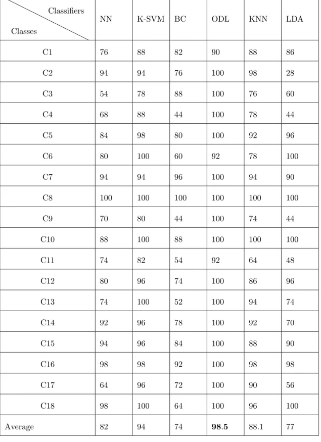
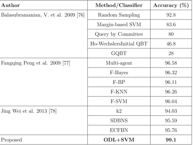
3.1 X-ray image classes: anatomical, direction. [6](A=Coronal, B=Axial, C=Other orientation D=Sagittal and E=Rotated) . . . 38 3.2 Merged classes of same images with different orientations. . . 39 3.3 Comparison of classification performance (%) using different classifiers. . . 40 4.1 Datasets used in experiments. . . 50 4.2 Performance comparison of multi-level classification with state-of-the-art
approaches on Wisconsin Breast Cancer Diagnostic dataset. . . 52 4.3 Performance comparison of multi-level classification with state-of-the-art
approaches on Wisconsin Breast Cancer original (WBC). . . 53 4.4 Comparison of performance of classification with state-of-the-art approaches
on Heart-StatLog dataset. . . 54 4.5 Performance comparison of multi-level classification with state-of-the-art
approaches on Pima Indians Diabetes dataset. . . 55 4.6 Performance comparison of multi-level classification with state-of-the-art
approaches on SPECTF (Heart) dataset. . . 56 4.7 Comparison of performance (in %) using individual classifiers on different
medical datasets.. . . 57 5.1 Classification accuracy (%) of multi-scale dictionary learning method using
wavelet decomposition based features and different dictionary sizes. . . 68 5.2 Classification accuracy (%) of the multi-scale dictionary learning method
with different classifiers on ICBM dataset. . . 71 5.3 Classification accuracy (%) of multi-scale dictionary learning method based
on individual and all combination of the sub-bands obtained from wavelet decomposition. . . 72
6.1 Comparison of classification performance (%) using individual classifiers without adaptive learning. . . 80 6.2 Comparison of classification performance (%) using individual classifiers
with adaptive learning. . . 80 6.3 Comparison of classification performance (%) using individual classifiers
without adaptive learning. . . 81 6.4 Comparison of classification performance (%) using individual classifiers
with adaptive learning. . . 81 7.1 Performance measure (%) of the proposed, fuzzy C-means and K-means
clustering methods obtained with the first feature extraction method and the Euclidean distance as similarity measure. . . 93 7.2 Performance measure (%) of the proposed, fuzzyC-means andK-means
clustering methods using second feature extraction method and Eu-clidean distance as similarity measure. . . 95 7.3 Performance measure (%) of the proposed, fuzzy C-means and K-means
clustering methods using first feature extraction method and cross correla-tion as similarity measure. . . 96 7.4 Performance measure (%) of the proposed, fuzzy C-means and K-means
clustering methods using second feature extraction method and cross cor-relation as similarity measure. . . 97 7.5 Performance measures (%) of the proposed, fuzzy C-means and K-Means
clustering methods using first feature extraction method and Mahalanobis distance as similarity measure. . . 98 7.6 Performance measure (%) of the proposed, fuzzy C-means and K-Means
clustering method using second feature extraction method and Mahalanobis distance as similarity measure. . . 99
7.7 Performance measure (%)of the proposed method with decreasing feature vector size (No.of concentric circles is 7) using Euclidean distance, cross correlation and Mahalanobis distance as similarity measure. . . 100 7.8 Performance measure(%) of the proposed method with increasing feature
vector size (No.of concentric circles=23) using Euclidean distance, cross correlation and Mahalanobis distance as similarity measure. . . 101 7.9 Performance measure(%) of the proposed method with different dictionary
LIST OF FIGURES
1.1 Process diagram for CBIR. . . 2
2.1 Traditional content based image classification and retrieval system. . . 11
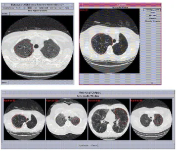
2.2 Some of the retrieved images with ASSERT tool. . . 12
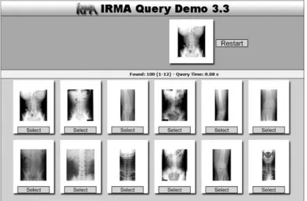
2.3 Some of the retrieved images with IRMA tool. . . 13
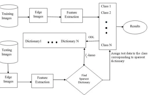
3.1 Block diagram of the proposed medical image classification. . . 29
3.2 (a) Samples of IRMA medical images. (b) Edge images of samples in (a). (c) Images are divided into equal size of patches. (d) A patch is divided into concentric circular regions. . . 30
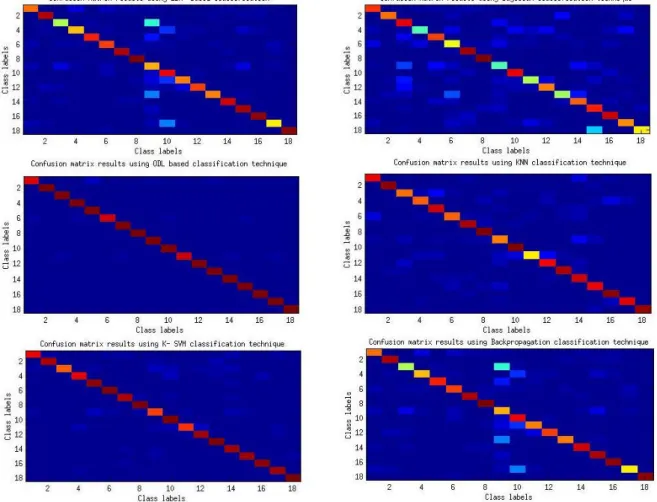
3.3 Confusion matrix using (a) LDA classification (b) Bayesian classification (c) ODL classification (d) KNN classification (e) K-SVM classification (f) NN classification . . . 34
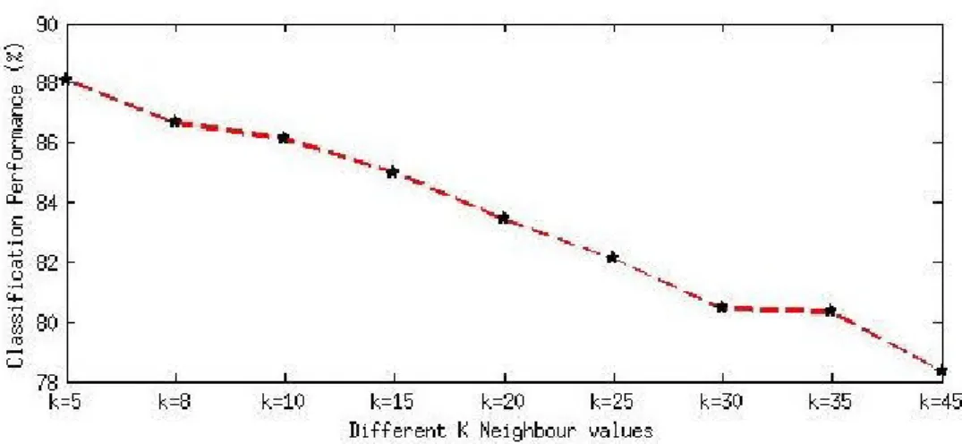
3.4 Performance of KNN classifier using different K values. . . 35
3.5 Classification performance of different types of SVM kernels. . . 35
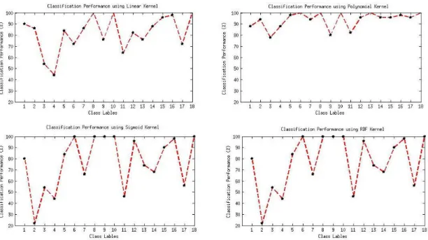
3.6 Classification performance of each class using (a) linear kernel SVM. (b) polynomial kernel SVM. (c) sigmoid kernel SVM. (d) RBF kernel SVM. . . 36
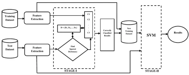
4.1 Block diagram of the multi-level classification framework using on-line dic-tionary learning and support vector machine. . . 46
4.2 Sensitivity measure of proposed method (ODL+SVM) on various UCI med-ical datasets. . . 57
4.3 Specificity measure of proposed method (ODL+SVM) on various UCI med-ical datasets. . . 58
5.1 Confusion matrix of medical modality image classification using SVM with haar wavelet feature. . . 69
5.2 Confusion matrix of medical modality image classification using neural net-work method with haar wavelet feature. . . 69 5.3 Confusion matrix of medical modality image classification Bayesian
classi-fication with haar wavelet feature. . . 70 5.4 Confusion matrix of medical modality image classification using multi-scale
dictionary learning. . . 70 6.1 Cardiac cycle of a typical heartbeat represented by the P-QRS-T wave form. 75 6.2 Examples of heartbeat shapes from the MIT-BIH data set. . . 76 7.1 Feature extraction.(a) Image is partitioned into concentric circular regions
of equal area. (b) Image is divided into sub-images and partitioned into concentric circular regions of equal area. . . 87 7.2 Some of the retrieved images, first column contains the query images and
remaining columns correspond to the retrieved images. . . 102 7.3 Comparision of average precision and recall of proposed, fuzzyC-means and
K-means clustering methods using first (I) and second (II) feature extrac-tion methods with three different distance similarity measures. (a) Highest precision recorded (%) using Euclidean distance as similarity measure. (b) Highest precision recorded(%)using cross correlation as similarity measure. (c) Highest precision recorded (%) using Mahalanobis distance as the sim-ilarity measure.(d) Highest recall recorded(%) using Euclidean distance as similarity measure. (e) Highest recall recorded (%) using cross-correlation as the similarity measure. (f) Highest recall recorded (%) using Maha-lanobis distance as similarity measure. Here, x-axis refers to different query images and the y-axis refers toF1 performance. . . 103 7.4 Comparison between retrieval time and feature vector size for different
B.1 Illustration of the idea of support vectors and an optimal hyperplane for linearly separable patterns. . . 116
ABBREVIATIONS
ED - Euclidean Distance
MRI - Magnetic Resonance Imaging CT - Computed Tomography
PET - Positron Emission Tomography MRI Magnetic Resonance Imaging ODL - On-line Dictionary Learning MOD - Method of Optimal Directions WSQ - Wavelet Scalar Quantization PCA - Principal Component Analysis SVD - Singular Value Decomposition LDA - Linear Discriminate Analysis k-NN k- Nearest Neighbour
ICA - Independent Component Analysis TBIR - Text Based Image Retrieval CBIR - Content Based Image Retrieval BC - Bayes Classifier
NN - Neural Network
IRMA - Information Retrieval in Medical Applications MLP - Multi-layer perceptron
SOM - Self-organizing Map DTI - Diffusion Tensor Imaging
MRA - Magnetic Resonance Angiography
FMRI - Functional Magnetic Resonance Imaging ICBM - International Consortium For Brain Mapping OMP - Orthogonal Matching Pursuit
CHAPTER 1
INTRODUCTION TO CONTENT BASED IMAGE
CLASSIFICATION AND RETRIEVAL
In the last few years, thousands of millions of images have become available on the Internet. The increase of these image collections is compelling people in various profes-sions, for example, medicine, architecture, geography, design, computer aided design, advertising and publishing to use them in various applications. Meanwhile, the study of image classification and retrieval, which is concerned with efficiently accessing sim-ilar type of images from large image collections, has become a more interesting and challenging task. Nevertheless, one cannot utilize the information in these image col-lections unless they are organized for efficient search and retrieval of data. Image classification and retrieval is all about techniques for storing and retrieving images both efficiently and effectively.
Previously, searching and retrieving similar images an image database was based on human annotation, i.e. each image in a database is given some keywords to de-note the semantic meaning of the image. Thus, classification and retrieving images was based on the keywords of images. This type of image retrieval is called as text based image retrieval (TBIR) [1]. Now, many search engines that claim to do image retrieval perform text based image retrieval like Google, QBIC and AltaVista. These search engines search the text around the image, such as captions, file names, and paragraphs located close to the image to search for relevant items to the query. This text based image retrieval method has many limitations, namely, as the size of image collection gets increasingly large manually annotating each image becomes very diffi-cult. Annotating an image based on human perception is very subjective. Different
people may assign different annotations to images with similar visual contents. The problem of searching for similar images in a large image repository based on content is called Content Based Image Retrieval (CBIR) [2]. The term content in CBIR refers to colors [3], shapes [3–5], textures [3, 4], or any other information that can be pos-sibly obtained from the image itself. Indexing remarkably affects the speed of data access besides supporting the accuracy for retrieval process and thus, is a significant factor in cataloging image database systems. Content based image indexing tends to facilitate automatic identification and abstraction of the visual content of an image. CBIR has the potential to greatly enhance the functionality of Picture Archiving and Communication Systems (PACS).
In the early 1990s Content Based Image Retrieval (CBIR) was proposed to over-come the limitations of text based image retrieval. There are many differences between content based image retrieval systems and classic information retrieval systems. The major differences are that in CBIR systems images are indexed using features extracted from the content itself and the objective of CBIR systems is to retrieve similar images to the query rather than exact matches. The similarity in most CBIR systems is quan-tified and the database entries are ranked based on their similarity to the query image. Similar images are retrieved as the result of a query. Different users may be interested in different parts of the same image. So, similarity based retrieval is more flexible than exact matching, and gives better performance in case of queries such as finding the images similar to the given image. The capability of present CBIR systems has been limited by their use of only primitive features like, color, shape, texture, spatial relationships among objects and these features can be used in most CBIR applications. In Section 1.1, we briefly describe various tasks involved in content based image classification and retrieval. In Section 1.2, we discuss certain issues related to medical image classification and retrieval that are addressed in this thesis. Section 1.3 outlines the organization of the thesis.
1.1 TASKS INVOLVED IN MEDICAL IMAGE CLASSIFICATION AND
RETRIEVAL
The objective of content based image retrieval is to develop techniques to automatically extract and retrieve relevant similar images from the large database. In conventional content based image retrieval systems, the query image is given to the CBIR system where the CBIR system will retrieve images from raw (unstructured) image database related to query image. Content based image retrieval involves three major tasks as shown in Fig. 1.1.
Fig. 1.1: Process diagram for CBIR.
The major functions of a CBIR are as follows:
• Analyze the contents of the source information and represent the contents of the analyzed sources in a way that will be suitable for matching user queries. This step is normally time consuming since it has to process all the source information (images) in the database.
• Analyze user queries and represent them in a form that will be suitable for matching with the source database, which is similar to the source images in the database.
• Define an approach to match the search queries with information in the stored database. Retrieve the images relevant to the query image.
1.1.1 Feature extraction
Feature extraction technique is the process of describing the image by considering parameters known as features (color, edge, texture etc.) from a given image. A feature is defined as a descriptive parameter that is extracted from an image [6]. The effectiveness of medical image classification and retrieval mainly depends on the effectiveness of features used for the representation of the content. An important issue is the choice of suitable features for a given task. Effective image retrieval can be achieved by collaboratively using color, edge density, boolean edge density, texture and histogram bins. These features are discussed in this section.
1.1.1.1 Color
Color has proven to be the most importent feature and almost all methods used color information. Although most of the images are in the RGB (Red, Green, Blue) color space, this space is only rarely used for indexing and querying as it does not relate well to the human color perception. It only works well for images taken under exactly the identical conditions each time. Other spaces such as HSV (Hue, Saturation, and Value) or the CIE Lab and Luv spaces are much better with respect to human perception and are used more commonly. This means that differences in the color space are close to the differences between colors that humans perceive. There are different types of color spaces available which are appropriate for different purposes. Some of the color spaces that we often come across are RGB, HSV, CIE Lab and Luv [3]. Color feature can be comprised of histogram bins or average, standard deviation or variance in an opted color space.
1.1.1.2 Texture
Texture [7] is another important property of images. Texture features [8] of images refer to the visual patterns that have properties of homogeneity that do not result from the presence of only a single color or intensity. Image texture content provides informa-tion of image properties such as smoothness, coarseness, and regularity which is useful in a CBMIR system. Basically, texture representation methods can be classified into two categories: structural and statistical. Structural methods including morphological operator and adjacency graph, describe texture by identifying structural primitives and their placement rules. They tend to be most effective when applied to textures that are very regular. Statistical methods, including Fourier power spectra, co-occurrence ma-trices, shift-invariant principal component analysis (SPCA), tamura feature, Markov random field, fractal model and multi-resolution filtering techniques such as Gabor [9] and wavelet transform, characterize texture by the statistical distribution of the image intensity.
1.1.1.3 Shape Retrieval
Shape features [8] of objects or regions have been used in many content-based im-age retrieval systems. Compared with color and texture, shape features are normally described after images have been segmented into regions. Since, accurate and ro-bust image segmentation is onerous to achieve. The use of shape features for image classification and retrieval has been restricted to some applications where objects or regions are readily available. The methods for shape description can be classified into boundary or region- based methods. A good shape representation feature for an object should be invariant to translation, rotation and scaling. More information is given in [8].
1.1.1.4 Semantics
Current CBIR systems retrieve similar images from a collection on the basis of the low level features of images, such as shape, color and texture. Nevertheless, some systems attempt to finding similar images that are semantically relative to a given query. Semantically similar is meant in the sense of human visual similarity perception (or called high level in CBIR).
1.1.1.5 Edge Information
Another choice for characterizing an image is its edge information. The advantage of this feature is that it is sufficiently invariant to illumination changes. Its main disadvantage is computational cost, noise sensitivity, and when not post-processed, high dimensionality.
1.1.2 Indexing for retrieval and browsing
Effective indexing and fast searching of images on basis of visual features pose a sig-nificant issue in content based image retrieval. Commonly, a tree structure is utilized to store image information since it has high dimensional metric space. R-tree [10], R*-tree [11], VP-tree structure [12] and Hybrid Tree [13] are some of the widely used tree structures. A majority of these multi-dimensional indexing methods perform sig-nificantly well for dimensions up to 20. A variant of R-tree employed in the indexing of spatial information is known as R*-tree. Both point and spatial data are supported at the same instant by an R*-tree but they are more complex compared to R-trees. Norbert Beckmann, Hans-Peter Kriegel, Ralf Schneider and Bernhard Seeger put forth the concept of R*-tree in 1990. Though R*-tree displays significant improvements over the R-tree variants its reinsertion method poses a considerable overhead. Database systems organizing both multidimensional points and spatial data can benefit from the R*-trees. Reduction of the area, margin and overlap of the directory rectangles are the basis for an R*-tree. The R*-tree utilizes an algorithm analogous to that of
the R-trees for query and delete operations. The primary difference lies in the insert algorithm. To be precise the mode of selection of which branch to insert the new node into and the methodology for splitting a full node in an R*-tree differs from that of the R-tree [11]. The Indexing tree structure can perform efficiently in dictionary operations. But, it can not be used for finding similarity among the images in the database. So, indexing tree structure was limited to structure the image database so that efficient retrieval is possible.
1.2 ISSUES ADDRESSED IN THIS THESIS
The previous section briefly described the various issues involved in content based image retrieval. The objective of this research work is to develop methods for clas-sification of medical data. The motivation for this objective stems from the need to organize large collection of medical images, for efficient classification and retrieval. The problem of image classification is addressed in the context of medical images. Classification of medical images into various genres or categories is an important task and most of the medical image classification systems available today classify medical images based on modality, body part, disease or orientation. In this thesis, we address two issues which are important for efficient classification and retrieval of medical data, namely, representation and classification. Representations can entangle and hide more or less the different explanatory factors of variations in the data. The objective of im-age classification is to categorize a given imim-age into one of the predefined categories. Medical image classification is an important task in content based medical image re-trieval. With the help of classification, the accuracy and retrieval speed of relevant images in content based image retrieval vastly improves. Classification of medical im-ages based on various body parts using on-line dictionary learning (ODL) andℓ1-lasso sparse representation on edge-based features is performed since different body parts are distinctly characterized by edge information.
med-ical datasets pose data imbalance problems which give poor classification performance with single classifiers. In this thesis, we propose a method to address the problem of data imbalance in medical images using level classification approach. A multi-level classifier combines correctly classified examples in the first multi-level with the training data and supplies them as input to the next level classifier.
Another issue in medical imaging is classification of medical images captured by different sensors. Capturing images using different modalities suffers from significant contrast variation between the images of the same organ or body part. Due to this large variation, existing image classification and retrieval algorithms do not perform well for different modality images. In this chapter, we propose a new classification tech-nique, namely, sparse representation based multi-scale dictionary learning to classify the different type of modality images. Wavelet features extracted from an image pro-vide discrimination useful for classification of medical images obtained from different sensors. Another important application area which is explored is automated detection of abnormal heartbeats captured by electronic cardiogram (ECG) signals. We employ an approach to classify abnormal heartbeat patterns from standard heartbeat patterns using adaptive dictionary learning on a standard ECG database.
Content based medical image retrieval (CBMIR) is the process of extracting rele-vant images to a query image, based on content rather than annotation. The key issues with CBMIR are the choice of features for representation of images, similarity/distance metric and a generic algorithm for retrieval of rotation invariant based similar images. The proposed CBMIR approach concentrates on retrieving rotation invariant resultant images and improving the accuracy of retrieved images with the help of clustering. A given image is partitioned into concentric circular regions of equal area and the mean and variance of each such area are considered as features for rotation invariant repre-sentation of images. These features are then used for the proposed dictionary learning based clustering and sparsest representation based classification algorithms.
1.3 ORGANIZATION OF THE THESIS
An overview of the existing approaches to image classification and retrieval is pre-sented in Chapter 2. Some research issues are identified in both these tasks which are addressed in this thesis. In Chapter 3, content based medial image classification is performed using on-line dictionary learning (ODL) and ℓ1-lasso sparse representation on edge-based features. In Chapter 4, the problem of imbalanced data problem in medical image classification is addressed by using multi-level classification approach. A new method for classification of medical images based on modality is proposed in Chapter 5, using the framework of multi-scale dictionary learning algorithm. In Chap-ter 6, the problem of automated detection of abnormal heartbeats captured by elec-tronic cardiogram (ECG) signals is addressed. An adaptive dictionary learning based classification technique is used to classify the normal and abnormal heartbeats from ECG medical database. In Chapter 7, dictionary learning based clustering method is proposed for content based medical image retrieval. An approach to group similar images into clusters that are sparsely represented by the dictionaries and simultane-ously learn dictionaries from the clusters using K-SVD method is proposed. A query image is matched with the existing dictionaries to identify the dictionary with the sparest representation using OMP algorithm. Then, images in the cluster associated with this dictionary are compared using a similarity measure to retrieve images similar to the query image. Considering mean and variance over concentric circular regions as features facilitates rotation invariance based image retrieval. Chapter 8 summarizes the research work carried out as part of this thesis, highlights the contributions of the work and discusses directions for future work.
CHAPTER 2
OVERVIEW OF APPROACHES FOR CONTENT BASED
MEDICAL IMAGE CLASSIFICATION
This chapter reviews some of the existing approaches to content based medical image classification and retrieval. The problem of content based image retrieval is briefly described in Section 2.1. The two important components of algorithms for image classification, namely, features for representation of images, and similarity/distance metric, are discussed in terms of the commonly made choices for these components. The existing algorithms for content based image retrieval are then reviewed. In Section 2.3, the existing approaches to image classification are reviewed, with particular focus on the classification of medical images. Some research issues arising out of the review of existing methods are identified, which are addressed in this thesis.
2.1 EXISTING METHODS FOR CONTENT BASED MEDICAL
IM-AGE CLASSIFICATION AND RETRIEVAL
There are hundreds of millions of images available on the Internet. Nevertheless, one cannot utilize the information in these image collections unless they are organized for efficient search and retrieval of data. Therefore, the need of an efficient method to retrieve digital images is recognized by the public. There are two approaches to image classification, namely, text based approach and content based approach. The former solution is a more traditional approach which indexes images by using keywords. The keyword indexing of digital images is useful but requires a considerable level of effort and often limited for describing image content. The alternate approach, the
content based image retrieval indexes images by using the low level features of the digital images and the searching depends on features being automatically extracted from the image. Content based image retrieval [2], is the term used to describe the process of retrieving images from a database on the basis of the internal features of images. In CBIR, digital images are indexed [11] by summarizing their visual contents through automatically extracted features such as texture, color, and shape. There exist different ways to express the query. The query can be defined by submitting one or more example images, providing a rough sketch of the desired item or by providing textual description of the object. CBIR retrieves stored digital images from a collection by comparing features extracted from the images. The most common features used are mathematical measures of color, texture or shape [2]. The CBIR system identities those stored images whose feature values match those of the query most closely and displays these found images to the user. In the following section, some of the frequently used types of features used for image retrieval will be described. The first step in content based medical image retrieval is to select an appropriate feature set for the image database. The selection of the feature set should be done in such a way that it should approximate images which are semantically similar to be as close to each other as possible in the feature space. The next step is to prepare a query image for retrieval i.e. extract features from the query image. Finally, an appropriate similarity measure is employed for retrieving the most similar images from the database. A block diagram of traditional content based image retrieval is shown in Fig. 2.1.
A common approach to feature extraction is to segment the images into regions [14] based on a certain similarity criterion. Regions from the segmentation result can then be used in region based queries for CBIR. This enables the user to include only the relevant regions when formulating a query. Chu et al. [15] described a knowledge based image retrieval of computed tomography (CT) and magnetic resonance imaging (MRI) images. In this approach, the brain lesions were automatically segmented and represented to form a knowledge based semantic model. Cai et al. [16] proposed a CBIR system for functional dynamic positron emission tomography (PET) images of
Fig. 2.1: Traditional content based image classification and retrieval system.
the human brain, where segmented clusters of tissue time activity curves from the temporal domain were used in the computation of similarity measure for retrieval. In [17], the delineations of the regions of interest were manually performed on the key frame from the stack of high resolution CT images and were used as features to represent the entire image. Some CBIR systems use segmentation to represent the regions, such as the ones used for retrieval of tumor shape and the shape of regions in spine X-ray images.
Guimond et al. [18] introduced user-selected volume of interest (VOI) for the re-trieval of pathological brain MRI images. In [19], group sparse representation with dictionary learning for medical image denoising and fusion was used. Wavelet opti-mization techniques for content based image retrieval in medical database were de-scribed in G. Quellec et al [20]. Linear discriminate analysis (LDA) based selection and feature extraction algorithm for classification and segmentation of one dimensional radar signals and two-dimensional texture and document images using wavelet packet was proposed by Etemand and Chellappa [21]. Recently, similar algorithms for simul-taneous sparse signal representation and discrimination were proposed [22]- [23]. In [24], Yi. Chen et al. proposed in-plane rotation and scale invariant clustering using
dictionaries. This approach provides Radon-based rotation and scale invariant clus-tering as applied to content based image retrieval on Smithsonian isolated leaf, Kimia shape and Brodatz texture datasets. Fei et al. [25] described a CT image denoising based on sparse representation using global dictionary. This approach improves low dose CT abdomen image quality through a dictionary learning based denoising method and accelerates the training time at the same time. Some of the existing medical CBIR systems as follows:
ASSERT : This system was developed in the school of electrical and computer engi-neering at Purdue University [17]. It is designed specifically for high resolution com-puted tomography images of the lung, since it uses some perceptual features specific to those images. It also includes gray-level features, such as the gray-level mean and standard deviation, texture features such as contrast, entropy and homogeneity and shape features such as the area. The feature vectors are indexed using the multi-hash method described in [26]. In Fig. 2.2, shows the some of the query related images with the ASSERT tool.
Fig. 2.2: Some of the retrieved images with ASSERT tool.
Tech-nology. It is focused on the querying of medical images using manually defined proto-types in a first stage and features are extracted from frequency, texture and structure analysis in regions segmented in a multi-scale blob-representation (blob tree). Those features are then indexed using a cluster-based approach.
The strategies adopted in the field of medicine often are (a) to use of more complex gray-level features (e.g. increase the number of gray level bins in the histogram), (b) to limit searches by creating prototypes for several well defined categories and (c) to use features that are specific of those images. MedGIFT uses the first strategy, IRMA uses the second and ASSERT uses the third. In Fig. 2.3, shows the some of the query related images with the IRMA tool.
Fig. 2.3: Some of the retrieved images with IRMA tool.
2.2 COMPONENTS OF CONTENT BASED IMAGE CLASSIFICATION
AND RETRIEVAL METHODS
An important component of content based image retrieval algorithms is the set of features extracted from a image or from a region of the image. Another component
is the similarity measure that is used to detect the presence of a image retrieval. We present below the different choices that can be made for each component, along with their advantages and disadvantages. A content based image retrieval algorithm can then be designed by suitably choosing each component.
2.2.1 Features used for representation of an image
Feature extraction is the process of describing the image by considering parameters known as features (color, edge, texture etc) from a given image. A feature is defined as a descriptive parameter that is extracted from an image [6]. The effectiveness of image retrieval depends on the effectiveness of features/attributes used for the representation of the content. An important issue is the choice of suitable features for a given task. Effective image retrieval can be achieved by collaboratively using color, edge density, boolean edge density, texture and histogram bins. These features are discussed in this section.
2.2.1.1 Extraction of gray-level features
Color has been the most effective and most widely used feature in CBIR [28, 29]. In medical CBIR, the color of each pixel is restricted to a gray levels intensity, which is already available, so it is quite straightforward to extract meaningful gray-level features. The objective is to transform the local gray-level information of each pixel into a global gray-level distribution of the full image, where visually similar images have similar representations.
Gray-level histogram
The most popular method of extracting gray level features of an image is to con-struct its histogram [29, 30]. A histogram is a statistical description that captures the gray levels distribution of an image. To construct an histogram, we discretized the intensity of the gray levels into a set of bins, and count the number of pixels whose intensity is in that bins range [31, 32]. In CBIR, the histogram is discretized into 256
bins, where the first bin has the number of black pixels (absence of color) and the last bin has the number of white pixels. Mainly, histograms suffer from two problems that limit their reliability. Perceptually, similar colors problem [30] is due to the very small difference between intensity values in neighboring bins. Sometimes, almost identical intensities are not assigned to the same bin, but to a neighbor. This means that the difference between the histograms of perceptually similar images (such as two images taken with different light conditions) can be quite big . An even bigger problem is the absence of any spatial information [33]. We can shuffle all the pixels in an image but the histogram remains untouched and therefore, the images are considered equal.
Partition-based histograms
Partition-based histograms incorporate spatial information by splitting the picture into k × k partitions, each one with its own histogram to store the local area gray-level information [34]. The spatial information emerges because only the corresponding pairs of local histograms are compared.
Color coherence vectors
Due to the absence of any spatial information in a histogram, an image with a large area of a given gray-level can be considered similar to another image that has many small areas of the same gray level scattered. To solve this problem, Pass et al. [33] proposed the Color Coherence Vectors (CCV) method. We start by identifying all the similar gray-level regions (connected components) in the image and count the number of pixels they have. If the number of pixels in a connected component is bigger than a given threshold, then they are classified as coherent. Otherwise, they are classified as incoherent. Not only a CCV has all the information present in histograms (i.e. we can convert a CCV into a histogram simply by adding the coherent and incoherent pixels for each pair), but it also measures if a gray-level is in a large area or scattered. A big number of coherent pixels are able to distinguish images with big similar gray levels areas from images with small scattered areas, even if both histograms are equal. Unfortunately, we still miss important spatial information. We do not know how many regions are present, how big they are or their location. Another potential problem is
the definition of the threshold used to classify the pixels coherence. Too low and even pixels in small regions will be coherent, too high and there will only be coherent pixels in large regions.
2.2.1.2 Extraction of texture features
Texture [35] is another important property of images. Texture features [36] of images refer to the visual patterns that have properties of homogeneity that do not result from the presence of only a single color or intensity. Image texture content provides information of image properties such as smoothness, coarseness, and regularity which is useful in a CBIR system. Basically, texture representation methods can be classified into two categories: structural and statistical. Structural methods including mor-phological operator and adjacency graph, describe texture by identifying structural primitives and their placement rules. They tend to be most effective when applied to textures that are very regular. Statistical methods, including Fourier power spectra, co-occurrence matrices, shift-invariant principal component analysis (SPCA), tamura feature, world decomposition, markov random field, fractal model and multi-resolution filtering techniques such as Gabor [37] and wavelet transform, characterize texture by the statistical distribution of the image intensity. Three classical approaches have been developed to describe textures, namely, structural, spectral and statistical. The structural approach assumes that the elements of a texture (textels) are placed under some rules. The spectral approach converts the image to the frequency domain to obtain features from its power spectrum. The statistical approach uses the statistical distribution of the pixels gray-level intensity to identify features. More recently, other methods were proposed inspired by human visual system (HVS) using multichannel filtering at different spatial frequencies and orientations.
2.2.1.3 Extraction of shape features
Shape features [36] of objects or regions have been used in many content-based image classification systems. Compared with color and texture features, shape features are usually described after images have been segmented into regions or objects. Since ro-bust and accurate image segmentation is difficult to achieve, the use of shape features for image retrieval has been limited to special applications where objects or regions are readily available. The methods for shape description can be classified into boundary or region-based and contour-based methods. A good shape representation feature for an object should be invariant to translation, rotation and scaling.
Region based features
Methods that extract region based features take into account all the pixels within the shape. Each shape is mapped onto a fixed sized grid or circle to achieve scale, rotation and translation invariance. This normalized shape is viewed as a probability density of a two-dimensional variable, from which orthogonal moments that describe some global properties of the shape can be computed [38, 39]. However, they are un-able to capture its local properties, thus failing to achieve partial occlusion invariance.
Contour based features
Methods that extract contour based features are more popular, since they extract both global and local features using only the shape boundary coordinates (x(t),(y(t)), t = 0, 1, ..., L-1 where, Lis a fixed number of samples (data points). All shapes are sampled into these data points so that (a) each shape signature (i.e. the representa-tion containing the features) has the same size, to facilitate the comparison between shapes and (b) to smooth the shape, reducing unwanted details, and increasing the computational efficiency [38]. Some of counter based features are complex coordinates, centroid distance, curvature and cumulative angular function.
2.2.2 Measure of similarity
Similarity measurement [3] is one of the key point in content based image classification and retrieval. An important step in most clustering is to select a distance measure, which will determine how the similarity of two elements is calculated. In CBIR, images are represented as features in the database. Once the features are extracted from the indexed images, the retrieval becomes the measurement of similarity between the features. Commonly used similarity measures are :
• The Euclidean distance (also called distance as the crow flies or 2-norm dis-tance). A review of cluster analysis in health psychology research found that the most common distance measure in published studies in that research area is the Euclidean distance or the squared Euclidean distance.
• The Manhattan distance (one-norm)
• The maximum norm (infinity norm)
• The Mahalanobis distance corrects data for different scales and correlations in the variables
• The angle between two vectors can be used as a distance measure when clustering high dimensional data.
• The Hamming distance measures the minimum number of substitutions required to change one member into another.
Euclidean distance [32] is the most common metric for measuring the distance between two vectors, and is given by the square root of the sum of the squares of the differences between vector components.
2.3 EXISTING METHODS FOR MEDICAL IMAGE CLASSIFICATION
Efficiently searching and retrieving of data in the large image collections poses sig-nificant technical challenges as the characteristics of the medical images differ from
other general purpose images. Some methods have been explored in recent years to automatically classify medical image collections into multiple semantic categories for efficient retrieval [40]. For example, in [41], the automatic categorization of 6231 radi-ological images into 81 classes is achieved by utilizing a combination of low level global texture features with low resolution scaled images and a K-nearest neighbor (KNN) classifier. Although, these approaches demonstrate promising results for medical im-age classification and retrieval, classification and searching similar imim-ages in a large database is still a challenge.
An X-ray image categorization and retrieval method using patch-based visual word representations is proposed in [42]. The feature extraction process is based on local patch representation of the image content and a bag-of-features approach for defining image categories, with a kernel based support vector machine (SVM) classifier. The method is especially effective in discriminating orientation and body regions in X-ray images, and in medical visual retrieval. In [43], a descriptor is proposed which combines local features with global shape features. The descriptor combines edge of whole image with edge density of sub-images and it is known as the edge density histogram descriptor (EDHD). The image retrieval and classification is then done based on euclidean distance and with the help of support vector machines.
A learning based classification framework based on local binary pattern (LBP) feature is proposed in [44]. Local binary pattern is extracted from each image in database with the help of an LBP operator which labels image pixels by thresholding neighborhood of each pixel with the center value and considers the result as a binary number, which is then classified using a maximum margin SVM. Moreover, a merging technique is applied on the overlapped classes. These overlapped classes are detected in merging scheme with the help of measures such as correctness rate of each class, similarity of imaging body organ and misclassification ratio. In [45], a least square support vector machines is used for breast cancer classification. Least square SVM (LSSVM) simplifies the required computation by solving a linear equation set. This equation set embodies all available information about the learning process. The most
important difference between SVM and LSSVM is that LSSVM uses a set of linear equations for training, while SVM uses a quadratic optimization problem which greatly reduces the computational cost. Wavelet optimization techniques for content based image retrieval in medical database are described in [20].
In, [46] shape and texture features are extracted from breast MRI images and genetic algorithm is applied to select the best feature to be used for classification process. To improve classification performance three different classifiers, namely, multi-layer perceptron (MLP), generalized regression neural network (GRNN) and support vector machine (SVM) are combined to from a multi-classifier system. Bartosz et al. [47] introduced under sampling balanced ensemble method to solve the imbalance problem. The construction of multiple independent classifiers is typically a non-trivial problem. In, [48] a cost-sensitive ensemble classification algorithm is proposed. The data imbalance problem is addressed by employing cost-sensitive decision trees as base classifiers which are trained on random feature subspaces to ensure diversity, and an evolutionary algorithm for simultaneous classifier selection and fusion. Marco Vannucci et al. [49] described a binary classification method named Labeled SOM Classification Unbalanced Sets (LASCUS) that can be applied to uneven datasets and sensitive problems such as malfunction detection. LASCUS method is based on the use of a self-organizing map (SOM) and fuzzy inference system (FIS). The SOM creates a set of clusters to be associated either to frequent or unfrequented situations while the FIS determines such association on the basis of data distribution.
Modality classification and its use in text based image retrieval in medical databases is proposed in [50]. Visual descriptors and text features are used for classifying the medical images. Medical image classification is then done with the help of support vector machines classifier. In [51], explore different type of medical image modality and retrieval strategies. Bags of visual words and fisher vectors representations are in-tegrated to perform medical modality classification. Wavelet optimization techniques for content based image retrieval in medical database are described in [20].
2.4 ISSUES ADDRESSED IN MEDICAL IMAGE CLASSIFICATION
This thesis is mainly focused on the issues related to the efficient classification and retrieval of medical images. Image classification and retrieval which is concerned with efficiently accessing similar type of images from large image collections, has become more interesting and more challenging as the medical datasets have grown over the years. The existing medical image search and retrieval techniques are not very efficient in terms of time and accuracy of search result because most of the existing tools for searching medical images use text based image retrieval techniques.
Text based image classification suffers from some serious limitations, namely, when the size of image collection gets increasingly large, annotating each image manually is very difficult. Also, different people may give different annotations to images with similar visual content. Improving the classification accuracy and reducing the retrieval time are important issues in medical images.
In most medical imaging systems, the same body part is captured from different orientations and magnification by the same sensor. Devise a rotation and scale invari-ant classification and retrieval system is a real challenge. Medical images are captured by different sensors (modalities). Images captured from various modalities suffer from significant contrast variation between the images of the same organ or body part. Due to this large variation, existing image classification and retrieval algorithms do not perform well for different modality images.
In addition, most of the medical datasets pose data imbalance problem i.e. un-equally distributed training samples among all the classes, which gives rise to poor classification performance with standard single classifiers. Finally, one of the most important problems in medical CBIR is to find images with similar anatomical regions and diseases which can greatly reduce the effort exerted by physicians to manually analyse and annotate the disease region.
2.5 SUMMARY
This chapter reviewed some of the existing approaches to content based medical image classification and retrieval. Various steps involved in content based image classification and retrieval system and the related work is briefly described. Also, the existing approaches for all the components of content based image retrieval system are reviewed. Some research issues arising out of the review of existing methods are addressed in this thesis.
CHAPTER 3
CLASSIFICATION OF MEDICAL IMAGES USING
EDGE-BASED FEATURES AND DICTIONARY
LEARNING
In this chapter, an approach for classification of medical images using edge-based features is proposed. We demonstrate that the edge information extracted from an image by dividing the image into patches and each patch into concentric circular re-gions provide discriminative information useful for classification of medical images by considering 18 categories of radiological medical images, namely, skull, hand, breast, cranium, hip, cervical spin, pelvis, radiocarpaljoint , elbow etc. The ability of on-line dictionary learning (ODL) to achieve sparse representation of an image is exploited to develop dictionaries for each class using edge-based feature. A low rate of misclassifi-cation error for these test images validates the effectiveness of edge-based features and on-line dictionary learning models for classification of medical images.
Digital image retrieval techniques are becoming increasingly important in the field of medical image databases. The increasing dependence on modern medical diagnos-tic techniques like radiology, histopathology and computerized tomography has led to an explosion in the number of medical images stored in hospitals. Images of vari-ous body parts and modalities are becoming an important source of anatomical and functional information for the diagnosis of diseases, medical research and education [52]. However, one cannot utilize the information in these image collections unless they are organized for efficient search and retrieval of data. Efficiently searching and retrieving of data in these large image collections poses significant technical challenges
as the characteristics of the medical images differ from other general purpose images. Some methods have been explored in recent years to automatically classify medical image collections into multiple semantic categories for effective retrieval [40]. For ex-ample, in [41], the automatic categorization of 6231 radiological images into 81 classes is achieved by utilizing a combination of low level global texture features with low res-olution scaled images and a K-nearest neighbor (KNN) classifier. Although these ap-proaches demonstrate promising results for medical image classification and retrieval, classification and search of similar images in a large database is still a challenge due to the enormity of the search space. Searching similar images in a large image repository on the basis of their visual content is called Content Based Image Retrieval (CBIR) [53]. The traditional text based image classification and retrieval (TBIR) approach has many practical limitations like the images in the collection have to be annotated manually which becomes very difficult as the size of the image collection increases and time consuming. Another important limitation of TBIC and TBIR is inadequacy in representing the image content [54]. Content based image classification and retrieval approaches are proposed to overcome the limitations of text based image classification and retrieval. Digital image retrieval techniques are crucial in the emerging field of medical image databases for clinical decision making process.
Medical image classification is an important task in content based medical image retrieval (CBMIR). Automatic medical image classification is a technique for assign-ing a medical image to an appropriate class among a number of medical image classes. In medical image classification, several methods have been proposed in the literature [55]- [56]. One approach to content based medical image retrieval is proposed in [55], in which medical images are classified based on body orientation, biological system, anatomical region and image modality. The performance of the classification is evalu-ated on IRMA database and the best classification result is achieved by using distorted tangent distance in a kernel density classifier. The CBMIR system can achieve better performance by filtering out the images of irrelevant classes from the medical database through classification. This significantly reduces the search space and time for
retriev-ing similar type of images. So, image classification is indeed an important stage in a CBMIR system.
The major limitations associated with existing text based image classification and retrieval techniques are: 1) It is time consuming as the physicians have to search through a large number of images for identifying similar images. 2) Most of the existing tools for searching medical images use text based image classification and retrieval techniques. These text based image classification suffer from several limitations [51] and the most important one is the need for manual annotation. Thus, the existing medical image search and retrieval techniques are not very efficient in terms of retrieval time and accuracy of search results.
In this chapter, we address the some issues in text based image classification and retrieval. The content based image classification techniques serve as an alternative to text based image classification. Moreover, CBMIR overcomes the need for manual annotation and human perception. Also, finding similar images in large volumes of medical image databases is a difficult task. Classification of medical images enables the efficient retrieval of relevant images from the large database and reduces the search space and time.
Selection of features for adequately representing the class specific information is an important process in medical image classification. An X-ray image categorization and retrieval method using patch-based visual word representations is proposed in [42]. The feature extraction process is based on local patch representation of the image content and a bag-of-features approach for defining image categories, with a kernel based SVM classifier. The method is especially effective in discriminating orientation and body regions in X-ray images, and in medical visual retrieval. In [43], a descriptor is proposed which combines local features with global shape features. The descriptor combines edge of whole image with edge density of sub-images and is known as the edge density histogram descriptor (EDHD). The image retrieval and classification is then done based on euclidean distance and with the help of support vector machines. A learning based classification framework based on local binary pattern(LBP) feature
is proposed in [44]. Local binary pattern is extracted from each image in database with the help of an LBP operator which labels image pixels by thresholding neighborhood of each pixel with the center value and considers the results as a binary number, which is then classified using a maximum margin SVM. Moreover, a merging technique is applied on the overlapped classes. These overlapped classes are detected in merging scheme with the help of measures such as correctness rate of each class, similarity of imaging body organ and misclassification ratio. In [45], a least square support vector machines is used for breast cancer classification. Least square SVM (LSSVM) simplifies the required computation by solving a linear equation set. This equation set embodies all available information about the learning process. The most important difference between SVM and LSSVM is that LSSVM uses a set of linear equations for training while SVM uses a quadratic optimization problem which greatly reduces the computational cost. The extracted feature database is constructed by merging some already existing features in the original database with some new visual content features that are extracted from the medical images using image processing techniques. Wavelet optimization techniques for content based image retrieval in medical database are described in [20].
In most cases, medical images can easily be classified based on edge information. In this chapter, we propose a novel feature extraction method using the edge information. Medical images of different body parts contains different edge information. An edge image is divided into patches and each patch into concentric circular regions. Mean and variance of pixel intensity values in each region is computed. Mean and variance are global measurements and these are more suitable with deterministic methods. In this method, different orientations of same shape images are combined into a single class, in order to achieve better classification. The reason for combining multiple classes to solve a given classification problem is due to the fact that in medical applications, numerous classes of any given medical image database have considerable overlap. Hence, a single class with limited features cannot classify images correctly [54].
and image processing. Sparse coding involves the representation of an image as a linear combination of some atoms in a dictionary [57]. Sparse representation is a powerful tool for efficiently representing data. This is mainly due to the fact that signals and images of interest tend to enjoy the property of being sparse in some dictionary. These dictionaries are often learned directly from the training data. Several algorithms like on-line dictionary learning (ODL) [58], K-SVD [59] and method of optimal directions (MOD) [60] have been developed to process training data. Sparse representation is used to match the input query image with the appropriate class. Linear discriminant analysis (LDA) based selection and feature extraction algorithm for classification using wavelet packet has been proposed by Etemand and Chellappa [21]. Recently, similar algorithms for simultaneous sparse signal representation and discrimination have also been proposed [22], [61]. In [62], a method for simultaneously learning a set of dictionaries that optimally represent each cluster is proposed. This approach was later extended by adding a block incoherence term in their optimization problem to improve the accuracy of sparse coding.
In this chapter, we propose an approach for classification of medical images on image retrieval in medical applications (IRMA) database [63] using on-line dictionary learning approach. Learned dictionaries are used to represent datasets in sparse model of IRMA medical images. Dictionaries are designed to represent each class. For a given N number of classes, we design N dictionaries to represent the classes. Each image associated with a dictionary provides the best sparsest representation. For every image in the given set of images {yi}ni=1, ODL is used to seek the dictionary D that has
the sparsest representation for the image. We define l(Dˆ,Φˆ) as the optimal value of the l1-lasso sparse coding problem [64]. This is accomplished by solving the following
optimization problem: l(Dˆ,Φˆ) = arg min D,Φ 1 N N X i=1 1 2kYi −DΦik 2 2 subject tokΦik1 ≤λ, (3.1)