Patellar Tendinopathy
Prevalence, ESWT treatment and Evaluation
Patellar Tendinopathy
Prevalence, ESWT treatment and Evaluation Zwerver, J
Dissertation University of Groningen, the Netherlands – With ref. – With summary in Dutch
ISBN: 978-90-367-4610-6
© J. Zwerver, Groningen, The Netherlands 2010
All rights reserved. No part of this publication may be reproduced or transmitted in any form or by any means, electronical or mechanical, including photocopy, recording or any information storage or retrieval system, without the prior written permission of the copyright owner.
Lay-out and cover: Helga de Graaf, Studio Eye Candy, Groningen (www.proefschrift.info) Printed by Ipskamp Drukkers, Enschede
RijkSunivERSiTEiT GRoninGEn
Patellar Tendinopathy
Prevalence, ESWT treatment and Evaluation
Proefschrift
ter verkrijging van het doctoraat in de
Medische Wetenschappen
aan de Rijksuniversiteit Groningen
op gezag van de
Rector Magnificus, dr. F. Zwarts,
in het openbaar te verdedigen op
woensdag 8 december 2010
om 16.15 uur
door
Johannes Zwerver
geboren op 3 september 1967
in de Noordoostpolder
Promotor Prof. dr. R.L. Diercks Copromotores Dr. I. van den Akker-Scheek
Dr. F.Hartgens Beoordelingscommissie Prof. dr. F.J.G. Backx
Prof. dr. S.K. Bulstra Prof. dr. K. Peers
Paranimfen Steef Bredeweg Hans de Vries
Contents
Chapter 1
General introduction
Chapter 2
Patellar tendinopathy (jumper’s knee):
a common and difficult-to-treat sports injury
Chapter 3
Prevalence of patellar tendinopathy in non-
elite-athletes
Chapter 4
Validity and reliability of the Dutch translation
of the VISA-P questionnaire for patellar
tendinopathy
Chapter 5
Biomechanical analysis of the single-leg
decline squat
Chapter 6
Extracorporeal shockwave therapy for patellar
tendinopathy: a review of the literature
Chapter 7
The TOPGAME study: effectiveness of
extracorporeal shockwave therapy in jumping
athletes with patellar tendinopathy. Design of
a randomised controlled trial
Chapter 8
No effect of extracorporeal shockwave
therapy on patellar tendinopathy in jumping
athletes during the competitive season: a
randomised clinical trial
Chapter 9
General discussion
Chapter 10
Summary
Appendices
A
Nederlandse samenvatting
B
VISA-P questionnaire
C
Nederlandse VISA-P vragenlijst
D
Dankwoord
E
Curriculum Vitae
F
List of publications
G
List of (inter)national presentations
H
Onderwijs en overige activiteiten
I
Share
9 15 29 41 51 63 79 91 109 127 133 141 145 149 155 159 165 173 179Chapter 1
General introduction 11
1
Background
The patellar tendon is commonly subject to overuse in sporting activities, especially in sports like basketball and volleyball which require explosive extension of the knee or ec-centric flexion. In 1973 Blazina introduced the term ‘jumper’s knee’ to describe the condi-tion of anterior knee pain in the patellar or quadriceps tendon or at the patellar or tibial attachment of the patellar tendon.1 Nowadays the term patellar tendinopathy is often
used in clinical practice for this chronic injury of the patellar tendon, which is clinically characterised by load-dependent pain at the inferior pole of the patella.2,3
Although there have been many advances in the understanding of the histopathology, imaging, and both conservative and surgical outcomes for this condition in the past two decades, successful management of athletes with patellar tendinopathy remains a major challenge for both practitioner and patient.2-6
This is why there is a definite need for studies into prevalence and etiological factors of patellar tendinopathy, feasible evaluation tools and functional tests to rate severity and outcome of interventions, and last but not least randomised controlled trials into the ef-fectiveness of different treatment options.
Aims
The scope of this thesis is to increase our knowledge of patellar tendinopathy in non-elite athletes and the role of extracorporeal shockwave therapy (ESWT) in the management of this condition. The first objective is to describe the prevalence of patellar tendinopathy in non-elite athletes and to find associated risk factors for this overuse injury. The second objective is to develop and study specific (Dutch) evaluation tools for patellar tendinopa-thy. The third objective is to evaluate the effectiveness of ESWT as treatment for patellar tendinopathy.
Outline of the thesis
First, in Chapter 2 an overview is given of the clinical characteristics, etiology, pathophysiol-ogy, diagnostic methods and treatment and rehabilitation options for patellar tendinopathy.
Chapter 3 presents the results of a cross-sectional survey into the prevalence of patel-lar tendinopathy among non-elite athletes from seven different sports and some potential risk factors for patellar tendinopathy.
The validity and reliability of the Dutch Translation of the VISA-P score, a question-naire that rates symptoms, function and sports participation of athletes with patellar
tendinopathy, are presented in Chapter 4.
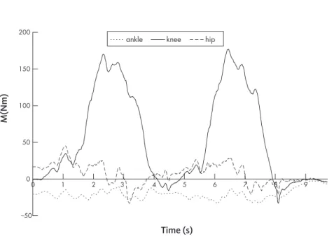
Chapter 5 describes the findings of a biomechanical study that analyses the load on the patellar tendon, knee moment and patellofemoral contact force during the single-leg decline squat, a functional assessment tool for patellar tendinopathy.
12 Chapter 1
In Chapter 6 a review is given of the literature on ESWT for patellar tendinopathy.
Chapter 7 describes the design of the TOPGAME study, a prospective multicenter study into the effectiveness of ESWT in active basketball, handball and volleyball play-ers who have patellar tendinopathy with symptoms for less than 12 months. The results
of this randomized controlled trial are presented in Chapter 8.
Chapter 9 combines the results of the different studies into a general discussion and conclusions are drawn. Practical recommendations and suggestions for further studies are presented.
General introduction 13
1
References
1. Blazina ME, Kerlan RK, Jobe FW, Carter VS, Carlson GJ (1973). Jumper’s knee. Orthop Clin North Am; 4:665-78.
2. Cook JL, Khan KM, Maffulli N, Purdam C (2000). Overuse tendinosis, not tendinitis part 2. Applying the new approach to patellar tendinopathy. Physician and Sportsmedicine; 28:31-+. 3. Khan KM, Cook JL, Bonar F, Harcourt P, Astrom M (1999). Histopathology of common
tendinopathies. Update and implications for clinical management. Sports Med; 27:393-408. 4. Coleman BD, Khan KM, Maffulli N, Cook JL, Wark JD (2000). Studies of surgical outcome after
patellar tendinopathy: clinical significance of methodological deficiencies and guidelines for future studies. Victorian Institute of Sport Tendon Study Group. Scand J Med Sci Sports; 10:2-11.
5. Cook JL, Khan KM (2001). What is the most appropriate treatment for patellar tendinopathy? Br J Sports Med; 35:291-4.
6. Peers KH, Lysens RJ (2005). Patellar tendinopathy in athletes: current diagnostic and therapeutic recommendations. Sports Med; 35:71-87.
Chapter 2
Patellar tendinopathy
(jumper’s knee):
a common and
difficult-to-treat sports injury
J. Zwerver
This chapter is a translated and adapted version of: Patellatendinopathie (‘jumper’s knee’); een veelvoorkomende
en lastig te behandelen sportblessure
16 Chapter 2
Abstract
Patellar tendinopathy is a common and difficult-to-treat overuse injury of the patellar tendon with a very negative impact on the careers of many athletes. It appears to involve a failed healing process in the tendon – not inflammation – and has consequences for the treatment strategy. Rehabilitation programs are based on the principles of load reduction and an eccentric exercise program to improve muscle-tendon function and optimise the kinetic chain. Prolonged rehabilitation is necessary because of slow tendon recovery. Anti-inflammatory treatment is often unsuccessful. Surgery does not guarantee a quick symptom-free return to sport at the original level either. Extracorporeal shockwave therapy, ultrasound-guided sclerosing of new vessels and tendinous and peri-tendinous injections of aprotinin and autologous growth factors seem to be promising new treatment options.
Patellar tendinopathy (jumper’s knee) 17
2
Introduction
Jumper’s knee, also called patellar tendinopathy, is a common condition among both rec-reational and professional athletes,1 and can influence the athlete’s career. In elite
basket-ball and volleybasket-ball players its prevalence is very high – 32 and 45% respectively.2 It is a
chronic injury of the patellar tendon, which is clinically characterised by load-dependent pain at the inferior pole of the patella.1 The high prevalence, the impairment of function
and the chronic character of this condition mean that jumper’s knee might have at least as much impact on an athlete’s career as an acute knee injury.2 For some athletes it is even
a reason to end their career.3 More than half of athletes with patellar tendinopathy still
has some symptoms even 10 years after their career ended.3
General practitioners, physical therapists, sports medicine physicians, sports and reha-bilitation physicians and orthopaedic surgeons frequently see athletes with this typical sports-related knee injury. They use many different treatments, often empirically based, but results are often frustrating for both athlete and doctor or physical therapist.4,5 The
poor results can be partly explained by the fact that until recently most treatments were targeted at reducing inflammation in the tendon. In recent years it has been demonstrated that the underlying pathology of patellar tendinopathy, just as in other tendinopathies, is not a tendinitis but rather a failed healing response, tendinosis.6,7 In clinical practice –
when no histopathology is available – it is better to speak of patellar tendinopathy. Patel-lar apexitis or patelPatel-lar tendinitis are also used. This article starts by reviewing the new insights on etiological factors and histopathology of patellar tendinopathy, because they have consequences for the treatment strategy to follow. This is followed by a description of the clinical characteristics and the conservative and surgical treatment of this typical sports injury.
Etiology
The multifactorial etiology of patellar tendinopathy has not yet been completely clari-fied. Men appear to be at a greater risk of getting this condition. Estrogens might protect
tendons from getting injured.8 Genetic predisposition seems to be an important factor as
well.9 Overload
A disturbed balance between load and loading capacity probably plays an important role. Because this injury is more common among elite than among recreational athletes, a link between training volume and frequency and the prevalence of patellar tendinopathy seems logic.2,10
Reduced loading capacity
A reduced loading capacity is also important. If an athlete is less capable of generating or absorbing forces this can result in a wrong jumping and/or landing technique, which gives an increased load on the patellar tendon. Bisseling et al. recently demonstrated
18 Chapter 2
that a stiffer landing strategy, with less motion in knee and ankle, increases the risk for patellar tendinopathy.11 Risk factors associated with this condition are reduced strength
of calf, quadriceps and gluteal muscles, inadequate core stability, reduced hamstring and quadriceps flexibility, and hyperpronation of the foot.12,13 Reduced dorsiflexion of the
ankle, for example remaining after an ankle distortion which frequently occurs in jump-ing athletes, can play a role.4,14
Pathology
In chronic tendinopathy a failed healing process results in a painful and weakened ten-don, which is then less capable of performing its most important functions, namely ab-sorbing and transducing forces. Repetitive microtrauma caused by overuse give rise to degenerative abnormalities in the tendon like changes in collagen structure and neuro-vascular proliferation.7 There is no inflammatory process. The histopathology is a
tendi-nosis, not a tendinitis. Vasculoneural ingrowth might play a role in the concomitant pain in tendinopathies.15
Clinical information
HistoryAthletes with patellar tendinopathy experience pain at the inferior pole of the patella. Pain usually starts insidiously and increases with activities like jumping, sprinting and landing. Symptoms often start after a period of increased training load. In the ini-tial phase symptoms disappear during the warm-up. Athletes tend to keep on going ‘through the pain’ and don’t seek medical help. When they continue to compete at the same level, the pain gradually increases and also remains during sporting activities; eventually, sport performance declines. Finally, there is even pain during daily activities and at rest.
To quantify the severity of the patellar tendinopathy during the treatment period one can use the Victorian Institute of Sport Assessment (VISA) score (see Appendix B). For
a Dutch translation see Appendix C.16 This questionnaire consisting of eight questions
was specifically designed for patellar tendinopathy and evaluates pain, function and sports participation, with a score of 100 reflecting an optimal symptom-free knee. VISA scores of athletes with patellar tendinopathy looking for medical help are around 50–70 points.
Patellar tendinopathy (jumper’s knee) 19
2
Physical examination
Circumscript palpation tenderness at the tendon insertion at the inferior pole of the pa-tella is the most characteristic finding during physical examination. The papa-tellar tendon can be thickened. A common finding is atrophy of the quadriceps muscle, especially the M. vastus medialis.
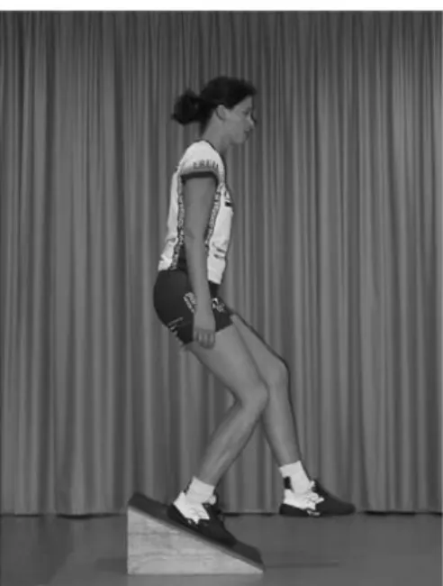
As mentioned before, it is important to evaluate potential etiological factors like reduced muscle-tendon function, inappropriate core stability and limited ranges of motion of sev-eral joints. The single leg decline squat is a functional test that loads the patellar tendon and can provoke pain. In this test the athlete, standing on one leg on a decline board at an angle of approximately 25 degrees, gradually flexes the knee (figure 2.1).17 The test can
be used to substantiate the diagnosis, but there is no gold standard.
Imaging techniques
Although MRI and ultrasound can increase the likelihood of a diagnosis of patellar tendi-nopathy, their value is limited (figures. 2.2 and 2.3).18-22 Patellar tendons of asymptomatic
athletes often show sonographic abnormalities, and symptoms and abnormalities can vary during the sports season.23,24 The prognostic and follow-up value of MRI and
ultra-sound are also limited because of the poor correlation between clinical symptoms and
imaging abnormalities in the tendon.21,25 There may be a correlation between
neovascu-larisation (figure 2.3b) on Doppler ultrasound and experienced pain.26,27
Figure 2.1‘Single leg decline squat’ test: pain-provoking test to increase the likelihood of patellar tendinopathy.
20 Chapter 2
Figure 2.2. Ultrasound characteristics of patellar tendinopathy: (a) thickened tendon with hypo-echogenic zones and calcifications; (b) colour-Doppler appearance of neovascularisation in the tendon.
femur
tibia patella
Figure 2.3. MRI of patellar tendinopathy, with increased signal intensity in the proximal part of the tendon; (b) in MRI using a fat-suppression technique.
femur
tibia patella
Patellar tendinopathy (jumper’s knee) 21
2
Conservative treatment
Several, mostly not evidence-based treatment options are used in the management of
patellar tendinopathy.4,5 With the current understanding that the underlying pathology
in tendinopathies is a tendinosis and not a tendinitis, one should reconsider the treatment strategy. A treatment program for patellar tendinopathy should aim mainly at restor-ing the balance between load and loadrestor-ing capacity and stimulatrestor-ing tendon regeneration
rather than reducing the inflammatory process.28
Explanation
Athletes with patellar tendinopathy need to get an explanation about the overuse and chronic character of their injury, and should be informed about the fact that a rehabilita-tion program often takes more than three months to achieve full recovery.
Pain reduction
Reduction of tendon load can reduce the pain to a tolerable level for the athlete. This does not mean that athletes should completely refrain from tendon-loading activities. Complete rest or even stronger complete immobilisation leads to further weakening of the muscle-tendon unit. It is better to load the muscle-tendon very carefully, thereby enabling the athlete to perform pain-free daily activities, participate in a rehabilitation program and adjust sporting activities. Ice packs, taping and bracing (patellar strap) and electrophysical mo-dalities sometimes give some short-term pain relief, but did not evidence a pain-reducing or regenerative effect at longer follow-up.
Exercise therapy to increase loading capacity
If strength and endurance of calf, quadriceps and gluteal muscles are weakened one should prescribe strength-training exercises to improve muscle performance. Core stabil-ity should also be optimised. Limitations in joint movements and other causal factors should be corrected. Sport-specific exercises should be included in the final part of the rehabilitation program.
Eccentric exercises
Eccentric, slightly painful exercises like the aforementioned single-leg decline squat ap-pear to be effective in the treatment of tendinopathies.6,29-31 During an eccentric
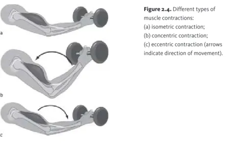
contrac-tion the muscle-tendon unit becomes elongated while the muscle contracts, in contrast to isometric and concentric contractions, in which the length respectively stays the same or becomes shorter. For example, during the single-leg decline squat the quadriceps muscle contracts while the patellar tendon-quadriceps unit elongates. Using this eccentric treat-ment strategy VISA scores improved by about 30 points.
A practical recommendation for athletes is to perform single-leg decline squats on the in-jured leg once or twice a day, in three series of 15 repetitions for a period of 12 weeks. The precise working mechanism is still unclear but it is likely that these exercises stimulate regeneration in the tendon. The old adage that exercise therapy should be painless is thus open to debate. Some pain during exercise therapy can lead to good treatment results
22 Chapter 2
Extracorporeal shockwave therapy
Extracorporeal shockwave therapy seems to be an appropriate additional treatment. Some randomised placebo-controlled studies demonstrate the effectiveness of ESWT on
pain and function in patellar tendinopathy.5,33 After ESWT treatment, athletes showed 30
to 40 points higher VISA scores than the control group. The beneficial effect of ESWT might be the result of an analgetic process, destruction of calcifications and stimulation of regeneration processes in the tendon. Unlike ESWT, low-intensity pulsed ultrasound
showed no additional benefit over an eccentric training program.34
Anti-inflammatory medication
Treatments aimed at reducing inflammation, like the commonly used NSAIDs and injec-tions with corticosteroids, seem to be illogic for degenerative tendons without inflamma-tion. At best, they give short-term pain relief, however their effectiveness in the long run has not been demonstrated. Because of their pain-reducing effect they can mask under-lying problems, resulting in even more extensive tendon abnormalities. Injections with corticosteroids have been controversial in recent decades since they influence collagen synthesis negatively and reduce tendon strength.
Ultrasound guided sclerotherapy
An interesting new treatment method is ultrasound-guided sclerosis of the neovessels in the tendon with polidocanol, a well-known sclerosant to treat varices. It is presumed that
tendinopathy pain is caused by neurovascular ingrowth in the tendon.35 The effectiveness
of this treatment method was recently demonstrated in a randomised clinical trial.36
Figure 2.4. Different types of muscle contractions: (a) isometric contraction; (b) concentric contraction; (c) eccentric contraction (arrows indicate direction of movement).
Patellar tendinopathy (jumper’s knee) 23
2
Other injection techniques
Tendinous and peritendinous injections with aprotinin, a protease inhibitor, or with autol-ogous blood or platelet-rich plasma and growth factors seem to be successful, but further research into the effectiveness of these treatments is necessary.37,38
Surgical treatment
Surgical treatment can be an option when despite a comprehensive and extensive reha-bilitation program conservative treatment fails. Several surgical procedures have been described. Success rates in the literature vary between 60 and 100% and are inversely
correlated with methodological quality.39 A recent clinical trial demonstrated that open
tenotomy has no advantage over eccentric training.40 Surgery does not guarantee a quick,
symptom-free return to sports at the original level. Also, after surgery a prolonged reha-bilitation period according the aforementioned guidelines is necessary.
Conclusion
Patellar tendinopathy is a common overuse injury of the patellar tendon with a very negative impact on the career of an athlete. Up to now no single treatment exists that guarantees a quick and symptom-free return to the original sports level. Therefore, a pro-longed rehabilitation program to restore the balance between load and loading capacity and to promote regeneration of the tendon is the best treatment.
24 Chapter 2
References
1. Blazina ME, Kerlan RK, Jobe FW, Carter VS, Carlson GJ (1973). Jumper’s knee. Orthop Clin North Am; 4:665-78.
2. Lian OB, Engebretsen L, Bahr R (2005). Prevalence of jumper’s knee among elite athletes from different sports: a cross-sectional study. Am J Sports Med; 33:561-7.
3. Kettunen JA, Kvist M, Alanen E, Kujala UM (2002). Long-term prognosis for jumper’s knee in male athletes. A prospective follow-up study. Am J Sports Med; 30:689-92.
4. Cook JL, Khan KM (2001). What is the most appropriate treatment for patellar tendinopathy? Br J Sports Med; 35:291-4.
5. Peers KH, Lysens RJ (2005). Patellar tendinopathy in athletes: current diagnostic and therapeutic recommendations. Sports Med; 35:71-87.
6. van Linschoten R, den Hoed PT, de Jongh AC (2007). [Guideline ‘Chronic Achilles
tendinopathy, in particular tendinosis, in sportsmen/sportswomen’]. Ned Tijdschr Geneeskd; 151:2319-24.
7. Khan KM, Cook JL, Bonar F, Harcourt P, Astrom M (1999). Histopathology of common tendinopathies. Update and implications for clinical management. Sports Med; 27:393-408. 8. Cook JL, Bass SL, Black JE (2007). Hormone therapy is associated with smaller Achilles
tendon diameter in active post-menopausal women. Scand J Med Sci Sports; 17:128-32. 9. Mokone GG, Gajjar M, September AV, Schwellnus MP, Greenberg J, Noakes TD, Collins M
(2005). The guanine-thymine dinucleotide repeat polymorphism within the tenascin-C gene is associated with achilles tendon injuries. Am J Sports Med; 33:1016-21.
10. Ferretti A (1986). Epidemiology of jumper’s knee. Sports Med; 3:289-95.
11. Bisseling RW, Hof AL, Bredeweg SW, Zwerver J, Mulder T (2007). Relationship between landing strategy and patellar tendinopathy in volleyball. Br J Sports Med; 41:e8. 12. Gaida JE, Cook JL, Bass SL, Austen S, Kiss ZS (2004). Are unilateral and bilateral patellar
tendinopathy distinguished by differences in anthropometry, body composition, or muscle strength in elite female basketball players? Br J Sports Med; 38:581-5.
13. Witvrouw E, Bellemans J, Lysens R, Danneels L, Cambier D (2001). Intrinsic risk factors for the development of patellar tendinitis in an athletic population. A two-year prospective study. Am J Sports Med; 29:190-5.
14. Malliaras P, Cook JL, Kent P (2006). Reduced ankle dorsiflexion range may increase the risk of patellar tendon injury among volleyball players. J Sci Med Sport; 9:304-9.
15. Alfredson H (2005). The chronic painful Achilles and patellar tendon: research on basic biology and treatment. Scand J Med Sci Sports; 15:252-9.
16. Visentini PJ, Khan KM, Cook JL, Kiss ZS, Harcourt PR, Wark JD (1998). The VISA score: an index of severity of symptoms in patients with jumper’s knee (patellar tendinosis). Victorian Institute of Sport Tendon Study Group. J Sci Med Sport; 1:22-8.
Patellar tendinopathy (jumper’s knee) 25
2
17. Zwerver J, Bredeweg SW, Hof AL (2007). Biomechanical analysis of the single-leg decline squat. Br J Sports Med; 41:264-8.
18. Cook JL, Kiss ZS, Khan KM (1999). Patellar tendinitis: the significance of magnetic resonance imaging findings. Am J Sports Med; 27:831.
19. Cook JL, Khan KM, Kiss ZS, Purdam CR, Griffiths L (2000). Prospective imaging study of asymptomatic patellar tendinopathy in elite junior basketball players. J Ultrasound Med; 19:473-9.
20. Cook JL, Khan KM, Kiss ZS, Coleman BD, Griffiths L (2001). Asymptomatic hypoechoic regions on patellar tendon ultrasound: A 4-year clinical and ultrasound followup of 46 tendons. Scand J Med Sci Sports; 11:321-7.
21. Khan K, Kannus P (2000). Use of imaging data for predicting clinical outcome. Br J Sports Med; 34:73.
22. Warden SJ, Kiss ZS, Malara FA, Ooi AB, Cook JL, Crossley KM (2007). Comparative accuracy of magnetic resonance imaging and ultrasonography in confirming clinically diagnosed patellar tendinopathy. Am J Sports Med; 35:427-36.
23. Malliaras P, Cook J, Ptasznik R, Thomas S (2006). Prospective study of change in patellar tendon abnormality on imaging and pain over a volleyball season. Br J Sports Med; 40:272-4. 24. Malliaras P, Cook J (2006). Patellar tendons with normal imaging and pain: change in
imaging and pain status over a volleyball season. Clin J Sport Med; 16:388-91.
25. Khan KM, Visentini PJ, Kiss ZS, Desmond PM, Coleman BD, Cook JL, Tress BM, Wark JD, Forster BB (1999). Correlation of ultrasound and magnetic resonance imaging with clinical outcome after patellar tenotomy: prospective and retrospective studies. Victorian Institute of Sport Tendon Study Group. Clin J Sport Med; 9:129-37.
26. Cook JL, Malliaras P, De Luca J, Ptasznik R, Morris ME, Goldie P (2004). Neovascularization and pain in abnormal patellar tendons of active jumping athletes. Clin J Sport Med; 14:296-9. 27. Cook JL, Malliaras P, De Luca J, Ptasznik R, Morris M (2005). Vascularity and pain in the
patellar tendon of adult jumping athletes: a 5 month longitudinal study. Br J Sports Med; 39:458-61.
28. Kountouris A, Cook J (2007). Rehabilitation of Achilles and patellar tendinopathies. Best Pract Res Clin Rheumatol; 21:295-316.
29. Jonsson P, Alfredson H (2005). Superior results with eccentric compared to concentric quadriceps training in patients with jumper’s knee: a prospective randomised study. Br J Sports Med; 39:847-50.
30. Young MA, Cook JL, Purdam CR, Kiss ZS, Alfredson H (2005). Eccentric decline squat protocol offers superior results at 12 months compared with traditional eccentric protocol for patellar tendinopathy in volleyball players. Br J Sports Med; 39:102-5.
31. Purdam CR, Jonsson P, Alfredson H, Lorentzon R, Cook JL, Khan KM (2004). A pilot study of the eccentric decline squat in the management of painful chronic patellar tendinopathy. Br J Sports Med; 38:395-7.
26 Chapter 2
32. Silbernagel KG, Thomee R, Eriksson BI, Karlsson J (2007). Continued sports activity, using a pain-monitoring model, during rehabilitation in patients with Achilles tendinopathy: a randomized controlled study. Am J Sports Med; 35:897-906.
33. Wang CJ, Ko JY, Chan YS, Weng LH, Hsu SL (2007). Extracorporeal shockwave for chronic patellar tendinopathy. Am J Sports Med; 35:972-8.
34. Warden SJ, Metcalf BR, Kiss ZS, Cook JL, Purdam CR, Bennell KL, Crossley KM (2008). Low-intensity pulsed ultrasound for chronic patellar tendinopathy: a randomized, double-blind, placebo-controlled trial. Rheumatology (Oxford); 47:467-71.
35. Ohberg L, Alfredson H (2002). Ultrasound guided sclerosis of neovessels in painful chronic Achilles tendinosis: pilot study of a new treatment. Br J Sports Med; 36:173-5.
36. Hoksrud A, Ohberg L, Alfredson H, Bahr R (2006). Ultrasound-guided sclerosis of neovessels in painful chronic patellar tendinopathy: a randomized controlled trial. Am J Sports Med; 34:1738-46.
37. James SL, Ali K, Pocock C, Robertson C, Walter J, Bell J, Connell D (2007). Ultrasound guided dry needling and autologous blood injection for patellar tendinosis. Br J Sports Med; 41:518-21.
38. Capasso G, Testa V, Maffuli N, Bifulco G (1997). Aprotinin, corticosteroids and normosaline in the management of patellar tendinopathy in athletes: a prospective randomised study. Sports Exerc Inj; 3:111-5.
39. Coleman BD, Khan KM, Kiss ZS, Bartlett J, Young DA, Wark JD (2000). Open and arthroscopic patellar tenotomy for chronic patellar tendinopathy. A retrospective outcome study. Victorian Institute of Sport Tendon Study Group. Am J Sports Med; 28:183-90. 40. Bahr R, Fossan B, Loken S, Engebretsen L (2006). Surgical treatment compared with eccentric
training for patellar tendinopathy (Jumper’s Knee). A randomized, controlled trial. J Bone Joint Surg Am; 88:1689-98.
Chapter 3
Prevalence of patellar
tendinopathy in
non-elite-athletes
J. Zwerver
S.W. Bredeweg
I. van den Akker-Scheek
30 Chapter 3
Abstract
Background: The prevalence of jumper’s knee among non-elite athletes from
different sports is unknown.
Aim: To determine the prevalence of jumper’s knee in non-elite athletes from
different sports and to determine potential risk factors for jumper’s knee.
Study design: Cross-sectional survey.
Methods: We interviewed 891 male and female non-elite athletes from seven
popular sports in the Netherlands: basketball, volleyball, handball, korfball, soccer, field hockey and athletics. Using a specially developed questionnaire information was obtained about individual characteristics (age, height and weight), training background, previous and actual knee problems and the VI-SA-P score.
Results: The overall prevalence of current jumper’s knee was 8.5% (78 out
of 891 athletes), showing a significant difference between sports with different loading characteristics and playing surfaces. Prevalence was highest among volleyball players (14.4%) and lowest among soccer players (2.5%); it was sig-nificantly higher among male athletes (51 out of 502, 10.2%) than female ath-letes (25 out of 389, 6.4%) (χ2=3.91, p=0.048). Mean duration of symptoms was
18.9 months (SD 21.6; range 2.0–59.8). The mean VISA-P score of the athletes with jumper’s knee was 71.4 (SD 13.8). Age, height and weight were signifi-cantly different between athletes with jumper’s knee and those without.
Conclusions: Prevalence of jumper’s knee is high among non-elite athletes
and varies between 2.5% and 14.4% for different sports. Jumper’s knee is al-most twice as common among male non-elite athletes compared to female ath-letes. Different sport-specific loading characteristics of the knee extensor appa-ratus, playing surface, age, height and weight seem to be risk factors associated with patellar tendinopathy.
Prevalence in non-elite athletes 31
3
Introduction
Jumper’s knee is characterised by activity-related anterior knee pain and focal patellar
or quadriceps tendon tenderness.1 Jumper’s knee often causes prolonged morbidity and
disability, and many athletes have to end their career because of this overuse injury.2,3
The etiology and underlying pathophysiology have not been completely elucidated so far. Repetitive microtrauma due to overuse causes (neuro)inflammatory and degenera-tive changes in the tendon, which finally fails to heal.4,5
Prevention seems to be of the utmost importance, since treatment is often hampered by a lack of scientific evidence directing the management of this condition. Several treatments have been described but which is the most appropriate treatment approach remains unclear.6,7 In order to develop preventive strategies for this condition, more
insight into its magnitude and etiology is necessary.8,9
Some research has been conducted into the prevalence of jumper’s knee, revealing an overall prevalence of 14% among elite athletes.3 Among elite volleyball and
basket-ball players a prevalence of 45% and 32% respectively has been reported.3 Little is
known about the prevalence of jumper’s knee in non-elite, recreational athletes though. Hence the aim of the present study was to estimate the prevalence of jumper’s knee in non-elite athletes from different sports and to determine potential risk factors for this condition.
Methods
Study designWe performed a cross-sectional evaluation among male and female athletes from 7 differ-ent popular sports in the northern Netherlands: basketball, volleyball, handball, korfball, soccer, field hockey and athletics. Teams and athletes were randomly chosen and asked to participate in this study. Only athletes who participated in the local or regional competi-tion or recreacompeti-tional athletes were included. Professional players or athletes at the nacompeti-tional elite level were excluded. After permission from the coaches the interviews were sched-uled right after a training session during the competitive season. All athletes who were present at the training agreed to fill out a specially developed questionnaire. The study was conducted according to the regulations of the Medical Ethical Committee at the Uni-versity Medical Center Groningen, participation was voluntary, and it was explained that completing the questionnaire would be seen as consent to participate.
Questionnaire
Each athlete filled out a specially designed questionnaire under the supervision of a trained interviewer. In the first part information was obtained about gender, age, height and weight. The second part included questions regarding sports participation: years of participation and number of hours per week. The third part was a structured medical history for previous and current knee problems. Each athlete went through this stan-dardised interview, and the information requested included:
32 Chapter 3
past knee injuries or complaints, previous diagnosis and treatment if a physician 1.
or physical therapist was consulted;
current knee problems: side of symptoms, diagnosis and treatment if a physician 2.
or physical therapist was consulted, localisation of pain in a diagram of the knee, duration of symptoms, acute or gradual onset, symptoms during or after athletic activities.
The diagnosis of jumper’s knee was deducted from several answers in the questionnaire combining: 1) a typical history of gradually developed activity-related anterior knee pain; and 2) a circumscript most painful spot just – pointed out in a diagram of the knee – at the upper or lower pole of the patella, in the patellar tendon or at its tibial insertion; and/ or 3) previous diagnosis of this condition by a physician or physical therapist.
To assess severity, those athletes with current symptoms suggestive of jumper’s knee also filled out the Dutch version of the VISA-P questionnaire (See Appendix C).10,11 This
questionnaire consists of eight questions, six of them rating pain during activities of daily living and simple tests of function on a visual analogue scale ranging from 0 to 10 points, with 10 representing optimal health. Two questions concern the ability to partici-pate in sporting activities. The maximum VISA score for an asymptomatic athlete is 100 points. The translated version of the VISA-P questionnaire is equivalent to its original version, has satisfactory test-retest reliability and is a valid score to evaluate symptoms, knee function and ability to play sports in Dutch athletes with patellar tendinopathy.
Data analysis
The prevalence of jumper’s knee was calculated for each sport separately and expressed in a percentage. A chi-square test was used to determine whether the prevalence differed between the 7 sports and between males and females. Descriptive statistics were used for the characteristics of the athletes per sport, and an ANOVA (with posthoc tests with Bonferroni correction) for a continuous variable or a chi-square test for a dichotomous variable was used to determine whether there were differences between the sports. Mean (±SD) VISA-P score and mean (±SD) duration of symptoms were calculated in athletes with jumper’s knee. Characteristics of athletes with and without jumper’s knee were com-pared using Student’s t-tests. A p-value < 0.05 was considered statistically significant. All analyses were performed using SPSS 16.0.
Results
Of a total 891 non-elite athletes, 76 currently had a jumper’s knee, which is an over-all prevalence of 8.5%. The prevalence differed between the 7 different kind of sports
(χ2=21.5, p=0.001), and was highest in volleyball players (14.4%) and lowest in soccer
players (2.5%) (Table 3.1). The prevalence was significantly higher in male athletes (51 out of 502, 10.2%) than in female athletes (25 out of 389, 6.4%) (χ2=3.91, p=0.048).
There was a significant difference in age between sports; hockey players were older than players of all other sports, except for soccer. There was also a significant difference in BMI; athletics had lower BMI compared to all other athletes, while handball players had
Prevalence in non-elite athletes 33
3
higher BMI compared to all other athletes except for hockey and soccer players. Themale/female distribution was significantly different between sports. There was also a difference in height, weight, current number of training and match hours, and total years playing sports.
The mean P score of the athletes with jumper’s knee was 71.4 (SD 13.8). Their VISA-P score was not significantly different between sports (ANOVA, p=0.94). Mean duration of symptoms was 18.9 months (SD 21.6; range 2.0–59.8). Age, height and weight were sig-nificantly different between athletes with jumper’s knee and those without. Athletes with jumper’s knee were significantly younger (22.8±3.1 years vs. 24.1±4.8 years; p=0.002), were taller (185±10.3 cm vs. 181±9.8 cm; p=0.001) and weighed more (77.4±11.1 kg vs. 73.6±11.6 kg; p=0.006). BMI, total years playing sports and current number of training and match hours did not differ between athletes with and without jumper’s knee.
Table 3.1. Prevalence of jumper’s knee in 7 different sports and athletes characteristics.
Basketball Volleyball Handball Korfball Soccer Field
hockey Athletics N 127 153 105 145 118 98 145 Prevalence JK 11.8% 14.4% 13.3% 4.8% 2.5% 5.1% 6.9% Men 89 (70.1%) 76 (49.7%) 45 (42.9%) 76 (52.4%) 92 (78%) 49 (50.0%) 75 (51.7%) Age (years) 23.6 (4.3) 22.9 (2.7) 23.8 (5.2) 23.2 (4.2) 24.7 (5.1) 26.6 (6.2) 24.0 (4.5) BMI (kg/m2) 22.5 (2.2) 22.2 (2.0) 23.5 (2.3) 22.5 (2.2) 23.0 (2.5) 22.7 (2.7) 21.1 (2.0) Height (cm) 186 (9.6) 182 (10.1) 179 (9.0) 180 (10.0) 181 (8.1) 179 (9.9) 178 (9.8) Weight (kg) 78.4 (11.1) 74.2 (10.9) 76.1 (11.9) 73.4 (11.2) 75.9 (11.0) 73.3 (12.4) 67.3 (10.1) Sport history (years) 9.3 (5.1) 9.9 (4.8) 13.8 (7.2) 15.1 (4.9) 16.5 (5.6) 15.5 (6.9) 8.7 (6.0) Hours sporting (/week) 4.6 (1.8) 5.0 (2.2) 5.7 (4.1) 4.0 (1.3) 4.7 (1.7) 4.2 (2.1) 5.7 (3.2)
34 Chapter 3
Discussion
This cross-sectional survey among non-elite, recreational athletes from 7 different sports showed that the overall prevalence of jumper’s knee varied between 2.5% (soccer) and 14.4% (volleyball). The overall prevalence in the sports included in this survey was 8.5%. Athletes with jumper’s knee reported a mean VISA-P score of 71.4 (SD 13.8) and had symptoms for 18.9 months (SD 21.6; range 2.0–59.8). No differences were found between the sports with regard to severity (VISA-P) or duration of jumper’s knee symptoms. Age, height and weight were significantly different between athletes with jumper’s knee and those without.
This the first study to describe the prevalence of jumper’s knee in non-elite athletes of different sports. The study’s reported prevalence of 8.5% indicates that jumper’s knee is also a common problem in this population and not only prevalent among elite athletes, albeit prevalence among the latter is higher. Lian et al. showed a prevalence of current jumper’s knee in male athletes of around 45% in volleyball, 32% in basketball, 23% in athletics, 15% in handball and 12% in soccer, whereas in our survey a lower prevalence of 14%, 12%, 7%, 13% and 3% respectively was found in these sports for both male and female athletes.3
Since jumper’s knee is considered to be an overuse injury that affects men more than women, the inclusion of female athletes in our study explains part of the difference in prevalence found between elite and non-elite athletes. In line with previous studies, we also found a significantly higher prevalence in male athletes (51 out of 502, 10.2%) than in female athletes (25 out of 389, 6.4%).3,12 It remains unclear why jumper’s knee affects
more men than women but some hypotheses have been postulated. One explanation could be that women have less quadriceps strength and inferior jumping capacity, so the
pa-tellar tendon is exposed to lower forces.13 Estrogens may play a protective role against
tendinopathies,12 although other studies have shown that estrogen inhibits
exercise-in-duced collagen synthesis in the human tendon and leads to a lower rate of tendon tissue repair.14,15 In any event, the difference between the prevalence of jumper’s knee found in
elite athletes and non-elite athletes cannot solely be explained by the fact that our study also included female athletes.
It is reasonable to assume that the number of training hours also plays a role. The elite athletes included in the study of Lian et al. practiced more than 12 hours/week, whereas in our study total number of weekly sporting hours was only 4 to 5. Ferretti already described a linear relationship between training volume and prevalence of jumper’s knee in volleyball players.16
The prevalence of jumper’s knee was highest in basketball, volleyball and handball, all indoor sports characterised by high demands on speed and power for the leg extensors. There seems to be an association between sport-specific knee-loading characteristics, playing surface and prevalence of jumper’s knee. Korfball, a typical Dutch sport also with many jumping, cutting and sprinting activities, had a much lower prevalence, which might be partly explained by the fact that most of the time it is played outdoors on a softer natural grass pitch. Field hockey, not a typical jumping sport but played on hard artificial turf, showed a higher prevalence than soccer. In a study among elite beach vol-leyball players, who jump and land in soft sand, a prevalence of 9% has been reported,
Prevalence in non-elite athletes 35
3
which is much lower than for indoor volleyball players.17 It therefore seems plausible thatthe higher the mechanical overload on the tendon the greater the risk for developing a jumper’s knee. Studies are needed to elucidate the exact underlying pathophysiological mechanism of patellar tendinopathy caused by this repetitive overloading of the ten-don.
Age, height and weight were found to be significantly different between non-elite athletes with and without a jumper’s knee. This result is in contrast to many other studies. We have recently reviewed the literature on etiological factors associated with patellar tendi-nopathy and found strong-to-moderate evidence that age,3,12,16,18-20 weight in females12,18,20-23
and height12,18-23 are not associated with patellar tendinopathy. Most of these studies
how-ever were performed on high-level athletes. There may be different risk factors for elite and non-elite athletes; more research on the latter group is thus needed.
When interpreting our results some methodological issues must be considered. The di-agnosis of jumper’s knee was deducted from the questionnaire and was based on the typical history, location of tenderness in a pain map and/or a previous diagnosis by an independent practitioner. No clinical examination by a (sports medicine) physician was performed and no MR or ultrasound imaging was done. This might have influenced the results of our study. However, several studies have used self-administered pain maps for the inclusion of subjects with jumper’s knee.24,25 The location of knee pain as indicated
on a self-administered pain map corresponds very well with the actual pain location.26
Furthermore, the diagnostic method with a self-administered pain map appeared to be quite reliable in a previous study.27 Out of a group of 268 athletes who based on the
questionnaire criteria were classified as having jumper’s knee, 45 athletes were invited to participate in an intervention study. Jumper’s knee was diagnosed by an experienced sports medicine physician in 44 of the 45 subjects, demonstrating that the number of false-positives was very low. As for the lack of MR and ultrasound imaging, it is also well-known that there is limited correlation between clinical symptoms and ultrasound or
MR imaging.28,29 Pain can exist without detectable tendon changes, but athletes can also
have imaging abnormalities of their tendon without symptoms of jumper’s knee. MR and ultrasound imaging thus increase the likelihood of a clinical diagnosis being made but are not the gold standard. Thus we believe that the method used in this study is a valid one to diagnose jumper’s knee in large cohorts.
Another limitation is that only those athletes who were able to practice filled out the ques-tionnaire, since the interviews were held directly after the training sessions. Anecdotal evidence shows that at least 3 athletes did not participate in the practice session because of a jumper’s knee problem and that 2 stopped for the same reason. Hence the prevalence found in this study might even be an underestimation of the problem.
In the sequence of sports injury prevention research it is therefore important to realize that jumper’s knee is a common, often chronic condition among recreational athletes too.8,9
Although it is not a time-loss injury, it can negatively influence an athlete’s career. Many athletes with jumper’s knee keep on playing sports, and based on VISA-P scores of 71 and symptoms duration of more than one-and-a-half years it can be concluded that their athletic performance is chronically hampered by their knee problem. This phenomenon of “no injuries, but plenty of pain” has recently been described by Bahr.30 The next step
po-36 Chapter 3
tential strategies that can reduce the load on the patellar tendon. Only then can preventive measures be developed and introduced which reduce the risk of sustaining this common and bothersome injury.
Conclusion
Prevalence of jumper’s knee is high among non-elite athletes and varies between 14.4% and 2.5% for different sports. Jumper’s knee is almost twice as common among male non-elite athletes than among female athletes. Different sport-specific loading characteristics of the knee extensor apparatus and playing surface seem to be risk factors associated with patellar tendinopathy.
Acknowledgement
We would like to thank Anouck Bletterman, Frank Buist, Martijn Doorn, Wendy van Faassen, Maaike Rozeman, Rene Scholten and Hester Verburg for their help in collecting the data and interviewing the athletes.
Prevalence in non-elite athletes 37
3
References
1. Blazina ME, Kerlan RK, Jobe FW, Carter VS, Carlson GJ (1973). Jumper’s knee. Orthop Clin North Am; 4:665-78.
2. Kettunen JA, Kvist M, Alanen E, Kujala UM (2002). Long-term prognosis for jumper’s knee in male athletes. A prospective follow-up study. Am J Sports Med; 30:689-92.
3. Lian OB, Engebretsen L, Bahr R (2005). Prevalence of jumper’s knee among elite athletes from different sports: a cross-sectional study. Am J Sports Med; 33:561-7.
4. Abate M, Gravare Silbernagel K, Siljeholm C, Di Iorio A, De Amicis D, Salini V, Werner S, Paganelli R (2009). Pathogenesis of tendinopathies: inflammation or degeneration? Arthritis Res Ther; 11:235.
5. Khan KM, Cook JL, Bonar F, Harcourt P, Astrom M (1999). Histopathology of common tendinopathies. Update and implications for clinical management. Sports Med; 27:393-408. 6. Cook JL, Khan KM (2001). What is the most appropriate treatment for patellar tendinopathy?
Br J Sports Med; 35:291-4.
7. Peers KH, Lysens RJ (2005). Patellar tendinopathy in athletes: current diagnostic and therapeutic recommendations. Sports Med; 35:71-87.
8. Finch C (2006). A new framework for research leading to sports injury prevention. J Sci Med Sport; 9:3-9.
9. van Mechelen W, Hlobil H, Kemper HC (1992). Incidence, severity, aetiology and prevention of sports injuries. A review of concepts. Sports Med; 14:82-99.
10. Visentini PJ, Khan KM, Cook JL, Kiss ZS, Harcourt PR, Wark JD (1998). The VISA score: an index of severity of symptoms in patients with jumper’s knee (patellar tendinosis). Victorian Institute of Sport Tendon Study Group. J Sci Med Sport; 1:22-8.
11. Zwerver J, Kramer T, Akker-Scheek I (2009). Validity and reliability of the Dutch translation of the VISA-P questionnaire for patellar tendinopathy. BMC Musculoskelet Disord; 10:102. 12. Gaida JE, Cook JL, Bass SL, Austen S, Kiss ZS (2004). Are unilateral and bilateral patellar
tendinopathy distinguished by differences in anthropometry, body composition, or muscle strength in elite female basketball players? Br J Sports Med; 38:581-5.
13. McNitt-Gray JL (2000). Musculoskeletal loading during landing. In: Zatziorsky V, editor. Biomechanics in Sport.Oxford, England: Blackwell Science.
14. Hansen M, Miller BF, Holm L, Doessing S, Petersen SG, Skovgaard D, Frystyk J, Flyvbjerg A, Koskinen S, Pingel J, Kjaer M, Langberg H (2009). Effect of administration of oral contraceptives in vivo on collagen synthesis in tendon and muscle connective tissue in young women. J Appl Physiol; 106:1435-43.
15. Miller BF, Hansen M, Olesen JL, Schwarz P, Babraj JA, Smith K, Rennie MJ, Kjaer M (2007). Tendon collagen synthesis at rest and after exercise in women. J Appl Physiol; 102:541-6. 16. Ferretti A (1986). Epidemiology of jumper’s knee. Sports Med; 3:289-95.
38 Chapter 3
17. Bahr R, Reeser JC (2003). Injuries among world-class professional beach volleyball players. The Federation Internationale de Volleyball beach volleyball injury study. Am J Sports Med; 31:119-25.
18. Crossley KM, Thancanamootoo K, Metcalf BR, Cook JL, Purdam CR, Warden SJ (2007). Clinical features of patellar tendinopathy and their implications for rehabilitation. J Orthop Res; 25:1164-75.
19. Lian O, Engebretsen L, Ovrebo RV, Bahr R (1996). Characteristics of the leg extensors in male volleyball players with jumper’s knee. Am J Sports Med; 24:380-5.
20. Witvrouw E, Bellemans J, Lysens R, Danneels L, Cambier D (2001). Intrinsic risk factors for the development of patellar tendinitis in an athletic population. A two-year prospective study. Am J Sports Med; 29:190-5.
21. Cook JL, Kiss ZS, Khan KM, Purdam CR, Webster KE (2004). Anthropometry, physical performance, and ultrasound patellar tendon abnormality in elite junior basketball players: a cross-sectional study. Br J Sports Med; 38:206-9.
22. Kujala UM, Osterman K, Kvist M, Aalto T, Friberg O (1986). Factors predisposing to patellar chondropathy and patellar apicitis in athletes. Int Orthop; 10:195-200.
23. Malliaras P, Cook JL, Kent PM (2007). Anthropometric risk factors for patellar tendon injury among volleyball players. Br J Sports Med; 41:259-63.
24. Cook JL, Khan KM, Kiss ZS, Griffiths L (2000). Patellar tendinopathy in junior basketball players: a controlled clinical and ultrasonographic study of 268 patellar tendons in players aged 14-18 years. Scand J Med Sci Sports; 10:216-20.
25. Cook JL, Khan KM, Kiss ZS, Purdam CR, Griffiths L (2001). Reproducibility and clinical utility of tendon palpation to detect patellar tendinopathy in young basketball players. Victorian Institute of Sport tendon study group. Br J Sports Med; 35:65-9.
26. Post WR, Fulkerson J (1994). Knee pain diagrams: correlation with physical examination findings in patients with anterior knee pain. Arthroscopy; 10:618-23.
27. Zwerver J, Verhagen E, Hartgens F, Akker-Scheek I, Diercks RL (2010). The TOPGAME-study: effectiveness of extracorporeal shockwave therapy in jumping athletes with patellar tendinopathy. Design of a randomised controlled trial. BMC Musculoskelet Disord; 11:28. 28. Cook JL, Khan KM, Kiss ZS, Coleman BD, Griffiths L (2001). Asymptomatic hypoechoic
regions on patellar tendon ultrasound: A 4-year clinical and ultrasound followup of 46 tendons. Scand J Med Sci Sports; 11:321-7.
29. Shalaby M, Almekinders LC (1999). Patellar tendinitis: the significance of magnetic resonance imaging findings. Am J Sports Med; 27:345-9.
30. Bahr R (2009). No injuries, but plenty of pain? On the methodology for recording overuse symptoms in sports. Br J Sports Med; 43:966-72.
Chapter 4
Validity and reliability of
the Dutch translation of
the VISA-P questionnaire
for patellar tendinopathy
J. Zwerver
T. Kramer
I. van den Akker-Scheek
42 Chapter 4
Abstract
Background: The VISA-P questionnaire evaluates severity of symptoms,
knee function and ability to play sports in athletes with patellar tendinopa-thy. This English-language self-administered brief patient outcome score was developed in Australia to monitor rehabilitation and to evaluate outcome of clinical studies.
Aim: To translate the VISA-P questionnaire into Dutch and to study the
reli-ability and validity of the Dutch version.
Methods: The questionnaire was translated into Dutch according to
interna-tionally recommended guidelines. Test-retest reliability was determined in 99 students with a time interval of 2.5 weeks. To determine discriminative valid-ity of the Dutch VISA-P, 18 healthy students, 15 competitive volleyball play-ers (at-risk population), 14 patients with patellar tendinopathy, 6 patients who had surgery for patellar tendinopathy, 17 patients with knee injuries other than patellar tendinopathy, and 9 patients with symptoms unrelated to their knees completed the Dutch VISA-P.
Results: The Dutch VISA-P questionnaire showed satisfactory test-retest
reliability (ICC = 0.74). The mean (± SD) VISA-P scores were 95 (± 9) for the healthy students, 89 (± 11) for the volleyball players, 58 (± 19) for patients with patellar tendinopathy, and 56 (± 21) for athletes who had surgery for patellar tendinopathy. Patients with other knee injuries or symptoms unrelated to the knee scored 62 (± 24) and 77 (± 24).
Conclusion: The translated Dutch version of the VISA-P questionnaire is
equivalent to its original version, has satisfactory test-retest reliability and is a valid score to evaluate symptoms, knee function and ability to play sports of Dutch athletes with patellar tendinopathy.
Dutch translation of the VISA-P questionnaire 43
4
Background
Jumper’s knee (patellar tendinopathy) is an insertional tendinopathy most commonly af-fecting the patellar tendon’s origin on the inferior pole of the patella.1 Prolonged
repeti-tive stress of the knee extensor apparatus can lead to this overuse tendinopathy of the patellar tendon in
athletes from different sports.2 Prevalence of patellar tendinopathy is especially high in
jumping sports.3
There is no consensus on what is the most appropriate treatment for patellar tendinopa-thy.4-6 Exercise-based conservative treatment, including specific eccentric strengthening
exercises, is considered to be useful in a rehabilitation program for patients with patel-lar tendinopathy.7 However, further research is necessary to determine the most effective
treatment strategies.
The VISA-P questionnaire has been introduced to quantify athletes’ disability due to pa-tellar tendinopathy, thereby facilitating research into this condition.8 This
self-adminis-tered brief questionnaire assesses symptoms, simple functional tests and ability to play sports. It has been proven to be a valid and reliable instrument for documentation of recovery from patellar tendinopathy.9,10 It is claimed to determine clinical severity but is
not a diagnostic tool.
The VISA-P questionnaire is an English-language questionnaire developed in Australia. It has already been translated into and adapted for a Swedish and an Italian version.11,12
Aim of this study was to translate the questionnaire into Dutch and to study the reliabil-ity and validreliabil-ity of the Dutch version of the VISA-P.
Methods
The VISA-P questionnaire
The VISA-P questionnaire consists of eight questions (see Appendix B). Six out of eight questions rate pain during activities of daily living and simple functional tests on an inversed visual analogue scale from 0 to 10 points, with 10 representing optimal health. Two questions concern the ability to participate in sporting activities. The maximum VISA score for an asymptomatic athlete is 100 points.
Translation procedure
The VISA Tendon Study Group in Australia was informed and gave their consent to a Dutch translation of the VISAP questionnaire (Jill Cook, personal communication, 2008). The VISA group slightly modified the original version by changing the time periods in question 8 (Jill Cook, personal communication, 2008). It was therefore decided to translate the modified version.
The English VISA-P was translated according to the method described by Beaton et al.13
This method recognises 5 stages: (1) translation, (2) synthesis, (3) back translation, (4) ex-pert committee review and (5) pre-testing. Two students (one informed, one uninformed) independently translated the questionnaire into Dutch (stage 1). At stage 2, a synthesis
44 Chapter 4
was made of these two translations. Back translation (stage 3) was done independently by two native English speakers fluent in Dutch, one with a medical background and one without. The expert committee consisting of two translators from stages 1 and 3, a sports medicine physician and a human movement scientist/epidemiologist drafted the final ver-sion (stage 4), which was pre-tested on eight persons.
Subjects
All subjects were asked for demographic characteristics (gender, age, height, weight and sport hours per week).
Reliability
To assess test-retest reliability, 99 students filled out the Dutch VISA-P twice, at an inter-val of 2.5 weeks.
Validity
To determine discriminative validity of the Dutch VISA-P, 89 persons completed it. They were divided into six groups: (1) 18 healthy students, (2) 15 competitive volleyball play-ers (at-risk population), (3) 14 patients with the of diagnosis patellar tendinopathy, (4) 6 patients who had surgery for patellar tendinopathy 6 months before, (5) 17 patients who had knee injuries other than patellar tendinopathy, and (6) 19 patients with symptoms unrelated to their knees. All patients were treated at the Center for Sports Medicine, Uni-versity Medical Center Groningen, The Netherlands. The study was conducted according to the regulations of the Medical Ethical Committee at University Medical Center Gron-ingen.
Statistics
Descriptive statistics (mean, SD) were used to describe the subject characteristics.
Reliability
Internal consistency was determined with Cronbach’s alpha. A principal component anal-ysis with varimax rotation was carried out to analyse the factor structure (eigen value above 1). Intraclass correlation coefficients (ICCs) were calculated to analyse test-retest reliability, and Bland and Altman plots were constructed.
Validity
Differences between the six groups were analysed with an ANOVA. The Bonferroni meth-od was applied to correct for multiple testing in the post-hoc analyses. All statistical anal-yses were carried out using SPSS version 16.0. A significance level of 5% was applied.
Dutch translation of the VISA-P questionnaire 45
4
Results
Translation
The expert committee agreed with the translation and back-translation. Only the transla-tion of the words in questransla-tions 4 and 5 was debated. Dutch athletes often use the English words ‘lunge’ and ‘squatting’ for these specific exercises instead of the Dutch translation (uitvalspas and hurkbeweging). Therefore it was decided to include both the English word and the Dutch translation. Pre-testing did not reveal any difficulties. For the Dutch VISA-P see Appendix C.
Subjects Reliability
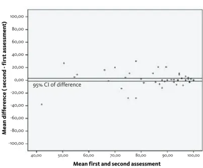
Of the 99 students who completed the VISA-P the First time, 71 filled it in the second time. The mean VISA-P score of the 28 drop-outs did not differ significantly from that of the first assessment of the 71 students. Of these 71 students, 53 (75%) were female. Mean age was 19.2 (± 0.9) years, mean height 1.75 (± 0.07) m, mean weight 67.4 (± 10.0) kg, and mean hours of sports activities per week 5.36 (± 4.1). The mean VISA-P score (± SD) was 89.5 (± 14.3) and 90.3 (± 14.2) at the first and second assessments, respectively. The ICC between the first and second assessments was 0.74 (P < 0.001). When looking at the individual questions, five out of eight questions had an ICC > 0.60 (range 0.45–0.82). Bland and Altman plot (Figure 4.1) shows that zero lies within the 95% CI of the mean difference, indicating that no bias had occurred.
Mean first and second assessment
Mean diff
er
ence ( second - first assessment
) 95% CI of difference 100,00 80,00 60,00 40,00 20,00 0,00 -20,00 -40,00 -60,00 -80,00 -100,00 40,00 50,00 60,00 70,00 80,00 90,00 100,00
46 Chapter 4
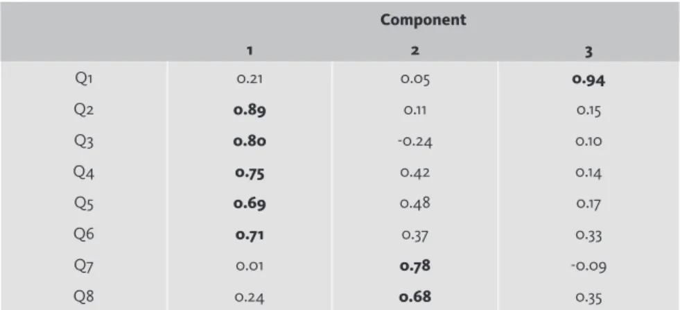
Table 4.1. Principal component analysis with varimax rotation, forced three-factor structure.
Component 1 2 3 Q1 0.21 0.05 0.94 Q2 0.89 0.11 0.15 Q3 0.80 -0.24 0.10 Q4 0.75 0.42 0.14 Q5 0.69 0.48 0.17 Q6 0.71 0.37 0.33 Q7 0.01 0.78 -0.09 Q8 0.24 0.68 0.35
Table 4.2. VISA-P scores and demographic characteristics (means (SD)) of the participants of the validity study. Healthy students At-risk population Injury other than knee
Knee injury Patellar tendino-pathy Surgery for patellar tendino-pathy Total N 18 15 19 17 14 6 89 VISA-P 95.3 (8.8) 88.6 (11.1) 76.6 (24.3) 61.9 (24.1) 58.2 (18.9) 56.0 (20.9) 75.3 (23.6) Male (%) 7 (38.9) 8 (53.3) 4 (21.1) 11 (64.7) 11 (78.6) 5 (83.3) 46 (51.7) Age (yrs) 20.0 (1.5) 25.2 (4.7) 19.2 (1.2) 24.7 (4.5) 25.1 (3.7) 32.5 (2.9) 23.0 (4.7) Height (m) 1.76 (0.08) 1.87 (0.08) 1.77 (0.07) 1.84 (0.09) 1.85 (0.09) 1.81 (0.08) 1.81 (0.09) Weight (kg) 69.9 (9.5) 82.0 (12.6) 69.2 (12.3) 82.1 (12.1) 80.5 (12.4) 93.3 (1.5) 77.0 (13.1) Sport (hours/ week) 5.1 (3.3) 8.0 (2.9) 5.6 (4.4) 3.8 (3.1) 4.5 (3.1) 1.0 (1.7) 5.2 (3.7)
Dutch translation of the VISA-P questionnaire 47
4
The Cronbach’s alpha was 0.73 for the first and 0.71 for the second assessment. Theprincipal component factor analysis yielded a two-factor structure, explaining 64.5% of the total variance. The question about sitting pain-free had the lowest factor score (0.53). Forcing a three-factor structure, explaining 74.6% of total variance, resulted in one com-ponent with five questions (pain during activities), a second comcom-ponent with two ques-tions (physical activity participation), and a third component with only one question (pain during sitting). The lowest factor score was 0.68 (see Table 4.1).
Validity
Mean VISA-P scores (± SD) and characteristics of the participants in the six different groups are displayed in Table 4.2. ANOVA revealed a significant difference between the six groups (F = 10.7, p < 0.001). When looking at the eight questions separately, on ques-tions 1 and 3 no significant difference was seen (p = 0.20 and 0.16, respectively). Posthoc analyses (Bonferroni correction) revealed that the mean VISA-P score of the group with patellar tendinopathy differed significantly from that of the healthy students group and the elite volleyball players (at-risk group), but not from the other three groups
Discussion
TranslationWe feel confident that the translated Dutch VISA-P questionnaire is linguistically equiva-lent to the original version, since our study shows that the VISA-scores of both healthy subjects (95) and athletes with patellar tendinopathy visiting a sports medicine clinic (58) are comparable with the results of Visentini (95 and 55 respectively).8 Also, the expert
translation committee judged the original and translated versions to be congruent.
Reliability
Over a time interval of 2.5 weeks, the Dutch version of the VISA-P score showed satisfac-tory test-retest reliability (ICC = 0.74). This is slightly lower than in previous studies,8,11,12
which had much shorter test-retest intervals, ranging from 1 hour to 1 week. We decided to take a longer time interval in order to prevent participants from copying the VISA-P from memory. A time interval of two weeks or more is commonly used in reliability studies. We are aware that a limitation of this study is that the test-retest reliability was investigated in asymptomatic students. One could argue that testing reliability in athletes with patellar tendinopathy would have been more appropriate. However, in the reliability study of Frohm the majority of participants (66%) who were asked to fill out the VISA-P
questionnaire were asymptomatic too.11 No differences with regard to reliability were
de-scribed between symptomatic and asymptomatic participants, therefore we believe that the reliability of the Dutch VISA-P questionnaire found in this study also applies to ath-letes with patellar tendinopathy.