International Journal of Emerging Technology and Advanced Engineering
Website: www.ijetae.com (ISSN 2250-2459, ISO 9001:2008 Certified Journal, Volume 7, Issue 12, December 2017)
Timing Analysis of Noisy MRI Segmentation using modified
Watershed Algorithm
Ravi Boda
1, B Rajendra Naik
2, D Srinivas
3, Boda Aruna
41,2ECE department & UCE Osmania university,Hyderabad, India 3,4ECE department & VJIT, Hyderabad, India
Abstract— Image segmentation is an important and
nontrivial task in image processing. The main purpose of the image segmentation is to partition the original image into uniform regions. Image segmentation can be applied as the pre-processing stage for the other image processing techniques. Segmentation of medical images has become
basic fundamental process to perform significant
quantitative analysis to analyze the human organs and lapse tissues. In this paper, we propose an algorithm for MRI segmentation which combines the watershed transform and topological gradient approach. Our aim is to improve the segmentation processing time by considering watershed of image gradient. The proposed method is fast, less sensitive to noise compared with Level set Method, active contour method and gray threshold method applicable on real time medical image processing.
Index Terms—Image segmentation, active contour, topological gradient, watershed, medical images. (key words),
I.INTRODUCTION
Image segmentation is the first step before going to the high level processing such as object recognition, human recognition and computer vision application. this task is highly challenging that most of the real time imaging devices gives corrupted images by intensity fluctuation. Particularly in medical images [1], image segmentation is essential for the feature extraction, image display and image dimensions.
Image segmentation techniques are divided into different types based upon the classification scheme. The most frequently used segmentation techniques are categorized into two types i.e. region based and edge based approach. The main concept of edge based approach is to identify the boundaries of the object by using the edge detection operator [2] and then extracting the boundaries using the edges information. The issue of edge detection is the existence of noise that results in random variations occurs from pixel to pixel. Thus in real images, ideal edges can never been encountered. The huge dissimilarity of the edge detection algorithms have come up with differences in their algorithmic and mathematical properties such as Canny, Laplacan, Prewitt, Sobel, Roberts, all of which are based on the variation of gray levels. The variations in gray levels are used to identify the discontinuity in gray levels.
On the other side, region based approach is based on the similarity of local image data. Most commonly used approaches are region growing, clustering, thresholding, splitting and merging. Yet, the performance evaluation of an image segmentation results is still a challenging task because they are failing to extract the exact boundaries of objects in a noisy images.
In present days, there are several image segmentation techniques are developed to work out the problem occurs in the detection of boundaries in an images e.g. Active Contour Method[3](ACM), Gray threshold Mechanism(GTM), Sobel Gradient Method (SGM) and level set method[4] etc. Chan and proposed a level set method which is not applicable for parallel programming because at every iteration average intensity inside and outside contour need to be computed, which increases the CPU time. For this purpose we propose a new algorithm which tries to overcome above drawbacks.
This paper is structured as mentioned: Section II we present different segmentation techniques In Section III, presents the methodology of implemented image segmentation technique. In section IV we show the experimental results, which are applied on head and brain MRI [5] images with 481x321 image sizes. The results of each image segmentation technique are compared using performance measure in time. Section V gives the conclusion of this paper.
II. IMAGE SEGMENTATION TECHNIQUES
This section details the concept of segmentation techniques are discussed and finally we implement the proposed method.
A.Active Contour Method (ACM)
International Journal of Emerging Technology and Advanced Engineering
Website: www.ijetae.com (ISSN 2250-2459, ISO 9001:2008 Certified Journal, Volume 7, Issue 12, December 2017)
90 Internal force is defined based on shape of snake where as external force defined from the image and /or from higher level image understanding criteria. The snake V(s) defined as
( ) ( ( ) ( )) (1)
Where
x
(
s
)
andy
(
s
)
be the x, y-coordinates that are related with contour, , -. Energy functional to be minimized can be represented as∫ ( ( ))
∫ ( ( ( )) ( ( )) ( ( )))
(2)
Where an internal energy by the reason of
bending, is an image force is an External
constrains force. Internal spline energy can be written as
( ) |
| ( ) | | ( )
Where ( ), ( ) are elasticity and stiffness of the snake. | ⁄ | is Continuity Force (diereses the distance between points) | ⁄ | is Curvature Force
(provides smoothness and removes oscillation of the snake). Image force is a weighted combination of three different functions presented, which attracts the snakes to line, edge and terminations.
( )
Where ( )
Here ( ) is denoted as image gray level at (x, y)
co-ordinates, represents whether snake is attracted to
light or dark lines.
Where | ( )|
It attracts the snake to contour with large image gradients i.e. to locate the strong edge. Line termination and corners may influence the snake using weighted energy function
E
termLet ‗g‘ be a smoothed form of image ‗f‘, ψ(x,y) is the gradient directions along the spline in smoothed image g, and let.
( ) ( ( ) ( ))
( ) ( ( ) ( ) )
are unit vector perpendicular and along the gradient directions ψ(x,y). Then the curvature of constant gray level contours in the smoothed image
⁄
⁄
The snake behavior is changed by varying the weights ωline, ωedge and ωterm. The third term of integral equation 2
comes from external constraints imposed either by user or some other higher level process.
B.Gray Threshold Mechanism (GTM)
It is a simple to compute image segmentation among all other image segmentation methods. Many objects (or) image regions are characterized by reflectivity (or) light absorption of their surfaces. A brightness constant also known as threshold is used to segment objects and background.
It is computationally less expensive and fast. Gray thresholding [8] one of the oldest image segmentation and still widely used in many applications like bio-medical imaging, SAR imaging etc. This thresholding can be done easily in real time using specialized hardware. A complete segmentation of an image ‗R‘ a finite set of R1, R2 …RS.
s i i R R 1 , , Ri
Rj 0 if i≠j (5)Complete gray level threshold mechanism results from thresholding
( ) { ( )
( )
Where g(i, j) output binary image and T is threshold g(i,j)=1 for image of object, g(i,j)=0 for image of background.
C.Sobel Gradient Method (SGM)
The sobel gradient of an image is computed for each pixel in an image sobel gradient is computed using the following steps:
step i. Creating the sobel masks for x-direction and y-
direction
step ii. Compute the x-direction derivative, which is subtracting the first row from the third row with the help of the mask.
step iii.Compute the y-direction derivative, which is subtracting the first column from the third column
with the help of the mask.
step iv.Calculate the gradient using the equation 2 1 2 2 ] [ )
( f Gx Gy
mag
f
(6)
III.PROPOSED METHOD
International Journal of Emerging Technology and Advanced Engineering
Website: www.ijetae.com (ISSN 2250-2459, ISO 9001:2008 Certified Journal, Volume 7, Issue 12, December 2017)
The method is split in to two parts the first part is topological gradient method and second part watershed transform
{ ( ) (7)
Topological Gradient Method
In this section, we adopt the topological gradient as an edge detection tool. First, we consider the principle of the topological asymptotic expansion
Let ω is an bounded domain of R2 and j(ω) =J(u ω) is a
cost function to be minimized. Where uω is solution of
partial differential equation[9] defined in ω. For a given function v in L2(ω). We need to find uH1 (ω) such that
Where c is constant For a given xo
ω and for a smallρ≥0 ,let ωp= ω/σp perturbed domain by insertion of a crack
)
(
0
n
x
p
. Wherex
0
,
(
n
)
is strait crack and n is unit vector which is normal to crack. The topological sensitivity theory gives an expression of))
(
(
)
,
(
)
(
)
(
)
(
j
f
G
x
0n
f
j
p
(8)Where f (
) is positive function going to zero with
and G(x0, n) is defined as the gradient at x0.For a function v in L2 (ω), consider the fallowing condition to find
u
H
1(
p)
so that
in
u
in
v
u
u
c
div
n0
)
(
(9)The basic goal is as fallows. If there is crack in a flat part of the image no edge detected. But, if a crack is along an edge(strong gradient) the potential energy decreases
As specified previously, the topological gradient is less sensitive to noise and small changes of the image. This is due to the topological gradient calculate in a global way whether a pixel is a chunk of an edge or not,
Since the topological gradient is the good tool for protecting the most necessary edges and eliminating all the other irrelevant ones, then the next idea is to change the morphological gradient by a topological gradient for the sake of minimize the set of minima of image, leading to better segmentation results. Our method is then consisting of two different steps; the first one is of detecting the essential edges of the image employing the topological gradient restoration process.
Watershed Transform
The second step consists of employ the watershed algorithm using the topological gradient calculated using topological gradient, rather than that of the morphological gradient classically used in watershed algorithms.
The Watershed is a unique algorithm used for biomedical image segmentation [10] i.e. subdividing different objects in an image It is sinking like landform defined hill points and ridge lines that go down into lower elevations and creek valleys [11]. The watershed segmentation method works well, if we can identify foreground objects and background objects. Watershed segmentation is done by following steps
1. Calculate the segmentation function
2. Compute the cutting edge markers. These are associated as
blobs or pixels within every object
3. Compute backdrop markers. These are the pixels, which
are not a part of any object
4. Change the segmentation function, so that it has only minima at the cutting edge and background marker location.
5. Compute the watershed transform of altered segmentation
function
The co-ordinate of points in the catchment basin related with regional minimum[12]( Mi. finally T[n] represents the set of coordinates (s, t) for which g(s, t) < n i.e.
, - *( )| ( ) + (10)
The binary Image is
( ) ( ) ⋂ , - (11)
Cn(Mi)=1 at location (x, y) if (x, y) C(Mi)AND(x, y)
T[n] Cn(Mi)=1 else. The AND operator is used toisolate the stage n of flooding the portion of the binary image T[n] which is related to regional minimum Mi.
IV.EXPERIMENTAL RESULTS
International Journal of Emerging Technology and Advanced Engineering
Website: www.ijetae.com (ISSN 2250-2459, ISO 9001:2008 Certified Journal, Volume 7, Issue 12, December 2017)
92
(a) Input (b) AC (c) GTM
(d) SGM (e) WST
The MRI segmented images of bone is shown in figure 1. (a) Shows the original input images, (b) shows the ACM segmented output images, (c) shows the GTM segmented output images, (d) shows the SGM segmented output, (e) Shows the WST segmented output MRI images.
International Journal of Emerging Technology and Advanced Engineering
Website: www.ijetae.com (ISSN 2250-2459, ISO 9001:2008 Certified Journal, Volume 7, Issue 12, December 2017)
(d) SGM (e) WST
The MRI segmented images of head . (a) Shows the original input images, (b) shows the ACM segmented output images, (c) shows the GTM segmented output images, (d) shows the SGM segmented output, (e) Shows the WST segmented output MRI images.
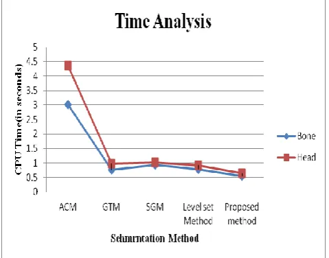
COMPARISON OF SEGMENTATION METHODS
S.No Segmentation method
CPU Time (in seconds) Bone Head
1 ACM 3.02 4.36
2 GTM 0.76 0.98
3 SGM 0.94 1.02
4 Level set Method 0.79 0.92
6 Praposed Method 0.55 0.64
Image dimentions 481x321
Table 1 shows the segmentation results with time comparison.The proposed topological gradient and watershed transform algorithm shows better performance over levelset Method
Time Analysis of segmentation methods
The graph in Fig.3. shows the segmentation results with time analysis.The proposed algorithm shows better performance over levelset Method, sobel Gradient Method,Gray threshold Method and Active contour
V.CONCLUSION
A new method for image segmentation with topological gradient and watershed transform has been presented in this work. First we detect the essential edges of the image then we compute the watershed of the gradient. This methodology has many advantages, particularly in real time medical applications. The results obtained are very promising particularly from the computing time point of view.
Acnowledgement
The author would like to acknowledge UGC, fellowship provided by Govt. of India. NewDelhi, India.
REFERENCES
[1] J.D. Lee, H.R. Su, P. E. Cheng, M. Liou, J. Aston, A. C. Tsai, and C.-Y. Chen, ―MR image segmentation using a power transformation approach,‖ IEEE Trans. Med. Imag., vol. 28, no. 6, pp. 894–905, Jun.2009.
[2] P. Jiantao, J. K. Leader, B. Zheng, F. Knollmann, C. Fuhrman, F. C. Sciurba, and D. Gur, ―A computational geometry approach to automated pulmonary fissure segmentation in CT examinations,‖ IEEE Trans. Med. Imag., vol. 28, no. 5, pp. 710–719, May 2009. [3] Bing Li, and Scott T. Acton, Active Contour External Force
Using Vector Field Convolution for Image Segmentation, IEEE Transactions On Image Processing, Vol. 16, No. 8, August 2007. [4] Souleymane Balla-Arabé, Xinbo Gao, Senior Member, IEEE, and
Bin Wang, ―A fast and Robust level set method for Image Segmentation using Fuzzy clustering and Lattice Boltzmann Method‖ IEEE transactions on cybernetics, vol. 43, NO. 3, JUNE 2013
[5] I. Isgum, M. Staring, A. Rutten, M. Prokop, M. A. Viergever, and B. van Ginneken, ―Multi-Atlas-based segmentation with local decision fusion—Application to cardiac and aortic segmentation in CT scans,‖ IEEE Trans. Med. Imag., vol. 28, no. 7, pp. 1000– 1010, Jul. 2009.
[image:5.595.48.288.514.702.2]International Journal of Emerging Technology and Advanced Engineering
Website: www.ijetae.com (ISSN 2250-2459, ISO 9001:2008 Certified Journal, Volume 7, Issue 12, December 2017)
94
[7] Charnchai Pluempitiwiriyawej, José M. F. Moura, Yi-Jen Lin Wu, and Chien Ho, ―STACS: New Active Contour Scheme for Cardiac MR Image Segmentation‖, IEEE TRANSACTIONS ON MEDICAL IMAGING, VOL. 24, NO. 5, MAY 2005.
[8] M. Sezgin and B. Sankur (2004). "Survey over image thresholding techniques and quantitative performance evaluation". Journal of Electronic Imaging 13 (1): 146–165. doi:10.1117/1.1631315.
[9] Chen Wei-bin, Zhang Xin, ―A New Watershed Algorithm for Cellular Image Segmentation based on Mathematical Morphology‖, IEEE:International Conference on Machine Vision and Human-machine Interface,2010
[10] H.P. Ng, S. Huang, S.H. Ong, K.W.C. Foong, P.S. Goh, W.L. Nowinski, ―Medical Image Segmentation Using Watershed Segmentation with Texture-Based Region Merging‖, 2008,pp. 4039-4042.
[11] Jean Cousty, Gilles Bertrand, Laurent Najman, and Michel Couprie. Watershed cuts: thinning, shortest path forests and topological watersheds. IEEE Transactions on Pattern Analysis and Machine Intelligence.32(5).2010.pp.925–939.