An Analysis of Skin Pixel Detection using Different
Skin Color Extraction Techniques
Gururaj P Surampalli
M.Tech (CSE) Student GNDEC Bidar-585401Dayanand J
Asst. Professor GNDEC Bidar-585401Dhananjay M
Asst. Professor GNDEC Bidar-585401ABSTRACT
Automated skin detection from a captured natural image has wide range of application. Detection of skin area in a given im-age is done through marking skin and non skin pixels. Process of identification of skin pixel is closely associated with color space being used. To select suitable method to extract skin re-gion has motivated this paper. We are using multiple color spaces in a paper to analyze and compare them. We have the different set of images to compare color space. The results indicate that YCbCr provide better performance compare to other color space.
General Terms:
Color space, Skin pixel
Keywords:
Skin pixel detection; Log opponent; HSV; YIQ; YCbCr
1. INTRODUCTION
The skin color is useful in most of the recent technologies which include face detection, localization, image content filtering, con-tent aware video compression and image color balancing ap-plications. There are many techniques for skin color modeling and recognition, which are proposed during the past years. Most commonly used is skin segmentation techniques, which involve the classification of individual image pixels into skin and non-skin categories on the basis of pixel color [6]. It is very hard task to extract regions of specific color from a given color image, the color of an object varies with changes in illumination color. It is found in present work that separating illumination from chromi-nance can produce good result in detecting skin pixel [2]. Generally human skin is characterized by a combination of red and melanin (yellow, brown) and there is somewhat a range of hue for skin and saturation that represent skin pixels [10]. The detection of human skin effected by various external factors, such as types of cameras used to capture the image, light-setting, human race and color spaces [1]. Skin color detection most im-portant process in vision systems, like gesture recognition, re-gion of interest hand tracking, video indexing, face detection, etc [7].
The skin pixel based detection can reduce the search space be-fore high level processing, however this is not an easy task. Skin pixels vary with ambient light, such as color lamps which acts as a filter, brightness and shadows, daylight, etc. Different cam-eras with variant resolution return different values for the same scene. Skin pixel detection is a cumbersome task. The main aim of skin color detection is to build a decision rule that will differ-entiate between skin and non-skin region based on pixels values.
Determining skin pixel color involves finding the range of val-ues for which most of the skin pixels would fall in a given color space. The theme of a color space is to facilitate the specifica-tion of colors in some standard way, generally accepted manner. A color space includes specification of a coordinate system and the subspace within a system where each color is represented by a single point. In later color spaces the orientation is toward hardware such as color monitors or toward applications where color manipulation is a goal such as the creation of color graph-ics or animation. Numerous color spaces are used for processing digital images. For some process, one color space may be more appropriate than other color space [7]. Most of the work has been done on finding faces in color images using methods such based on chrominance pesent in image [4], detecting face based on skin color [8] [11] and based on AdaBoost [12].
[image:1.595.335.510.477.602.2]The block diagram of the skin detection using Log opponent, HSV, YIQ, YCbCr techniques discussed in present paper as shown: Generally, a good skin color model must offer a high
Fig. 1. Block diagram of skin detection
detection rate and a low false positive rate. That is, skin color model must detect more number of skin pixels while minimizing the number of non-skin pixels classified as skin [9].
Pixel classification is complicated and there are number of sug-gested methods for classifying pixels color as skin or non-skin color in an attempt to achieve the maximum performance [3].
2. SOME SKIN PIXEL DETECTION TECHNIQUES
2.1 Log opponent
opponent hues, yellow-blue and green-red, which will get can-cel each other superimposed. Log opponent color space for skin pixel color detection is suggested by Fleck et al [6].
Where n is a random noise value generated from a uniform distri-bution over the range[0,1]and the constant 105 is used to scale the range to the interval[255,255].
The algorithm for the log opponent is as follows:
Step 1: Take input image, which contains RGB values. Step 2: Intensity in the image is calculated by multiplying the parameter value (0.596, -0.274, -0.322) to the RGB components. Step 3: The hue present in the image is calculated by taking in-verse tangent of the(Rg, By).
Step 4: Check whether intensity lies between (20, 90) and hue lies between (100, 150) [6].
Step 5: If the pixel satisfies Step: 4, the detected pixel is skin pixel. Otherwise the pixel detected is not skin pixel.
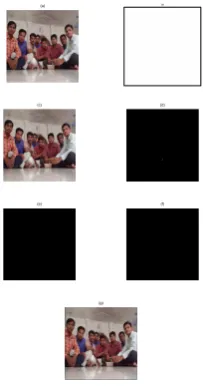
The result using log opponent as shown
(a)
(c) (d)
(e) (f)
[image:2.595.378.478.284.474.2](g)
Fig. 2. (a) Input image. (b) Segmented. (c) Color segmented image.
(d) Edge detection. (e) Binarilization. (f) Morphological operation. (g) Skin detected.
From the results, it can be observed that segmented image is white because the all pixel present in image considered as skin pixel. Hence, futher processing steps also fail to give correct re-sult.
2.2 HSV (Hue, Saturation, Value)
The HSV color space defines color with intuitive values, based on the artist idea of characteristics such as tint, saturation and tone. It was introduced when it is needed to mention color properties numerically. Hue defines the dominant color as de-fined by wavelength, let us for instance the difference between red and yellow colors. Saturation is the measures of colorful-ness area in proportion to the brightcolorful-ness on the area, such as the distinction between red and pink color. The value indicates the color luminance, the distinction between a dark red and a light red. In skin detection, the numeric value of component is discarded to eliminate the undesirable effect of not evenly dis-tributed illumination[6].
There are some of the studies, which shows that HSV is invariant to highlights at white light sources, to matte surfaces, and ambi-ent lighting. However, hue non continuities and the computation of the luminance component conflict badly with the properties of color vision. The cyclic behavior of Hue-Saturation spaces also
makes it inconvenient for parametric skin color models that need a rigid cluster of skin performance, for optimum performance. The selected range of H control segmentation ton reddish colors and the saturation range selected ensures the exclusion of pure red and very dark red colors, both of which are caused by small variations in lighting conditions. The threshold on value(V) is introduced to reject dark colors.
The algorithm for the HSV is as follows:
Step 1: Take input image, which contains RGB values. Step 2: Convert RGB to HSV components.
Step 3: Check whether saturation lies between (0.20, 0.75), value greater than 0.35 and hue lies between (0, 25) [6]. Step 4: If the pixel satisfies Step: 3, the detected pixel is skin pixel. Otherwise the pixel detected is not skin pixel.
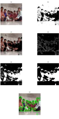
The result using HSV as shown
(a) (b)
(c) (d)
[image:2.595.118.220.298.493.2](g)
Fig. 3. (a) Input image. (b) Segmented. (c) Color segmented image.
(d) Edge detection. (e) Binarilization. (f) Morphological operation. (g) Skin detected.
The results shown indicate that all the pixel in an image consid-ered as non skin pixel, therefore a black image. The black image converted into binary image (f), which is white. Hence, morpho-logical image is also white.
2.3 YIQ (Luma, Inphase, Quadrature)
It is also known as modified log opponent technique [6]. The YIQ system is intended to take advantage of human color-response characteristics. The algorithm for the YIQ is as follows:
Step 1: Take input image, which contains RGB values. Step 2: Intensity in the image is calculated by multiplying the parameter value (0.5957, 0.2745, -0.3213) to the RGB compo-nents [6].
Step 3: Check whether intensity lies between (20, 90) and hue lies between (100, 150) [6].
Step 4: If the pixel satisfies Step: 3, the detected pixel is skin pixel. Otherwise the pixel detected is not skin pixel.
The result using YIQ as shown
(a)
(c) (d)
(e) (f)
[image:3.595.118.220.95.291.2](g)
Fig. 4. (a) Input image. (b) Segmented. (c) Color segmented image.
(d) Edge detection. (e) Binarilization. (f) Morphological operation. (g) Skin detected.
2.4 YCbCr (Luma, Blue Chroma, Red Chroma)
YCbCr color space has been defined to meet the increasing requirements of digital algorithms in handling video information and has become the most frequently used color space in digital videos. YCbCr is consist of three components, two of them is of chrominance and one is of luminance[5].
In order to optimize the performance of skin color clustering, the present work uses YCbCr space to build a skin color model, since it is also known that, as the chrominance components are almost independent of luminance component in the space there are non-linear relations between chrominance (Cb, Cr) and luminance(Y) of skin pixel color in the high and low luminance region. As in RGB space, the triple component (r,g,b) expresses not only color but also luminance. Luminance may change across a person’s face due to the ambient lighting and is not a reliable measure in discriminating skin from non-skin region. YCrCb is actually an encoded nonlinear RGB signal, commonly used by European television studios and for image compression work. Color is represented by luma (that is luminance, computed from nonlinear RGB), it is constructed as a weighted sum of the RGB values, and two color difference values Cr and Cb that are calculated by subtracting luma from RGB red and blue components. The conversion simplicity and explicit separation of luminance and chrominance components makes this color space attractive for skin color modeling. In YCbCr color space, the two chroma components Cr, and Cb can be efficiently used to define explicitly skin region. The thresholds be selected as (Crmax; Crmin) and (Cbmax; Cbmin) , a pixel value is classified as skin pixel, if the values (Cr,Cb) fall within the thresholds. The luminance has to be removed from the color representation in the chromatic color space. Chromatic colors known as “pure” colors in the absence of luminance.
The YCbCr conversion from RGB color space can be accom-plished by following matrix.
" Y
Cb Cr
#
=
" 1 1 1
0.148 −0.291 0.439 0.439 −0.368 −0.071
# "R
G B
#
+
" 16
128 128
#
For the skin pixel using YCbCr, red chrominance value lies be-tween (140, 165) , blue chrominance value lies bebe-tween (140, 195) and hue value lies between (0.01, 0.1). The result using YCbCr as shown
From results of YCbCr, it is found that the segmented and color
(a) (b)
(c) (d)
(e) (f)
(g)
Fig. 5. (a) Input image. (b) Segmented. (c) Color segmented image.
(d) Edge detection. (e) Binarilization. (f) Morphological operation. (g) Skin detected.
segmented images are formed on the basis of skin pixel. The fur-ther processing is also carried out, the skin region detected is shown in image (g).
3. RESULTS AND DISCUSSION 3.1 Single image analysis
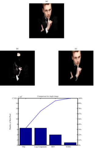
Consider the image for applying all the techniques discussed it is found that YCbCr is more capable in finding more valid skin pixel as shown.
(a) (b) (c)
[image:3.595.379.478.96.293.2](d) (e)
Fig. 6. Input image and results of Log opponent HSV YIQ YCbCr
For single image it has been observed from the results that, the YCbCr technique is able to recognize skin pixel more as com-pared to other techniques. The HSV technique also produces some good result. As shown below.
YIQ Log Component HSV YCbCr 0
0.2 0.4 0.6 0.8 1 1.2 1.4 1.6 1.8 2x 10
5 (f)
Color spaces
Number of Skin Pixel
[image:3.595.347.511.468.564.2]0% 10% 20% 30% 40% 50% 60% 70% 80% 90% 100%
Fig. 7. Graph shows Number of skin pixel detected in different
colorspaces
[image:3.595.381.479.653.731.2]the vector y are drawn as bars in descending order. Each bar is labeled with the associated skin detection technique. The line above the bars shows the cumulative percentage. It is observed that with several test images, the region of skin can be detected efficiently by using YCbCr technique. Log opponent, HSV and YIQ fails to detect the correct skin pixel. The table 1 below shows number of skin pixel detected in images shown in results.
Table 1. Number of Skin Pixel Detected for Test Images
Image Log opponent HSV YIQ YCbCr
1 65025 0 65025 9352
2 65025 16092 65025 27552
3 65025 6 65025 21474
4 65025 0 65025 1546
5 65025 0 65025 5916
6 65025 39378 65025 9583
7 65025 119 65025 22628
8 65025 251 65025 19980
9 65025 21 65025 8708
10 65025 0 65025 17638
3.2 Comparison of Log Opponent, HSV, YIQ and YCbCr Techniques
When each technique is applied to set of test images, following graph can drawn indication number of skin pixel and non skin pixel present in the image.
0 5 10 15 20 25 30 35
0 1 2 3 4 5 6 7x 10
4
Images
Detection(Pixel)
Skin Pixel Non Skin Pixel
0 5 10 15 20 25 30 35
0 1 2 3 4 5 6 7x 10
4
Images
Detection(Pixel)
Skin pixel Non Skin pixel
0 5 10 15 20 25 30 35
0 1 2 3 4 5 6 7x 10
4
Images
Detection(Pixel)
Skin Pixel Non Skin Pixel
0 5 10 15 20 25 30
0 1 2 3 4 5 6 7x 10
4
Images
Detection(Pixel)
[image:4.595.77.273.415.763.2]Skin Pixel Non Skin Pixel
Fig. 8. Comparison of Log opponent, HSV, YIQ, YCbCr
From above graphs the following can be determined:
—Log opponent:
It has been observed that log opponent technique provides the result in which it considers the all pixel present in the image as skin pixel. The graph shows the blue bars indicating skin pixel present in the some test images processed. Log opponent technique fails to provide good result as observed but the im-provement can be achieved by changing the parametric values.
—HSV:
The HSV technique provide good result as compared to the log opponent, the red bars represents the non skin pixels present in the test image. It is observed that the re-gion of skin area provided by this technique is not satisfactory.
—YIQ:
YIQ results are as shown. By observation the results are similar to the log opponent. That is the number of pixel present in the image considering as skin pixel shown by blue bars. Blue bars indicate number of skin pixel present in an image.
—YCbCr:
The study and experimental results proves that YCbCr tech-nique or color space provides correct skin pixel among other discussed. The result shows number of pixel detected as skin and non skin pixel.
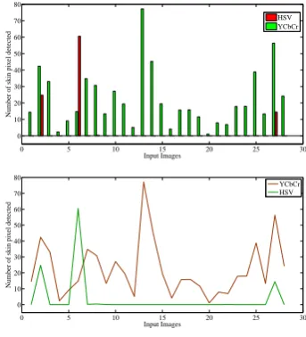
As per the above set of graphs and by the observation, it is found that Log opponent, YIQ are fail to detect the exact skin pixel present in the test images. Hence values which are provided by these techniques are not adequate. For the image of size 255 x 255 providing result 65025, that is whole area of the image is considering as skin area which not true.
In another case where HSV and YCbCr are providing good result. Hence comparing the two techniques on the basis of there detection ability, following graph can be drawn.
0 5 10 15 20 25 30
0 10 20 30 40 50 60 70 80
Input Images
Number of skin pixel detected
HSV YCbCr
0 5 10 15 20 25 30
0 10 20 30 40 50 60 70 80
Input Images
Number of skin pixel detected
[image:4.595.341.513.503.692.2]YCbCr HSV
Fig. 9. Comparison of HSV, YCbCr techniques
Table 2. Percentage of skin region detected for some images with size 255 x 255
Image HSV HSV in % YCbCr YCbCr in %
1 0 0 9352 14.384
2 16092 24.747 27552 42.374
3 6 0.009 21474 33.024
4 0 0 1546 2.377
5 0 0 5916 9.098
6 39378 60.558 9583 14.737
7 119 0.183 22628 34.798
8 251 0.386 19980 30.726
9 21 0.032 8708 13.391
10 0 0 17638 27.124
efficiency of YCbCr is more compared to HSV, Log opponent and YIQ. The percentage of skin detected for some images of
(a)
(b) (c)
YIQ Log Component HSV YCbCr 0
2 4 6 8 10 12 14 16 17.9011x 10
4 Comparison for single image
Color spaces
Number of Skin Pixel
[image:5.595.92.250.280.523.2]0% 11% 22% 34% 45% 56% 67% 78% 89% 100%
Fig. 10. (a). Input image, Output of (b). HSV , (c). YCbCr, (d).
Comparison graph showing number of skin pixel detected
size 255x255 from database by using HSV and YCbCr shown in table 2. The reason for percentage change in the HSV for image 6 as shown in figure 10.
4. CONCLUSION
Skin detection plays very important and crucial part in many bio-metric systems. The work has been carried out to understand and analyze the log opponent, HSV, YIQ and YCbCr skin pixel de-tection techniques. Each technique perform the pixel dede-tection using different set of parameters for detecting skin pixel and non skin pixel.
The proper analysis is carried out during the present work, which involve applying each technique to set of images present in
database. The comparison of each technique for the single im-age is performed. Further work extends for comparing the log opponent, HSV, YIQ, and YCbCr for skin pixel detection ability. Results show that log opponent, YIQ fails to detect the correct skin pixel. Hence analysis is emphasized on HSV, YCbCr. Dur-ing the comparison of HSV, YCbCr it is found that skin pixel detection efficiency of HSV is 3.57% and for YCbCr is 96.42% for the set of images used from database.
5. ACKNOWLEDGMENTS
I owe my thanks much more than the words can express to my parents and friends without whose co-operation and inspiration this paper would have been a distant dream.
6. REFERENCES
[1] E Angelopoulou. Understanding the color of human skin. In Proc. SPIE Conf. On Human Vision and Electronic Imaging VI (SPIE), 4299:243–251, 2001.
[2] J Brand, S Mason, M Roach, and M Pawlewski. Enhanc-ing face detection in colour images usEnhanc-ing a skin probabil-ity map.Int. Conf. on Intelligent Multimedia, Video and Speech Processing, pages 344–347, 2001.
[3] Cynthia A Brewer. Color use guidelines for data represen-tation.Alexandria: American Statistical Association, pages 55–56, 1999.
[4] D N Chandrappa, M Ravishankar, and D R RameshBabu. Automated detection and recognition of face in a crowded scene.International Journal of Computer and Network Se-curity, 2(6):65–70, June 2010.
[5] D N Chandrappa, M Ravishankar, and D R RameshBabu. Face detection in color images using skin color model al-gorithm based on skin color information.IEEE, pages 254– 258, 2010.
[6] Tarek Abd El-Hafeez. A new system for extracting and detecting skin color regions from pdf documents. Inter-national Journal on Computer Science and Engineer-ing(IJCSE), 2(9):2838–2846, 2010.
[7] B Hajar, E Sanaa, J Abdelilah, and A Driss. Recognition of adult video by combining skin detection features with motion information.IEEE, 2010.
[8] Jiang Qiang-rong and Li Hua-lan. Robust human face de-tection in complicated color images.IEEE Trans, 2010. [9] S Sanjay, D S Chauhan, V Mayank, and S Richa. A robust
skin color based face detection algorithm.Tamkang Jour-nal of Science and Engineering, 6(4):227–234, 2003. [10] M R Tabassum, A U Gias, M M Kamal, H M Muctadir,
M Ibrahim, A K Shakir, A Imran, S Islam, M G Rabbani, S M Khaled, M S Islam, and Z Begum. Comparative study of statistical skin detection algorithms for sub-continental human images.Institute of Information Technology, Uni-versity of Dhaka, pages 1–8.
[11] Randazzo Vincenzo and Usai Lisa. An improvement of ad-aboost for face-detection with motion and color informa-tion.IEEE 14th International Conference on Image Analy-sis and Processing (ICIAP), 2007.