Structure-Guided Modeling and Binding Studies of
GABA
A
Receptor Subunit Beta-3
ABSTRACT
The major inhibitory GABA systems are known to take part a vital role in epilepsy, associated with excessive neuronal circuitry excitation. This excitatory action reflects seizures which result from GABA inhibitory circuit dysfunction. GABA mediates its fast inhibition through GABAA receptors
by activating it and then opens the chloride channels. This opening allows chloride ions to flow into the interior of the cell which inhibits the excitability. This inhibitory action devises anticonvulsive properties as a consequence. So GABAA receptors are primary targets in the pathophysiology
of epilepsy. Enhancement in the the action of GABAA
receptors is the basis of epileptic seizures reduction on which many antiseizure drugs act. Modulation of GABAA receptor
can be shown by many synthetic and natural compounds. Valerenic acid, a plant origin compound shows modulatory effects on GABAA receptors. In our present study, the
homology model of the GABAA receptor subunit beta-3 is
build and docking studies are performed with Valerenic acid. Docking analysis is done to know the binding interactions and their binding affinity. The results revealed the interacting amino acids which are involved in binding of GABAA
receptor subunit beta-3 with Valerenic acid are ASP35, GLN56, PHE55, TYR120, ARG111 and TYR 54.
Keywords
GABA, GABAA receptor, epilepsy, antiseizure, Valerenic
acid
1.
INTRODUCTION
The γ-aminobutyric acid type A receptor (GABAA), a member
of the Cys-loop ligand-gated ion channels, is the major mediator of GABAergic synaptic inhibition in the brain [1]. GABA-mediated inhibition through GABAA receptors is of
major importance in the normal functioning of the nervous system and have also been the target of several clinically relevant anticonvulsant drugs [2, 3].When GABA molecules or GABA-like compounds bind to the receptor and activate it, this channel temporarily opens and allows the passage of negatively charged molecules, such as chloride ions (Cl-), to pass from the cell’s exterior to its interior. This ion flow decreases the cell’s excitability. This cumulative neuronal inhibition caused by GABA binding to many neurons results anticonvulsive properties [4].
The GABAA receptors are composed of a pentameric
structure, with five subunits arranged in a ring enclosing a central chloride ion channel. The five subunits arise from seven subunit families that contain multiple subtypes (α1–6,
β1–3, γ1–3, δ, ε, π, θand ρ1–3) [1, 5]. The majority of GABAA
receptors contain two α subunits, two β subunits, and a γ or δ
subunit [6] and that there is regional heterogeneity of the subunit composition of GABAA receptors in the mammalian
brain[1,7]. The formation of functional GABAA receptors
requires the coexpression of a least two subunit types, α and β subunits, with both of these subunits contributing to the GABA binding site. GABAA receptors are activated by
binding of agonist to recognition sites located at α/β subunit interfaces [8, 9]. The functional and pharmacological properties of the GABAA receptors are determined in large
part by their subunit composition [10]. These differential regulatory properties also underlie their role for fine tuning of neuronal circuits and genesis of network oscillations which represent a major facet of homeostatic synaptic plasticity and contribute to the excitation/inhibition (E/I) balance under physiological conditions and upon pathological challenges [11]. Imbalance between excitatory and inhibitory synaptic transmission in key brain areas are implicated in the pathophysiology of epilepsy, in which there is a decrease in the GABA mediated inhibition. [11]. Furthermore, these distinct receptor subtypes are preferentially expressed in specific regions and neuronal populations and they exhibit different sensitivities to modulators including neurosteroids, benzodiazepines, ethanol and barbiturates [12, 13]. By taking this diversity role of GABAA receptors, in our present study,
Valerenic acid, a modulator of these receptors, is taken and performed docking studies with GABA receptor. To stimulate the structure-based design for new drugs that target restricted neuronal networks implicated in GABA related epilepsy medication and minimize side effects, it is vitally important to find the 3D structures of GABAA receptors
subtypes. The present study initiates an attempt to find the 3D structures for the extracellular domain of beta-3 subunit of GABAA receptor and docking studies are carried out with
Valerenic acid respectively. The results are further analyzed to know the interactions and binding mode of the receptor with Valerenic acid.
2.
METHODOLOGY
2.1 Sequence Retrieval and Homology
Modeling
The protein sequence of human GABAA receptor subunit
beta-3(GABRB3) is obtained from the SWISS-PROT database that has 473 amino acids. The extracellular domain sequence is retrieved and searched to find out the homologus protein structures to be used as a template by the BLAST program against Protein Data Bank database. The 3D homology model of GABRB3 is modeled using crystal structural coordinates of templates on the basis alignment of target and template. These procedures are performed by ICM MOLSOFT [14]. The
B. Rajasekhara Reddy
Professor of ECE
K G Reddy College of Engineering and Technology, Moinabad, Ranga Reddy
District, PIN: 501504, India.
L. Pratap Reddy,
PhD.Professor of ECE JNTU College of Engineering
JNTUH, Kukatpally Hyderabad – 500085, India.
Sravani Saragandla
Research AssociateAsterace Labs Windsor Plaza
modeled structure is further optimized by energy minimization using Discovery Studio.
2.2 Model Evaluation
2.2.1 PROCHECK
A versatile protein structure analysis program [15] PROCHECK is used in validation of protein structure and models by verifying the parameters like Ramachandran plot quality, peptide bond planarity, Bad nonbonded interactions, main chain hydrogen bond energy, Calpha chirality and over-all G factor and the side chain parameters like standard deviations of chi1 gauche minus, trans and plus, pooled standard deviations of chi1 with respect to refined structures [16].
2.2.2 ProSA
This program compares Z scores between target and template structure. The Z scores of model is a measure of compatibility between its sequence and structure. The model Z score should be comparable to the Z scores obtained from the template [17].
2.2.3 RMSD
Root Mean Squared Deviation (RMSD) is commonly used to represent the distance between two objects. In a structural sense, this value indicates the degree to which two three dimensional structures are similar. The lower the value, the more similar the structures are. The RMSD value [18] between the template 3RHW and our model structure is calculated using SPDBV program (Figure 39a0 & (b)).
2.3 Ligand Preparation
The structure of the Valerenic Acid is downloaded from pubchem and imported into Discovery Studio. Hydrogen bonds are added and the energy is minimized using CHARMm force field. The 3D structure is generated using catalyst.
2.4 Molecular Docking
The docking method used in this study is LigandFit in Discovery Studio software and the binding site of the proteins were identified by Eraser algorithm from the receptor site parameter of the tool. To perform docking process, a protocol called “Dockligands” (LigandFit) is selected among those listed under receptor-ligand interaction protocol cluster. The ligand compound is given as input in the parameter meant for “input ligands” and the protocol is run for each of the proteins selected for the study. The various conformations for ligand in this docking procedure were generated by Monte Carlo trials. The final energy refinement of the ligand pose (or) pose optimization in ligandfit occurs by Broyden-Flecher Gold Farbshanno (BFGS) method. The determination of the ligand binding affinity is calculated using LigScore and PLP, JAIN and Dock score and the ligand binding energies were calculated based on the high Dock score of best conformation.
3.
RESULTS AND DISCUSSION
The extracellular domain (26-245) of human GABAA receptor
subunit beta-3 is retrieved in FASTA format from the SWISS-PROT database (Accession No: P28472, Entry name: GBRB3_HUMAN, Protein name: Gamma-aminobutyric acid receptor subunit beta-3). For modeling of GABAA receptor
subunit beta-3, homology search for the best template is
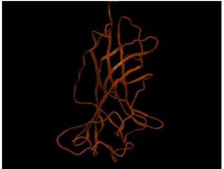
[image:2.595.318.540.181.326.2]carried out using PDB-BLAST and identified 3RHW from C.Elegans having 38% identity and 90% similarity with our target sequence. The 3D homology model of human GABRB3 is predicted using the crystal structure coordinates set of 3RHW on the basis of sequence structure alignment (Figure 1). The model building is done by ICM molsoft. The model structure (Figure 2) is further optimized by energy minimization using Discovery Studio by applying charmm forcefields.
Figure 1: Alignment of the template 3RHW and GABRB3
Figure 2: The 3D structure of human GABRB3modelled using ICM MOLSOFT
[image:2.595.315.537.365.533.2]overall general similarities and subtle differences among predicted model of GABARB3 and the 3D structure of template 3RHW can be seen from the backbone superposition (Figure 5). As evident from superposition, general folding topology of the structure is similar; however, some structural differences appear between the predicted model and template. These differences are mainly due to insertion and deletions in different loop regions. The RMSD (Root Mean Square Deviation) between predicted model and template is 0.56 Å. The low RMSD between the target and template reflects the presence of strong homology (The lower the value, the more similar the structures are).
The 3D structure of Valerenic Acid, which is generated by catalyst after energy minimization using CHARMm forcefield, is shown in Figure 6.
Figure 3(a): Ramachandran Map of GABRB3 from Homo
sapiens
The Protein-Ligand interaction plays a significant role in structural based drug designing. In the present work, to study
[image:3.595.320.514.73.284.2]Figure 3(b): Ramachandran Map of 3RHW from C. Elegans
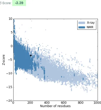
Figure 4: The plan of Z-Score shows spots of Z score values of proteins determined by NMR (represented in dark blue color) and by X- ray (represented in light blue
color) using ProSA program. The black dot represents Z-Scores of our model.
the binding modes of the Valerenic acid in the active site of gabrb3 Molecular docking study was performed by ligand fit program. The binding modes are calculated using intermolecular flexible docking simulations of LigandFit program. As a result, ten different conformations were generated, but only top ranked docked complex score compound is taken for binding affinity analysis. The different score values include Ligscore1, Ligscore2, PLP1, PLP2, JAIN, PMF and Dockscore as represented in Table 2. Ligscores indicates the protein ligand affinity energy; higher PLP score indicates stronger receptor-ligand binding, high PMF score indicates a stronger receptor-ligand binding affinity and DockScore is used to estimate the ligand-binding energies. Candidate ligand poses are evaluated and prioritized according to the DockScore functions.
[image:3.595.60.271.254.488.2] [image:3.595.323.535.529.708.2] [image:3.595.58.267.564.741.2]Figure 6: structure of Valerenic acid
Two important parameters have been considered for selecting potential compounds among the given input. They are
(i) Hydrogen bond details of the top-ranked pose and (ii) prediction of binding energy between the docked ligand and the protein using the mentioned scores as obtained for the analysis (Table 2).
[image:4.595.55.283.565.738.2]From the ten conformations, the compound with the highest dockscore is taken for interaction analysis of the hydrogen bonding. By enlarging this interaction analysis the hydrogen bond interaction is contributed as major parameter. The Hydrogen bonding interaction of the valeneric acid with GABRB3 (Figure 2) are analyzed. Results are analyzed using Hbond Monitor of Discovery studio.2.1 involved in hydrogen bond formation with amino acids. The results reveal that docked complex with a dock score of 47.665 with two hydrogen bonds and it is the best conformation. The binding mode of the compound has been shown in Figure 7.
Figure 7 shows the amino acid residues involved in hydrogen bond interactions with protein GABRB3 and the ligand Valeneric acid. The interacting amino acids are ASP35, GLN56, PHE55, TYR120, ARG111 and TYR 54. The hydrogen bonds are formed between oxygen atom of ligand molecule interacting with hydrogen of GLN56 amino acid (O2:L…HE22:GLN56) and hydrogen atom of ligand interacting with oxygen of ASP35 (H39:L…. OD1:ASP35). The other interactions are O1 of ligand molecule interacting with OD2 of ASP35 (O1:L…OD2:ASP35) and PHE55, TYR120, TYR54 and ARG111 show non-bonded interactions respectively.
Figure 7: Docking of valerenic acid with GABRB3: Hydrogen bond interactions
4.
CONCLUSION
The present study demonstrates that based on the selected template 3RHW, the 3D structural model of GABAA receptor
subunit beta-3 is predicted and optimally minimized. The refined structure is further validated by PROCHECK, ProSA and RMSD. In Ramachandran plot analysis, it is observed that maximum residues (84%) lie in the most favored and no residues in disallowed region. Further docking studies are carried out with the valeneric acid to study the binding mechanism between GABAA receptor subunit beta-3 and
valeneric acid. Based on the docking score, out of ten conformations of the receptor ligand complex, the complex structure having the highest dockscore is taken for interaction analysis. The results show that the interacting amino acids are ASP35, GLN56, PHE55, TYR120, ARG111, and TYR54 and hydrogen bonding forming with amino acids GLN56 and ASP35. This study would be useful in both understanding the activity mode of valeneric acid the most favorable binding mode of the top – ranking complex will be useful in designing new derivatives as human GABRB3 modulators.
5.
REFERENCES
[1] McKernan RM, and Whiting PJ (1996) Which GABAA-receptor subtypes really occur in the brain? Trends Neurosci 19:139–143.
[2] Choi DW, Farb DH, and Fischbach CD (1977) Chlordiazepoxide selectively augments GABA action in spinal cord cell cultures. Nature (Lond) 269:342-344.
[3] Macdonald RL, and Barker JL (1981) Benzodiazepines specifically modulate GABA-mediated postsynaptic inhibition in cultured mammalian neurones. Nature (Lond) 271 :563-564
[4] Whiting PJ, Mckernan RM, and Wafford KA. Structure and pharmacology of vertebrate GABAA receptor subtypes.In: International Review of Neurobiology (Bradley RJ., and Harris RA, eds.). San Diego: Academic Press, 1995. p. 95.
[5] Mody I (2005). Aspects of the homeostaic plasticity of GABAA receptor-mediated inhibition. J Physiol 562, 37–
46.
[6] Baumann SW, Baur R, and Sigel E (2002). Forced subunit assembly in α1β2γ2 GABAA receptors. Insight into the absolute arrangement. J Biol Chem 277, 46020– 46025.
[7] Fritschy J.-M., Benke D., Mertens S., Oertel W. H., Bachi T. and Mohler H. (1992) Five subtypes of type A gamma-aminobutyric acid receptors identified in neurons by double and triple immunofluorescence staining with subunitspecific antibodies. Proc. Natn. Acad. Sci. U.S.A. 89, 6726–6730.
[8] Smith GB, Olsen RW (1995) Functional domains of GABAA receptors. Trends Pharmacol Sci 16:162–168. [9] Berezhnoy D, Gravielle MC, and Farb DH (2007)
Pharmacology of the GABAA receptor, in Handbook of Contemporary Neuropharmacology (Sibley D, Hanin I, Kuhar M, and Skolnick P eds) vol 1, pp 465–568, John Wiley & Sons/Wiley- Interscience, Hoboken, NJ. [10]Korpi, E.R., Gründer, G., Lüddens, H., 2002. Drug
interactions at GABAA receptors. Prog. Neurobiol. 67, 113–159.
[11]Fritschy J: Epilepsy, E/I balance and GABAA receptor plasticity. Front Mol Neurosci 2008, 1:5.
[13]Fritschy JM, Bru¨nig I. Formation and plasticity of GABAergic synapses:physiological mechanisms and pathophysiological implications. Pharmaco Ther 2003; 98: 299–323.
[14]Abagyan RA, Totrov MM, Kuznetsov DA. ICM: A New Method For Protein Modeling and Design: Applications to Docking and Structure Prediction From The Distorted Native Conformation. J. Comp. Chem. 1994; 15:488– 506.
[15]Laskowski RA et al. AQUA and PROCHECK-NMR : Programs for checking the quality of protein structures
solved by NMR,Journal of Biomolecular NMR. 1996, 8(4): 477-86
[16] Morris AL et al. Stereochemical quality of protein structure coordinates. Proteins. 1992, 12(4): 345-64 [17]Sippl MJ. Knowledge-based potentials for proteins.
Current Opinion in Structural Biology.1995, 5(2): 229-35 [18] Zhang Y & Skolnick J. Scoring function for automated assessment of protein structure template quality. Proteins. 2004, 57: 702
Table 1: Percentage of residues in the core region of the Ramachandran plot for the model built and template.
Table 2: Summary of docking information of the ten conformations of the docked poses of the Valeneric acid with GABRB3.
Compound Protein Lig
Score 1
Lig Score 2
PLP1 PLP2 JAIN PMF Dock
score
Pose number
Valerenic
Acid
GABRB3
1.63 2.72 29.62 30.26 -0.91 52.26 47.665 1
1.74 2.78 38.92 39.5 0.01 45.77 45.346 2
1.81 2.47 30.3 34.78 -0.22 36.34 44.883 3
1.81 2.47 30.3 34.78 -0.22 36.34 44.883 4
2.35 3.2 27.6 28.37 -0.65 55.24 41.855 5
1.78 2.8 27.14 28.38 -1.22 53.5 40.661 6
1.28 1.98 19.49 26.23 -0.87 45.9 40.515 7
2.35 2.87 25.58 28.12 -0.82 55 39.059 8
1.67 3.34 39.91 38.18 -0.25 48.54 38.261 9
1.67 3.34 39.91 38.18 -0.25 48.54 38.261 10
Structure Core Allowed Generous Disallowed
GABRB3 84.0 15.5 0.6 0.0