R E S E A R C H
Open Access
Antithrombin attenuates myocardial dysfunction
and reverses systemic fluid accumulation
following burn and smoke inhalation injury:
a randomized, controlled, experimental study
Sebastian Rehberg
1,2*, Yusuke Yamamoto
1, Eva Bartha
1, Linda E Sousse
1, Collette Jonkam
1, Yong Zhu
1,
Lillian D Traber
1, Robert A Cox
3,4, Daniel L Traber
1,4and Perenlei Enkhbaatar
1,4Abstract
Introduction:We hypothesized that maintaining physiological plasma levels of antithrombin attenuates myocardial dysfunction and inflammation as well as vascular leakage associated with burn and smoke inhalation injury. Therefore, the present prospective, randomized experiment was conducted using an established ovine model. Methods:Following 40% of total body surface area, third degree flame burn and 4 × 12 breaths of cold cotton smoke, chronically instrumented sheep were randomly assigned to receive an intravenous infusion of 6 IU/kg/h recombinant human antithrombin (rhAT) or normal saline (control group;n= 6 each). In addition, six sheep were designated as sham animals (not injured, continuous infusion of vehicle). During the 48 h study period the animals were awake, mechanically ventilated and fluid resuscitated according to standard formulas.
Results:Compared to the sham group, myocardial contractility was severely impaired in control animals, as suggested by lower stroke volume and left ventricular stroke work indexes. As a compensatory mechanism, heart rate increased, thereby increasing myocardial oxygen consumption. In parallel, myocardial inflammation was induced via nitric oxide production, neutrophil accumulation (myeloperoxidase activity) and activation of the p38-mitogen-activated protein kinase pathway resulting in cytokine release (tumor necrosis factor-alpha, interleukin-6) in control vs. sham animals. rhAT-treatment significantly attenuated these inflammatory changes leading to a myocardial contractility and myocardial oxygen consumption comparable to sham animals. In control animals, systemic fluid accumulation progressively increased over time resulting in a cumulative positive fluid balance of about 4,000 ml at the end of the study period. Contrarily, in rhAT-treated animals there was only an initial fluid accumulation until 24 h that was reversed back to the level of sham animals during the second day.
Conclusions:Based on these findings, the supplementation of rhAT may represent a valuable therapeutic approach for cardiovascular dysfunction and inflammation after burn and smoke inhalation injury.
Keywords:capillary leakage, cardiovascular hemodynamics, mitogen-activated protein kinase, myocardial oxygen consumption, tumor necrosis factor, left ventricular dysfunction
* Correspondence: [email protected]
1Investigational Intensive Care Unit, Department of Anesthesiology, The
University of Texas Medical Branch, 301 University Blvd., Galveston, TX 77555, USA
Full list of author information is available at the end of the article
Introduction
Burn- and smoke inhalation-induced shock not only implies hemodynamic instability and vascular leakage but also severe myocardial dysfunction [1], which already occurs two hours after the injury [2]. Contrary to septic cardiomyopathy [3], for example, the clinical relevance of burn- and smoke-induced myocardial dys-function is only poorly recognized. However, there is a significant correlation between left ventricular dysfunc-tion and in-hospital mortality of critically burned patients [4]. In addition, myocardial stress caused by burn injuries persists for up to three years, thereby also influencing long-term outcome [5]. Therefore, effective treatment strategies for burn- and smoke-induced myo-cardial dysfunction are warranted.
The proposed pathogenesis of burn- and smoke-induced myocardial dysfunction is based on inflammatory mechan-isms, including activation of the p38-mitogen-activated protein kinase (p38-MAPK) pathway as well as production of tumor necrosis factor alpha (TNF-a) and nitric oxide (NO) [6-8]. The plasma-derived glycoprotein antithrombin III (AT) has been shown to reduce neutrophil activation [9] and to attenuate all of these inflammatory cascades in lung injury, for example [10-12]. However, potential bene-fits of AT on cardiovascular inflammation and dysfunction following burns and smoke inhalation have not been investigated, yet. A second rationale for the use of AT in these patients is an AT deficiency following burn injuries [13], that represents an independent predictor of length of hospital stay and mortality [14,15].
Accordingly, we hypothesized that maintaining physio-logical plasma levels of AT reduces myocardial inflam-mation and attenuates cardiovascular dysfunction associated with burn and smoke inhalation injury. Therefore, the present prospective, randomized, con-trolled laboratory experiment was designed to elucidate the effects of an intravenous infusion of recombinant human AT (rhAT) on myocardial neutrophil accumula-tion, activation of the p38-MAPK pathway, myocardial TNF-asecretion and systemic NO production as well as cardiovascular hemodynamics and systemic fluid accu-mulation in an established ovine model [16,17].
Materials and methods
The study was approved by the Animal Care and Use Committee of the University of Texas Medical Branch at Galveston and conducted in compliance with the guidelines of the National Institutes of Health.
Instrumentation and surgical procedures
Eighteen female sheep were anesthetized and instrumen-ted for chronic hemodynamic monitoring using an established protocol [16,17]. Details are provided in Additional file 1, Materials and methods.
Experimental protocol
Following baseline measurements (BL) in the healthy sheep, a tracheostomy was performed and a urinary bladder catheter was placed under deep anesthesia. Twelve animals were then subjected to 40% of total body surface area third degree flame burn and 4 × 12 breaths of cold cotton smoke under deep anesthesia using an established protocol [16,17]. The arterial car-boxyhemoglobin level was determined immediately after smoke inhalation to quantify the degree of injury. Although third degree burns are considered painless, 0.03 mg buprenorphine i.v. was administered before the burn and every 12 h subsequently to provide analgesia for the edges of the burn area, which may be second-degree burns. The sheep were then randomly assigned to the control group (injured, continuous infusion of vehicle (NaCl 0.9%),n= 6) or the rhAT group (injured, continuous infusion of 6 IU/kg/h rhAT (GTC Biothera-peutics Inc., Framingham, MA, USA) from 1 h post injury until the end of the 48-h study period, n = 6). During the experiment the investigators were unaware of the animal’s group assignment. Six animals were assigned to the sham group (not injured, continuous infusion of vehicle (NaCl 0.9%)). Due to the obvious lack of burn injury, this group assignment could not be blinded.
All animals were mechanically ventilated with a tidal volume of 15 mL/kg and a positive end-expiratory pres-sure of 5 cmH2O throughout the entire experiment. In
this context, it is important to consider the study design and the physiological differences between sheep and humans (see in detail Additional file 1, Materials and methods). The inspiratory oxygen fraction was set at 100% for the first three hours post injury, and was then adjusted to maintain oxygenation (arterial oxygen saturation >90%, partial pressure of oxygen >90 mmHg), whenever possible. The respiratory rate was adjusted according to individual blood gas analyses to ensure normocapnia.
Resuscitation was performed with lactated Ringer’s solution according to the Parkland formula (4 mL/kg/% burned body surface area within 24 h) [18]. To compen-sate for the initial fluid loss, sheep received one half of the total calculated amount for 24 h within the first 8 h after injury. At the end of the 48-h study period, sheep were deeply anesthetized with ketamine (15 mg/kg) and killed by injection of 60 mL of saturated potassium chloride.
Hemodynamic monitoring and laboratory analyses
at specific time points. Details are provided in Additional file 1, Materials and methods.
Immunohistochemical analysis and Western blots
Samples from the left ventricle (anterior wall) were used for quantification of myeloperoxidase activity, p38-MAPK, TNF-aand interleukin-6 (IL-6). Myeloperoxidase activity was determined using a commercially available assay (Myeloperoxidase Activity Assay, Northwest Life Science Specialties, Vancouver, BC, Canada) according to the manufacturer’s protocol. Details are provided in Addi-tional file 1, Materials and methods.
Statistical analyses
Sigma Stat 3.1 software (Systat Software, Inc., San Jose, CA, USA) was used for statistical analyses. Analysis of variance on ranks methodologies appropriate for non-normally distributed variables with repeated measures were used. Each variable was analyzed separately for dif-ferences among groups, difdif-ferences across time, and for group by time interaction. After confirming the signifi-cance of different group effects over time,post hoc pair-wise comparisons among groups were performed using the Student-Newman-Keuls procedure to adjust for the elevated false positive rate found otherwise in multiple testings. Finally, a rank sum test was applied to compare values at each time point. Western blot analyses and myeloperoxydase activity were compared with the rank sum test. Data are expressed as median with interquar-tile range (25th; 75th). Differences were considered as statistically significant whenPwas less than 0.05.
Results
Baseline characteristics
The mean body weight of the sheep was 33 kg (29; 38). There were no differences among study groups in any of the investigated variables at BL. Carboxyhemoglobin values after smoke inhalation, as an index of the severity of injury, did not differ among groups (control: 69% (58; 74) vs. rhAT: 73% (58; 80),P= 0.610).
Cardiovascular hemodynamics
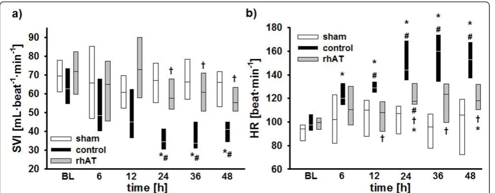
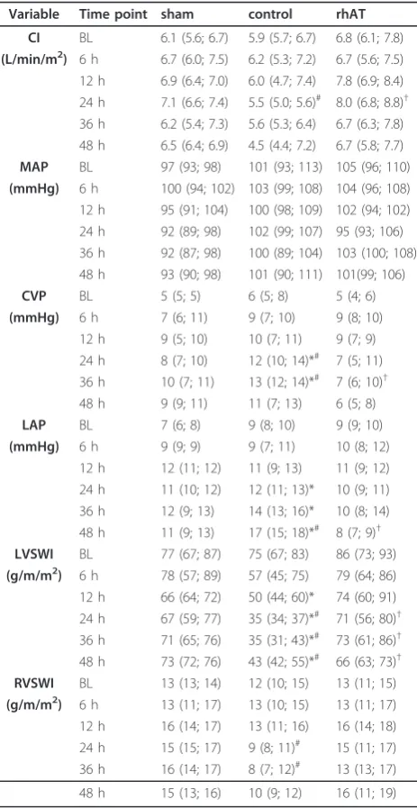
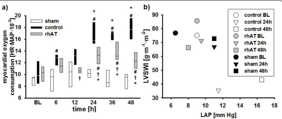
Stroke volume index (SVI; 24 to 48 h:P<0.05 vs. BL and 24 to 48 h:P<0.05 vs. sham, Figure 1a) and left ventricular stroke work index (LVSWI, 12 to 48 h:P<0.05 vs. BL and 24 to 48 h:P<0.05 vs. sham, Table 1) decreased by about 50% vs. BL in the control group within 24 h. In these ani-mals, cardiac index was maintained (no statistical differ-ences vs. BL) by a progressive increase in heart rate (HR; 6 to 48 h:P<0.05 vs. BL and 12 to 48 h:P<0.001 vs. sham, Figure 1b). Myocardial oxygen consumption [19] increased significantly (24 to 48 h:P<0.05 vs. BL and 6 h, 24 to 48 h: P<0.05 vs. sham, Figure 2a). rhAT treatment attenuated the decreases in SVI (24 to 48 h:P<0.05 vs. control) and
LVSWI (24 to 48 h:P< 0.05 vs. control each), resulting in a lower HR and oxygen consumption than in control ani-mals (12 to 48 h:P< 0.05 each). With one exception at 24 h (rhAT and sham vs. control:P<0.05 each), there were no significant differences in cardiac index between study groups (Table 1). Right ventricular stroke work index was lower in control animals as compared to sham from 24 to 36 h (P<0.05 each). Left atrial pressure (LAP) increased over time in the control group (24 to 48 h:P< 0.05 vs. BL each). rhAT infusion was associated with a lower LAP (48 h:P= 0.016 vs. control). Similarly, central venous pressure (CVP) increased in control animals (24 to 36 h:P< 0.05 vs. BL;P< 0.05 vs. sham), whereas CVP did not increase in rhAT-treated animals (P= 0.01 vs. control at 36 h). Left ventricular contractility, expressed as a corre-lation of LVSWI and LAP, an index of left ventricular pre-load (Figure 2b), was markedly impaired in the control group, whereas contractility in rhAT-treated animals was comparable to sham animals.
Vascular leakage and fluid accumulation
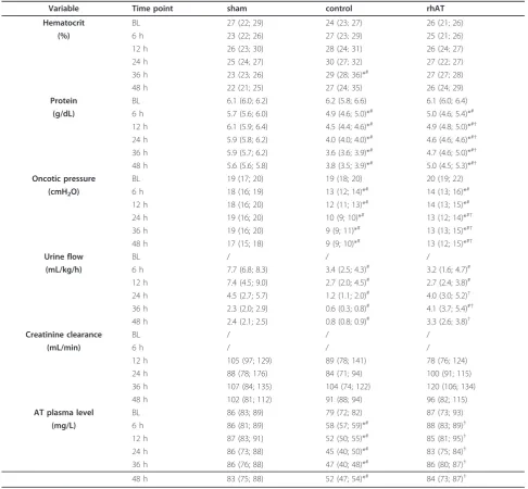
Vascular leakage was evidenced in control animals by an increased hematocrit (36 h: P = 0.017 vs. BL andP = 0.048 vs. sham, Table 2) in parallel with decreases in plasma protein concentration and plasma oncotic pres-sures (6 to 48 h:P< 0.05 vs. BL and 6 to 48 h:P < 0.05 vs. sham, Table 2). Accordingly, cumulative net fluid balance progressively increased resulting in a systemic fluid accumulation of about 4,000 mL in the control group at 48 h (12 to 48 h: P< 0.05 vs. sham, Figure 3a). In rhAT-treated animals, there was an initial decrease in plasma protein concentration and plasma oncotic pres-sures without any further progression afterwards (P < 0.01 vs. BL andP <0.05 vs. sham each). Hematocrit did not change compared with BL or sham animals in the rhAT group. Cumulative net fluid balance initially increased in rhAT-treated animals from 12 to 24 h (P< 0.05 vs. sham), but came back to sham level at 48 h (P= 0.004 vs. control, Figure 3a).
Urine flow in control animals progressively decreased over the study period and was lower than in the sham group (6 to 48 h: P <0.05 each, Table 2). In the rhAT group, urine flow was lower than in sham animals from 6 to 12 h (P<0.05 each), but higher than in control ani-mals from 24 to 48 h (P <0.05 each).
Gas exchange and global oxygen transport
oxygen transport between the study groups (see Addi-tional file 2, Table S1).
Laboratory analyses
In control animals burn and smoke inhalation was asso-ciated with an immediate decrease of AT plasma concen-trations (6 to 48 h:P< 0.05 vs. BL) resulting in lower values than in sham animals (6 to 48 h: P< 0.05 each, Table 2). The continuous infusion of rhAT prevented this decrease and kept AT plasma concentrations at BL level and higher than in control animals (6 to 48 h:P< 0.05 each).
No significant differences in activated clotting time, activated partial thromboplastin time and platelet count could be shown over time or between study groups. There was an increase in prothrombin time in both injured groups as compared to BL (36 to 48 h:P< 0.05) and sham animals (12 to 48 h:P< 0.05); however, with-out statistical differences between the control and the rhAT group (see Additional file 2, Table S1).
There were no significant differences between study groups in creatinine clearance during the study period (Table 2).
In control animals, plasma levels of nitrates and nitrites (NOx) increased five-fold within 12 h (6 to 12 h:P< 0.05 vs. BL and sham each). Then, NOx plasma levels decreased from 12 to 24 h, before they increased again from 24 to 48 h (P<0.05 vs. BL and sham each). rhAT infusion attenuated the first increase (12 h:P= 0.032 vs. control) and prevented the second increase of plasma NOx compared with control animals (36 to 48 h:P < 0.05 vs. control, Figure 3b).
Immunohistochemical analyses and Western blots
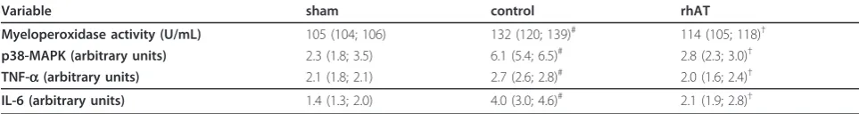
Immunohistochemical analyses of myocardial tissues revealed a significant increase in myeloperoxydase activ-ity in control vs. sham animals (P= 0.026, Table 3). In rhAT-treated animals, myeloperoxidase activity was lower than in the control group (P= 0.041).
Western blots performed in myocardial tissues showed higher concentrations of p38-MAPK in the control vs. the sham group (P= 0.028, Table 3). In addition, there was an increase in myocardial concentrations of the inflammatory cytokines TNF-a and IL-6 in control vs. sham animals (P< 0.05 each, Table 3). Contrarily, in the rhAT group concentrations of p38-MAPK (P= 0.01) as well as TNF-a (P = 0.046) and IL-6 (P = 0.038), were lower than in control animals.
Discussion
The major findings of the present study are that 1) burn and smoke inhalation injury was associated with myo-cardial dysfunction and inflammation, and that 2) a con-tinuous infusion of rhAT attenuated the impairment of myocardial contractility, 3) reduced myocardial inflam-mation, 4) limited NO production, and 5) reversed sys-temic fluid accumulation 6) without compromising plasmatic coagulation in a clinically relevant ovine model.
[image:4.595.57.544.88.278.2]Burn- and smoke-induced myocardial dysfunction is typically characterized by an impaired left ventricular contractility [1,20]. In the present study, a reduced myo-cardial function in control animals was evidenced by lower SVI and LVSWI as compared to BL and sham ani-mals. Although no direct measurement of contractility
Figure 1Stroke volume index (a) and heart rate (b). *P< 0.05 vs. baseline;#P< 0.05 vs. sham;†P< 0.05 vs. control; data are represented as
was performed, the Starling-based relation of LVSWI with the individual preload at this time (LAP) highly sug-gests an impaired myocardial contractility in the control vs. sham group. Notably, the time courses for SVI and HR in control animals nicely demonstrate the“two-hit” character of the injury [21]: the first one, immediately fol-lowing the injury, is caused by the burn-induced shock and the associated systemic inflammatory response syn-drome (SIRS). The second one is most probably attributed
to an inflammatory response to the smoke inhalation usually occurring 24 h post injury [21,22].
As a compensatory mechanism, HR increased in control animals thereby maintaining cardiac output. However, myocardial oxygen consumption, as suggested by the (heart) rate-(mean arterial) pressure product [19], was consistently higher than in sham animals. In the acute phase of the injury, this increased myocardial oxygen con-sumption and the impairment of myocardial function potentially aggravate burn-induced shock as well as the associated SIRS, thereby eventually promoting multiple organ failure. Previously, burn- and smoke-induced myo-cardial dysfunction was thought to be transient and to resolve after successful treatment of the originating trauma [2]. Meanwhile, there is convincing evidence that it also influences long-term outcome. An increased HR and myo-cardial oxygen consumption, for example, were found to be present even three years after burn trauma in children [5,23]. Hypovolemia as a cause of reduced SVI and increased HR in the present study seems unlikely within the first 24 h, because in this case ventricular filling pres-sures as well as mean arterial pressure and cardiac index would have been decreased in control animals.
rhAT infusion markedly improved contractility, as suggested by higher SVI and LVSWI values as compared to control animals. In accordance with these findings, HR and myocardial oxygen consumption were signifi-cantly lower in rhAT-treated than in control animals. Whereas beneficial effects of AT on pulmonary gas exchange [10] and wound healing [24] in patients suffer-ing from burn and smoke inhalation have been reported before, this is the first time that a therapeutic effect of AT on myocardial function is described.
Since“cardiac molecular signalling after burn trauma” involves several inflammatory pathways [7], the anti-inflammatory effects of rhAT represent a potential mechanism of action. As part of the burn- and smoke-induced SIRS, there is a pronounced activation of neutro-phils leading to transendothelial migration into the organs and promotion of inflammatory cascades, like NO produc-tion and p38-MAPK pathway activaproduc-tion [6-8]. Neutrophil accumulation in heart tissues was suggested by an increase in myeloperoxidase [25] in control vs. sham animals that was attenuated by rhAT. This finding is well explained by the interaction of AT with the syndecan-4 receptor result-ing in reduced neutrophil activation and chemotaxis [9,26].
[image:5.595.57.289.99.548.2]In addition, activation of the p38-MAPK cascade plays a central role in burn-induced myocardial dysfunction [6,7,27-29]. It regulates apoptosis of cardiomyocytes in the early stage after severe burns [6,28] and promotes inflammation by increasing the production of TNF-a and downstream cytokines like IL-6 [1,6,27], both being significantly increased in the control vs. the sham group
Table 1 Variables of cardiovascular hemodynamics
Variable Time point sham control rhAT
CI BL 6.1 (5.6; 6.7) 5.9 (5.7; 6.7) 6.8 (6.1; 7.8)
(L/min/m2) 6 h 6.7 (6.0; 7.5) 6.2 (5.3; 7.2) 6.7 (5.6; 7.5) 12 h 6.9 (6.4; 7.0) 6.0 (4.7; 7.4) 7.8 (6.9; 8.4) 24 h 7.1 (6.6; 7.4) 5.5 (5.0; 5.6)# 8.0 (6.8; 8.8)†
36 h 6.2 (5.4; 7.3) 5.6 (5.3; 6.4) 6.7 (6.3; 7.8) 48 h 6.5 (6.4; 6.9) 4.5 (4.4; 7.2) 6.7 (5.8; 7.7)
MAP BL 97 (93; 98) 101 (93; 113) 105 (96; 110)
(mmHg) 6 h 100 (94; 102) 103 (99; 108) 104 (96; 108) 12 h 95 (91; 104) 100 (98; 109) 102 (94; 102) 24 h 92 (89; 98) 102 (99; 107) 95 (93; 106) 36 h 92 (87; 98) 100 (89; 104) 103 (100; 108) 48 h 93 (90; 98) 101 (90; 111) 101(99; 106)
CVP BL 5 (5; 5) 6 (5; 8) 5 (4; 6)
(mmHg) 6 h 7 (6; 11) 9 (7; 10) 9 (8; 10)
12 h 9 (5; 10) 10 (7; 11) 9 (7; 9)
24 h 8 (7; 10) 12 (10; 14)*# 7 (5; 11)
36 h 10 (7; 11) 13 (12; 14)*# 7 (6; 10)†
48 h 9 (9; 11) 11 (7; 13) 6 (5; 8)
LAP BL 7 (6; 8) 9 (8; 10) 9 (9; 10)
(mmHg) 6 h 9 (9; 9) 9 (7; 11) 10 (8; 12)
12 h 12 (11; 12) 11 (9; 13) 11 (9; 12)
24 h 11 (10; 12) 12 (11; 13)* 10 (9; 11)
36 h 12 (9; 13) 14 (13; 16)* 10 (8; 14)
48 h 11 (9; 13) 17 (15; 18)*# 8 (7; 9)†
LVSWI BL 77 (67; 87) 75 (67; 83) 86 (73; 93)
(g/m/m2) 6 h 78 (57; 89) 57 (45; 75) 79 (64; 86) 12 h 66 (64; 72) 50 (44; 60)* 74 (60; 91) 24 h 67 (59; 77) 35 (34; 37)*# 71 (56; 80)†
36 h 71 (65; 76) 35 (31; 43)*# 73 (61; 86)†
48 h 73 (72; 76) 43 (42; 55)*# 66 (63; 73)†
RVSWI BL 13 (13; 14) 12 (10; 15) 13 (11; 15)
(g/m/m2) 6 h 13 (11; 17) 13 (10; 15) 13 (11; 17)
12 h 16 (14; 17) 13 (11; 16) 16 (14; 18)
24 h 15 (15; 17) 9 (8; 11)# 15 (11; 17)
36 h 16 (14; 17) 8 (7; 12)# 13 (13; 17)
48 h 15 (13; 16) 10 (9; 12) 16 (11; 19)
*,P<0.05 vs. BL;#
,P<0.05 vs. sham;†,P<0.05 vs. control; data are represented as median with interquartile range (25th
; 75th
in the present study. Inhibition of p38-MAPK has been shown to improve myocardial function after burns in vitro[29] and in rats [27]. An inhibitory effect of AT on the p38-MAPK pathway, as suggested in the present study, is supported by an experiment on acute lung injury in rats [11]. Interestingly, the beneficial effects of AT in the latter study were abolished, if concomitant heparin was infused. Since heparin is known to block the interaction of AT with the syndecan-4 receptor [30], this mechanism may be involved in the reduction of p38-MAPK via AT. This hypothesis, however, requires further verification in future studies.
In accordance with the changes in p38-MAPK expres-sion, concentrations of TNF-a and IL-6 were lower in rhAT-treated than in control animals. TNF-aand IL-6 have both been repeatedly reported to correlate with cardiac contraction and relaxation deficits after burn trauma [4,31,32] but also in heart failure patients [33]. Myocardial secretion of TNF-amay be directly respon-sible for an impaired myocardial contractility, reduced ejection fraction and biventricular dilation [7]. One mechanism of TNF-a-mediated cardiodepression includes the induction of NO synthesis [34]. The critical role of NO in burn- and smoke-induced myocardial dys-function [8,35] is supported by the biphasic increase in NOx plasma levels that almost reflect the changes in SVI in control animals.
Vascular leakage, another hallmark of burn and smoke inhalation injury, was evidenced by marked decreases in protein concentration and oncotic pressures in injured animals. In this context, dilution due to over-resuscita-tion can be excluded, since hematocrit was maintained
or even increased. Control animals progressively accu-mulated about 4,000 mL of fluid during the study per-iod. rhAT-treated animals also had an initial drop in protein concentration and oncotic pressure that was probably caused by an irreversible loss via the burn wound. However, rhAT prevented a progression of pro-tein leakage and reversed the initial fluid accumulation resulting in a fluid balance similar to sham animals at 48 h. The clinical relevance of this finding is emphasized by the correlation of fluid accumulation with negative outcome not only in burned patients [36] but also in critical ill patients in general [37]. Since all animals received the same amount of fluid (after adjustment for the individual body weight) and there were no differ-ences in renal function (creatinine clearance) between groups, these findings suggest a reduction in vascular leakage by rhAT. This hypothesis is supported by sev-eral experimental studies reporting beneficial effects of AT on endothelial function. Potential mechanisms of action include interactions with the syndecan-4 receptor [9,26], preservation of the endothelial glycocalyx [38] and inhibition of endothelial cell activation [39]. The remaining difference in total urine output between sham and rhAT-treated animals may be explained by the addi-tional fluid loss due to evaporation over the burn wound in the injured rhAT group.
[image:6.595.62.538.89.290.2]The clinical applicability of the presented therapeutic approach is potentially limited by the early start of the infusion, the availability and unwanted side effects of AT. However, starting AT infusion one hour post-injury may be feasible, because contrary to most other syn-dromes patients with burn and smoke inhalation injury
are transported to the hospital immediately and can be diagnosed rapidly. In addition, the recombinant prepara-tion of AT ensures independence from blood donaprepara-tions and a reduced risk of transmitted infections. Finally, based on the present results, there seems to be no impairment of plasmatic coagulation with the low dose of 6 IU/kg/h rhAT.
This study has some limitations that we want to acknowledge. Due to the“two hit”character of the experi-mental model it is hard to differentiate the effects of rhAT on the burn-induced changes and the symptoms caused
[image:7.595.55.542.110.560.2]by smoke inhalation. However, the combined injury is of high clinical relevance. Another limitation is the estima-tion of LVSWI by standard formulas and the use of a cor-relation of LVSWI and LAP as an index of myocardial contractility, because inotropy should be assessed by load independent measures. However, these are only available by echocardiography. To avoid the compromising effects of anesthetics on myocardial function, the animals were awake during the experiment necessitating a ventilation regime that differed from the recommendation of the acute respiratory distress syndrome (ARDS) network.
Table 2 Hematocrit, plasma protein concentration, oncotic pressure, urine flow, creatinine clearance and antithrombin plasma levels
Variable Time point sham control rhAT
Hematocrit BL 27 (22; 29) 24 (23; 27) 26 (21; 26)
(%) 6 h 23 (22; 26) 27 (23; 29) 25 (21; 26)
12 h 26 (23; 30) 28 (24; 31) 26 (24; 27)
24 h 25 (24; 27) 30 (27; 32) 27 (22; 27)
36 h 23 (23; 26) 29 (28; 36)*# 27 (27; 28)
48 h 22 (21; 25) 27 (24; 35) 26 (24; 29)
Protein BL 6.1 (6.0; 6.2) 6.2 (5.8; 6.6) 6.1 (6.0; 6.4)
(g/dL) 6 h 5.7 (5.6; 6.0) 4.9 (4.6; 5.0)*# 5.0 (4.6; 5.4)*#
12 h 6.1 (5.9; 6.4) 4.5 (4.4; 4.6)*# 4.9 (4.8; 5.0)*#†
24 h 5.9 (5.8; 6.2) 4.0 (4.0; 4.0)*# 4.6 (4.6; 4.6)*#†
36 h 5.9 (5.7; 6.2) 3.6 (3.6; 3.9)*# 4.7 (4.6; 5.0)*#†
48 h 5.6 (5.6; 5.8) 3.8 (3.5; 3.9)*# 5.0 (4.5; 5.3)*#†
Oncotic pressure BL 19 (17; 20) 19 (18; 20) 20 (19; 22)
(cmH2O) 6 h 18 (16; 19) 13 (12; 14)*
#
14 (13; 16)*#
12 h 18 (16; 20) 12 (11; 13)*# 14 (13; 15)*#
24 h 19 (16; 20) 10 (9; 10)*# 13 (12; 14)*#†
36 h 19 (16; 20) 9 (9; 11)*# 13 (13; 15)*#†
48 h 17 (15; 18) 9 (9; 10)*# 13 (12; 15)*#†
Urine flow BL / / /
(mL/kg/h) 6 h 7.7 (6.8; 8.3) 3.4 (2.5; 4.3)# 3.2 (1.6; 4.7)#
12 h 7.4 (4.5; 9.0) 2.7 (2.0; 4.5)# 2.7 (2.4; 3.8)#
24 h 4.5 (2.7; 5.7) 1.2 (1.1; 2.0)# 4.0 (3.0; 5.2)†
36 h 2.3 (2.0; 2.9) 0.6 (0.3; 0.8)# 4.1 (3.7; 5.4)#†
48 h 2.4 (2.1; 2.5) 0.8 (0.8; 0.9)# 3.3 (2.6; 3.8)†
Creatinine clearance BL / / /
(mL/min) 6 h / / /
12 h 105 (97; 129) 89 (78; 141) 78 (76; 124)
24 h 88 (78; 176) 84 (71; 94) 100 (91; 115)
36 h 107 (84; 135) 104 (74; 122) 120 (106; 134)
48 h 102 (81; 112) 91 (88; 94) 96 (82; 115)
AT plasma level BL 86 (83; 89) 79 (72; 82) 87 (73; 93)
(mg/L) 6 h 86 (81; 89) 58 (57; 59)*# 88 (83; 89)†
12 h 87 (83; 91) 52 (50; 55)*# 85 (81; 95)†
24 h 86 (73; 88) 45 (40; 50)*# 83 (75; 84)†
36 h 86 (76; 88) 47 (40; 48)*# 86 (80; 87)†
48 h 83 (75; 88) 52 (47; 54)*# 84 (73; 87)†
*,P<0.05 vs. BL;#
,P<0.05 vs. sham;†,P<0.05 vs. control; data are represented as median with interquartile range (25th
; 75th
Finally, the smoke arising from house or industrial fires contains a multitude of toxic gases potentially resulting in differences in lung injury compared with the cotton smoke tested in the present study.
Conclusions
In summary, this is the first study demonstrating therapeu-tic effects of rhAT substitution on myocardial dysfunction and inflammation as well as systemic fluid accumulation following burn and smoke inhalation injury in a clinically relevant animal model. Based on these findings, the supple-mentation of rhAT may represent a potential therapeutic approach for burn- and smoke inhalation-induced cardio-vascular dysfunction.
Key messages
• Combined burn and smoke inhalation injury is associated with a marked myocardial dysfunction. To compensate for the reduced stroke volume, heart rate and eventually myocardial oxygen consumption are increased.
•Impairment of myocardial function after burn and smoke inhalation injury is characterized by an
activation of the p38-mitogen-activated protein kinase pathway, myocardial neutrophil accumulation and increased nitric oxide production.
•The therapeutic infusion of recombinant human antithrombin restored myocardial function to the level of sham (not injured) animals, resulting in higher stroke volumes and reduced myocardial oxygen consumption.
•The therapeutic infusion of recombinant human antithrombin reversed cumulative systemic fluid accu-mulation back to the level of sham animals at 48 h.
•The attenuated inflammatory response, as sug-gested by a reduced activation of the p38-mitogen-activated protein kinase pathway resulting in lower cytokine release, less myocardial neutrophil accumu-lation and less nitric oxide production, presents a potential mechanism of action.
Additional material
Additional file 1: Supplemental file (word): detailed Materials and methods. This file provides detailed information about the
[image:8.595.60.539.89.288.2]instrumentation, mechanical ventilation, hemodynamic monitoring, Western blots as well as laboratory and immunohistochemical analyses.
Figure 3Cumulative net fluid balance (a) and concentration of NOx in the plasma (b). *P<0.05 vs. baseline;#P<0.05 vs. sham;†P<0.05 vs. control; data are represented as box plots with median and interquartile range (25th; 75th);n= 6 per group. BL, baseline; NOx, nitrates and nitrites; rhAT, recombinant human antithrombin III.
Table 3 Immunohistochemical analyses and Western blots
Variable sham control rhAT
Myeloperoxidase activity (U/mL) 105 (104; 106) 132 (120; 139)# 114 (105; 118)†
p38-MAPK (arbitrary units) 2.3 (1.8; 3.5) 6.1 (5.4; 6.5)# 2.8 (2.3; 3.0)†
TNF-a(arbitrary units) 2.1 (1.8; 2.1) 2.7 (2.6; 2.8)# 2.0 (1.6; 2.4)†
IL-6 (arbitrary units) 1.4 (1.3; 2.0) 4.0 (3.0; 4.6)# 2.1 (1.9; 2.8)†
#
,P<0.05 vs. sham;†,P<0.05 vs. control; data are represented as median with interquartile range (25th; 75th); each groupn= 6
[image:8.595.56.538.358.422.2]Additional file 2: Supplemental file (word): additional data. This file provides additional data that may be of interest for the reader, but where not relevant in respect to the message of the study.
Abbreviations
ARDS: acute respiratory distress syndrome; AT: antithrombin III; BL: baseline; CVP: central venous pressure; HR: heart rate; IL-6: interleukin-6; LAP: left atrial pressure; LVSWI: left ventricular stroke work index; NO: nitric oxide; NOx: nitrates and nitrites; p38-MAPK: p38-mitogen-activated protein kinase; rhAT: recombinant human antithrombin III; SIRS: systemic inflammatory response syndrome; SVI: stroke volume index; TNF-α: tumor necrosis factor-alpha
Authors’contributions
SR designed and performed the experiment, summarized and analyzed the data, and wrote the manuscript. YY, EB, LES, CJ and YZ performed the experiment, and summarized the data and edited the manuscript. LDT and DLT designed the experiment, analyzed the data and edited the manuscript. RAC performed the experiment, analyzed the data and edited the manuscript. PE designed and performed the experiment, analyzed the data and edited the manuscript. All authors read and approved the final manuscript.
Competing interests
The authors declare that they have no competing interests.
Acknowledgements
This work is dedicated to Prof. D.L. Traber, who passed away in September, 2012. The authors thank the technicians of the Investigational Intensive Care Unit for expert technical assistance during the study.
This work was supported by Shriners of North America (grant no. 85500, 84050, 85220), NIH (GM097480-02, T32-GM8256),“Deutsche
Forschungsgemeinschaft”and the Open Access Publication Fund of the University of Muenster.
Author details
1Investigational Intensive Care Unit, Department of Anesthesiology, The
University of Texas Medical Branch, 301 University Blvd., Galveston, TX 77555, USA.2Department of Anaesthesiology and Intensive Care, University of Muenster, Albert-Schweitzer-Campus 1, 48149 Muenster, Germany. 3Department of Pathology, The University of Texas Medical Branch, 301
University Blvd. 33, Galveston, TX 77555, USA.4Shriners Hospital for Children, 815 Avenue D, Galveston, TX 77550, USA.
Received: 28 January 2013 Revised: 18 March 2013 Accepted: 11 May 2013 Published: 11 May 2013
References
1. Horton JW:Left ventricular contractile dysfunction as a complication of thermal injury.Shock2004,22:495-507.
2. Horton JW, Garcia NM, White DJ, Keffer J:Postburn cardiac contractile function and biochemical markers of postburn cardiac injury.J Am Coll Surg1995,181:289-298.
3. Muller-Werdan U, Buerke M, Ebelt H, Heinroth KM, Herklotz A, Loppnow H, Ruß M, Schlegel F, Schlitt A, Schmidt HB, Söffker G, Werdan K:Septic cardiomyopathy - a not yet discovered cardiomyopathy?Exp Clin Cardiol 2006,11:226-236.
4. Lin CY, Wu CK, Yeong EK, Lin HH, Huang YT, Lee JK, Lin YH, Chiang FT, Tang YB, Tsai CT:Prognostic significance of left ventricular diastolic function in burn patients.Shock2012,37:457-462.
5. Jeschke MG, Gauglitz GG, Kulp GA, Finnerty CC, Williams FN, Kraft R, Suman OE, Mlcak RP, Herndon DN:Long-term persistance of the pathophysiologic response to severe burn injury.PLoS One2011,6: e21245.
6. Zhang JP, Ying X, Liang WY, Luo ZH, Yang ZC, Huang YS, Wang WC:
Apoptosis in cardiac myocytes during the early stage after severe burn. J Trauma2008,65:401-408, discussion 408.
7. Carlson DL, Horton JW:Cardiac molecular signaling after burn trauma. J Burn Care Res2006,27:669-675.
8. White J, Carlson DL, Thompson M, Maass DL, Sanders B, Giroir B, Horton JW:Molecular and pharmacological approaches to inhibiting nitric oxide after burn trauma.Am J Physiol Heart Circ Physiol2003,285: H1616-1625.
9. Dunzendorfer S, Kaneider N, Rabensteiner A, Meierhofer C, Reinisch C, Romisch J, Wiedermann CJ:Cell-surface heparan sulfate proteoglycan-mediated regulation of human neutrophil migration by the serpin antithrombin III.Blood2001,97:1079-1085.
10. Enkhbaatar P, Esechie A, Wang J, Cox RA, Nakano Y, Hamahata A, Lange M, Traber LD, Prough DS, Herndon DN, Traber DL:Combined anticoagulants ameliorate acute lung injury in sheep after burn and smoke inhalation. Clin Sci (Lond)2008,114:321-329.
11. Sun HM, Hong LZ, Shen XK, Lin XQ, Song Y, Shi Y:Antithrombin-III without concomitant heparin improves endotoxin-induced acute lung injury rats by inhibiting the activation of mitogen-activated protein kinase.Chin Med J (Engl)2009,122:2466-2471.
12. Komura H, Uchiba M, Mizuochi Y, Arai M, Harada N, Katsuya H, Okajima K:
Antithrombin inhibits lipopolysaccharide-induced tumor necrosis factor-alpha production by monocytesin vitrothrough inhibition of Egr-1 expression.J Thromb Haemost2008,6:499-507.
13. Kowal-Vern A, Walenga JM, McGill V, Gamelli RL:The impact of antithrombin (H) concentrate infusions on pulmonary function in the acute phase of thermal injury.Burns2001,27:52-60.
14. Niedermayr M, Schramm W, Kamolz L, Andel D, Romer W, Hoerauf K, Zimpfer M, Andel H:Antithrombin deficiency and its relationship to severe burns.Burns2007,33:173-178.
15. Lavrentieva A, Kontakiotis T, Bitzani M, Papaioannou-Gaki G, Parlapani A, Thomareis O, Tsotsolis N, Giala MA:Early coagulation disorders after severe burn injury: impact on mortality.Intensive Care Med2008,
34:700-706.
16. Lange M, Connelly R, Traber DL, Hamahata A, Cox RA, Nakano Y, Bansal K, Esechie A, von Borzyskowski S, Jonkam C, Traber LD, Hawkins HK, Herndon DN, Enkhbaatar P:Combined neuronal and inducible nitric oxide synthase inhibition in ovine acute lung injury.Crit Care Med2009,
37:223-229.
17. Enkhbaatar P, Connelly R, Wang J, Nakano Y, Lange M, Hamahata A, Horvath E, Szabo C, Jaroch S, Holscher P, Hillmann M, Traber LD, Schmalstieg FC, Herndon DN, Traber DL:Inhibition of neuronal nitric oxide synthase in ovine model of acute lung injury.Crit Care Med2009,
37:208-214.
18. Baxter CR, Shires T:Physiological response to crystalloid resuscitation of severe burns.Ann N Y Acad Sci1968,150:874-894.
19. Gobel FL, Norstrom LA, Nelson RR, Jorgensen CR, Wang Y:The rate-pressure product as an index of myocardial oxygen consumption during exercise in patients with angina pectoris.Circulation1978,57:549-556. 20. Adams HR, Baxter CR, Izenberg SD:Decreased contractility and
compliance of the left ventricle as complications of thermal trauma.Am Heart J1984,108:1477-1487.
21. Soejima K, Schmalstieg FC, Sakurai H, Traber LD, Traber DL:
Pathophysiological analysis of combined burn and smoke inhalation injuries in sheep.Am J Physiol Lung Cell Mol Physiol2001,280:L1233-1241. 22. Traber DL, Enkhbaatar P:Thermal lung injury and acute smoke inhalation.
InFishman’s Pulmonary Diseases and Disorders..4 edition. Edited by: Fishman DA. New York: McGraw-Hill Medical Publishing Company; 2008:. 23. Williams FN, Herndon DN, Suman OE, Lee JO, Norbury WB, Branski LK,
Mlcak RP, Jeschke MG:Changes in cardiac physiology after severe burn injury.J Burn Care Res2011,32:269-274.
24. Lavrentieva A, Kontakiotis T, Bitzani M, Parlapani A, Thomareis O, Scourtis H, Tsotsolis N, Lazaridis L, Giala MA:The efficacy of antithrombin
administration in the acute phase of burn injury.Thromb Haemost2008,
100:286-290.
25. Makita H, Nishimura M, Miyamoto K, Nakano T, Tanino Y, Hirokawa J, Nishihira J, Kawakami Y:Effect of anti-macrophage migration inhibitory factor antibody on lipopolysaccharide-induced pulmonary neutrophil accumulation.Am J Respir Crit Care Med1998,158:573-579.
26. Kaneider NC, Forster E, Mosheimer B, Sturn DH, Wiedermann CJ: Syndecan-4-dependent signaling in the inhibition of endotoxin-induced endothelial adherence of neutrophils by antithrombin.Thromb Haemost 2003,90:1150-1157.
inflammatory cytokine TNF-alpha.Am J Physiol Heart Circ Physiol2001,
280:H1970-1981.
28. Cao W, Xie YH, Li XQ, Zhang XK, Chen YT, Kang R, Chen X, Miao S, Wang SW:Burn-induced apoptosis of cardiomyocytes is survivin dependent and regulated by PI3K/Akt, p38 MAPK and ERK pathways. Basic Res Cardiol2011,106:1207-1220.
29. Lv GF, Dong ML, Hu DH, Zhang WF, Wang YC, Tang CW, Zhu XX: Insulin-mediated inhibition of p38 mitogen-activated protein kinase protects cardiomyocytes in severe burns.J Burn Care Res2011,32:591-599. 30. Justus AC, Roussev R, Norcross JL, Faulk WP:Antithrombin binding by
human umbilical vein endothelial cells: effects of exogenous heparin. Thromb Res1995,79:175-186.
31. Bryant D, Becker L, Richardson J, Shelton J, Franco F, Peshock R, Thompson M, Giroir B:Cardiac failure in transgenic mice with myocardial expression of tumor necrosis factor-alpha.Circulation1998,97:1375-1381. 32. Giroir BP, Horton JW, White DJ, McIntyre KL, Lin CQ:Inhibition of tumor
necrosis factor prevents myocardial dysfunction during burn shock.Am J Physiol1994,267:H118-124.
33. Wu CK, Lee JK, Chiang FT, Yang CH, Huang SW, Hwang JJ, Lin JL, Tseng CD, Chen JJ, Tsai CT:Plasma levels of tumor necrosis factor-alpha and interleukin-6 are associated with diastolic heart failure through downregulation of sarcoplasmic reticulum Ca2+ ATPase.Crit Care Med 2011,39:984-992.
34. Muller-Werdan U, Engelmann H, Werdan K:Cardiodepression by tumor necrosis factor-alpha.Eur Cytokine Netw1998,9:689-691.
35. Soejima K, Schmalstieg FC, Traber LD, Szabo C, Salzman A, Traber DL:Role of nitric oxide in myocardial dysfunction after combined burn and smoke inhalation injury.Burns2001,27:809-815.
36. Belba M, Aleksi A, Nezha I, Tafaj S, Shtylla M, Belba G:Net fluid accumulation and outcome. A randomized clinical trial.Ann Burns Fire Disasters2009,22:16-21.
37. Vincent JL, Sakr Y, Sprung CL, Ranieri VM, Reinhart K, Gerlach H, Moreno R, Carlet J, Le Gall JR, Payen D:Sepsis in European intensive care units: results of the SOAP study.Crit Care Med2006,34:344-353. 38. Chappell D, Jacob M, Hofmann-Kiefer K, Rehm M, Welsch U, Conzen P,
Becker BF:Antithrombin reduces shedding of the endothelial glycocalyx following ischaemia/reperfusion.Cardiovasc Res2009,83:388-396. 39. Uchiba M, Okajima K, Kaun C, Wojta J, Binder BR:Inhibition of the
endothelial cell activation by antithrombinin vitro.Thromb Haemost 2004,92:1420-1427.
doi:10.1186/cc12712
Cite this article as:Rehberget al.:Antithrombin attenuates myocardial dysfunction and reverses systemic fluid accumulation following burn and smoke inhalation injury: a randomized, controlled, experimental study.Critical Care201317:R86.
Submit your next manuscript to BioMed Central and take full advantage of:
• Convenient online submission
• Thorough peer review
• No space constraints or color figure charges
• Immediate publication on acceptance
• Inclusion in PubMed, CAS, Scopus and Google Scholar
• Research which is freely available for redistribution