N A N O E X P R E S S
Open Access
Preparation TiO
2
core-shell nanospheres and
application as efficiency drug detection sensor
Jingli Yue
1,2, Zhenhua Chen
2*, Yifeng E
2, Lianshan Chen
1,2, Jing Zhang
2, Yimeng Song
3and Yuchun Zhai
1*Abstract
In this paper, we report the facile preparation of monodisperse titanium dioxide-diltiazem/tetrachlorobismuth core-shell nanospheres (TiO2@DTMBi), in which, diltiazem (DTM)/tetrachlorobismuth (BiCl4) complexes were
employed as electroactive materials. The morphology, size, formation, and structure of the obtained TiO2@DTMBi
spheres were investigated by transmission electron microscopy, scanning electron microscopy, dynamic light scattering, Fourier transform infrared spectroscopy, and X-ray diffraction. The optimal condition of obtained monodisperse 40-nm TiO2@DTMBi spheres was researched. The results of using TiO2@DTMBi nanospheres as proposed drug sensor
indicate a wide linear range (10−7to 10−1M) and a very low detection limit of 0.20μg/mL.
Keywords:Titanium dioxide; Nanoparticles; Sensors; Diltiazem hydrochloride
Background
Monodisperse nanoparticles have continued to arouse interests due to their broad range of applications in bio-logical and biomedical applications, such as drug and gene delivery vectors, bioimaging agents, chemical, and biological sensors [1-5]. The sensing of biological agents, diseases, toxic materials, and drugs is always an important goal for biomedical diagnosis and forensic analysis [4]. Be-cause the attachment of metallic and semiconductor nanoparticles onto electrodes drastically enhances the conductivity and electron transfer from the redox analytes, these nanoparticles have been widely applied to electroan-alytical sensing [6]. Among various metal oxide nano-particles, owing to its low cost, good electrochemical performance, and hydrophilic surface, TiO2nanoparticles
have been typically and widely used in biological and elec-troanalytical sensing fields, especially in aqueous systems [4,5]. Thus, fabrication of monodisperse TiO2
nanoparti-cles have always attracted much attention [5,7-9]. How-ever, so far there is lack of knowledge regarding using TiO2nanoparticles as drug detection sensor. Here in, the
present work aims to investigate TiO2 nanospheres as
high-efficiency sensor for detection of diltiazem, a drug
commonly used in the treatment of hypertension, angina pectoris, and some types of arrhythmia.
Recently, a few investigations focused on potentiomet-ric membrane as sensors used for the analysis of differ-ent kinds of drugs including of diltiazem: the detection concentration range is approximately 10−5 to 10−1 M, and the detection limit was about several micrograms per milliliter [10,11]. Though the carbon nanotubes were introduced into the research [11], it seemed to widen the detection concentration range and lowering the de-tection limit is still a big challenge. By the virtue of TiO2
in sensing field [5-7], in the present work, we intend to prepare a sensor with wider linear range and lower de-tection limit as sub micrograms per milliliter.
Methods
Preparation of TiO2nanoparticles (TiO2NPs)
The synthesis of TiO2nanoparticles follows the titanium
(IV) butoxide Ti (OC4H9) hydrolysis method reported
be-fore with some modification [7,12]. Briefly, Ti (OC4H9)
(97%, Sigma-Aldrich, St. Loius, MO, USA) was dissolved in distilled water at room temperature to form an aqueous solution of 0.12 mol/L. After stirring for 12 h, the pre-pared solution was kept in a water bath under approxi-mately 80°C without stirring for 3 h. The obtained white precipitates were alternately rinsed by distilled water and ethanol thoroughly, then, they were ultrafiltered through 0.22-μm pore-size filters to remove the insoluble
* Correspondence:[email protected];[email protected]
2
College of Pharmacy, Liaoning Medical University, 121001 Jinzhou, People’s Republic of China
1
School of Material and Metallurgy, Northeastern University, 110004 Shenyang, People’s Republic of China
Full list of author information is available at the end of the article
impurities. Finally, after centrifugally separated from solution, the fabricated nanoparticles were dried at 120°C for 20 h and sintered at 600°C for 4 h for further characterization and application.
Preparation of TiO2@DTMBi core-shell nanospheres
In a typical procedure (T1 system, Table 1), 0.01 mol TiO2 NPs were added into a 50.0-mL solution which
contain 0.01 mol Bi (NO3)3· 5H2O (98%, Sigma-Aldrich,
[image:2.595.305.540.90.211.2]St. Loius, MO, USA) and 0.1 mol HCl to form a mixture under ultrasound conditions. Subsequently, the mixture was added into a 50.0-mL, 0.01-mol/L diltiazem hydro-chloride (Fluka, structure shown in Figure 1) solution drop by drop under vigorous stirring. The resulted pre-cipitates were thoroughly rinsed by distilled water and ethanol alternately. After dried at 60°C for 10 h, the products were collected for further characterization and application. The other systems follow the same steps with different molar ratio of DTMBi/TiO2 as listed in
Table 1.
Preparation of TiO2@DTMBi nanospheres modified
membrane electrodes
According to the literature [10], the general procedure to prepare TiO2@DTMBi nanospheres (NSs) modified
polyvinylchloride (PVC) membrane was as follows:
5.0-mg TiO2@DTMBi NSs along with 30.0-mg PVC, and
65.0-mg dibutyl phthalate (DBP) were dispersed in 5.0-mL tetrahydrofuran (THF) to form a mixture. The resulting mixture was transferred into a glass dish. The solvent was evaporated slowly until an oily concentrated mixture was obtained. A Pyrex tube (4 mm o.d.) was dipped into the mixture for approximately 8 s so that a transparent mem-brane of about 0.3-mm thickness is formed. The tube was then filled with 1.0-mM DTM solution and soaked in 1.0-mM DTM solution for 24 h before used as mem-brane electrode.
Preparation of standard diltiazem hydrochloride solutions
A stock solution of 0.1 M diltiazem hydrochloride was prepared. The working solutions (10−7to 10−1 M) were prepared by serial appropriate dilution of the stock solution.
Characterization
To identify the composition of the synthetic products, Fourier transform infrared spectroscopy (FTIR) was performed by using a SHIMADZU spectrum system (SHIMADZU, Kyoto, Japan) with a resolution of 4.00 cm−1. The structure of the products was characterized by X-ray diffraction (XRD) using a SHIMADZU X-lab 6000 X-ray powder diffractometer with Cu Kα radiation. The morphologies of the products were studied by scan-ning electron microscopy (SEM, Hitachi, S4800, Tokyo, Japan) and transmission electron microscopy (TEM, JEM-1200EX, Tokyo, Japan). The mean diameter of the corresponding sample was performed by using dynamic light scattering (DLS, Malvern, Nano ZS90, Worcestershire, UK). The electrochemical data were obtained using a CHI660C electrochemical workstation using cyclic volt-ammetry and electromotive force measurements. The typical cell for electrochemical data measurement was assembled as follows:
Ag-AgCl | internal solution, 1 mM DTM | PVC mem-brane electrode | sample solution | Hg-Hg2Cl2, KCl (satd.).
Results and discussion Morphology of TiO2@DTMBi NSs
Figure 2a shows the schematic Ti (OC4H9) hydrolysis route
of preparation of TiO2 nanoparticles and TiO2@DTMBi
core-shell NSs. The TEM image in Figure 2b reveals the obtained TiO2NPs having the size of approximately 30 nm.
DLS result (Figure 2b insert) further confirms the average diameter of TiO2NPs that is 31.5 nm. Figure 2c indicates
the obtained TiO2@DTMBi nanospheres having the
size of approximately 40 nm. The magnified TEM images (Figure 2c inserts) show the selected spheres (indicated by the rectangles) having approximately 30 nm TiO2 core
and approximately 5-nm thickness shell.
Sensor properties of TiO2@DTMBi NSs
The cyclic voltammograms in Figure 1d reveal that the electrode modified by TiO2@DTMBi NSs exhibits
[image:2.595.57.290.655.733.2]sig-nificantly more electron transfer and current compared to the unmodified one. SEM images show the obvious Table 1 Key parameters of obtained TiO2@DTMBi NSs
and drug detection results
Sample DTMBi/TiO2
(molar ratio)
Morphology Detection
limit (μg/mL)
T0 No TiO2 Aggregates 1.53
T1 1:1 Core-shell spheres 0.20
T2 2:1 Aggregates 1.12
T3 1:2 Aggregates 0.94
difference between electrode surface with or without TiO2@DTMBi NSs modified; the unmodified electrode
surface presents the aggregates of DTMBi complexes with uncertain shape (Figure 2e), while for the modified electrode, TiO2@DTMBi NSs can be clearly discerned
(Figure 2f ). It is obvious that these TiO2@DTMBi NSs
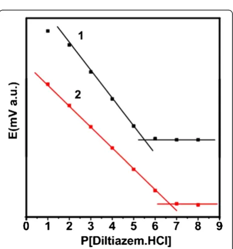
enhance the conductivity and electron transfer of the modified electrode, thus, the enhanced electro transfer would increase the sensitivity to diltiazem. Figure 3 shows the calibration curves of using direct DTMBi and TiO2@DTMBi core-shell NSs as detection sensors. By
extrapolating the linear parts of the calibration curves, it can be calculated that the detection range and limit for DTMBi sensor (T0 sample) are 10−1 to 10−5 M and 1.53μg/mL, respectively. These results are consistent with the reported results that the detection limits for the most selective electrodes sensors are in the range of 10−5 to 10−6 M [10]. While for TiO2@DTMBi core-shell NSs
as detection sensor, in which TiO2 nanoparticles were
introduced, a wider detection range of 10−1to 10−7M and a much lower detection limit of 0.20μg/mL than the re-ported results not using TiO2 nanoparticles were
[image:3.595.55.540.89.402.2]ob-tained. These data suggest that TiO2@DTMBi core-shell
Figure 2Schematic illustrations, TEM, cyclic voltammograms, and SEM images. (a)Schematic illustration of preparation of TiO2nanoparticles
and TiO2@DTMBi core-shell nanospheres.(b)TEM image of TiO2nanoparticles; the insert is size distribution.(c)TEM images of TiO2@DTMBi core-shell
nanospheres; the inserts are two magnified spheres.(d)Cyclic voltammograms of electrodes (1), T0 and (2) T1. SEM images of the electrode surface(e), T0 and(f)T1.
[image:3.595.306.539.457.706.2]NSs can be used as a proposed high-performance sensor for diltiazem detection.
Formation, structure, and optimal preparation condition of TiO2@DTMBi NSs
FTIR spectra of TiO2@DTMBi NSs clearly show the
characteristic absorption peaks ascribed to DTM ran-ging from 1,230 to 1,650 cm−1(Figure 4a (spectrum 1), indicated by the arrows). XRD reflection also shows TiO2@DTMBi NSs having the feature peaks of DTM
(Figure 4b (spectrum 1), indicated by the arrows). XRD reflections in Figure 4b also indicate that the crystal
structure of the obtained TiO2 NSs and TiO2@DTMBi
NSs both mainly belong to anatase titanium dioxide [13], though the small peaks belong to rutile TiO2 also been
found.
[image:4.595.59.539.89.304.2]In Figure 4b, XRD peaks of DTM are only visible for T1 sample. This is because T3 sample contains very low con-tent of DTM. This inference is consiscon-tent with the FTIR re-sults showed in Figure 4a. FTIR spectrum for T3 sample presents very weak absorption from 1,230 to 1,650 cm−1 as-cribed to DTM characteristic peaks (Figure 4a (spectrum 2). We deduce that the very low content of DTM in T3 sam-ple was because of the rinsing process. For T1 samsam-ple, Figure 4Infrared spectra and XRD reflection. (a)Infrared spectra of samples (1) T1, (2) T3, and (3) T0;(b)XRD reflection of (1) T1, (2) T3, (3) TiO2NPs, and (4) T0.
[image:4.595.57.542.510.715.2]because the initial ratio of DTMBi/TiO2 is much higher
than T3 sample, T1 sample contains more amount of DTM after the rinsing process.
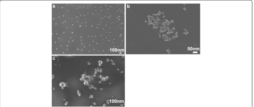
As illustrated in Figure 2a, there are three preparation steps for TiO2@DTMBi NSs, during the third step, it is
clear that the DTMBi/TiO2 ratio will play an important
role in controlling the morphology. We also investigate the effect of different DTMBi/TiO2 (molar ratio, listed
in Table 1) on the obtained TiO2@DTMBi products. As
SEM images shown in Figure 5, we can find the
mono-disperse TiO2@DTMBi NSs only been obtained at
DTMBi/TiO2= 1:1; the lower or higher ratio both
pro-duced much larger aggregates. This might ascribe to the interaction between TiO2 and DTM molecules
(struc-ture shown in Figure 1) such as hydrogen bond interac-tions are depended on different DTMBi/TiO2ratio. This
inference is according to the literature reports about the H-bond interactions between organic molecules, and crystal particles can modify the growth and assemble of crystal particles [14,15].
Mechanism for response improvement in the TiO2-based
system
As far as the mechanism for response improvement in the TiO2-based system is concerned, take T1 sample for
typ-ical example, we think that evident response improvement is mainly caused by two reasons. One is the response sur-face area for T1 and T0 (the control) is different. Figure 2e, f reveals that electrode surface for T0 and T1 are totally different; it is obvious that T1 with many nanospheres have bigger response surface area than T0 without TiO2
nanoparticles. The other is that those TiO2nanoparticles
enhance the conductivity and electron transfer of the modified electrode, thus, the enhanced electro transfer would increase the sensitivity to diltiazem drug. The re-sults listed in Table 1 also indicate that the morphology of the obtained TiO2@DTMBi samples play a very
im-portant role on the detection limit. T1 sample with mono-disperse morphology has a much lower detection limit of 0.20μg/mL than those of T2 (1.12 μg/mL) and T3 sam-ples (0.94 μg/mL) with aggregate morphology (shown in Figure 5). We deduce that this difference is mainly caused by different response surface area of T1 to T3 samples, monodisperse nanospheres having bigger response surface area than those aggregate ones.
Conclusions
In summary, monodisperse, core-shell TiO2@DTMBi NSs
with size of approximately 40 nm were facile prepared. The obtained TiO2@DTMBi NSs were also investigated as
sen-sor to detect diltiazem. The results reveal that when these core-shell NSs are used as detection sensor, they can pro-vide a wider detection range of 10−1to 10−7M and much lower detection limit of 0.20μg/mL than the literature data.
These data demonstrate that TiO2@DTMBi core-shell NSs
can be used as proposed high-performance sensor for dilti-azem detection.
Competing interests
The authors declare that they have no competing interests.
Authors’contributions
JY, YE, LC, JZ, and YS took the tasks of experimental, data collection, and draft writing; ZC gave his contributions on the experimental design and guidance, data analysis, as well as the main paper organization; and YZ took the contributions on the research guidance, discussion, and paper modification. All authors read and approved the final manuscript.
Authors’information
ZC is a Ph.D. major in Biomedical Engineering, Sichuan University, China. He has focused his research interest on the biomaterials especially on the nanoparticles synthesis and application for more than 7 years. His published papers involved the inorganic and organic nanoparticles toward
multifunctional nanocarriers and sensors and biomineralization.
Acknowledgements
This work is supported by the National Natural Science Foundation of China (No. 51202199 and 51074205), Natural Science Foundation of Liaoning Province (No.2014022038), Excellent Talents Program of Liaoning Provincial Universities (No. LJQ2013089), Liaoning S & T Project (No.2013225305), Liaoning Provincial University Students Researching Training Programs (No. 201210160012), and Liaoning Medical University Principal Fund (No. XZJJ20130104-01).
Author details
1School of Material and Metallurgy, Northeastern University, 110004
Shenyang, People’s Republic of China.2College of Pharmacy, Liaoning
Medical University, 121001 Jinzhou, People’s Republic of China.3College of
Chemistry, Nankai University, 300071 Tianjin, People’s Republic of China.
Received: 14 July 2014 Accepted: 27 August 2014 Published: 3 September 2014
References
1. De M, Ghosh PS, Rotello VM:Applications of nanoparticles in biology.
Adv Mater2008,20(22):4225–4241.
2. Chen Z, Wang C, Chen J, Li X:Biocompatible, functional spheres based on oxidative coupling assembly of green tea polyphenols.J Am Chem Soc
2013,135(11):4179–4182.
3. Basu S, Basu PK:Nanocrystalline metal oxides for methane sensors: role of noble metals.J Sens2009,2009:861968.
4. Diamond D: InPrinciples of Chemical and Biological Sensors.Edited by Diamond D. New York: John Wiley & Sons; 1998:1–10.
5. Li Y, Yu X, Yang Q:Fabrication of TiO2nanotube thin films and their gas
sensing properties.J Sens2009,2009:402174.
6. Luo X, Morrin A, Killard AJ, Smyth MR:Application of nanoparticles in electrochemical sensors and biosensors.Electroanal2006,18(4):319–326. 7. Lee G-H, Kim M-S:Crystal structure of TiO2thin films grown on sapphire
substrates by RF sputtering as a function of temperature.Electron Mater Lett2010,6(2):77–80.
8. Eun T-H, Kim S-H, Jeong W-J, Jeon S-J, Kim S-H, Yang S-M:Single-step fabrication of monodisperse TiO2hollow spheres with embedded
nanoparticles in microfluidic devices.Chem Mater2009,21(2):201–203. 9. Chen Y, Yang SY, Kim J:Phase transformation comparison of TiO2
nanorods and TiO2thin film after annealing.Electron Mater Lett2012,
8(3):301–304.
10. Ganjali MR, Razavi T, Dinarvand R, Riahi S, Norouzi P:New diltiazem potentiometric membrane sensor stands on theoretical calculations as a useful device for diltiazem hydrochloride analysis in pharmaceutical formulation and urine.Int J Electrochem Sci2008,3(12):1543–1558. 11. I-Nashar RM, Abdel Ghani NT, Hassan SM:Construction and performance
characteristics of new ion selective electrodes based on carbon nanotubes for determination of meclofenoxate hydrochloride.
12. Barringer EA, Bowen HK:Formation, packing, and sintering of monodisperse TiO2powders.J Am Ceram Soc1982,65(12):199–201.
13. Park HK, Moon YT, Kim DK, Kim CH:Formation of monodisperse spherical TiO2powders by thermal hydrolysis of Ti (SO4)2.J Am Ceram Soc1996,
79(10):2727–2732.
14. Chen Z, Wang C, Zhou H, Sang L, Li X:Modulation of calcium oxalate crystallization by commonly consumed green tea.Cryst Eng Comm2010,
12(3):845–852.
15. Chen Z-H, Ren X-L, Zhou H-H, Li X-D:The role of hyaluronic acid in biomineralization.Front Mater Sci2012,6(4):283–296.
doi:10.1186/1556-276X-9-465
Cite this article as:Yueet al.:Preparation TiO2core-shell nanospheres
and application as efficiency drug detection sensor.Nanoscale Research
Letters20149:465.
Submit your manuscript to a
journal and benefi t from:
7Convenient online submission
7Rigorous peer review
7Immediate publication on acceptance
7Open access: articles freely available online
7High visibility within the fi eld
7Retaining the copyright to your article