Highly polarized HLA class II antigen
processing and presentation by human
intestinal epithelial cells.
R M Hershberg, … , P E Framson, G T Nepom
J Clin Invest.
1998;
102(4)
:792-803.
https://doi.org/10.1172/JCI3201
.
The high concentration of foreign antigen in the lumen of the gastrointestinal tract is
separated from the underlying lymphocytes by a single cell layer of polarized epithelium.
Intestinal epithelial cells can express HLA class II antigens and may function as
antigen-presenting cells to CD4(+) T cells within the intestinal mucosa. Using tetanus toxoid specific
and HLA-DR-restricted T lymphocytes, we show that polarized intestinal epithelial cells
directed to express HLA-DR molecules are able to initiate class II processing only after
internalization of antigen from their apical surface. Coexpression of the class II
transactivator CIITA in these cells, which stimulates highly efficient class II processing
without the characteristic decline in barrier function seen in polarized monolayers treated
with the proinflammatory cytokine gamma-IFN, facilitates antigen processing from the
basolateral surface. In both cases, peptide presentation to T cells via class II molecules was
restricted to the basolateral surface. These data indicate a highly polarized functional
architecture for antigen processing and presentation by intestinal epithelial cells, and
suggest that the functional outcome of antigen processing by the intestinal epithelium is
both dependent on the cellular surface at which the foreign antigen is internalized and by
the underlying degree of mucosal inflammation.
Research Article
Find the latest version:
J. Clin. Invest.
© The American Society for Clinical Investigation, Inc. 0021-9738/98/08/0792/12 $2.00
Volume 102, Number 4, August 1998, 792–803 http://www.jci.org
Highly Polarized HLA Class II Antigen Processing and Presentation by Human
Intestinal Epithelial Cells
Robert M. Hershberg,*‡ Diane H. Cho,* Adel Youakim,i M. Brigid Bradley,¶ Janet S. Lee,¶ Paul E. Framson,*
and Gerald T. Nepom*§
*Virginia Mason Research Center, Seattle, Washington 98101; ‡Division of Medical Genetics, and §Department of Immunology, University of Washington School of Medicine, Seattle, Washington 98195;iImmunex Corporation, Seattle, Washington 98101; and ¶Immunology Program, Memorial Sloan Kettering Cancer Center, New York 10021
Abstract
The high concentration of foreign antigen in the lumen of the gastrointestinal tract is separated from the underlying lymphocytes by a single cell layer of polarized epithelium. Intestinal epithelial cells can express HLA class II antigens
and may function as antigen-presenting cells to CD41 T
cells within the intestinal mucosa. Using tetanus toxoid spe-cific and HLA-DR–restricted T lymphocytes, we show that polarized intestinal epithelial cells directed to express HLA-DR molecules are able to initiate class II processing only af-ter inaf-ternalization of antigen from their apical surface. Co-expression of the class II transactivator CIITA in these cells, which stimulates highly efficient class II processing without the characteristic decline in barrier function seen in polarized monolayers treated with the proinflammatory
cy-tokine g-IFN, facilitates antigen processing from the
baso-lateral surface. In both cases, peptide presentation to T cells via class II molecules was restricted to the basolateral surface. These data indicate a highly polarized functional architec-ture for antigen processing and presentation by intestinal epithelial cells, and suggest that the functional outcome of antigen processing by the intestinal epithelium is both de-pendent on the cellular surface at which the foreign antigen is internalized and by the underlying degree of mucosal in-flammation. (J. Clin. Invest. 1998. 102:792–803.) Key words: intestinal epithelium • HLA class II • cell polarity • oral
toler-ance • inflammatory bowel disease
Introduction
The intestinal epithelium is composed of polarized epithelial cells that provide a crucial barrier function. This single cell layer separates a myriad of bacterial and food antigens in the lumen of the gastrointestinal tract from the largest comple-ment of lymphocytes in the body, which defines the
gut-associ-ated lymphoid tissue (GALT).1 In addition to its crucial role in
barrier function and in nutrient and ion transport, the intesti-nal epithelium is an active component in the complex immuno-regulation of the GALT. Intestinal epithelial cells (IECs) se-crete a wide variety of cytokines, both constitutively and after invasion with bacterial pathogens (1). Moreover, IECs have extensive cellular contact with several distinct populations of T lymphocytes both within the epithelium (intestinal epithelial lymphocytes) and in the underlying lamina propria (lamina propria lymphocytes). Whereas several molecules that medi-ate the physical interaction between T cells and intestinal epi-thelial cells have been identified (2), the functional conse-quences of these interactions are not well understood.
Recent studies in experimental systems of both oral toler-ance (3–5) and inflammatory bowel disease (6) have under-scored the importance of mucosal CD41 lymphocyte re-sponses. Because the surface expression of class II molecules is essential in cells capable of functioning as antigen-presenting cells (APCs) to CD41 T cells (7), it is noteworthy that numer-ous reports have described the low level of HLA class II anti-gens on the surface of normal IEC and the increased expres-sion of these molecules in a variety of pathological conditions, including inflammatory bowel disease (8), graft-versus-host disease (9), and Celiac disease (10). The cellular machinery for efficient processing and presentation of antigen to CD41 T lymphocytes via HLA class II includes a variety of cellular pro-teases, as well as the invariant chain (Ii) and HLA-DM het-erodimer (reviewed in references 11 and 12). Indeed, Kaiser-lian et al. have shown that murine IECs can function as APCs to CD41 cells (13). We have recently characterized the class II pathway in several human IEC lines, and shown that these cells can process and present antigen to CD41 T cells in a path-way modulated by the expression of Ii and HLA-DM (14).
An important issue arises when considering the in vivo APC function of IECs due to the tight junctions that restrict the passage of macromolecules between individual cells. Spe-cifically, the apical (or luminal) surface of the IEC is selec-tively exposed to high concentrations of foreign antigen; yet, the expression of HLA class II antigens on epithelial cells in tissue sections of intestinal mucosa from human (15, 16) and rat (17) was observed to be restricted to the basolateral sur-face. The situation is not static, however, because in the pres-ence of inflammatory cytokines such as g-interferon (g-IFN),
Address correspondence to Robert M. Hershberg, Virginia Mason Research Center, 1000 Seneca Street, Seattle, WA 98101. Phone: 206-583-6525; FAX: 206-223-7543; E-mail: [email protected]
Received for publication 24 February 1998 and accepted in revised form 11 June 1998.
the barrier function of the epithelium is significantly impaired (18). The resulting increase in paracellular transport of luminal antigens would result in the concurrent exposure of both the apical and basolateral surfaces of the cell to foreign antigens. In this regard, differences in the cellular trafficking of antigen after exposure to either the apical or basolateral surfaces of polarized, HLA class II positive epithelial cells may have func-tional consequences in the regulation of the adjacent and sub-jacent T lymphocytes. Conceivably, these differences might tilt the delicate balance in the GALT away from immune toler-ance towards the inflammatory response seen in the intestinal mucosa in a wide variety of clinical disorders.
Here we describe a model system to study the physiologic interaction between polarized epithelial cells and CD41 T lym-phocytes, and investigate the polar nature of HLA class II an-tigen processing and presentation by IECs. Using the polar-ized human IEC line T84 engineered to constitutively express HLA-DR molecules, we demonstrate the restricted expression of HLA class II antigens at the basolateral surface of the cell, mimicking the topologically restricted expression pattern seen in vivo. Using tetanus toxoid specific and HLA-DR–restricted T cells, we demonstrate that, in the absence of Ii and HLA-DM, only exposure of antigen at the apical surface of the T84 HLA-DR transfectants is capable of initiating HLA class II processing and presentation. However, when the T84 HLA-DR transfectants are engineered to also overexpress the class II transactivator (CIITA), resulting in the induced expression of invariant chain and HLA-DM, both the apical and basolat-eral surface can initiate processing. These data are consistent with a model in which both the polarized exposure of antigen and the underlying level of mucosal inflammation may dra-matically modulate the functional outcome of the class II anti-gen processing by the intestinal epithelium.
Methods
Cells and transfectants. The T84 cell line (ATCC CCL-248) was de-termined by reverse dot blot hybridization with HLA-DR–specific probes to be: DRB1*0101/09012 and DRB4*0101. HLA-DRB1*0401– expressing transfectants were generated by retroviral infection as previously described (14). Where indicated, the T84 HLA-DR trans-fectants were superinfected with a recombinant retrovirus expressing the class II transactivator, CIITA (19). Dividing T84 HLA-DRB1*0401 transfectants were exposed to fresh recombinant CIITA virus daily for 7 d and infection was verified as described in Results. T84 cells were grown in DME supplemented with 10% vol/vol FBS, non-essen-tial amino acids (GIBCO BRL, Gaithersburg, MD), 20 mM glutamine, and penicillin/streptomycin. The EBV-transformed B-LCL BSM and the B3T hybrid T2 (containing a homozygous deletion of the HLA class II locus including the region encoding HLA-DM) were grown in RPMI with 10% vol/vol FBS and 50 mM b-mercaptoethanol.
Confocal microscopy. Cells were grown on borosilicate chamber coverslips (Lab-Tek, Nunc, Napierville, IL) or on polycarbonate filter inserts (6.5 mm diameter, 0.4 m pore size; Costar, Cambridge, MA) and maintained for 3–5 d after confluence (transepithelial resistance [TER] . 1,000 ohms cm2). Processing of the cells for
immunofluores-cence was done at room temperature. Cells were washed three times with PBS, fixed for 15 min in 4% paraformaldehyde in PBS, washed twice in PBS, and incubated for 30 min in 50 mM NH4Cl to quench
unreacted aldehyde groups. The cells were washed twice more with PBS and then permeabilized and blocked in PBS containing 0.1% Triton X-100 and 5% BSA (PTB) for 30 min. Subsequently, the cells were incubated with primary antibodies (anti–E-cadherin and anti– ZO-1; Zymed Laboratories, San Francisco, CA); anti–HLA class I
(W6/32, kindly provided by Daniel Geraghty, Fred Hutchinson Can-cer Research Center, Seattle, WA); anti–HLA-DR (L243), diluted in PTB for 1–2 h, washed three times with PBS, and then incubated for 1 h with the appropriate fluorescent-conjugated secondary antibody (Oregon green goat anti–rabbit IgG for the anti–ZO-1 antibody, and Rhodamine goat anti–mouse IgG for the others; Molecular Probes, Eugene, OR) diluted in PTB. After washing with PBS three times, the coverslips or filters (cut out of their holders and placed on slides) were mounted and then covered with DABCO/glycerol/PBS, and in the case of the filters, coverslipped and sealed before microscopy. The samples were analyzed by on a Molecular Dynamics Laser Scan-ning Microscope Model 2001 (Molecular Dynamics, Sunnyvale, CA) equipped with an Kr/Ar laser and a Nikon 603 NA 1.4 oil lens. Im-ages were collected using an SGI Iris Indigo 4000 Workstation (Sili-con Graphics, Mountain View, CA). The final images were prepared using Adobe Photoshop V3.0. All images were processed identically.
Cell surface–specific biotinylation, immunoprecipitation, and im-munoblotting. For biotinylation, cells were grown on polycarbonate filter inserts (24 mm diameter, 0.4 m pore size; Costar, Cambridge, MA) until TER were . 1,000 ohms cm2. The cells were labeled either
apically or basolaterally with biotin (Calbiochem, San Diego, CA) as previously described (20). After biotinylation, cells were solubilized in Lysis buffer (2.5% n-octylglucoside, 0.1 M Tris, pH 7.4, 0.15 M NaCl, 2 mM MgCl2, 0.02% NaN3, 1 mg/ml leupeptin, 1 mg/ml
pepsta-tin, and 1 mM PMSF) for 90 min at 48C with frequent titration. The lysates were centrifuged at 14,000 g for 10 min and the supernatants retained for immunoprecipitation. 100 mgof the supernatants were incubated with 20 mg of antibody overnight at 48C. 50 microliters of Protein G agarose (prewashed in Lysis buffer containing 5% BSA) was added to the samples and they were further incubated at 48C for 90 min. The Protein G agarose was washed four times with Lysis buffer and the bound material eluted by boiling for 10 min in reduc-ing SDS-PAGE sample buffer. The eluted samples were fractionated on a 10% Tris-glycine gel, transferred to nitrocellulose, blocked over-night in PBS containing 0.1% Tween 20 and 5%BSA at 48C, and the biotinylated proteins were detected using streptavidin–horseradish peroxide (HRP; Jackson Labs, Bar Harbor, ME) and a chemilumi-nescent substrate for peroxidase (ECL; Amersham Corp., Arlington Heights, IL) followed by exposure to film.
Immunoblot analyses of invariant chain using the mAb Pin-1 and HLA-DMb using anti–HLA-DMb rabbit serum (both kindly pro-vided by Peter Cresswell, Yale University, New Haven, CT), and for HLA-DMa using anti–HLA-DMa rabbit serum (kindly provided by Alexander Rudensky, University of Washington, Seattle, WA) were performed using goat anti–mouse or goat anti–rabbit IgG 1 IgM (Jackson ImmunoResearch Laboratories, Inc.) and chemilumines-cence as described (14).
Analysis of barrier function. 300 3 103 T84 cells were plated on
upright transwell filters (3.0 mm pore size; Costar) exposing the apical surface to the top chamber, or on upside down filters that were subse-quently inverted exposing the basolateral surface to the top chamber. Cells were cultured until TER exceeded 1,000 ohms cm2 (generally
5–7 d) before use in antigen presentation or processing experiments. TER was assessed using an epithelial voltometer (EVOM, Sarasota, FL) and converted to ohms cm2 based on the surface area of the
trans-well filter. Bulk protein transport was assessed using intact horserad-ish peroxidase, as described (21).
TCR Va and Vb primers as described (23). Clone 2 was found to use the gene segment encoding Vb5.2 and clone 4 was found to use the gene segment encoding Vb13. For clone 4, these data were confirmed using a Vb13 subfamily-specific monoclonal antibody (Endogen, Inc., Cambridge, MA).
For antigen-processing and/or presentation assays, T84 cells were plated on transwells, and the monolayers were verified to have a TER
. 1,000 ohms cm2 at the start of the experiment. For the peptide
pre-sentation experiments, HSA peptide was added to the top chamber of upright or inverted transwell cultures for 30 min, followed by the ad-dition of 100 3 103 HSA-specific hybridoma cells. Supernatent from
the co-culture was collected after 24 h and assayed for IL-2 using the HT-2 cell line as previously described (14). For the processing experi-ments, TT was applied to the upper or lower chamber of inverted transwell monolayers as shown in Fig. 4, resulting in basolateral or apical exposure to antigen, respectively. After 4 h, the antigen was re-moved and 100 3 103 TT-specific T cell hybridoma or T cell clones
were applied to the top chamber and co-cultured with the T84 cells for 24 h. The supernatant was collected after 24 h and assayed for IL-2 using HT-2 cells. For the drug experiments, cells were prepulsed with drug from both the apical and basolateral surfaces for 30 min before adding antigen and the drug was present during antigen pulsing. Drug was removed from both chambers with the removal of antigen. All drugs were purchased from Sigma Chemical Co. (St. Louis, MO).
For the assays with the T cell clones, the T cells were removed from the transwells after 24 h, transferred to 96-well flat bottom plates, and cultured for 48 h in RPMI supplemented with 10% vol/vol FBS without the addition of exogenous IL-2. 1.0 mCi of 3
[H]-thymi-dine was present during the final 16 h of culture, and radiolabeled
incorporation was measured by scintillation spectroscopy. g-IFN (Genzyme, Cambridge, MA) was prepared and used according to manufacturer’s instructions.
Results
HLA expression is largely restricted to the basolateral surface of polarized T84 cells.The intestinal epithelial cell line T84 forms polarized monolayers in vitro and has been used exten-sively as a model to investigate a variety of physiologic and im-munologic properties of the intestinal epithelium (e.g., 1, 18, 24). To study the HLA class II antigen processing pathway in a manner independent of the cytokines required to induce class II expression in this cell line, we used retroviral-mediated gene transfer to generate T84 cells that constitutively express sur-face HLA-DR molecules, and demonstrated that these cells could both process and present antigen to CD41 T lympho-cytes (14).
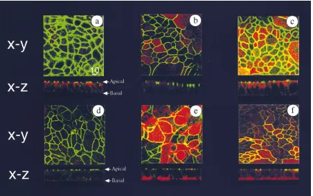
[image:4.612.58.510.374.658.2]Previous studies addressing the distribution of HLA class II on the intestinal epithelium in vivo have observed the ex-pression of these molecules to be restricted to the basolateral surface (15–17). To determine whether the T84 HLA-DR transfectants would be a suitable model to study polarized as-pects of HLA class II antigen processing and presentation, we used confocal microscopy to examine the polarized expression of HLA-DR in these cells. As seen in Fig. 1, the wild-type T84
Figure 1. Expression of HLA-DR molecules in polarized T84 cells is restricted to the basolateral surface. Shown are confocal microscopic
im-ages of wild-type T84 cells (top, bottom left), T84 HLA-DRB1*0401 (bottom middle), and T84 HLA-DRB1*0401/CIITA transfectants (bottom
right) stained with a variety of mAb. Depicted are horizontal (X–Y, or en face) images, with representative vertical (X–Z) sections below. In all
pictures cells are stained with anti–ZO-1, and secondary antibody for anti–ZO-1 was goat anti–rabbitOregon green. All other antibodies listed used
goat anti–mouserhodamine as secondary antibody. Co-staining in A: anti-E cadherin; B: anti-MDR; C: anti-HLA class I; D–F: anti-HLA class II.
Cells were grown on transwells until TER . 1,000 ohm cm2, and fixed, permeabilized, and stained with saturating concentrations of antibody as
cells (Fig. 1, a–d) as well as the T84 HLA-DR transfectants in the absence (Fig. 1 e) or presence (Fig. 1 f) of the directed overexpression of the class II transactivator CIITA (see be-low) form monolayers that demonstrate the polarized charac-teristics of a number of proteins and the presence of discrete tight junctions. Specifically, staining with antibodies directed against the tight junction complex molecule ZO-1 was apical and junctional (Fig. 1, a–f), whereas the staining with an anti-body directed against E-cadherin showed the characteristic ba-solateral pattern, with more staining evident along the lateral aspects of the cell (where it functions in cell adhesion) than along the basal portion of the cell (Fig. 1 a). An identical pat-tern of E-cadherin staining was seen in the T84 HLA-DR transfectants (data not shown).
Staining with several monoclonal antibodies specific for HLA-DR revealed predominantly basolateral surface staining, with the most prominent staining seen along the basal aspects of the cell (Fig. 1 e and f). As seen in the horizontal X-Y (en face) images, intracellular class II staining was also evident and vesicular in nature (Fig. 1, e and f). This pattern was not HLA-DR allele-specific as T84 cells expressing their endogenous HLA-DRB1*0101 allele (either after treatment with g-IFN or with directed expression of CIITA alone in the absence of the transfected DR4 allele) showed an identical pattern (data not shown). The staining seen with a monoclonal antibody di-rected against HLA class I revealed a pattern similar to that seen with antibodies against E-cadherin, with more prominent surface expression along the lateral aspect of the cell and no apical surface expression (Fig. 1 c). Less intracellular staining was evident with HLA class I than HLA class II antibodies. The staining with an antibody directed against the P-glycopro-tein (MDR1, used as an apical marker) was faint but restricted to the apical surface.
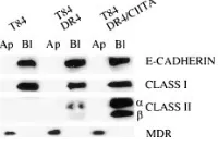
To confirm that the distribution of the various proteins by confocal microscopy was indeed polarized, we used selective cell surface biotinylation to independently assess and quanti-tate the polarized expression of HLA-DR in the T84 transfec-tants (20, 25, 26). The surface of intact, mature monolayers (with TER values . 1,000 ohm cm2) were labeled apically or
basolaterally with biotin, after which the cells were lysed, im-munoprecipitated using antibodies to the specific proteins, an-alyzed by SDS-PAGE, and transferred to nitrocellulose. The biotinylated proteins were detected using peroxidase-conju-gated streptavidin and chemiluminescence.
As seen in Fig. 2, class II expression was only seen on the basolateral surface of the transfected cells, whereas none was seen on the apical surface. The expression of E-cadherin (a well characterized marker of the basolateral surface) and class I were similarly localized. In contrast, MDR1a could only be detected when the cells were biotinylated apically. The lack of surface biotinylation of class II on the apical surface suggests that the small amount of staining seen on the apical aspect of the cells by confocal microscopy represented intracellular pools of the protein. These data indicate that the polarized ex-pression of class II in T84 on the basolateral surface mimics the staining pattern for class II seen in vivo, and neither requires matrix components of the underlying basement membrane nor contact with adjacent (intraepithelial lymphocytes) or subja-cent (lamina propia lymphocytes) T lymphocytes.
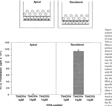
Antigen presentation to CD41 T lymphocytes is limited to the basolateral surface, but antigen processing is predominantly initiated from the apical surface in T84 HLA-DR
transfec-tants. To address the question of which surface or surfaces of polarized epithelial cells were capable of stimulating CD41 T cells, T84 HLA-DRB1*0401 transfectants were grown on semi-permeable transwell filters and tested for their ability to stimulate a peptide-specific, HLA-DRB1*0401-restricted T cell hybridoma. As schematically outlined in Fig. 3, T84 cells were grown with their apical or basolateral surface exposed to the top chamber of the transwell insert, and cultured to allow the formation of a monolayer with intact tight junctions and high electrical resistance (. 1,000 ohms cm2). The T84 DR4
cells were pulsed for 1 h with peptide, after which T cell hybri-domas were added. T cells were always added to the top cham-ber to maximize IEC-T cell contact. Consistent with the pat-tern of cell surface expression of class II antigens observed by confocal microscopy and selective surface biotinylation, only the basolateral surface of the T84 cells could present peptide antigen to T lymphocytes (Fig. 3). No stimulation was seen when the peptide and T cells were added to the apical surface of the T84 DR4 transfectants. The stimulation was HLA-DR allele-specific, as the T84 transfected with HLA-DRB1*1101 (DR5) failed to stimulate the hybridoma.
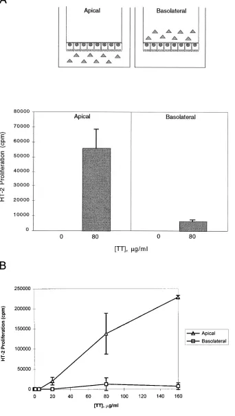
[image:5.612.316.416.61.128.2]APCs normally must process intact protein to generate the antigenic peptides that bind to nascent or recycled class II mol-ecules. To determine which surface or surfaces of the entero-cyte could initiate antigen processing of an intact protein via HLA class II molecules, we exposed either the apical or baso-lateral surface of electrically resistant, polarized T84 DRB1*0401 monolayers to an intact protein antigen (TT). As shown in Fig. 3, because antigen presentation occurred only at the basolat-eral surface due to the restricted expression of HLA-DR, the T84 transfectants were always plated “bottoms up” with their basolateral surface exposed to the top chamber. Hence, the cells were pulsed with intact antigen by addition to the bottom (apical) or top (basolateral) chamber. Processing was quanti-tated using a TT-specific, HLA-DRB1*0401–restricted T cell hybridoma, added after antigen pulsing and washing. As shown schematically in Fig. 4 A, the T cells were always added to the top chamber where basolateral contact (and antigen presentation) occurs. Significant processing occurred only af-ter exposure of the apical surface to antigen at the concentration of 80 mg/ml that we had defined as the optimal concentration from previous work with these transfectants (14) (Fig. 4 A). To ensure that the results obtained were not restricted to a single concentration of antigen, we performed dose response curves for apical and basolateral antigen processing. As shown in Fig. 4 B, minimal to no processing was seen when intact antigen
Figure 2. Selective cell surface
bi-otinylation reveals polarized ex-pression of HLA class II molecules at the basolateral surface. T84 wild type, T84 DRB1*0401, and T84 DRB1*0401/CIITA transfectants were plated on transwells until a TER . 1,000 ohm cm2 was
was added to the basolateral surface over a wide range of TT concentrations. As with the peptide-specific presentation seen in Fig. 3, no class II-mediated antigen presentation of processed antigen was seen at the apical surface (data not shown). Note that these observations are consistent with our previous results on 96-well plates (in contrast to the transwell system described here), demonstrating that T84 DR4 are capable of limited class II processing in the absence of Ii and HLA-DM (14).
To ensure that the differences observed did not simply re-flect an inability of the basolateral surface of the T84 DR4 cells to internalize protein antigen, we directly measured the uptake of intact HRP by both the apical and basolateral surface at var-ious time points including 15, 30, and 60 min. At all time points tested in several independent experiments, the basolateral sur-face was more efficient at internalizing HRP than the apical surface under identical conditions (data not shown).
Overexpression of the class II transactivator, CIITA, results in efficient antigen processing without affecting the barrier function of polarized T84 cells.We have recently shown that efficient antigen processing by T84 cells requires the expres-sion of not only class II antigens and cellular proteases, but also the Ii and the HLA-DM heterodimer (14). The role of these molecules in class II biosynthesis and antigen processing has been extensively reviewed (11, 12). The expression of both Ii and HLA-DM is induced by g-IFN in T84, likely mediated by the transcriptional activation of these genes by the CIITA, the expression of which is also induced by g-IFN (27).
T84 DRB1*0401 transfectants superinfected with a retrovi-rus directing the overexpression of CIITA resulted in class II antigen processing and presentation to CD41 T cells similar to that seen after treatment of the cells with g-IFN (Fig. 5 A). The induced expression of Ii, HLA-DMa and HLA-DMb was con-firmed in the T84 CIITA transfectants by immunoblotting (Fig. 5 B). As previously shown (14) and confirmed here, the wild-type T84 cells do not express Ii, HLA-DMa, or HLA-DMb. We also used an HLA-DR1–specific mAb and flow cy-tometry to demonstrate the expression of the endogenous HLA-DRB1*0101 allele induced by the overexpression of CIITA (data not shown).
In addition to stimulating antigen processing by T84 cells,
g-IFN treatment has been observed to elicit a profound and relatively rapid decrease in TER in T84 monolayers without an immediate alteration in cell morphology or integrity of the monolayer (18). This alteration in barrier function after g-IFN treatment would limit the ability to selectively expose one sur-face of the cell to intact antigen and to independently study the polarity of antigen processing and presentation. Surprisingly, we observed that the T84 HLA-DRB1*0401 transfectants overexpressing CIITA were able to maintain a high TER (Fig. 5 C), despite showing many of the characteristics of transfec-tants treated with g-IFN with regards to class II antigen pro-cessing. Treatment of the T84 CIITA transfectants with g-IFN resulted in the characteristic decline in TER seen in the wild-type T84 cells (Fig. 5 C), suggesting that the directed
overex-Figure 3. HLA-DR–restricted peptide
pre-sentation occurs only at the basolateral surface of T84 cells. (Top) Schematic view of transwell antigen presentation assay. T84 cells are plated on upright transwell filters (3.0 m pore size), exposing the apical surface to the top chamber, or on upside-down filters that are subsequently inverted, exposing the basolateral surface to the top chamber. T cells are always added to the top chamber to maximize T cell-T84 con-tact. (Bottom) T84 HLA-DRB1*0401 transfectants plated in the apical or ba-solateral orientation were co-cultured with HSA-specific, HLA-DRB1*0401– restricted T cell hybridoma cells for 24 h in the absence or presence of 10 mM of the HSA peptide for which the hybridomas are specific. T84 transfectants expressing simi-lar levels of the HLA-DRB1*1101 (DR5) allele were used as a negative control. IL-2 production of the hybridomas was quanti-tated using the supernatant from the top chamber by [3H]thymidine uptake (cpm) of
[image:6.612.62.428.60.409.2]pression of CIITA was not “dominant” to the effects seen with
g-IFN in altering barrier function. The pattern of TER eleva-tion after plating of the cells on transwells and the kinetics and character of the response to g-IFN were identical in the
[image:7.612.59.401.68.673.2]wild-type T84 cells and the CIITA transfectants (data not shown). Moreover, as seen in Fig. 1, there was no discernible difference in morphology, ZO-1 staining, or class II localization in the CIITA transfectants compared to the HLA-DR transfectants.
Figure 4. Class II antigen processing of
in-tact tetanus toxoid in T84 HLA-DR trans-fectant is markedly more efficient after ex-posure of intact antigen from the apical than from the basolateral surface. A. (Top) Schematic view of transwell antigen pro-cessing assay. Because HLA-DR expres-sion and antigen presentation is restricted to the basolateral surface (Figs. 1–3), the T84 HLA-DR4 transfectants are plated with their basolateral surface exposed to the top chamber. The polarized, electri-cally resistant (. 1,000 ohm cm2)
The intact barrier function of the T84 cells overexpressing CIITA was further verified by their inability to allow passage of intact HRP (Fig. 5 D). The addition and 24-h co-culture of antigen and antigen-specific, HLA-DRB1*0401–restricted T cell hybridomas to either the T84 DR4 or T84 DR4 CIITA transfectants did not result in any transport of HRP across the monolayer (5 D, “T cells 1 Ag”), demonstrating that the monolayers restricted passage of protein during the typical course of the antigen-processing experiments. Hence, overex-pression of CIITA resulted in efficient HLA class II antigen processing, but had no apparent effect on barrier function in T84 cells. These results demonstrate that g-IFN–induced changes in APC activity can be dissociated at a molecular level from the barrier-disrupting activity.
The overexpression of CIITA facilitates HLA class II anti-gen processing from the basolateral surface of polarized T84 monolayers. Next we assessed the ability of the T84
HLA-DRB1*0401/CIITA transfectants to process intact protein an-tigen in the polarized processing assays. Note that increased class II expression in the CIITA transfectants (compared to
[image:8.612.57.558.55.366.2]the T84 DR4 transfectants alone) corresponds to that from the endogenous HLA-DRB1*0101 allele and does not inter-fere with the functional studies detailed using T cells that are restricted to the transfected HLA-DRB1*0401 allele. In con-trast to the restricted ability of the HLA-DR transfectants to initiate processing of intact TT only at the apical surface, the T84 HLA-DR4/CIITA transfectants could initiate processing from either the apical or basolateral surface (Fig. 6). The anti-gen dose response curves done under identical conditions consistently reveal that the apical processing in the DR4/ CIITA transfectants is more efficient at low antigen doses than the basolateral processing in the DR4/CIITA transfec-tants, or the apical processing in the DR4 transfectants alone (Figs. 4 B and 6). The data indicate that in the presence of CIITA expression (mirroring the response of g-IFN facilitat-ing efficient antigen processfacilitat-ing), the highly restricted polar initiation of processing is no longer evident, and both polar surfaces can initiate antigen processing. In both instances, the presentation to CD41 T cells remains restricted to the baso-lateral surface of the cell.
Figure 5. The overexpression of the class II transactivator CIITA in T84 HLA-DR transfectants results in efficient antigen processing without
affecting barrier function. (A) IL-2 production by the TT-specific, HLA-DRB1*0401–specific hybridoma stimulated by T84 HLA-DRB1*0401 transfectants in the presence (right) or absence (left) of 80 mg/ml TT. Assays were performed on 96-well flat bottom plates (not on transwells) as described previously. T84 cells used were T84 HLA-DRB1*0401 (stippled bars), T84 HLA-DRB1*0401 pulsed with g-IFN for 48 h before addi-tion of TT (striped bars), and T84 HLA-DRB1*0401/CIITA transfectants in the absence of g-IFN (filled bars) (B) Immunoblot analysis using antibodies specific for Ii, HLA-DMa, or HLA-DMb in the cells listed. (C) Overexpression of CIITA does not impair the ability of T84 cells to develop a high TER. Both curves represent TER data from T84 cells transfected with and expressing CIITA. Media from both the top and bot-tom chambers were changed daily with (closed circles) or without (open circles) the addition of 500 m/ml g-IFN to the basolateral surface of the cell. (D) Overexpression of CIITA does not result in the paracellular transport of proteins across the T84 monolayer. Bulk protein transport was assessed using intact HRP as described. To ensure transport was not affected by the addition of the T lymphocytes to the monolayer during the processing assays, the HRP assays were done immediately after co-cultivation of the T84 monolayers with 100 3 103 T cells for 24 h in the
Class II processing initiated from both the apical and baso-lateral surface requires endosomal acidification but only the apical processing is inhibited by disruption of the actin-based cytoskeleton. We next compared the sensitivity of the apical
processing in the T84 DR4 transfectants and the apical and ba-solateral processing in the T84 DR4/CIITA transfectants to several drugs known to inhibit endocytosis or intracellular traf-ficking events involved in class II processing to investigate the class II pathways from the distinct polarized surfaces. Based on the data obtained from the TT dose response curves, all drug inhibition experiments were performed at a TT concen-tration of 80 mg/ml. The drug concentrations used were deter-mined to be noncytopathic by light microscopy, and TER was checked at the conclusion of the experiment, and although re-duced, was found to be . 300 ohms cm2 at the concentrations
and exposure times indicated.
Cytochalasin D interferes with the function of actcon-taining microfilaments and has been shown to selectively in-hibit apical endocytosis in Madine-Darby canine kidney (MDCK) cells (28, 29). Analogously, cytochalasin D selectively inhibited the antigen processing of apically added antigen in a dose-dependent manner (Fig. 7). In contrast, processing from the basolateral surface was unaffected by cytochalasin D. The macrolide antibiotic bafilomycin A1, which inhibits the vacu-olar H1 ATPase resulting in inhibition of endosomal acidifica-tion (30), showed a dose-dependent inhibiacidifica-tion of class II anti-gen processing from both the apical and basolateral surfaces, although the inhibition at low concentration for the apical pro-cessing in the DR4 and DR4/CIITA transfectants was less than for the basolateral processing.
Stimulation of different human T cell clones after class II an-tigen processing initiated from the apical or basolateral surface of IECs. To determine whether the polarized class II
path-ways might result in the stimulation of different subsets of T lymphocytes, we tested the responsiveness of several CD41, TT-specific, HLA-DRB1*0401–restricted human T cell clones for their ability to proliferate against T84 DR4/CIITA trans-fectants exposed to intact TT from their apical or basolateral surface. In previous studies, we demonstrated that T84 HLA-DR transfectants could be used as efficient APCs for human T
cell clones (14). For the majority of clones studied, pretreat-ment of the T84 HLA-DR transfectants with g-IFN or the di-rected expression of CIITA in the APC (to induce Ii and HLA-DM) before the addition of antigen was required for op-timal T cell stimulation. In some T cell clones, however, pre-treatment with g-IFN or expression of CIITA in the APC ac-tually inhibited the T cell responses seen, and optimal T cell proliferation was observed without any manipulation of the T84 HLA-DR transfectants before antigen pulsing and T cell co-cultivation.
Accordingly, we chose two independent T cell clones for these studies: one clone (designated clone 2) that was en-hanced by expression of CIITA in the T84 DR4 transfectant, and another (designated clone 4) in which stimulation was op-timal without CIITA expression in the APC. The clonality of these cells was confirmed using RT-PCR and Va- and Vb -spe-cific primers (see Methods section). When assayed using polar-ized T84 transfectants on transwells, these two clones showed distinct patterns of responsiveness to T84 DR4/CIITA trans-fectants that depended on which side (i.e., apical or basolat-eral) antigen was added to the T84 cells. As seen in Fig. 8, clone 2 was stimulated to proliferate by T84 DR4/CIITA cells that processed antigen from the basolateral surface but not from identical cells exposed to antigen from the apical surface. In marked contrast, clone 4 proliferated more vigorously against T84 DR4/CIITA cells that processed antigen from the apical rather than from the basolateral surface. Although the precise peptides recognized by these individual T cell clones are not known, these data suggest that the same cell exposed to an identical antigen in a polarized context may result in the stimulation of distinct (but potentially overlapping) subsets of T cells.
Discussion
[image:9.612.58.392.65.268.2]IECs exist in a highly polarized anatomical context, with high concentrations of dietary and bacterial antigens at the mucosal (apical) surfaces and high concentrations of lymphoid cells at the basolateral surface. HLA class II expression is polarized in
Figure 6. Expression of CIITA in the T84
these cells, restricted to the basolateral membrane, and we now demonstrate that there is a remarkable functional polarity to the antigen-processing and presentation pathway in these cells.
Using three independent approaches—confocal micros-copy, selective surface biotinylation, and HLA class II–medi-ated peptide presentation to antigen-specific T cells—we dem-onstrate that the surface expression of HLA class II expression in polarized intestinal epithelial cells is restricted to the baso-lateral surface. These data are consistent with the pattern of surface expression of MHC class II antigens in the human (15,
31) and rat (17) intestinal epithelium seen using immunohis-tochemistry, and, more recently, in MDCK, a polarized kidney epithelial cell line, cells transfected with HLA-DR1 (32). With regard to the precise sequences required for basolateral target-ing of class II molecules, several groups have recently shown that sequences within the cytoplasmic domain of the Ii can di-rect the expression of a chimeric molecule to the basolateral surface of polarized MDCK cells (32, 33). The Ii is physically associated with nascent HLA class II molecules, but is progres-sively trimmed to the CLIP fragment (which does not contain the basolateral targeting sequence) as the nascent class II com-plex migrates to an acidic, peptide-loading compartment re-ferred to as CIIV (or MIIC) before transport to the cell surface (reviewed in 11 and 12). Hence, the physiological significance of Ii in directing class II molecules to the basolateral surface of polarized cells remains unclear. Our data using T84 cells engi-neered to express HLA-DR in the absence of Ii clearly reveal that basolateral targeting of class II molecules does not require Ii. These data are in agreement with the findings that class II expression is mostly restricted to the basolateral surface in MDCK cells expressing HLA-DR in the absence of Ii (32). The precise molecular basis of the spatially-restricted pattern of class II expression at the basolateral surface without Ii has not been investigated. Interestingly, the cytoplasmic tail of the b chain of class II contains a di-leucine motif that has been implicated in recycling from the cell surface (34, 35), en-dosomal localization (36), and basolateral targeting (36 and reviewed in 37). It is also possible that sequences in the cyto-plasmic tail of the a chain of the class II heterodimer, which have also been implicated in recycling of mature class II mol-ecules from the cell surface (35), may contribute to basolat-eral targeting.
In addition to the restricted expression of HLA class II molecules at the basolateral surface, there was remarkable po-larity with regard to the ability of the epithelial cell to process intact antigen with the processing of TT initiated only from the apical surface in T84 cells directed to express HLA-DR mole-cules in the absence of Ii and HLA-DM. To be able to study the potential effects of g-IFN on polarized aspects of class II processing in IEC in the absence of the untoward effects of this cytokine in disrupting the barrier function of T84 monolayers, we directed the overexpression of CIITA (with the concomi-tant induction of Ii and HLA-DM expression) in the T84 HLA-DR transfectants. We observed that antigen processing was more efficient in the CIITA “co-transfectants” and that, strikingly, both polar surfaces were capable of initiating class II antigen processing.
In this context, several features of the CIITA transfectants are noteworthy. First, in contrast to the report of Siegrist et al., which detailed the inability of melanoma cells overexpressing CIITA to efficiently process intact antigen (38), the overex-pression of CIITA in T84 cells was sufficient to confer efficient class II antigen processing that consistently equaled or ex-ceeded that seen after treatment with g-IFN. These data sug-gest that the effect of CIITA is not identical in all “nonprofes-sional” antigen-presenting cells. IECs, such as T84 cells, may be especially proficient in class II processing in the setting of CIITA expression because they constitutively express high levels of cysteine and aspartyl proteases (14) including cathep-sin S (R. Hershberg and H. Chapman, unpublished data). Sec-ond, the directed overexpression of CIITA enhanced antigen processing in T84 cells without a concomitant decrease in
bar-Figure 7. Drug inhibition of class II processing from the apical and
[image:10.612.58.298.56.482.2]Figure 8. Different human T cell clones
are selectively stimulated after class II processing initiated from the apical or basolateral surface with the same anti-gen. Stimulation of two different TT-spe-cific HLA-DRB1*0401–restricted T cell clones (designated clone 2 and 4, respec-tively) by T84 DR4 CIITA transfectants on transwells pulsed with 80 mg/ml TT from the apical or basolateral surface, as indicated. After 24 h of co-culture, the T cell clones were removed from the top chamber of the transwells and cultured in 96-well round bottom plates (without further addition of antigen or APC) for 48 h. 1.0 mCi 3[H]thymidine was present
during the last 18 h of culture, and
3[H]thymidine uptake was measured. All
points were done in triplicate and stan-dard error is indicated.
[image:11.612.58.555.243.669.2]rier function. This demonstrates that the effects of g-IFN in the regulation of “physiological” epithelial cell functions, such as tight junction permeability, may be dissociated at the molec-ular level from its effect on the induced expression of immuno-logically relevant molecules, such as Ii and HLA-DM.
The polarized trafficking of antigen via HLA class II in ep-ithelial cells highlight the complexity of endosomal structures in polarized epithelial cells, with distinct apical and basolateral structures that communicate somewhat through a “common” endosomal pathway (39, 40). Based on the inhibition of both apical and basolateral processing by the proton pump inhibitor bafilomycin, processing of antigen from both polarized sur-faces requires transit through an acidic compartment. These data are consistent with several studies detailing the trafficking of nascent and recycled class II through acidic endosomal structures in the presence or absence of Ii (34, 41). The selec-tive stimulation of several CD41 T cell clones depending on the route of antigen uptake suggests that processing from the apical or basolateral surface has the capacity to generate dif-ferent HLA class II-peptide complexes from the same antigen. This is reminiscent of other reports that the processing and presentation of specific peptide epitopes within the same pro-tein antigen (34, 41) or virus particle (42) appear highly depen-dent on the presence or absence of Ii and/or HLA-DM (re-viewed in 43). The data presented here provide the first polarized context for such Ii and HLA-DM independent class II processing events, and are likely to be of physiological sig-nificance, given both the high concentration and large number of food and microbial antigens constantly in contact with the intestinal epithelium.
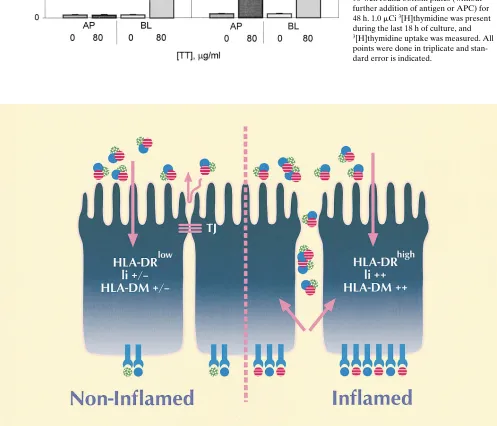
In summary, we identify distinct features of HLA class II processing after exposure of either the apical or basolateral surface to a protein antigen, and a schematic model is pre-sented in Fig. 9. As shown, the spatially restricted pattern of class II expression on the basolateral surface of polarized IECs facilitates the presentation of antigens to T cells within the epi-thelium and in the underlying lamina propria. This polarized peptide presentation via HLA class II at the basolateral sur-face occurs whether the intact antigen is internalized and pro-cessed from the apical or basolateral surface of the cell. The combined effect of proinflammatory cytokines such as g-IFN within the intestinal mucosa in altering barrier function (allow-ing increased paracellular passage of luminal antigens across the epithelium), coupled with the facilitation of efficient anti-gen processing and presentation by intestinal epithelial cells, may help to explain the exaggerated and often intractable in-flammatory response seen in the intestinal mucosa of patients with inflammatory bowel disease. This may result in the pro-cessing and presentation of distinct peptide epitopes from lu-minal antigens by epithelial cells from inflamed (in contrast to “noninflamed”) mucosa. Whether the “sampling” of luminal antigens via apical processing and presentation by class II mol-ecules of certain peptide epitopes by the intestinal epithelium to CD41T cells in the lamina propria in the noninflamed mu-cosa contributes to the generation of oral tolerance remains a matter for speculation. Consistent with the data presented here, proinflammatory cytokines are likely to dramatically al-ter the delicate balance between tolerance and responsiveness in the intestinal mucosa. In this context it is notable that g-IFN administration to mice can abrogate the tolerogenic effect of orally administered antigen (44), yet oral tolerance can occur in g-IFN–receptor2/2mice (45).
Acknowledgments
We gratefully acknowledge Linda Wicker and Dennis Zaller (Merck Research Laboratories) for T cell hybridomas, Jane Buckner and Steve Ziegler (Virginia Mason Research Center) for critical review of the manuscript, Nicky Ducommun for help with preparation of the manuscript, and Mari Hall for expert assistance with preparation of figures.
This work was supported by a First Award from Crohn’s and Colitis Foundation of America (R.M. Hershberg); National Institutes of Health (Bethesda, MD) grant AI50902 (J.S. Lee); National Insti-tutes of Health grant AI38913 (G.T. Nepom).
References
1. Jung, H.C., L. Eckmann, S.-K. Yang, A. Panja, J. Fierer, E. Morzycka-Wroblewska, and M.F. Kagnoff. 1995. A distinct array of proinflammatory cy-tokines is expressed in human colon epithelial cells in response to bacterial in-vasion. J. Clin. Invest. 95:55–65.
2. Cepek, K.L., S.K. Shaw, C.M. Parker, G.J. Russell, J.S. Morrow, D.L. Rimm, and M.B. Brenner. 1994. Adhesion between epithelial cells and T lym-phocytes mediated by E-cadherin and the alpha E beta 7 integrin. Nature. 372: 190–193.
3. Chen, Y., J. Inobe, and H.L. Weiner. 1995. Induction of oral tolerance to myelin basic protein in CD8-depleted mice: both CD41 and CD81 cells mediate active suppression. J. Immunol. 155:910–916.
4. Garside, P., M. Steel, F.Y. Liew, and A.M. Mowat. 1995. CD41 but not
CD81 T cells are required for the induction of oral tolerance. Int. Immunol. 7: 501–504.
5. Barone, K.S., S.L. Jain, and J.G. Michael. 1995. Effect of in vivo deple-tion of CD41 and CD81 cells on the induction and maintenance of oral toler-ance. Cell. Immunol. 163:19–29.
6. Powrie, F. 1995. T cells in inflammatory bowel disease: protective and pathogenic roles. Immunity. 3:171–174.
7. Germain, R.N. 1994. MHC-dependent antigen processing and peptide presentation: providing ligands for T lymphocyte activation. Cell. 76:287–299.
8. Mayer, L., D. Eisenhardt, P. Salomon, W. Bauer, R. Plous, and L. Picci-nini. 1991. Expression of class II molecules on intestinal epithelial cells in hu-mans. Differences between normal and inflammatory bowel disease.
Gastroen-terology. 100:3–12.
9. Bland, P.W., and C.V. Whiting. 1992. Induction of MHC class II gene products in rat intestinal epithelium during graft-versus-host disease and effects on the immune function of the epithelium. Immunology. 75:366–371.
10. Ciclitira, P.J., J.M. Nelufer, H.J. Ellis, and D.J. Evans. 1986. The effect of gluten on HLA-DR in the small intestinal epithelium of patients with Coe-liac disease. Clin. Exp. Immunol. 63:101–104.
11. Wolf, P.R., and H.L. Ploegh. 1995. How MHC class II molecules acquire peptide cargo: biosynthesis and trafficking through the endocytic pathway.
Annu. Rev. Cell Dev. Biol. 11:267–306.
12. Cresswell, P. 1994. Assembly, transport, and function of MHC class II molecules. Annu. Rev. Immunol. 12:259–293.
13. Kaiserlian, D., K. Vidal, and J.-P. Revillard. 1989. Murine enterocytes can present soluble antigen to specific class II-restricted CD41 T cells. Eur. J.
Immunol. 19:1513–1516.
14. Hershberg, R.M., P.E. Framson, D.H. Cho, L.Y. Lee, S. Kovats, J. Beitz, J.S. Blum, and G.T. Nepom. 1997. Intestinal epithelial cells utilize two distinct pathways for HLA class II antigen processing. J. Clin. Invest. 100:204–215.
15. Hirata, I., L.L. Austin, W.H. Blackwell, J.R. Weber, and W.O. Dobbins, III. 1986. Immunoelectron microscopic localization of HLA-DR antigen in con-trol small intestine and colon and in inflammatory bowel disease. Dig. Dis. Sci. 31:1317–1330.
16. Sarles, J., J.P. Gorvel, D. Olive, S. Maroux, C. Mawas, and F. Giraud. 1987. Subcellular localization of class I (A,B,C) and class II (DR and DQ) MHC antigens in jejunal epithelium of children with coelic disease. J. Pediatr.
Gastroenterol. Nutr. 6:51–56.
17. Mayrhofer, G., and L.D. Spargo. 1990. Distribution of class II major his-tocompatibility antigens in enterocytes of the rat jejunum and their association with organelles of the endocytic pathway. Immunology. 70:11–19.
18. Madara, J.L., and J. Stafford. 1989. Interferon-g directly affects barrier function of cultured intestinal epithelial monolayers. J. Clin. Invest. 83:724–727. 19. Bradley, M.B., J.M. Fernandez, G. Ungers, T. Diaz-Barrientos, V. Steimle, B. Mach, R. O’Reilly, and J.S. Lee. 1997. Correction of defective expression in MHC class II deficiency (bare lymphocyte syndrome) cells by retroviral trans-duction of CIITA. J. Immunol. 159:1086–1095.
20. Hughson, E.J., and R.P. Hirt. 1996. Assessment of cell polarity. In Epi-thelial Cell Culture. A.J. Shaw, editor. IRL Press, Oxford, UK. 37–66.
role in abrogating the effect of a T cell cytokine. J. Immunol. 153:5730–5739. 22. Woods, A., H.Y. Chen, M.E. Trumbauer, A. Sirotina, R. Cummings, and D.M. Zaller. 1994. Human major histocompatibility complex class II-restricted T cell responses in transgenic mice. J. Exp. Med. 180:173–181.
23. Genevée, C., A. Diu, J. Nierat, A. Caignard, P.-Y. Dietrich, L. Ferra-dini, S. Roman-Roman, F. Triebel, and T. Hercend. 1992. An experimentally validated panel of subfamily-specific oligonucleotide primers (Va1-w29/Vb1-w24) for the study of human T cell receptor variable V gene segment usage by polymerase chain reaction. Eur. J. Immunol. 22:1261–1269.
24. Colgan, S.P., C.A. Parkos, J.B. Matthews, L. D’Andrea, C.S. Awtrey, A.H. Lichtman, C. Delp-Archer, and J.L. Madara. 1994. Interferon-g induces a cell surface phenotype switch on T84 intestinal epithelial cells. Am. J. Physiol. 36:C402–C410.
25. Gottardi, C.J., L.A. Dunbar, and M.J. Caplan. 1995. Biotinylation and assessment of membrane polarity: caveats and methodological concerns. Am. J.
Physiol. 268:F285–F295.
26. Strohmeier, G.R., W.I. Lencer, T.W. Patapoff, L.F. Thompson, S.L. Carlson, S.J. Moe, D.K. Carnes, R.J. Mrsny, and J.L. Madara. 1997. Surface ex-pression, polarization, and functional significance of CD73 in human intestinal epithelia. J. Clin. Invest. 99:2588–2601.
27. Chang, C.-H., and R.A. Flavell. 1995. Class II transactivator regulates the expression of multiple genes involved in antigen presentation. J. Exp. Med. 181:765–767.
28. Gottlieb, T.A., I.E. Ivanov, M. Adesnik, and D.D. Sabatini. 1993. Actin microfilaments play a critical role in endocytosis at the apical but not the baso-lateral surface of polarized epithelial cells. J. Cell Biol. 120:695–710.
29. Jackman, M.R., W. Shurety, J.A. Ellis, and J.P. Luzio. 1994. Inhibition of apical but not basolateral endocytosis of ricin and folate in Caco-2 cells by cytochalasin D. J. Cell Sci. 107:2547–2556.
30. Lencer, W.I., G. Strohmeier, S. Moe, S.L. Carlson, C.T. Constable, and J.L. Madara. 1995. Signal transduction by cholera toxin: processing in vesicular compartments does not require acidification. Am. J. Physiol. 269:G548–G557.
31. Pociot, F., A.G. Wilson, J. Nerup, and G.W. Duff. 1993. No independent
association between a tumor necrosis factor-a promoter region polymorphism
and insulin-dependent diabetes mellitus. Eur. J. Immunol. 23:3050–3053. 32. Simonsen, A., E. Stang, B. Bremnes, M. Roe, K. Prydz, and O. Bakke. 1997. Sorting of MHC class II molecules and the associated invariant chain (li) in polarized MDCK cells. J. Cell Sci. 110:597–609.
33. Odorizzi, G., and I.S. Trowbridge. 1997. Structural requirements for ma-jor histocompatibility complex class II invariant chain trafficking in polarized Madin-Darby canine kidney cells. J. Biol. Chem. 272:11757–11762.
34. Zhong, G., P. Romagnoli, and R.N. Germain. 1997. Related leucine-based cytoplasmic targeting signals in invariant chain and major histocompati-bility complex class II molecules control endocytic presentation of distinct de-terminants in a single protein. J. Exp. Med. 185:429–438.
35. Pinet, V., M. Vergelli, R. Martin, O. Bakke, and E.O. Long. 1995. Anti-gen presentation mediated by recycling of surface HLA-DR molecules. Nature. 375:603–604.
36. Hunziker, W., and C. Fumey. 1994. A di-leucine motif mediates endocy-tosis and basolateral sorting of macrophage IgG Fc receptors in MDCK cells.
EMBO (Eur. Mol. Biol. Organ.) J. 13:2963–2969.
37. Mellman, I. 1995. Molecular sorting of membrane proteins in polarized and nonpolarized cells. Cold Spring Harbor Symp. Quant. Biol. LX:745–752.
38. Siegrist, C.A., E. Martinez-Soria, I. Kern, and B. Mach. 1995. A novel antigen-processing-defective phenotype in major histocompatibility complex class II-positive CIITA transfectants is corrected by interferon-g. J. Exp. Med. 182:1793–1799.
39. Knight, A., E. Hughson, C.R. Hopkins, and D.F. Cutler. 1995. Mem-brane protein trafficking through the common apical endosome compartment of polarized caco-2 cells. Mol. Biol. Cell. 6:597–610.
40. Odorizzi, G., A. Pearse, D. Domingo, I.S. Trowbridge, and C.R. Hop-kins. 1996. Apical and basolateral endosomes of MDCK cells are intercon-nected and contain a polarized sorting mechanism. J. Cell Biol. 135:139–152.
41. Momburg, F., S. Fuchs, J. Drexler, R. Busch, M. Post, G.J. Hammerling, and L. Adorini. 1993. Epitope-specific enhancement of antigen presentation by invariant chain. J. Exp. Med. 178:1453–1458.
42. Pinet, V., M.S. Malnati, and E.O. Long. 1994. Two processing pathways for the MHC class II-restricted presentation of exogenous influenza virus anti-gen. J. Immunol. 152:4852–4860.
43. Ceman, S., and A.J. Sant. 1995. The function of invariant chain in class II-restricted antigen presentation. Semin. Immunol. 7:373–387.
44. Zhang, Z., and J.G. Michael. 1990. Orally inducible immune unrespon-siveness is abrogated by IFN-g treatment. J. Immunol. 144:4163–4165.
45. Kjerrulf, M., D. Grdic, L. Ekman, K. Schon, M. Vajdy, and N.Y. Lycke.
1997. Interferon-g receptor-deficient mice exhibit impaired gut mucosal