Available online on www.ijpsr.com 742
IJPSR (2011), Vol. 2, Issue 4 (Review Article)
Received on 16 December, 2010; received in revised form 25 February, 2011; accepted 03 March, 2011
KANENGISER’S EVALUATION METHOD: AN EFFICIENT METHOD FOR DETERMINING OCULAR IRRITANT POTENTIAL IN COSMETICS
Swapnil S. Khadke* and Swapnil D. Mahajan
Department of Pharmacology, JSPM’s Rajarshi Shahu College of Pharmacy and Research, Tathawade, Pune, Maharashtra, India
ABSTRACT
A growing interest in usage of cosmetics has created the need for greater precision in evaluation and has stimulated research into cosmetic medicine. Ocular irritation testing represents an important step in the safety evaluation of cosmetic products. The objective of this study was to introduce a method of assessing ocular irritancy in human subjects and to illustrate the significance of a new expanded grading system, designed by Kanengiser, for the precise evaluation of ocular lesions and quantitative assessment of ocular surface responses to cosmetic products. Evaluation procedure is carried out using acute instillation method. A scoring scale for Palpebral Conjunctival Irritation, Eyelid Irritation, Lachrymation, Subjective Irritation, Bulbar Conjunctival Irritation, Corneal Abnormalities, Palpebral and Bulbar Conjunctival, Caruncular, and Corneal Fluorescein Ophthalmic Staining was designed by Kanengiser & observations are interpreted graphically. From the observations it is evident that frequency of occurrence of adverse events is minimal in the human ocular instillation studies. The area of corneal fluorescein staining was judged on a 0 to +4 scale using the same terminology as for corneal cloudiness (Draize). The statistical analysis of correlation between subjective irritation levels and area levels of corneal staining demonstrated better correlation for Kanengiser’s scoring system (r = 0.82) than for Draize’s scoring system (r = 0.74). From the above data we can illustrate that Kanengiser’s ocular grading system is an efficient, ‘evaluation method for determining the ocular irritant potential of cosmetic products in human eyes’.
Keywords: Melatonin, Immunity, Antioxidant, Pineal gland
Correspondence to Author: Swapnil S. Khadke
Available online on www.ijpsr.com 743 INTRODUCTION: A growing interest in usage of
cosmetics has created the need for greater precision in preparation and evaluation and has stimulated research into cosmetic medicine. The in-process & final product quality assurance testing continues throughout the entire production process. These tests include Ocular Irritancy Testing, Finished Product Auditing, Anaerobic Testing, Infrared Spectroscopy (FTIR), etc. Ocular irritation testing represents an important step in the safety evaluation of cosmetic products 1.
Objective: The objective of this study was to introduce a method of assessing ocular irritancy in human subjects and to illustrate the significance of a new expanded grading system, designed by Kanengiser, for the precise evaluation of ocular lesions and quantitative assessment of ocular surface responses to cosmetic products (i.e. shampoos, soaps, sunscreens, eye area, and facial cosmetics).
Evaluation Procedure 2:
Acute Instillation: 0.1 ml of test material is instilled in the human eye. Ophthalmic examinations were performed at 30 seconds, 5 minutes, 15 minutes, 60 minutes, and 120 minutes post-instillation. The subjects are screened for ocular symptoms, objective ophthalmic irritation, and ocular surface fluorescein staining.
Scoring scales: Scoring scales are designed as Objective, Subjective Ophthalmic Scoring Scale & Fluorescein Ophthalmic Staining Scale. The scales are as given below:
Scoring scale:
Subjective Ophthalmic Scoring Scale: Subjective Irritation (stinging, burning, itching, dryness, and/or foreign body sensation)
0= None; 1=Slight; 2=Mild; 3=Moderate; 4=Severe
Objective Ophthalmic Scoring Scale (Slit Lamp Biomicroscope Examination):
Lachrymation Tear Film Break-up
Time
0 = Normal tear production ≥10 = normal (No excess wetness)
1 = Trace increase in wetness 2 = Mild increase in wetness
(No distinct formed tears) 3 = A few formed tears
(Contained within the cul de sac and on surface of globe)
4 = Intense tearing
(Leaving cul de sac and globe, wetting lids and face)
Eyelid Irritation (redness, scaling, swelling, and/or meibomian secretions):
0 = No evidence of inflammation 1 = Trace inflammation
2 = Mild inflammation 3 = Moderate inflammation 4 = Severe inflammation
Palpebral Conjunctival Irritation:
0 = No evidence of inflammation
1 = Trace redness (very mild inflammation) 2 = Mild redness (mild inflammation)
Available online on www.ijpsr.com 744 Bulbar Conjunctival Irritation:
0 = No evidence of inflammation
1 = Trace redness (very mild inflammation) 2 = Mild redness (mild inflammation)
3 = Moderate redness, some dilation of blood vessels (moderate inflammation)
4 = Marked, intense redness several dilated blood vessels (severe inflammation)
Corneal Abnormalities (opacities, oedema,
infiltrates, vascularization, and/or epithelial defects):
0 = Normal, no abnormality 1 = Trace, very mild abnormality 2 = Mild abnormality
3 = Moderate abnormality 4 = Severe abnormality
Palpebral and Bulbar Conjunctival, Caruncular, and Corneal Fluorescein Ophthalmic Staining Scale:
Area
No Ocular Irritancy
0 = No staining
Mild Ocular Irritancy Range:
1 = >0 and < 10% 2 = >10% and < 20% 3 = >20% and < 30%
Moderate Ocular Irritancy Range:
4 = >30% and < 40% 5 = >40% and < 50%
6 = >50% and < 60%
Severe Ocular Irritancy Range:
7 = >60% and < 70% 8 = >70% and < 80% 9 = >80% and < 90% 10 = >90% and < 100%
11 = Mild superficial tissue abrasion 12 = Moderate superficial tissue abrasion 13 = Severe deeper tissue abrasion Density:
1 = Occasional, scattered punctate staining
2 = More uniform pattern of diffusely scattered punctate staining
3 = Dense foci of punctate staining within the areas of diffuse punctate staining
4 = General pattern of dense punctate staining Observations:
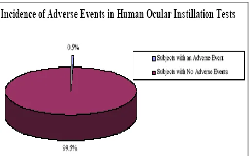
FIG. 1: INCIDENCE OF ADVERSE EVENTS IN HUMAN OCULAR INSTILLATION TESTS
[image:3.612.318.575.469.629.2]Available online on www.ijpsr.com 745 performed 2. Of 196 human subjects who
participated in ocular instillation studies since 1998, only 1 subject experienced an adverse event. This event, which occurred prior to test material instillation, was unrelated to the test material or to the study procedures. Other evidence of the safety of this methodology is provided by the repeated willingness of subjects to enroll in these studies as well as the resolution of all observed ocular irritation during the course of the studies.
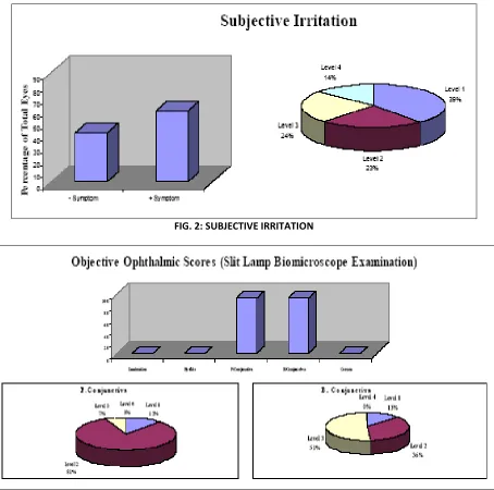
Approximately 59% of the total 228 individual eye evaluations, performed post instillation, demonstrated reports of subjective irritation, including stinging, burning, itching, dryness, and/or foreign body sensation 3. The distribution of subjective irritation levels was as follows: 39% reported level 1, 23% reported level 2, 24% reported level 3, and 14% reported level 4.
[image:4.612.84.538.225.675.2]FIG. 2: SUBJECTIVE IRRITATION
Available online on www.ijpsr.com 746 Approximately 94% of the total 228 individual eye
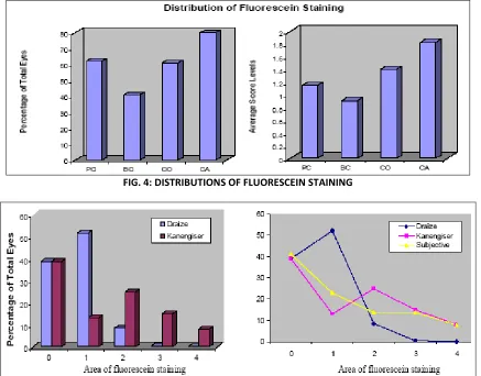
evaluations revealed both palpebral and bulbar conjunctival irritation. No lachrymation, eyelid or corneal irritation was observed. The levels of palpebral conjunctival irritation were 12%, 81%, 7%, and 0% at levels 1, 2, 3, and 4, respectively5. The levels of bulbar conjunctival irritation were 13%, 36%, 51%, and 0% at levels 1, 2, 3, and 4, respectively. The percentages of fluorescein staining, with both area and density scores, in the total 228 individual eye evaluations were 62%, 41%,
[image:5.612.92.529.238.580.2]61%, and 80% on palpebral and bulbar conjunctiva, cornea, and caruncle, respectively. The distributions of the average score levels for palpebral and bulbar conjunctivae, cornea, and caruncle staining were 1.15, 0.91, 1.4, and 1.83, respectively. Higher fluorescein staining of the caruncle than that of other ocular tissues 6, this is due to the effect of gravity, the tear fluid dynamics of the blinking eye, and the flow of tears through the nasolachrimal system, causing the instilled test material to be directed.
FIG. 4: DISTRIBUTIONS OF FLUORESCEIN STAINING
FIG. 5: AREA LEVELS OF FLUORESCEIN STAINING ON CORNEA (KANENGISER VS. DRAIZE) The area of corneal fluorescein staining was judged
on a 0 to +4 scale using the same terminology as for corneal cloudiness (Draize) 7. Corneal fluorescein staining was concurrently assessed by Kanengiser’s scoring scale. The percentages of fluorescein staining observed in the total 228 eye evaluations
Available online on www.ijpsr.com 747 demonstrated better correlation for Kanengiser’s
scoring system (r = 0.82) than for Draize’s scoring system (r = 0.74) 8.
CONCLUSION:
Human ocular instillation is an effective and safe
in vivo methodology for the assessment of cosmetic irritancy.
Kanengiser’s grading system assesses subjective human responses in addition to objective irritation and ocular surface tissue staining, which is scored on a scale with thirteen area and four density classifications 9.
When scoring methodologies are compared in assessing corneal irritation, Kanengiser’s scoring system has a higher level of correlation with subjective irritation responses than Draize’s scoring system.
Human ocular instillation represents a reliable, predictable and reproducible ocular irritant testing methodology to assess the safety of many substances. Kanengiser’s ocular grading system is an efficient, ‘evaluation method for
determining the ocular irritant potential of cosmetic products in human eyes 10.
REFERENCES:
1. Home Office. Statistics of scientific procedures on living animals, Great Britain – 2002.
2. Section 5(4) Animals (Scientific Procedures) Act 1986.
3. Supplementary note to the Home Secretary’s response to the Animal Procedures Committee interim report on the review of the operation of the Animals (Scientific Procedures) Act 1986.
4. Stuard, Caudill and Lehman-McKeeman: Characterization of the effects of musk ketone on mouse hepatic cytochrome P450 enzymes. Fund. & Appl. Toxicol 1997; 40:264-271.
5. Details taken from documents submitted to the US Environment Protection Agency in the early 1990s, under the Toxic Substances Control Act.
6. Details taken from documents submitted to the US Environmental Protection Agency in 1992, under the Toxic Substances Control Act.
7. Greenough et al: Food & Chem. Toxicol 1996; 34:161-166.
8. Details taken from documents submitted to the US Environment Protection Agency in 1992, under the Toxic Substances Control Act.
9. Anderson & Anderson: Toxic effects of air freshener emissions. Arch. Environ. Health 1997; 52:433-441. 10. Cosmetics & toiletries magazine act 2004: vol. 119:
no. 10, 32-35 362.