Development 139, 3950-3961 (2012) doi:10.1242/dev.082024 © 2012. Published by The Company of Biologists Ltd
INTRODUCTION
During Xenopusearly embryogenesis, Nodal/Activin, Wnt, BMP and FGF signaling pathways play key roles in promoting germ-layer formation. Nodal/Activin is the primary signal to induce mesoderm and endoderm in a dose-dependent fashion. FGF signaling also participates in mesoderm formation (Amaya et al., 1991; Amaya et al., 1993), mainly through providing competence for the embryonic cells to Nodal/Activin. BMP and Wnt pathways are active at the ventral side of embryo (Christian et al., 1991; Dale and Wardle, 1999) and are principally responsible for ventro-posteriorization of germ layers (Maéno et al., 1994; Suzuki et al., 1994; Schmidt et al., 1995; Dale and Wardle, 1999). At the dorsal side, they are blocked by antagonists secreted from the Spemann organizer: notably Noggin, Chordin, Cerberus, Dkk1, Xnr3, etc. (De Robertis et al., 2000). Thus the two groups of signals establish a balance for patterning body plan.
In Xenopus, the Nodal ligand genes, Xnr1-6, are induced by the maternal transcription factor VegT in vegetal cells (Clements et al., 1999; Hyde and Old, 2000; Takahashi et al., 2000; Hilton et al., 2003). Upon ligand gene transcription, Nodal signal is transmitted downstream and induces transcription of mesoderm- and endoderm-specific genes: Xbra, Mix1, Mix2, Goosecoid, Milk, Mix.1, Mixer, Sox17and GATA4-6, for example (Xanthos et al., 2001; Shivdasani, 2002; Zorn and Wells, 2007). Endoderm-specific genes, meanwhile, inhibit mesoderm genes such that mesoserm and endoderm formation is restricted within correct locations. Maternal
-catenin signaling is enriched in dorsal-vegetal cells and induces Siamoistranscription in the Nieuwkoop centre (Wodarz and Nusse,
1998), which subsequently induces gene transcription in the Spemann organizer (Wessely et al., 2001) to antagonize ventral signals. -Catenin also works in synergism with VegT to enhance transcription of Nodal-related genes (Agius et al., 2000; Takahashi et al., 2000), hence establishing a gradient of Nodal signal, with higher activity dorsally and lower activity ventrally. In addition, complex autoregulatory loops play important roles in the regulation of the activity of Nodal signaling (Schier, 2003).
Differentiation of early embryonic cells into germ layers is accompanied by the loss of pluripotency, which is maintained by pluripotency factors. In mammals, these factors are typically Oct4, Sox2, Nanog, cMyc and Klf4 (Niwa et al., 2000; Zaehres et al., 2005; Avilion et al., 2003; Fong et al., 2008; Nakatake et al., 2006). XenopusOct4 homologous factors Oct60, Oct25 and Oct91 inhibit mesendoderm germ-layer formation via inhibition of the activities of VegT, -catenin and Nodal (Cao et al., 2006; Cao et al., 2007; Cao et al., 2008). Sox2 is well known for its role in neural fate specification. Although these factors are crucial for the maintenance of pluripotency and self-renewal of embryonic stem (ES) cells, they exhibit distinct functions in ES cell differentiation assays and in embryonic development. Here, we report the identification and characterization of Kruppel-like factor 4 (Klf4) during Xenopus early embryogenesis. It promotes endoderm differentiation in both Nodal/Activin-dependent and -independent mechanisms. Moreover, it is involved in body axis patterning via activation of a subset of Spemann organizer genes, which code for Nodal/Activin, Wnt and BMP antagonists. In addition, loss of Klf4 function leads to failure of germ-layer differentiation. Thus we propose that Klf4 confers the competence of early embryonic cells to the activities of inducing signals such as Nodal/Activin so that embryonic cells can differentiate properly. Our results gain novel insights into the functions of Klf4 and the regulatory network for germ-layer differentiation and axis patterning in Xenopusembryos.
MATERIALS AND METHODS Embryos and explants
Xenopus laevis embryos and embryonic explants were obtained and cultured using conventional methods. To block endogenous Nodal activity, uninjected or injected embryos were incubated in culture medium 1Model Animal Research Center of Nanjing University and MOE Key Laboratory of
Model Animals for Disease Study, 12 Xuefu Road, Pukou High-Tech Zone, 210061 Nanjing, China. 2School of Medical Lab Science, Wenzhou Medical College, 325035 Wenzhou, China.
*These authors contributed equally to this work ‡Author for correspondence ([email protected])
Accepted 12 August 2012 SUMMARY
Klf4 is a transcription factor of the family of Kruppel-like factors and plays important roles in stem cell biology; however, its function
during embryogenesis is unknown. Here, we report the characterization of a Klf4 homologue in Xenopus laevis during
embryogenesis. Klf4is transcribed both maternally and zygotically and the transcript is ubiquitous in embryos during germ-layer
formation. Klf4 promotes endoderm differentiation in both Nodal/Activin-dependent and -independent manners. Moreover, Klf4
regulates anteroposterior body axis patterning via activation of a subset of genes in the Spemann organizer, such as Noggin, Dkk1
and Cerberus, which encode Nodal, Wnt and BMP antagonists. Loss of Klf4 function leads to the failure of germ-layer differentiation, the loss of responsiveness of early embryonic cells to inducing signals, e.g. Nodal/Activin, and the loss of transcription of genes involved in axis patterning. We conclude that Klf4 is required for germ-layer differentiation and body axis patterning by means of rendering early embryonic cells competent to differentiation signals.
KEY WORDS: Kruppel-like factor 4 (Klf4), Germ-layer differentiation, Body axis patterning, Transcriptional regulation, Xenopus laevis
Klf4 is required for germ-layer differentiation and body axis
patterning during
Xenopus
embryogenesis
Qing Cao1,*, Xuena Zhang1,*, Lei Lu1, Linan Yang2,Jimin Gao2, Yan Gao1, Haihua Ma1and Ying Cao1,2,‡
D
E
V
E
LO
P
M
E
N
terminal zinc fingers missing was PCR amplified to make construct pCS2+Klf4ZF. The C-terminal DNA-binding domain (DBD) aa 270-404 was subcloned to make pCS2+Klf4(DBD). For the test of efficiency of the antisense morpholino against Klf4, the ORF including the morpholino binding site was ligated to pCS2+eGFPmcs and pCS2+6MTmcs vectors to make pCS2+Klf4-eGFP and pCS2+Klf4-MT, respectively. The repression and activation form of Klf4 were made by ligating Klf4 DNA binding domain to pCS2+evemcs and pCS2+VP16mcs (Cao et al., 2008), thus resulting in plasmids pCS2+eve-Klf4(DBD) and pCS2+VP16-Klf4(DBD). A plasmid containing complete cDNA of mouse Klf4 (mKlf4) was purchased from IMAGE Consortium (Berlin) and the coding region was subcloned to make pCS2+mKlf4.
Whole-mount in situ hybridization
Whole-mount in situ hybridization on whole embryos or animal caps was carried out essentially as described (Harland, 1991).
In vitro transcription, antisense morpholino oligonucleotides (MOs) and microinjection
Antisense RNA probes for whole-mount in situ hybridization and mRNAs for microinjection were prepared as described (Cao et al., 2006). To prepare antisense RNA probes for whole-mount in situ hybridization, plasmids for Cerberus, Chordin, Dkk1, Gsc, Klf4, Mix2, Mixer, Noggin, Siamois, Sox17, Sox2, XAG2, Xbra, Xnr1, Xnr5 and Xvent2 were linearized and transcribed with T7 RNA polymerase. To prepare mRNAs for microinjection, plasmids pCS2+Klf4, pCS2+Klf4-eGFP, pCS2+Klf4-MT, pCS2+mKlf4, pCS2+Klf4ZF, pCS2+Klf4(DBD), pCS2+VP16-Klf4(DBD), pCS2+eve-pCS2+VP16-Klf4(DBD), pCS2+dnTCF3, pCS2+NLS-LacZ, pSP64T-activinB, pSP64T-dnXAR1, tBR-64T, pXFD (dnFGFR) and pSP64T-Xnr2 were linearized and transcribed with Sp6 mMessage mMachine kits (Ambion). All probes and mRNAs were cleaned up with an RNeasy Kit (Qiagen). An antisense morpholino oligonucleotide (MO), K4MO: TTCCCTCCACCTCTCATTAATCTGG – which targets 36/–12bp of 5⬘UTR – was designed to knock down endogenous Klf4 in Xenopus laevis. A six-base mismatched MO, K4MO6mis: TTCtCTCgACCTaTCATgAATaTGc (mismatched bases are in lowercase), and the standard control MO (ctrlMO), CCTCTTACCTCAGTTACAATTTATA, were used as controls. All MOs were purchased from GeneTools. Injected doses of mRNAs or MOs are described in the text.
Quantitative RT-PCR
Total RNAs and cDNAs were prepared using exactly the same procedure as described (Cao et al., 2006). Quantitative RT-PCR (qPCR) was performed on an ABI 7300 system and primers are listed in supplementary material Table S1. Amplification parameters were as follows: one cycle of predenaturation at 95°C for 10 seconds, followed by 40 cycles of denaturation at 95°C for 5 seconds, annealing and extension at 60°C for 31 seconds and an additional cycle for the melting curve. Crosspoints were calculated using ABI 7300 system SDS software. Final results were presented as histograms with relative units.
1% NP-40) supplemented with protease inhibitors (Roche). Homogenates were incubated on ice for 10 minutes and centrifuged at 12,000 rpm for 10 minutes, and supernatants were transferred to fresh tubes, boiled in 1⫻ Laemlli buffer, and centrifuged again at 12,000 rpm for 5 minutes. Supernatants were collected and 10 l of each sample were loaded into SDS-PAGE for electrophoresis. Western blotting was performed using the conventional method. A myc-tag antibody was used to detect the expression of Klf4-MT and an -actin antibody was used for detection of actin.
X-gal staining
Embryos injected with lacZmRNA were fixed in HEMFA and subjected to X-gal staining (Coffman et al., 1990). After staining, embryos were washed in PBS, fixed again in HEMFA and stored in 100% ethanol at –20°C, until processed for whole-mount in situ hybridization.
RESULTS
Spatial-temporal expression of Klf4during
Xenopusembryonic development
The identified cDNA encodes a protein of 404 amino acids. The sequence has the highest similarities to Klf4 in other species: for instance, 94% in Xenopus tropicalis, 51% in zebrafish and 55% in mouse (supplementary material Fig. S1A,B). Three classical zinc-finger motifs are present at the carboxyl terminus, which are typical for Kruppel-like factors (Pearson et al., 2008) and nearly identical among Klf4 proteins in different species (supplementary material Fig. S1A). There is a record for Xenopus laevis Klf4under accession number NM_001086359 in GenBank; however, this gene product shares the highest identity to Xenopus Klf17 [or Neptune (NM_001088664)] and mouse Klf2, but not Klf4. In the genome of Xenopus tropicalis, Klf4 gene locates upstream sequentially to rad23b, znf462 and tmem38b. When the order of these genes is reversed, the arrangement is identical to that in both zebrafish and mouse (Zfp462is synonymous with znf462) (supplementary material Fig. S1C). These comparisons suggested that the sequence we identified is orthologous to Klf4in other species.
Klf4 is maternally transcribed as it is present in the animal region of early cleavage stages, e.g. stages 3 and 6.5 (supplementary material Fig. S2A,B). During midblastula, Klf4 was detected ubiquitously in embryos but slightly enriched at one side of the embryos (supplementary material Fig. S2C). Later, the enrichment was found in the dorsal marginal zone in gastrula embryos (supplementary material Fig. S2D,E). Bisection of a gastrula embryo showed that Klf4 was present in ectoderm and the marginal zone, but enriched slightly in the dorsal margin of the organizer, prechordal mesoderm and endomesoderm (supplementary material Fig. S2F). During neurulation, Klf4
D
E
V
E
LO
P
M
E
N
localizes to two narrow lines within the neural folds (supplementary material Fig. S2G) and the anlage of cement gland (supplementary material Fig. S2H). Neural expression of Klf4 soon disappears but the cement gland expression persists until the tadpoles hatch (supplementary material Fig. S2I,J). Klf4is also specifically present in trigeminal nerve and lung primordium at stage 34 (supplementary material Fig. S2J), and the prospective duodenum/stomach at stage 43 (supplementary material Fig. S2K). During embryogenesis, maternal Klf4 is more abundant than zygotic Klf4 in gastrulae and neurulae. During the tailbud stages, the expression level rises up again (supplementary material Fig. S2L). Klf4 transcript is present in both animal and vegetal blastomeres at the eight-cell embryo stage (supplementary material Fig. S2M). At stage 8.5 when zygotic transcription and germ-layer differentiation starts, transcript was detected in animal, equatorial and vegetal regions (supplementary material Fig. S2N). Therefore, Klf4transcription is ubiquitous in early embryos. In summary, spatiotemporal expression patterns of Klf4suggest that it might be involved in early embryonic development.
Klf4 gain-of-function analyses in Xenopus embryos
The blastopore formed normally in uninjected control embryos and tended to close at stage 11.5. In embryos injected with Klf4mRNA, gastrulation was severely interrupted, as there was no clear blastopore formation (Fig. 1A,C). At stage 32, the majority of these embryos showed severely reduced anteroposterior body axis, pronounced belly protrusion with heavy pigmentation and seemingly exaggerated cement glands (Fig. 1B,C). In injected embryos, expression of the pan-mesoderm marker Xbra was strongly inhibited, suggesting that mesoderm formation was blocked (Fig. 1D,G). The endoderm gene Sox17was detected only in the vegetal area of normal embryos, but it was ectopically
activated in equatorial and animal regions in injected embryos (Fig. 1E,G). The neuroectoderm gene Sox2was expressed at the dorsal side of control gastrula embryos; however, Klf4RNA injection led to expansion of the Sox2expression domain to the ventral side, thus suggesting an increment in neuroectoderm (Fig. 1F,G). Furthermore, we injected one ventral-animal blastomere at the eight-cell stage with lacZ RNA alone or lacZ and Klf4 RNAs together. Klf4-induced ectopic expression of Sox17 or Sox2 occurred within the lacZ-labeling regions (Fig. 1H-J), implying an autonomous effect of Klf4.
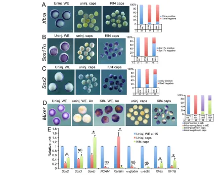
Isolated Xenopus blastula ectoderm, i.e. the animal caps, differentiates into epidermis. It can be induced to adopt different cell fates by inducers. At the gastrula stage, animal caps without Klf4injection did not exhibit any discernible Xbraand Sox17
[image:3.612.51.348.58.363.2]expression (Fig. 2A,B). Caps injected with Klf4 showed no difference from uninjected caps with respect to Xbraexpression. However, there was strong activation of Sox17in caps injected with Klf4(Fig. 2B). We observed repeatedly weak Sox2expression in uninjected caps, but Klf4 overexpression clearly led to an increase (Fig. 2C). These results are in agreement with the data observed in whole embryos. In addition, Mixer, another gene that is required for endoderm induction (Henry and Melton, 1998), was also strongly stimulated by Klf4 overexpression in both whole embryos and animal caps (Fig. 2D). Therefore, Klf4 is capable of inhibiting mesoderm while promoting endoderm and neuroectoderm formation. At neurula stage, Klf4-injected caps still showed higher levels of genes that specify neural precursors, e.g. Sox2, Sox3 and SoxD, but no neural tissue differentiation was observed, as revealed by NCAMexpression (Fig. 2E). Epidermal differentiation was nearly completely blocked in Klf4 caps (Fig. 2E). Genes marking mesodermal tissues, -globinand -actin, were detected only in background levels in both control and Klf4 caps (Fig. 2E). Instead, significant increases in expression of the
Fig. 1. Overexpression of Klf4in Xenopus early embryos.(A,B)The effect of Klf4mRNA injection on gastrulation (A) and on body axis formation (B). (C)Quantification of phenotypes shown in A and B in triplicate. (D-F) The influence of Klf4mRNA injection on mesoderm (D), endoderm (E) and ectoderm (F). Embryos in D were placed in vegetal views; those in E and F were placed in vegetal view (v) and animal view (a), separately. (G)Quantification of embryos with gene expression observed in D,E,F. In these experiments, 400 pg of Klf4mRNA were injected into the equatorial region of all blastomeres of two-cell or four-cell embryos. (H,I)lacZlabeling of targeted injection into one animal-ventral blastomere at the eight-cell stage and whole-mount in situ hybridization detection of Sox17(H) and Sox2(I) expression. Embryos were also placed in animal (a) and vegetal (v) views, respectively, as indicated at the top of the panels. (J)Quantification of embryos with normal or ectopic gene expression in H and I. lacZmRNA was injected at 20 pg/nl; Klf4mRNA was injected at 40 pg/nl. The arrows indicate the blastopore (bl). In all the panels, dorsal is up for Sox2 -stained embryos.
D
E
V
E
LO
P
M
E
N
liver marker genes Xhex and XPTB (Chen et al., 2003) demonstrated that endodermal tissue differentiation occurred in Klf4 caps (Fig. 2E). The result suggested that Klf4 is capable of promoting the formation of neural precursor cells, but is not able to induce neural tissue differentiation on its own.
Klf4 loss-of-function analyses
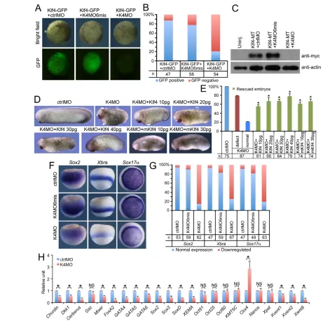
We designed an antisense morpholino oligonucleotide (K4MO) to knock down Xenopus laevisKlf4 by targeting the 5⬘UTR of its mRNA. K4MO could efficiently inhibit translation of the mRNA for the fusion protein GFP (Fig. 3A,B) and mRNA for Klf4-MT fusion protein in embryos (Fig. 3C). By contrast, both the six-base mismatched control MO (K4MO6mis) and the standard control MO (ctrlMO) did not inhibit protein translation (Fig. 3A-C), showing the specificity of K4MO.
At the tailbud stage, the Klf4 morphant displayed a severely reduced anteroposterior body axis and head size (Fig. 3D,E). This phenotype was rescued by co-injection of 10, 20, 30 or 40 pg Klf4
mRNA, as co-injection of the mRNA reversed the shortening of the body axis to different degrees, with a better rescuing effect at higher doses (Fig. 3D,E). The rescued embryos were obviously better in body axis formation than the Klf4 morphant. Moreover, co-injection of 10 or 30 pg mouse Klf4RNA (mKlf4) also resulted in a similar rescuing effect (Fig. 3D,E), suggesting a conserved function of Xenopusand mouse Klf4.
[image:4.612.54.485.59.405.2]Injection of ctrlMO or K4MO6mis in embryos didn’t affect expression of Xbra, Sox17and Sox2. However, they were inhibited in the Klf4 morphant (Fig. 3F,G). In addition, the mesoderm genes Chordin, Xvent1, Xvent2and Wnt8, the endoderm genes Mixer, FoxA2, GATA4, GATA5and GATA6, and the ectoderm genes Sox2, Sox3, SoxDand XEMAwere all repressed (Fig. 3H). The repression effect was specific for these genes because other genes such as Goosecoid(Gsc), Oct25, Oct60, Oct91, KMT5C, Cbx4and the germ cell genes Nanosand Xpat were not significantly altered or even upregulated (Fig. 3H). These results implied that Klf4 is a prerequisite for the differentiation of early embryonic cells to germ layers. Fig. 2. Assays on Klf4 function with animal caps.(A-C)Uninjected control whole embryos (Uninj. WE), uninjected control animal caps (uninj. caps), and caps injected with Klf4mRNA (Klf4 caps), were assays for the expression of Xbra(A), Sox17(B) and Sox2(C), and their respective quantification. (D)Mixerexpression in uninjected control whole embryos (Uninj. WE), Klf4mRNA-injected whole embryos (Klf4. WE), uninjected control animal caps (uninj. caps) and injected caps (Klf4 caps), and respective quantification. In A-D, embryos were placed vegetally to view normal expression of marker genes; except those in D, embryos were also orientated to animal view (An) to show the staining for Mixerin animal pole. Graphs represent the numbers of WE or caps with (positive) or without (negative) gene expression in three experiments. (E)qPCR detection of gene expression to analyse tissue differentiation in animal caps injected with Klf4RNA. Error bars represent s.d. in triplicate. A Student’s t-test was conducted to compare the changes in gene expression between uninjected (Uninj. caps) and Klf4-injected (Klf4) caps. Asterisks indicate P<0.01. NS: not significant. In the experiments above, 400 pg of Klf4mRNA was injected close to the animal pole of all blastomeres of four-cell embryos, and animal caps were removed at stage 8.5. For whole-mount in situ hybridization assays, caps were cultured until sibling control embryos reached stage 10.5. For qPCR assays, caps were cultured until sibling control embryos reached stage 15.
D
E
V
E
LO
P
M
E
N
Klf4 regulates Nodal/Activin pathway
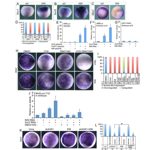
In the classic point of view, the Nodal/Activin pathway is the primary inducing signal for endoderm specification. Thus we have investigated whether Klf4 has any influence on Nodal/Activin
[image:5.612.53.524.57.515.2]activity. Overexpression of Klf4induced ectopic transcription of Xnr1and Xnr5(Fig. 4A,B,D). Accordingly, the Nodal/Activin direct target gene, Mix2, was stimulated (Fig. 4C,D). In HEK293T cells, transfection of the Xnr5 expression plasmid slightly Fig. 3. Loss-of-function analyses on Klf4.(A)Effect of ctrlMO, K4MO6mis or K4MO on the translation of co-injected mRNA for Klf4-GFP in embryos. Embryos were observed at stage 11 under brightfield or green fluorescent protein (GFP) and photos were taken using the same exposure parameters. (B)Quantification of embryos without (GFP-negative) or with (GFP-positive) green fluorescence. (C)Effect of ctrlMO, K4MO6mis and K4MO on the translation of Klf4-MT using western blotting (WB). Uninjected (Uninj.) and injected embryos were collected at stage 10.5 for WB. A myc-tag (MT) antibody (anti-myc) was used to detect the Klf4-MT fusion protein. -Actin was used as a loading control. In A and C, each MO was injected at 10 ng, Klf4-GFPor Klf4-MTmRNA was injected at 400 pg. (D)The rescuing effect of injected Xenopus Klf4mRNA (Klf4) or mouse Klf4 (mKlf4) mRNA on the Klf4 morphant. MOs were injected at 10 ng each. The dose of mRNA was indicated at the top of each panel.
(E)Quantification of normal, Klf4 morphant and rescued embryos in D. Error bars represent s.d. in three experiments. Student’s t-test was used to compare the ratio of rescued embryos against the ratio of unaffected embryos among K4MO-injected embryos. The asterisks indicate P<0.01. (F)Effect of injection of ctrlMO, K4MO6mis and K4MO on expression ofSox2, Xbraand Sox17. Embryos for Sox2expression were orientated to a dorsolateral view; those for Xbrato a lateral view, and Sox17to a vegetal view. (G)Statistical numbers of embryos in three experiments with normal or downregulated gene expression in F. (H)Gene expression analysis with qPCR. In F and H, 20 ng of ctrlMO or K4MO was injected into the equatorial region of all blastomeres at the four-cell stage and collected at gastrulation for whole-mount in situ hybridization or qPCR. Error bars represent s.d. in triplicate. A Student’s t-test was used to compare the change in gene expression between ctrlMO- and K4MO-injected embryos. Asterisks indicate P<0.01. NS: not significant.
D
E
V
E
LO
P
M
E
N
Fig. 4. The regulatory effect of Klf4 on the Nodal/Activin pathway. (A-C)The effect of Klf4overexpression on transcription of Xnr1(A), Xnr5 (B) and Mix2(C). Klf4mRNA (300 pg) was injected into the equatorial region of all blastomeres at the four-cell stage. Control and injected embryos at stage 8.5 were collected for detection of Xnr1and Xnr5expression, while embryos at stage 10 were collected for detection of Mix2expression. In A and B, embryos were in lateral view. In C, vegetal view was shown for ‘ctrl’ to reveal normal expression of Mix2, and animal view was shown for ‘Klf4’ to reveal ectopic Mix2expression in ectoderm. (D)Quantification of embryos in A-C in three experiments. (E)Luciferase assays in HEK293T cells transfected with ARELuc reporter plasmid and expression plasmids for Xnr5, FAST1 or Klf4. (F)Luciferase assays in embryos injected with ARELuc reporter plasmid and mRNAs for Xnr2 or Klf4. (G)Luciferase assays on pGL3-basic vector in embryos injected with Xnr2or Klf4mRNAs. Error bars represent s.d. in eight experiments. In F and G, plasmids were injected at 40 pg, Xnr2mRNA was injected at 10 pg, and Klf4mRNA was injected at 300 pg. (H)Mix2expression in uninjected (Uninj.) and Klf4RNA-injected (Klf4) embryos, and treated separately with different chemicals as indicated. All embryos are in vegetal view. Klf4mRNA (400 pg) was injected vegetally at the four-cell stage, treated with chemicals and collected at stage 10.5 for whole-mount in situ hybridization. (I)Quantification of embryos in H with normal, downregulated or upregulated Mix2expression in three experiments. (J)Mix2Luc(–712) luciferase assay with untreated embryos or embryos treated with SB431542. Plasmid was injected at 40 pg, Xnr2mRNA was at 10 pg, and Klf4mRNA was injected at 300 pg. Error bars represent s.d. in seven experiments. (K)The rescuing effect of Klf4 on Mix2expression. All embryos were in vegetal view. dnXAR1RNA (1.5 ng) and Klf4RNA (400 pg) were injected separately or together into the vegetal pole of four-cell embryos. Embryos were collected at stage 11 for whole-mount in situ hybridization. (L)Quantification of embryos with normal or altered Mix2expression observed in K. Error bars represent s.d. in triplicate. Student’s t-test showed the significance of the ratio of rescued Mix2expression in embryos with dnXAR1+Klf4RNA injection compared with background (ratio of embryos with unaffected Mix2
expression in dnXAR1-injected embryos). Asterisks indicate P<0.01.
D
E
V
E
LO
P
M
E
N
stimulated ARELuc, the Nodal/Activin responsive luciferase reporter (Pierreux et al., 2000; Germain et al., 2000). Co-transfection of the Klf4 expression plasmid stimulated the reporter somewhat more strongly. FAST1, a key nuclear signal transducer for the Nodal/Activin pathway, could stimulate ARELuc significantly. Addition of Klf4 plasmid to the cells led to much stronger stimulation of the reporter (Fig. 4E). Stimulation of ARELuc reporter by Klf4 was recapitulated nicely in embryos via injection of the Nodal ligand mRNA alone or together with Klf4 mRNA (Fig. 4F). Injection of these mRNAs had no significant effect on the pGL3-basic plasmid (Fig. 4G) that was used for constructing the ARELuc reporter, excluding the unspecific stimulation of ARELuc by Klf4. Therefore, Klf4 promoted Nodal/Activin activity. In uninjected embryos, blocking protein translation with CHX treatment or blocking Nodal/Activin with SB431542, a specific chemical inhibitor of the Nodal/Activin type I receptor, led to dramatic inhibition of Mix2expression. It was completely eradicated in embryos treated with both CHX and SB431542 (Fig. 4H,I). Vegetal injection of Klf4 RNA enhanced Mix2transcription compared with uninjected embryos. Treatment of injected embryos with CHX, SB431542 or both resulted in significant reduction in Mix2; however, the expression levels were much higher than in uninjected embryos, respectively (Fig. 4H,I). Therefore, even in the absence of protein translation and/or the Nodal feedback loop, Klf4 was still able to stimulate Nodal target gene expression. Klf4 exhibited the same effect on Mix2promoter activity. A luciferase reporter, Mix2Luc(–712), which contains –712/+13 fragment of Xenopus Mix2promoter (Cao et al., 2008), was stimulated in embryos by Klf4or Xnr2overexpression. The stimulation grew much stronger when Klf4 and Xnr2 were simultaneously overexpressed (Fig. 4J). SB431542 treatment dampened the stimulation, however, the promoter activity in embryos with Klf4overexpression was still much higher than the background level in treated embryos without Klf4overexpression (Fig. 4J). Both gene expression and promoter analyses demonstrated that Klf4directly regulates Nodal/Activin target gene expression. As a support, Klf4could rescue the decrease of Mix2 expression resulting from injection of dnXAR1(Fig. 4K,L), the dominant-negative Xenopus Activin receptor I (Hemmati-Brivanlou and Melton, 1992).
Klf4 promotes transcription of genes responsible for endoderm differentiation
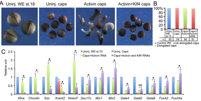
We tested the effect of Klf4 on mesoderm and endoderm differentiation induced by Nodal/Activin in animal caps. At the
neurula stage, control animal caps differentiated into epidermis and showed no elongation; however, those injected with Activin mRNA showed obvious elongation (Fig. 5A,B). By contrast, caps injected with both Activin and Klf4mRNAs did not elongate (Fig. 5A,B). qPCR revealed that, at the gastrula stage, animal caps injected with Activin mRNA showed high levels of mesoderm genes such as Xbra, Chordin, Gsc, Xvent2and XmenF. Meanwhile, the Nodal target and endoderm genes such as Mix1, Mix2, Sox17, Gata4-6, FoxA2and FoxA4awere also induced in these caps (Fig. 5C). When Klf4mRNA was co-injected, the mesoderm genes were dramatically inhibited, whereas the Nodal target genes and endoderm genes were enhanced (Fig. 5C). In summary, these analyses showed that Klf4 promotes endoderm differentiation while inhibiting mesoderm differentiation.
Crosstalk between Klf4 and Nodal/Activin signaling in germ-layer differentiation
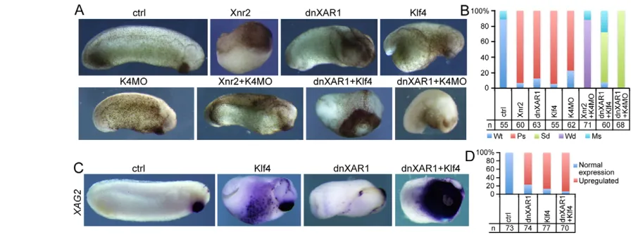
Activation of Nodal via injection of Xnr2mRNA or inhibition of Nodal activity via injection of dnXAR1 mRNA led to different developmental defects (Fig. 6A,B), as previously reported (Hemmati-Brivanlou and Melton, 1992). Ventral injection of Klf4 resulted in belly protrusion with heavy pigmentation, resembling an anteriorized phenotype. K4MO injection generated embryos with a significantly shortened anteroposterior axis (Fig. 6A,B). Interestingly, co-injection of Xnr2RNA and K4MO led to nearly normal embryos. By contrast, co-injection of Klf4and dnXAR1 brought about an extremely anteriorized phenotype, which showed a severely decreased anteroposterior body axis but with hugely exaggerated cement glands (Fig. 6A,B). Finally, when dnXAR1 RNA and K4MO were injected together ventrally, embryos bent towards the ventral side, probably owing to a lack of tissue differentiation (Fig. 6A,B).
[image:7.612.51.387.62.231.2]In congruence with the phenotypes above, injection of Klf4or dnXAR1RNA alone resulted in ectopic expression of XAG2, an anterior marker gene. When they were injected together, much stronger XAG2 expression was observed (Fig. 6C,D). This confirms the idea that overexpression of Klf4in the absence of Nodal/Activin leads to extreme anteriorization of the body axis. The effect was also observed in embryos with Klf4overexpression and inhibition of either BMP, FGF or Wnt (supplementary material Fig. S3A,B). Therefore, Klf4 anteriorizes body axis and enhances anteriorization in response to inhibition of posteriorization signals. We examined how the crosstalk between Klf4 and Nodal/Activin affected germ-layer differentiation. First, injection of dnXAR1 mRNA resulted in decreased Sox17 in the vegetal region. Fig. 5. Influence of Klf4overexpression on tissue differentiation induced by Activin. (A)Activin BmRNA (0.2 pg) was injected alone or together with 300 pg of Klf4mRNA into the animal pole of all blastomeres of two-cell or four-two-cell embryos. At stage 8.5, animal caps were removed from uninjected or injected embryos and cultured until the neurula stage. (B)Quantification of embryos and caps in A. (C)Gene expression analysis with qPCR on the caps described in A, but collected at stage 10. Error bars represent s.d. in three experiments. Asterisks indicate P<0.01. NS: not significant.
D
E
V
E
LO
P
M
E
N
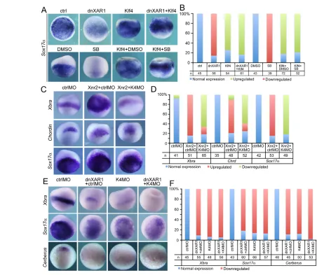
Overexpression of Klf4again activated ectopic Sox17. When both Klf4 and dnXAR1 were injected together, Sox17 was still ectopically stimulated in the animal region (Fig. 7A,B). Such an effect could be reproduced in embryos in which Nodal/Activin was blocked with SB431542 (Fig. 7A,B). Mixer, another gene involved in endoderm differentiation, could also be induced by overexpression of Klf4 in embryos with SB431542 treatment (supplementary material Fig. S4A,B). These effects are similar in that Klf4 still activates Mix2 expression in the absence of Nodal/Activin. By contrast, Klf4 knockdown compromised the upregulation of mesendoderm genes Xbra, Chordinand Sox17
resulting from Xnr2RNA injection (Fig. 7C,D). This result was in agreement with the fact that Klf4 knockdown was able to rescue the Xnr2overexpression phenotype (Fig. 6A). Finally, blocking either Nodal/Activin or Klf4led to a decrease in Xbra, Sox17and Cerberus expression. When both signals were inhibited, gene expression was nearly totally lost (Fig. 7E,F). This means that there is a synergistic effect between the two signals and both are a prerequisite for germ-layer formation.
Klf4 induces anteriorizing signals
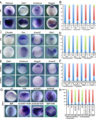
Injection of Klf4alone or meanwhile in the absence of Nodal/Activin activity led to anteriorization of embryos. Accordingly, Klf4 overexpression stimulated transcription of Siamois(Fig. 8A,B) at midblastula, which is required for induction of the organizer precursor (Nieuwkoop centre) and organizer gene transcription. Organizer genes such as Cerberus, Dkk1and Nogginwere activated prematurely at midblastula, when there were no or very weak transcription of these genes (supplementary material Fig. S5A,B). During gastrulation, these genes were expressed in much broader or ectopic regions in Klf4-injected embryos (Fig. 7A,B). Activation of the organizer genes was a direct effect. In uninjected embryos, CHX treatment totally eliminated gene expression. By contrast, Klf4 overexpression was able to activate the genes in both DMSO- and CHX-treated embryos (supplementary material Fig. S6A,B).
Klf4did not stimulate all organizer genes, as Chordinand Gsc were inhibited in response to Klf4 overexpression (Fig. 8C,D).
Moreover, the ventral gene Xvent2was also significantly inhibited in Klf4-injected embryos (Fig. 8C,D). Similar to Xenopus Klf4, mouse Klf4(mKlf4) likewise induced Dkk1activation in Xenopus embryos, supporting the idea that they are functionally homologous (Fig. 8C,D). Opposite to the effect of overexpression, Klf4 knockdown resulted in downregulation of Dkk1, Cerberusand Noggin(Fig. 8E,F). Upregulation of ventral genes such as Xvent2 was not detected in Klf4 morphant (Fig. 8E,F), this was possibly due to the failure of germ-layer differentiation (Fig. 7E). Hence, Klf4is sufficient and necessary for the transcription of the subset of organizer genes, which are known as anterior fate inducers. Overexpression of Klf4 enhanced anteriorization when Nodal, BMP or Wnt was blocked. Accordingly, expression of Dkk1was also strongly augmented in such embryos during gastrulation (Fig. 8G,H).
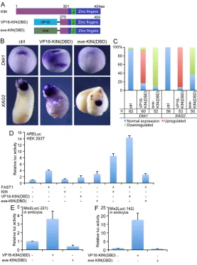
[image:8.612.56.504.60.227.2]Regulation of genes in germ-layer differentiation and axis patterning by Klf4gave rise to the question of whether it acts as a repressor or an activator. Klf4 consists of the transcriptional regulation domain at the N-terminus and the DNA-binding domain (DBD) of three zinc fingers at the C-terminus (supplementary material Fig. S7A). Injection of mRNA for the N-terminal region without zinc fingers (Klf4ZF) led to no significant change in embryogenesis. Injection of mRNA for the C-terminal region [Klf4(DBD)] had a minor effect on embryonic development. The affected embryos showed decreased tail region and slight belly protrusion (supplementary material Fig. S7B,C). Therefore, both regions are required for Klf4 function. As the C-terminus is responsible for DNA binding, we replaced the N-terminus of Klf4 with VP16 transcriptional activation domain or Even-skipped transcriptional repression domain to make VP16-Klf4(DBD) and eve-Klf4(DBD) fusion constructs, respectively (Fig. 9A). Injection of mRNA for VP16-Klf4(DBD) led to stimulation of Dkk1and XAG2, similar to the wild-type Klf4. However, eve-Klf4(DBD) caused an opposite effect (Fig. 9B,C). Luciferase assays in cells exhibited that VP16-Klf4(DBD) acted similarly to Klf4 to enhance FAST1 stimulated ARELuc activity, but eve-Klf4(DBD) could not exert such an effect (Fig. 9D). As CHX treatment showed that Mix2 Fig. 6. Correlation between Nodal and Klf4 during embryonic development.(A)Injection of Xnr2mRNA, dnXAR1mRNA, Klf4mRNA, or K4MO individually or in different combinations, as labeled above each panel, generated different effects on embryogenesis. (B)Quantification of phenotypes observed in A in three experiments. Wt, wild type; Ps, phenotypic changes after single injection; Sd, stronger phenotype after double injection; Wd, weaker phenotype after double injection; Ms, other phenotypes, such as those dead, or similar to phenotypes after single injection. (C)Expression of XAG2in uninjected control embryos at stage 28 and injected embryos as indicated. (D)Quantification of XAG2expression in embryos in C in four experiments. In all experiments above, 1.5 pg Xnr2mRNA, 1.5 ng dnXAR1mRNA, 300 pg Klf4mRNA or 5 ng K4MO was injected. Injections of Xnr2alone or Xnr2plus K4MO together were radial, while others were ventral injections made at the four-cell stage.
D
E
V
E
LO
P
M
E
N
is a direct target of Klf4, a fragment of Mix2promoter –221/+13 that contains the FoxH1 and Smad binding sites (the Activin response element) and a fragment of –142/+13 that does not contain the binding sites (Cao et al., 2008) were also used for testing the activity of the fusion constructs in embryos. VP16-Klf4(DBD) showed strong stimulating effect on both reporters; by contrast, eve-Klf4(DBD) was repressive (Fig. 9E,F). These experiments demonstrated that Klf4is an activator for transcription of these genes.
DISCUSSION
We have observed for the first time in the present study that during Xenopus embryogenesis: (1) Klf4 promotes endoderm differentiation; (2) Klf4functions in pattern formation of the body axis; and (3) Klf4acts as a competence factor for early embryonic cells to differentiate.
Klf4 is required for germ-layer differentiation
[image:9.612.52.514.56.449.2]We identified the cDNA for the mammalian Klf4orthologue in Xenopus laevis. This is supported not only by sequence comparison and synteny analysis, but also by the fact that mouse Klf4can rescue the Xenopus Klf4 morphant and that overexpression of Xenopus or mouse Klf4 generates a similar effect on gene expression. Xenopus Klf4is maternally expressed. In mammals, there is little maternal transcript of Klf4according to microarray data (Hamatani et al., 2008); however, significant maternal inheritance of Klf4is observed in medaka and zebrafish (Wang et al., 2011; Li et al., 2011; Luo et al., 2011). This might be because of the divergence of gene regulation between lower and high vertebrates during evolution. Zygotic transcription of Klf4 is detected in both lower vertebrate and mammalian early embryos, but the abundance of zygotic transcripts is lower than maternal Fig. 7. Combinatorial effects of Nodal/Activin and Klf4 on germ-layer differentiation.(A)The effect of blocking Nodal/Activin via injection of dnXAR1or treatment with SB431542 (SB) on Klf4-induced endoderm gene expression. The ‘ctrl’ and ‘dnXAR1’ embryos were orientated in vegetal view; the others were in lateral view to show staining in both vegetal and animal regions. (B)Quantification of embryos with normal or altered Sox17expression in A in triplicate. (C)The effect of K4MO or/and Xnr2mRNA injection on mesoderm and endoderm gene expression. The embryos are in lateral view. (D)Quantification of embryos with normal or altered gene expression in C in triplicate. (E)The effect of injection of dnXAR1or/and K4MO on mesendoderm gene expression. The embryos for ‘Xbra’ staining are in lateral view, whereas the rest are in vegetal view. (F)Quantification of embryos with normal or altered gene expression in E in three experiments. In all experiments above, 1.5 ng dnXAR1mRNA, 300 pg Klf4mRNA, 15 ng ctrlMO or K4MO, 2 pg Xnr2mRNA were injected. Injected or treated embryos were collected at stage 10.5 for whole-mount in situ hybridization.
D
E
V
E
LO
P
M
E
N
ones, as revealed in Xenopusand medaka fish (Wang et al., 2011). Ubiquitous transcription of Klf4in early cleavage, blastula and gastrula embryos fits well with its function in germ-layer formation described in the present study. Besides, the localized expression of Klf4in later stages suggests that Klf4might also function in tissue differentiation or organ formation.
Overexpression and knockdown experiments in whole embryos and animal caps demonstrate that Klf4 plays critical roles for germ-layer differentiation and body axis patterning. In animal caps, Klf4 promotes neuroectoderm and endoderm; however, only endodermal tissue differentiation and not neural tissue differentiation occurs. Thus, Klf4 can drive endodermal differentiation and maintain the identity of neural precursors. The fate choice should be context-dependent. Although Klf4 induces transcription of Dkk1, Cerberus and Noggin, which neuralize ectoderm, the promotion of neuroectoderm by Klf4 seems to be cell-autonomous. In dissociated animal caps with Klf4 knockdown, neuralization of cap cells is not affected significantly (data not shown). In ES cells, Klf4 and Sox2 are components of the core regulatory circuitry for the maintenance of pluripotency via protein-protein interaction, thus promoting transcription of each other (Orkin et al., 2008; Wei et al., 2009). By analogy, Klf4 may directly interact with Sox2 in Xenopus embryos to promote neuroectoderm formation.
Klf4 enhances the activity of the Nodal pathway. The effect is reflected by the fact that Klf4 activates transcription Nodal ligand genes and target genes, and enhances promoter/reporter activity. This provides at least in part a mechanism for how Klf4 promotes endoderm differentiation. Luciferase assay with ARELuc in cells and embryos suggests that Klf4 might regulate the Nodal pathway indirectly, possibly via a Nodal feedback loop. Blocking the activity of Nodal receptor leads to a decrease in Klf4-induced Mix2 expression and promoter activity. Therefore, the feedback loop plays a role in Klf4-regulated Nodal activity. Meanwhile, Mix2 expression and promoter activity in response to CHX or/and SB431542 treatment also imply that Klf4regulates Nodal target genes directly. Hence, both direct and indirect effects play a role in the regulation of Nodal/Activin by Klf4. This is consistent with the idea that Klf4still induces ectopic endoderm gene expression, but the endogenous endoderm genes are repressed when Nodal receptor is blocked. As endoderm genes, e.g. Sox17and Mixer, also inhibit mesoderm gene expression, it is plausible that endoderm differentiation dominates over mesoderm in both animal caps and whole embryos with overexpression of Klf4.
Klf4is involved in body axis patterning
Overexpression of Klf4caused an anteriorized phenotype resembling the effect of the simultaneous blocking of BMP, Wnt and Nodal
were collected for detecting Siamois. (E)Dkk1, Cerberus, Nogginand Xvent2expression in response to K4MO injection. Embryos were in vegetal view. ctrlMO and K4MO were injected at 20 ng and embryos were collected at stage 10.5 for whole-mount in situ hybridization.
(F)Quantification of embryos with normal or altered expression in E in triplicate. (G)The effect of injecting Klf4(at 300 pg), dnXAR1(1.5 ng), dnWnt8(1 ng) or tBRmRNA (1 ng) alone or in combination on Dkk1expression, as indicated above each panel. Embryos were collected at stage 10.5 for whole-mount in situ hybridization. (H)Quantification of embryos with normal or altered expression in G in triplicate. An: Animal view; La: lateral view; Ve: vegetal view.
D
E
V
E
LO
P
M
E
N
[image:10.612.53.373.58.457.2](Piccolo et al., 1999). The effect can be explained by the idea that Klf4 is sufficient and necessary for the activation of a subset of organizer genes, e.g. Dkk1, Cerberusand Noggin, which code for antagonists for BMP4, Xnrs and Wnt8. However, Klf4is not able to induce a complete secondary body axis. This is probably due to the fact that Klf4 induces some organizer genes but at the same time represses others; hence it is insufficient to drive the formation of a complete secondary axis. This can be supported by the idea that the anteriorized phenotype grows much stronger in embryos with Klf4 overexpression and Nodal, Wnt or BMP inhibition.
The mechanism of differential regulatory effects of Klf4 on organizer genes remains to be elucidated. Previous studies demonstrated that Klf4 can function as both a transcriptional activator and a repressor, depending on the transcriptional corepressors or coactivators it recruits (Ai et al., 2007; Evans et al., 2007; Evans et al., 2010). Thus Klf4 might regulate different organizer genes in cooperation with different transcriptional corepressors or coactivators. In addition, organizer genes and Nodal, BMP or Wnt pathways regulate each other and consist of a regulatory network for axis patterning. Disturbance of one or more signals in the network by Klf4 will inevitably lead to changes in other signals. According to the present knowledge, there are more than a dozen of genes expressed in the organizer. It will be interesting to investigate thoroughly the differential regulatory effects of Klf4on these genes, which will give us more insights into the molecular mechanisms that control body axis patterning.
Klf4 functions as a competence factor
Loss of Klf4 function results in failure of the differentiation of germ layers, suggesting that Klf4is required for the initiation of a differentiation program. This is because, in the absence of Klf4, Nodal/Activin activity is impaired. Therefore, target gene expression and mesendoderm differentiation is inhibited. Blocking both Klf4 and Nodal/Activin activity results in more severe inhibition. By contrast, Klf4 activates Nodal/Activin target gene expression and promoter activity. Thus, a synergistic effect exists between Klf4 and Nodal/Activin to induce target gene transcription. This dual regulation might exemplify a model for the correlation between Klf4 and other signaling, especially the Wnt pathway.Dkk1is a known Wnt target, and Klf4 is also required for Dkk1 transcription: likewise Wnt and Klf4 might cooperate to regulate Dkk1. The model remains an intriguing topic and the detailed mechanisms need further investigation. In summary, we propose that Klf4 serves as a competence factor and enables early embryonic cells to be responsive to inducing signals for germ-layer differentiation and body axis patterning.
Acknowledgements
We thank Drs W. Wu, Y. L. Chen, M. Taira and D. Melton for gifts of plasmids.
Funding
[image:11.612.51.328.59.431.2]This work was supported by the Ministry of Science and Technology (2011CB943804) and the National Science Foundation of China (30971649) to Y.C.
Fig. 9. Assays on the transcriptional activity of Klf4. (A)Construction of fusion constructs of Klf4. (B)The effect of the injection of 150 pg of VP16-Klf4(DBD) or eve-Klf4(DBD) mRNA on gene expression. Embryos at stage 10.5 were used for detection of Dkk1and embryos at stage 26 were used for detection of XAG2. In the panels for Dkk1expression, the middle one was lateral view and the other two were vegetal view. In the panels for XAG2expression, the first two from the left are ventral view and the third is anterior view. (C)Quantification of embryos with normal or altered gene expression observed in B in triplicate. (D)The effect of fusion constructs on ARELuc using luciferase assays in HEK 293T cells. (E,F)The effect of fusion constructs on Mix2
promoter/luciferase reporters in embryos. VP16-Klf4(DBD) or eve-Klf4(DBD) mRNA (150 pg), and 40 pg of each reporter plasmid were injected. Embryos were collected at stage 10 for luciferase activity measurement. Error bars represent s.d. in eight (D) or six (E,F) experiments.
D
E
V
E
LO
P
M
E
N
Avilion, A. A., Nicolis, S. K., Pevny, L. H., Perez, L., Vivian, N. and Lovell-Badge, R.(2003). Multipotent cell lineages in early mouse development depend on SOX2 function. Genes Dev. 17, 126-140.
Cao, Y., Siegel, D. and Knöchel, W.(2006). Xenopus POU factors of subclass V inhibit Activin/Nodal signaling during gastrulation. Mech. Dev. 123, 614-625.
Cao, Y., Siegel, D., Donow, C., Knöchel, S., Yuan, L. and Knöchel, W.(2007). POU-V factors antagonize maternal VegT activity and beta-Catenin signaling in Xenopus embryos. EMBO J. 26, 2942-2954.
Cao, Y., Siegel, D., Oswald, F. and Knöchel, W.(2008). Oct25 represses transcription of Nodal/Activin target genes by interaction with signal transducers during Xenopus gastrulation. J. Biol. Chem. 283, 34168-34177.
Chen, Y., Jurgens, K., Hollemann, T., Claussen, M., Ramadori, G. and Pieler, T.(2003). Cell-autonomous and signal-dependent expression of liver and intestine marker genes in pluripotent precursor cells from Xenopus embryos.
Mech. Dev. 120, 277-288.
Christian, J. L., McMahon, J. A., McMahon, A. P. and Moon, R. T.(1991). Xwnt-8, a Xenopus Wnt-1/int-1-related gene responsive to mesoderm-inducing growth factors, may play a role in ventral mesodermal patterning during embryogenesis. Development111, 1045-1055.
Clements, D., Friday, R. V. and Woodland, H. R.(1999). Mode of action of VegT in mesoderm and endoderm formation. Development126, 4903-4911.
Coffman, C., Harris, W. and Kintner, C.(1990). Xotch, the Xenopus homolog of
Drosophila Notch. Science 249, 1438-1441.
Dale, L. and Wardle, F. C.(1999). A gradient of BMP activity specifies dorsal-ventral fates in early Xenopus embryos. Semin. Cell Dev. Biol. 10, 319-326.
De Robertis, E. M., Larraín, J., Oelgeschläger, M. and Wessely, O.(2000). The establishment of Spemann’s Organizer and patterning of the vertebrate embryo.
Nat. Rev. Genet. 1, 171-181.
Evans, P. M., Zhang, W., Chen, X., Yang, J., Bhakat, K. K. and Liu, C.(2007). Kruppel-like factor 4 is acetylated by p300 and regulates gene transcription via modulation of histone acetylation. J. Biol. Chem. 282, 33994-34002.
Evans, P. M., Chen, X., Zhang, W. and Liu, C.(2010). KLF4 interacts with beta-catenin/TCF4 and blocks p300/CBP recruitment by beta-catenin. Mol. Cell. Biol. 30, 372-381.
Fong, H., Hohenstein, K. A. and Donovan, P. J.(2008). Regulation of self-renewal and pluripotency by Sox2 in human embryonic stem cells. Stem Cells 26, 1931-1938.
Germain, S., Howell, M., Esslemont, G. M. and Hill, C. S.(2000).
Homeodomain and winged-helix transcription factors recruit activated Smads to distinct promoter elements via a common Smad interaction motif. Genes Dev. 14, 435-451.
Hamatani, T., Yamada, M., Akutsu, H., Kuji, N., Mochimaru, Y., Takano, M., Toyoda, M., Miyado, K., Umezawa, A. and Yoshimura, Y.(2008). What can we learn from gene expression profiling of mouse oocytes? Reproduction135, 581-592.
Harland, R. M.(1991). In situ hybridization: an improved whole-mount method for Xenopus embryos. Methods Cell Biol. 36, 685-695.
Hemmati-Brivanlou, A. and Melton, D. A.(1992). A truncated Activin receptor inhibits mesoderm induction and formation of axial structures in Xenopus
embryos. Nature359, 609-614.
Henry, G. L. and Melton, D. A.(1998). Mixer, a homeobox gene required for endoderm development. Science281, 91-96.
cells. Mol. Cell. Biol. 26, 7772-7782.
Nieuwkoop, P. and Faber, J.(1975). External and internal stage criteria in the development of Xenopus laevis. Amsterdam, The Netherlands: Elsevier.
Niwa, H., Miyazaki, J. and Smith, A. G.(2000). Quantitative expression of Oct-3/4 defines differentiation, dedifferentiation or self-renewal of ES cells.Nat. Genet. 24, 372-376.
Orkin, S. H., Wang, J., Kim, J., Chu, J., Rao, S., Theunissen, T. W., Shen, X. and Levasseur, D. N.(2008). The transcriptional network controlling pluripotency in ES cells. Cold Spring Harb. Symp. Quant. Biol. 73, 195-202.
Pearson, R., Fleetwood, J., Eaton, S., Crossley, M. and Bao, S.(2008). Krüppel-like transcription factors: a functional family. Int. J. Biochem. Cell Biol. 40, 1996-2001.
Piccolo, S., Agius, E., Leyns, L., Bhattacharyya, S., Grunz, H., Bouwmeester, T., De Robertis, E. M.(1999). The head inducer Cerberus is a multifunctional antagonist of Nodal, BMP and Wnt signals. Nature397, 707-710.
Pierreux, C. E., Nicolás, F. J. and Hill. C. S.(2000). Transforming growth factor beta-independent shuttling of Smad4 between the cytoplasm and nucleus. Mol. Cell. Biol. 20, 9041-9054.
Schier, A. F.(2003). Nodal signaling in vertebrate development. Annu. Rev. Cell Dev. Biol. 19, 589-621.
Schmidt, J. E., Suzuki, A., Ueno, N. and Kimelman, D.(1995). Localized BMP-4 mediates dorsal/ventral patterning in the early Xenopus embryo. Dev. Biol. 169, 37-50.
Shivdasani, R. A.(2002). Molecular regulation of vertebrate early endoderm development. Dev. Biol. 249, 191-203.
Suzuki, A., Thies, R. S., Yamaji, N., Song, J. J., Wozney, J. M., Murakami, K. and Ueno, N.(1994). A truncated bone morphogenetic protein receptor affects dorsal-ventral patterning in the early Xenopus embryo. Proc. Natl. Acad. Sci. USA91, 10255-10259.
Takahashi, S., Yokota, C., Takano, K., Tanegashima, K., Onuma, Y., Goto, J. and Asashima, M.(2000). Two novel Nodal-related genes initiate early inductive events in Xenopus Nieuwkoop center. Development127, 5319-5329.
Wang, D., Manali, D., Wang, T., Bhat, N., Hong, N., Li, Z., Wang, L., Yan, Y., Liu, R. and Hong, Y.(2011). Identification of pluripotency genes in the fish medaka. Int. J. Biol. Sci. 7, 440-451.
Wei, Z., Yang, Y., Zhang, P., Andrianakos, R., Hasegawa, K., Lyu, J., Chen, X., Bai, G., Liu, C., Pera, M. and Lu, W.(2009). Klf4 interacts directly with Oct4 and Sox2 to promote reprogramming. Stem Cells 27, 2969-2978.
Wessely, O., Agius,., E, Oelgeschlager, M., Pera, E. M. and De Robertis, E. M.
(2001). Neural induction in the absence of mesoderm: -Catenin dependent expression of secreted BMP antagonists at the blastula stage in Xenopus. Dev. Biol. 234, 161-173.
Wodarz, A. and Nusse, R.(1998). Mechanisms of Wnt signaling in development.
Annu. Rev. Cell Dev. Biol. 14, 59-88.
Xanthos, J. B., Kofron, M., Wylie, C. and Heasman, J.(2001). Maternal VegT is the initiator of a molecular network specifying endoderm in Xenopus laevis.
Development 128, 167-180.
Zaehres, H., Lensch, M. W., Daheron, L., Stewart, S. A., Itskovitz-Eldor, J. and Daley, G. Q.(2005). High-efficiency RNA interference in human embryonic stem cells. Stem Cells23, 299-305.
Zorn, A. M. and Wells, J. M.(2007). Molecular basis of vertebrate endoderm development. Int. Rev. Cytol. 259, 49-111.

![A diastereoisomer of furo[3,2 c]quinoline1](data:image/gif;base64,R0lGODlhAQABAIAAAP///wAAACH5BAEAAAAALAAAAAABAAEAAAICRAEAOw==)