Superior T memory stem cell persistence
supports long-lived T cell memory
Enrico Lugli, … , Genoveffa Franchini, Mario Roederer
J Clin Invest. 2013;
123(2)
:594-599.
https://doi.org/10.1172/JCI66327
.
Long-lived memory T cells are able to persist in the host in the absence of antigen;
however, the mechanism by which they are maintained is not well understood. Recently, a
subset of human T cells, stem cell memory T cells (T
SCMcells), was shown to be
self-renewing and multipotent, thereby providing a potential reservoir for T cell memory
throughout life. However, their in vivo dynamics and homeostasis still remain to be defined
due to the lack of suitable animal models. We identified T cells with a T
SCMphenotype and
stem cell–like properties in nonhuman primates. These cells were the least-differentiated
memory subset, were functionally distinct from conventional memory cells, and served as
precursors of central memory. Antigen-specific T
SCMcells preferentially localized to LNs
and were virtually absent from mucosal surfaces. They were generated in the acute phase
of viral infection, preferentially survived in comparison with all other memory cells following
elimination of antigen, and stably persisted for the long term. Thus, one mechanism for
maintenance of long-term T cell memory derives from the unique homeostatic properties of
T
SCMcells. Vaccination strategies designed to elicit durable cellular immunity should target
the generation of T
SCMcells.
Brief Report
Immunology
Find the latest version:
Brief report
Superior T memory stem cell persistence
supports long-lived T cell memory
Enrico Lugli,1,2 Maria H. Dominguez,1 Luca Gattinoni,3,4 Pratip K. Chattopadhyay,1
Diane L. Bolton,1 Kaimei Song,1 Nichole R. Klatt,5 Jason M. Brenchley,5
Monica Vaccari,6 Emma Gostick,7 David A. Price,7 Thomas A. Waldmann,8
Nicholas P. Restifo,3 Genoveffa Franchini,6 and Mario Roederer1
1ImmunoTechnology Section, Vaccine Research Center (VRC), National Institute of Allergy and Infectious Diseases (NIAID), NIH,
Bethesda, Maryland, USA. 2Unit of Clinical and Experimental Immunology, Humanitas Clinical and Research Center, Rozzano, Italy. 3Center for Cancer Research, National Cancer Institute (NCI), 4Experimental Transplantation and Immunology Branch, NCI, 5Laboratory of Molecular
Microbiology, NIAID, and 6Animal Models and Retroviral Vaccines Section, NCI, NIH, Bethesda, Maryland, USA. 7Institute of Infection and Immunity,
Cardiff University School of Medicine, Cardiff, United Kingdom. 8Metabolism Branch, NCI, NIH, Bethesda, Maryland, USA.
Long-lived memory T cells are able to persist in the host in the absence of antigen; however, the mechanism
by which they are maintained is not well understood. Recently, a subset of human T cells, stem cell memory T
cells (T
SCMcells), was shown to be self-renewing and multipotent, thereby providing a potential reservoir for
T cell memory throughout life. However, their in vivo dynamics and homeostasis still remain to be defined
due to the lack of suitable animal models. We identified T cells with a T
SCMphenotype and stem cell–like
properties in nonhuman primates. These cells were the least-differentiated memory subset, were functionally
distinct from conventional memory cells, and served as precursors of central memory. Antigen-specific T
SCMcells preferentially localized to LNs and were virtually absent from mucosal surfaces. They were generated in
the acute phase of viral infection, preferentially survived in comparison with all other memory cells
follow-ing elimination of antigen, and stably persisted for the long term. Thus, one mechanism for maintenance of
long-term T cell memory derives from the unique homeostatic properties of T
SCMcells. Vaccination strategies
designed to elicit durable cellular immunity should target the generation of T
SCMcells.
Introduction
Long-lived memory T cells are able to persist in the host in the absence of antigen (1). In mice, lymphocytic choriomeningitis virus–specific CD8+ T cells are maintained for life after the acute
infection (2). Similarly, in humans, vaccinia virus–specific T cells can be found for many decades after vaccination (3). However, it is unclear whether these memory cells are long lived per se, or differ-entiate regularly from a rarer, long-lived antigen-specific precursor population undergoing slow homeostatic turnover (4).
Dozens of subsets form the memory T cell compartment (5). Conventionally, antigen-experienced T cells have been divided into central memory (TCM) cells and effector memory (TEM)
cells, according to their phenotype, function, differentiation history, and anatomical localization (6). Previously, TCM cells as
a whole were thought to exhibit stem cell–like behavior, given their capacity to self-renew and to generate more differentiated progeny in response to multiple stimuli (7). However, this con-cept was recently challenged by the discovery of an earlier stage of memory T cell differentiation in humans, termed T stem cell memory (TSCM) (8). TSCM cells are precursors of other memory
cells including TCM cells, and display enhanced self-renewal
capacity; TSCM cells can also generate multiple subsets of
mem-ory cells in vitro, and, despite sharing multiple functional attrib-utes with conventional memory cells, they maintain a largely naive-like phenotype, with a core of expressed genes charac-teristic of naive cells (8). To date, mouse TSCM cells have been
described (9, 10), but those specific for viral or tumor antigens have not been identified, making their relevance in physiology
and pathology elusive. To address these questions in a relevant animal model, we attempted to characterize TSCM cells (either as
a bulk population or antigen-specific) in healthy nonhuman pri-mates (NHPs) and during the course of SIV infection. The iden-tification of such a population in the NHPs, the most widely used animal model for HIV infection, is directly relevant to the design of an effective HIV vaccine.
Results and Discussion
Human CD8+ T
SCM cells display a largely naive-like phenotype, but
express high levels of CD95, CXCR3, CD122, and LFA-1 (8, 11). In order to characterize the role of TSCM cells in the generation of
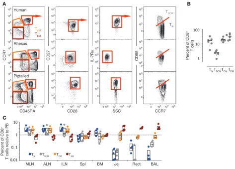
T cell memory in vivo, we sought to determine whether a similar subset of cells exists in NHPs. In both healthy rhesus macaques (RMs) and pigtail macaques (PTMs), we identified CD95hi CD8+
T cells in the CD45RA+CCR7+CD27+CD28+IL-7Rα+ naive-like
compartment (Figure 1A). Similarly to those in humans, NHP TSCM cells constitute about 2%–3% of circulating CD8+ T cells
(Fig-ure 1B). We also identified a CD4+ TSCM subset in PBMCs, with a
phenotype and frequency similar to CD8+ T
SCM cells
(Supplemen-tal Figure 1, A and B; supplemen(Supplemen-tal material available online with this article; doi:10.1172/JCI66327DS1). The NHP model allows a detailed examination of cellular distributions in tissues; we found that CD8+ T
SCM cells from healthy RMs are most abundant in LNs,
less so in the spleen and bone marrow, and are virtually absent at mucosal surfaces, i.e., the jejunum, the rectum, and the BAL, where only TCM and TEM cells are present (Figure 1C). CD4+ TSCM cells
displayed a similar distribution in the body, although less skewed toward the LNs (Supplemental Figure 1C). Thus, TSCM cells have a
tropism for secondary lymphoid tissues, with a distribution most similar to naive T (TN) cells.
Conflict of interest: The authors have declared that no conflict of interest exists.
brief report
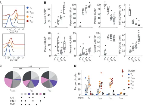
We next investigated whether NHP TSCM cells have features of
memory cells and precede TCM and TEM cells in terms of
differ-entiation. Immunophenotypic analysis of activation and memory markers (8) indicated that NHP CD8+ T
SCM cells from healthy RMs
are a discrete subset (Figure 2, A and B). Indeed, they are interme-diate between TN and TCM cells, according to the expression of
pro-teins that are progressively upregulated (CCR5, IL-18Rα) or down-regulated (CD38, CD130) with differentiation (Figure 2B). The human TSCM core phenotypic markers CXCR3, CD122 (Figure 2,
A and B), and LFA-1 (Supplemental Figure 2, A and B) were also upregulated in TSCM cells compared with TN cells. Unexpectedly, a
relatively large proportion of TSCM cells were proliferating (Ki-67+)
in the PBMCs (Figure 2A), but not in the LNs, spleen, or BM (Sup-plemental Figure 2C). In all sites, TSCM cells were mostly HLA-DR
negative. Similar phenotypic data were obtained for CD4+ TSCM
cells (Supplemental Figure 1D).
To assess the cytokine production capability of NHP TSCM cells,
we stimulated PBMCs from healthy RMs with staphylococcal enterotoxin B (SEB). Following stimulation, all subsets of memory T cells produced IFN-γ, TNF, and IL-2 (Figure 2C). The patterns of cytokine expression show that TSCM cells differ from TCM and
TEM cells, with a decreased proportion of cells producing a
com-bination of IL-2 and TNF, but not IFN-γ, and an increased
pro-portion of cells producing IFN-γ and IL-2 or IFN-γ only. Overall, their quality of cytokine production differed from conventional memory T cells (Figure 2C). TSCM cells also display attributes of
memory in vivo, as proliferating (Ki-67+) T
SCM cells incorporated
BrdU following SIVmac239 infection in PTMs similarly to
conven-tional TCM and TEM cells (Supplemental Figure 2D). In contrast,
BrdU incorporation by TN cells was negligible.
To further assess whether TSCM cells from healthy RMs constitute
discrete, less-differentiated memory cells, we evaluated their multi-potency (i.e., to generate other subsets) and self-renewing capability (i.e., to maintain a TSCM phenotype) in response to TCR stimulation
in vitro with αCD3/CD28 antibodies (Figure 2D). In the prolifer-ating (CFSE-diluted) fraction, TSCM phenotype cells could only be
recovered from sort-purified TN or TSCM cells. In addition, TSCM cells
were also able to generate cells with a TCM, TEM, and terminal effector
T (TTE) phenotype (Figure 2D). Importantly, TCM cells could not
gen-erate TSCM cells, but did generate TCM and TEM cells. Together, these
data indicate that TSCM cells constitute a discrete memory subset
and support the concept that TSCM cells serve as precursors of other
memory cells, according to the relationship TSCM→TCM→TEM→TTE.
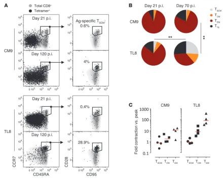
To determine the dynamics of antigen-specific T cell subsets during a natural infection in vivo, we enumerated SIV-specific
[image:3.585.56.529.81.420.2]CD8+ T cells in RMs using Mamu-A*01 pMHCI multimers
Figure 1
brief report
presenting SIV-derived Gag CM9 or Tat-TL8 peptides (Supple-mental Figure 3A). Antigen-specific TSCM cells were detected
within both CM9- and TL8-specific cells at day 21 after infection, thus demonstrating that they are elicited early (Figure 3A). The antigen-specific TSCM cells were not merely “bystander” cells,
but displayed evidence of activation (HLA-DR+, CD38bright) and
proliferation (Ki-67+) (Supplemental Figure 3B). At day 21 after
infection, CM9- and TL8-specific CD8+ T cells were dominated by
TEM-like cells, while TSCM, TCM, and TTE cells constituted a small
proportion of the total response (Figure 3, A and B).
The biology of the CM9 and TL8 Mamu-A*01–restricted epitopes is highly divergent. Unlike CM9, which is generally main-tained intact throughout the chronic phase of infection, the TL8 uniformly undergoes escape mutation within 4–5 weeks after SIVmac239 or SIVmac251infection in 100% of animals (12–14).
Tat-TL8 sequence variants do not stimulate Tat-TL8-specific T cells due to their reduced capability to bind the pMamu-A*01 class I mole-cule (12) or to signal through the TCR (14). Following TL8 escape, there is at least a 105-fold decrease in the relative antigen load of
TL8 versus CM9. Using this model, it is thus possible to investigate
the relative antigen dependence of the different T cell subsets. As expected, viral escape from TL8-directed CTLs resulted in a dra-matic decrease in the frequency of total circulating TL8-specific CD8+ T cells between days 21 and 70–120 after infection
(Supple-mental Figure 3C). Conversely, CM9-specific CD8+ T cells were
maintained at high levels throughout the course of the infection (Supplemental Figure 3C). Notably, despite the near-complete elimination of antigen (extremely low levels from residual “archi-val,” unmutated virus may persist) (12), TL8-specific CD8+ T cells
did not disappear, but remained detectable beyond day 335 after infection (Supplemental Figure 3C).
Phenotypic analysis of the TL8-specific population revealed a significant difference in the dynamics of memory subsets in response to viral escape mutation. Indeed, the proportion of TL8-specific TEM cells decreased dramatically in favor of the less
differentiated TSCM and TCM cells (Figure 3, A and B). By day
70 after infection, TSCM cells constituted approximately 25% of
the total TL8-specific response (Figure 3B). In contrast, mem-ory subset distribution within the CM9-specific CD8+ T cells
[image:4.585.53.528.80.431.2]remained unchanged between days 21 and 70 after infection
Figure 2
brief report
(Figure 3B). Furthermore, as would be expected from a loss of antigen, TL8-specific, but not CM9-specific TSCM and TCM cells,
became quiescent (Ki-67–, HLA-DR–) at day 70 after infection
(Supplemental Figure 3B).
Following antigen loss, TL8-specific TCM and TEM cells
under-went considerable attrition (respectively, ~10- and ~100-fold decrease in the absolute count at day 70 versus day 14), while TSCM cells did not (Figure 3C and Supplemental Figure 3D).
Sim-ilarly, CM9-specific TSCM counts did not change over time, while
the CM9-specific TCM and TEM cells contracted, albeit at markedly
slower rates compared with TL8-specific CD8+ T cells
(Supple-mental Figure 3D), reflecting the 1-log decrease in viral load fol-lowing peak viremia by day 70 after infection (i.e., establishment of the viral set point).
CM9- and TL8-specific subsets, FACS-sorted from a chroni-cally SIV-infected RM, were fully functional as they proliferated following cognate peptide stimulation in vitro (Supplemental Figure 4A). Importantly, each T cell subset regenerated cells with the same phenotype and simultaneously derived more, not less, differentiated progeny according to the relationship
TSCM→TCM→TEM (Supplemental Figure 4B), thus recapitulating
the results depicted in Figure 2D and further demonstrating the multipotency and self-renewing capability of TSCM cells in vitro.
The distribution of memory subsets observed at day 70 after infection within both the CM9- and TL8-specific populations was similar in 4 animals at more than 335 days after infection (Supplemental Figure 5A).
Following escape mutation of the TL8 epitope, the frequency of SIV-specific T cell subsets did not change in the inguinal LNs over time in 2 animals, suggesting that their lymphoid localiza-tion is not time dependent (Supplemental Figure 5B). Moreover, the same cells maintained a stable pattern of localization, as described in Figure 1C, during late chronic infection (Supple-mental Figure 5C). Collectively, these findings indicate that TSCM
cells are stable, long-lived memory cells with enhanced survival capacity compared with conventional memory cells when little or no antigen is present.
Freshly isolated CM9-specific TSCM cells were indeed less
proapop-totic in vitro than TCM and TEM cells during chronic infection
[image:5.585.71.511.78.432.2](Sup-plemental Figure 6, A and B). Similarly, fewer TL8-specific TSCM cells Figure 3
brief report
(AAALAC) and meet NIH standards (Guide for the Care and Use of Labora-tory Animals. NIH publication no. 85-23. Revised 1985). See Supplemental Methods for animal procedures.
Antibodies, Mamu-A*01 tetramers, and flow cytometry. Surface and intracellular analysis of protein expression and cytokine production was performed as described (8, 18). Tetramer staining was conducted at 37°C for 10 minutes. Cells were analyzed and sorted using a modified LSR II and FACSAria II, respectively (BD Biosciences).
Apoptosis studies. Fresh PBMCs were cultured in complete RPMI 1640 medium supplemented with αCD49d and αCD28 antibodies (both at 1 μg/ml; BD Biosciences) at 37°C for 42 hours. Surface phosphatidylserine expres-sion was revealed as described (18).
Proliferation studies. Cell proliferation was determined by dilution of CFSE (Life Technologies) as described (8). Labeled cells were stimulated for 6 days with 2 μg/ml CM9 and TL8 peptides in the presence of sorted, CD3–
autologous antigen-presenting cells (1:1 ratio to T cells) or with plate-bound αCD3 (10 μg/ml; clone FN18) plus soluble αCD28 (1 μg/ml; clone CD28.2) antibodies.
In vivo BrdU labeling. Administration of BrdU (Sigma-Aldrich) and analy-sis of its incorporation were performed as described (18).
Gene expression analysis. CD8+ T cell subsets were sorted as depicted in
Supplemental Figure 7. See Supplemental Methods for gene expression quantification procedures.
Statistics. Analyses were performed using Prism (GraphPad Software Inc.), JMP (SAS Institute Inc.), and SPICE (NIAID, NIH) (19). Nonparametric Wilcoxon rank tests were used to compare distributions. When possible, a paired Student’s t test (2 tailed) was used. In some cases, nonparametric 1-way ANOVA (Kruskal-Wallis test) was used to compare 3 or more groups. Distributions shown with pie charts were compared with SPICE. P values were considered significant when less than 0.05.
Study approval. All animal procedures were reviewed and approved by the Institutional Animal Care and Usage Committee of the VRC, the NIAID, the NCI, and Oregon Health and Science University (Portland, Oregon, USA). The anonymous human blood sample (Figure 1A) was obtained from the NIH blood bank under institutional review board exemption.
Acknowledgments
We thank Joanne Yu for antibody conjugation; the VRC Flow Cytometry Core for cell sorting; Richard Koup, Constantinos Pet-rovas, Takuya Yamamoto, Louis Picker, and Andrew Sylwester for sharing MamuA*01 samples; the VRC NHP Immunogenicity Core for sample processing; and Daniel Douek for critical discussion. This research was supported by the NIAID and NCI Intramural Research Programs at the NIH.
Received for publication August 13, 2012, and accepted in revised form November 1, 2012.
Address correspondence to: Enrico Lugli or Mario Roederer, ImmunoTechnology Section, Vaccine Research Center, NIAID, NIH, 40, Convent Dr., Bethesda, Maryland 20892, USA. Phone: 301.594.8491; Fax: 301.480.2788; E-mail: [email protected] (E. Lugli). E-mail: [email protected] (M. Roederer).
bound annexin V compared with the total memory fraction (there were insufficient T cells for evaluation of TL8-specific TCM and TEM
cells individually; Supplemental Figure 6B). Consistent with the lack of chronic antigen exposure, TL8-specific cells were less pro-apoptotic than CM9-specific cells (Supplemental Figure 6B).
We thus reasoned that the preferential expression of antiapop-totic molecules in TSCM cells could be associated with their
preferen-tial survival. We could not quantify gene expression in antigen-spe-cific cells due to their paucity. Instead, we sorted “resting” versus “blasting” (presumably antigen-responding) CD8+ T cell subsets
according to their scatter properties (Supplemental Figure 7A). The “blasting” lymphocytes uniformly expressed high levels of HLA-DR, indicative of activation in vivo (Supplemental Figure 7B). Notably, genes such as LEF1 (regulating self-renewal), BCL2, and
MCL1 (both antiapoptotic) were not differentially expressed among “resting” memory subsets, but were specifically upregulated to high levels in “blasting” TSCM cells (Supplemental Figure 7C).
In summary, our data show that NHP TSCM cells are closely
related to human TSCM cells, and constitute a discrete memory
T cell subset, distinct from TCM cells, on the basis of: (a) surface
immunophenotype, (b) localization in the body, (c) cytokine pro-duction, and (d) in vivo turnover. Although TCM cells were
demon-strated to possess stem cell–like properties in multiple experimen-tal conditions (7), our data suggest that TSCM cells are superior
to TCM cells in this regard owing to their superior self-renewing
capability and multipotency, their relative antigen dependence in vivo, and their apoptotic refractoriness. Notably, these properties are not unique to TSCM cells, but rather are highly preferentially
associated with the TSCM subset. And importantly, the superior
persistence of TSCM cells following antigen loss suggests that they
are the main precursors of T cell memory in the postantigen phase. Maintenance of antigen-specific TSCM cells is likely intrinsically
programmed. We exclude the possibility that the TSCM pool is
maintained by the continuous recruitment of newly generated naive T cells by thymic output (15) in our model, as thymic out-put is severely impaired in chronic, untreated HIV and SIV infec-tions (16, 17). In addition, we find equivalent maintenance of CM9- and TL8-specific TSCM cells despite a profound difference
in antigen availability.
Our data strongly suggest that TSCM cells play a crucial role in
supporting long-term cellular immunity in vivo. Future studies aimed at identifying antigen-specific TSCM cells in adoptive
trans-fer models are required to define whether they are uniquely suited for this function. On the basis of these properties, we propose that future vaccination strategies designed to generate durable immunity should target the induction of TSCM cells. Nevertheless,
cellular immunotherapy strategies will need to exploit TSCM
prop-erties to support the persistence of in vivo–transferred virus- and tumor-specific T cells.
Methods
Animals. Animals were handled in accordance with the standards of the American Association for the Accreditation of Laboratory Animal Care
1. Zinkernagel RM, Bachmann MF, Kundig TM, Oehen S, Pirchet H, Hengartner H. On immunolog-ical memory. Annu Rev Immunol. 1996;14:333–367. 2. Murali-Krishna K, et al. Counting
antigen-spe-cific CD8 T cells: a reevaluation of bystander activation during viral infection. Immunity. 1998; 8(2):177–187.
3. Hammarlund E, et al. Duration of antiviral
immu-nity after smallpox vaccination. Nat Med. 2003; 9(9):1131–1137.
4. Surh CD, Sprent J. Homeostasis of naive and mem-ory T cells. Immunity. 2008;29(6):848–862. 5. Perfetto SP, Chattopadhyay PK, Roederer M.
Seventeen-colour flow cytometry: unravelling the immune system. Nat Rev Immunol. 2004; 4(8):648–655.
6. Sallusto F, Geginat J, Lanzavecchia A. Central mem-ory and effector memmem-ory T cell subsets: function, generation, and maintenance. Annu Rev Immunol. 2004;22:745–763.
7. Stemberger C, Neuenhahn M, Gebhardt FE, Schie-mann M, Buchholz VR, Busch DH. Stem cell-like plasticity of naive and distinct memory CD8+ T cell
brief report
8. Gattinoni L, et al. A human memory T cell sub-set with stem cell-like properties. Nat Med. 2011; 17(10):1290–1297.
9. Gattinoni L, et al. Wnt signaling arrests effector T cell differentiation and generates CD8+ memory
stem cells. Nat Med. 2009;15(7):808–813. 10. Zhang Y, Joe G, Hexner E, Zhu J, Emerson SG.
Host-reactive CD8+ memory stem cells in
graft-ver-sus-host disease. Nat Med. 2005;11(12):1299–1305. 11. Lugli E, et al. Identification, isolation and in vitro
expansion of human and nonhuman primate T stem cell memory cells. Nat Protoc. 2012;8(1):33–42. 12. Allen TM, et al. Tat-specific cytotoxic T lym-phocytes select for SIV escape variants during
resolution of primary viraemia. Nature. 2000; 407(6802):386–390.
13. O’Connor DH, et al. Acute phase cytotoxic T lym-phocyte escape is a hallmark of simian immu-nodeficiency virus infection. Nat Med. 2002; 8(5):493–499.
14. Price DA, et al. T cell receptor recognition motifs gov-ern immune escape pattgov-erns in acute SIV infection. Immunity. 2004;21(6):793–803.
15. Vezys V, et al. Continuous recruitment of naive T cells contributes to heterogeneity of antiviral CD8 T cells during persistent infection. J Exp Med. 2006; 203(10):2263–2269.
16. Douek DC, Picker LJ, Koup RA. T cell dynamics
in HIV-1 infection. Annu Rev Immunol. 2003; 21:265–304.
17. Richardson MW, et al. T-cell receptor excision cir-cles (TREC) in SHIV 89.6p and SIVmac251 models of HIV-1 infection. DNA Cell Biol. 2004;23(1):1–13. 18. Lugli E, Mueller YM, Lewis MG, Villinger F, Kat-sikis PD, Roederer M. IL-15 delays suppression and fails to promote immune reconstitution in virally suppressed chronically SIV-infected macaques. Blood. 2011;118(9):2520–2529.