© 2018, IRJET | Impact Factor value: 6.171 | ISO 9001:2008 Certified Journal | Page 2355
A Review on Automatic Detection of Dental Tooth Decay in Bitewing
Radiography
Sharanjit Kaur
1, Navjot Kaur
2,Navpreet Rupal
31 Student, Dept. of Computer Science Engineering, GIMET, Punjab, India 2,3 Assistant Professor, Dept. of Computer Science Engineering, GIMET, Punjab, India
---***---Abstract –Detection and prevention of dental diseases is critical at early stages. Aggravation of such diseases could lead to painful events for the patient. Dental solutions to the patients can be given only at doctor end and solution is difficult to achieve with the help of technology. In this manner, dental specialists routinely favor bitewing radiograph which is a technique for finding of the carious injuries caused within teeth. Numerous radiologic components can influence the capacity to precisely identify the carious problems; presentation parameters, picture receptor used to receive the picture from camera such as x-ray imaging, image preparation for examination, framework for analysis of disease , visual conditions and visual effects. Adjacent to these radiologic factors, different morphologic techniques used to convert coloured images to gray scale form for analysis , for example, pits and crevices(special dental conditions), dental irregularities, for example, hypo-plastic pits and concavities and obtained changes of dentition, for example, scraped spot and disintegration can mirror the presence of a carious sore. In this way, the result of a false positive analyze is the start of treatment corresponding to disease predicted. The techniques used to analyze dental conditions and a technique which is best for the analysis of dental problems can be checked using comparative analysis given through this literature.
Key Words : Dental tooth decay, radiography, diagnostic
imaging, microscopy.
1.INTRODUCTION
Dental treatments are required to check problems within the teeth. Teeth is vital part of body that is required to be monitored in order for successful digestion [1]. The net demineralization may begin not long after tooth discharge in tooth decay feeble children without being seen by dental specialists. This system may progress additionally achieving a dental cavity that is the sign and also the reaction of the carious procedure. Tooth decay is by the day's end a continues sequence which might be assessed untrustworthily when only a particular time point is considered. Fig 1 demonstrates the carious procedure. Tooth decay investigation recommends something past recognizing injuries. In this manner, tooth decay finding - as an insightful method is the affirmation of the closeness and level of tooth decay damage. Other than the judgment of its development is a fundamental bit of conclusion. Since assurance is a psychological laying place on the way to deal
with treatment decision, it is by and by associated with the treatment plan to be taken after.
[image:1.595.323.546.233.386.2].
Fig-1: General diagram of dental investigation
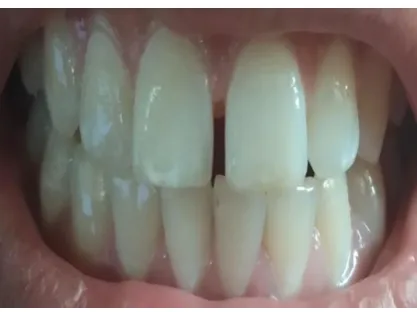
© 2018, IRJET | Impact Factor value: 6.171 | ISO 9001:2008 Certified Journal | Page 2356 Fig-2: Teeth after dental care.
Clinical-visual assurance may be affable to longitudinal seeing in spite of the way that the assessment is subjective. It is less requesting to have a contraption that would not simply recognize demineralization however measures it as well. By then watching development or arrestment would be fundamental; use the contraption again and find in what heading the numbers change. The thought is hugely captivating so no enormous shock researchers have tried such undertakings to make, test and perfect such contraptions[3][4]. In [5] every one of these techniques for tooth decay disclosure relies upon the explanation of no less than one physical signs. These are causally related to no less than one feature of a conveys sore. In the first place, the signs must be gotten using a receptor device and ordered. The plan of a banner is a bit of the analytic fundamental initiative process. In any case, none of the systems is fit for setting up each one of these signs to a status that could be called conclusion. "The art of perceiving an affliction from its signs and symptoms" is a methodology that can't be supplanted by a machine or a device. Tooth decay estimation should be found concerning the objectives of present day clinical tooth decay organization and the continues sequence of disease points of interest, reaching out from sub-surface carious changes through to additionally created injuries. Huge dental tooth decay are routinely apparent to the stripped eye, however smaller wounds can be contrast religion to recognize. Visual and material appraisal close by radiographs can be used as regularly as conceivable among dental pros. Once in a while, tooth decay may be vary religion to perceive [6][7].
Microbes can enter the polish to accomplish dentin, yet then the outside surface maybe at first site set up. These tooth decay, as a less than dependable rule insinuated as "covered tooth decay", in the preliminary stage X-pillar are the most ideal approach to recognize them, regardless of the visual examination of the tooth showed the complete set up or irrelevantly punctured. Without X-bars it isn't possible to perceive these issues until the point that they had ended up being not kidding which besides, caused certified mischief [8].
1.1 GUIDELINE :
There are three guideline sorts of dental X-bars to play out the dental tooth decay acknowledgment what's progressively, unique contaminations:
The all encompassing dental X-beam: Panoramic X-pillars exhibit an extensive point of view of the jaws, teeth, sinuses, nasal region, and temporomandibular (jaw) joints. These X-bars do show issues, for instance, influenced teeth, bone varieties from the standard, pimples, solid advancements (tumors), illnesses, breaks and dental tooth decay [9].
The bitewing dental X-pillar: Bitewing X-shafts exhibit the upper and lower back teeth and how the teeth touch each other in a lone view. These X-shafts are utilized to check for decay between the teeth and to demonstrate how well the upper what's all the more, cut down teeth line up. They in like manner show bone incident when genuine gum ailment or, of course a dental illness is accessible [10].
Periapical X-pillars: Periapical X-shafts show the entire tooth, from the revealed crown to the complete of the root and the bones that assistance the tooth. These X-pillars are used to find dental issues underneath the gum line or in the jaw, for example, influenced teeth, abscesses, wounds, tumors, and bone changes associated with a couple of ailments [11].
1.2Computer Vision:
PC vision is the science and advancement that contrast with the vision of machines. As a legitimate angle, PC vision is the technique to get information from pictures by building a produced structure. This photos data can be displayed to the system in numerous structures, the most surely understood points of view from a lone camera, video course of action, sees from various cameras, or multi-dimensional data from a restorative scanner [12]. In [13] [14] the built up issue in PC vision is that of choosing if the photo data contains some specific dissent, feature, or activity. This endeavor can consistently be handled enthusiastically and without exertion by a human, yet is so far not elegantly clarified in PC vision for the general case: optional inquiries in self-confident conditions. The current systems for dealing with this issue can, most ideal situation handle it only for specific articles, for instance, essential geometric things, human appearances, printed or physically composed characters, or vehicles, and specifically conditions, normally depicted in regards to particularly described lighting up, establishment, and position of the inquiry in regard to the camera in. There is an arrangement of affirmation issue, for instance,
Recognition: This issue involves in the affirmation of articles that were pre-shown or learned by the system.
© 2018, IRJET | Impact Factor value: 6.171 | ISO 9001:2008 Certified Journal | Page 2357 this can be fulfill by the use of the individual face, fingertips,
teeth shape, et cetera.
Detection: Our work fits in this affirmation issue, in light of the fact that the standard goal is to perceive, in the photo, territories containing dental tooth decay. For this circumstance the reason for getting the photos, is successfully limited for area of dental tooth decay.
The relationship of a PC vision structure is exceptionally application subordinate. A couple of structures are stay singular applications which unwind a specific estimation or acknowledgment issue, while other constitute a sub-course of action of a greater diagram which, for case, in like manner contains sub-systems for control of mechanical actuators, organizing, information databases, man-machine interfaces, et cetera. The specific use of a PC vision structure also depends upon if its helpfulness is pre-demonstrated or if some bit of it can be learned or changed in the midst of activity. There are, nevertheless, normal limits which are found in various PC vision structures:
Picture Acquisition: An propelled picture is made by one or a couple of picture sensors, which, other than various sorts of light-fragile cameras, consolidate run sensors, tomography contraptions, radar, ultra-sonic cameras, et cetera. Dependent upon the sort of sensor, the ensuing picture data is a typical 2D picture, a 3D volume, or a photo progression [15], [16]. The pixel regards frequently contrast with light power in one or a couple of horrible gatherings (diminish pictures or shading pictures), yet can in like manner be related to various physical measures, for instance, significance, ingestion or reflectance of sonic or electromagnetic waves, or nuclear alluring resonation .
Pre-Processing: This organize is perform before a PC vision procedure can be associated with picture data remembering the true objective to evacuate some specific information, it is regularly vital to process the data to ensure that it satisfies certain assumptions induced by the method [17], [18]. For example bustle reducing in demand to ensure that isn't familiarize false information with the system, separate change to ensure that pertinent information can be distinguished and scale-space depiction to increase vital information in appropriate scales. Middle channel is utilized to accomplish this progression.
Highlight Extraction: Image features at various levels of versatile quality are removed from the photo data. There are two sorts of feature extraction, the low-level that identifies with the basic features that can be expelled subsequently from a photo with no shape information. Moreover, the unusual state feature extraction concerns finding shapes in PC pictures [19], [20].
Identification/Division: In [21] PC vision, division insinuates the methodology of isolating a modernized picture into various segments (sets of pixels) . The goal of division is to unravel and/or, then again change the
depiction of a photo into something that is more huge and less requesting to inspect. Picture division is usually used to discover articles and cutoff points (lines, twists, et cetera.) in pictures. More unequivocally, picture division is the technique of selecting a name to every pixel in a photo to such a degree, to the point that pixels 9+with a similar stamp share certain visual qualities.
High Level Pre-handling: In this movement the data is generally an instructive accumulation containing a specific inquiry. This relies upon the features that portray the region [22].
For example, in a method to perceive afflictions, the commitment to this movement is the features that best fit on the ailment arrange. Identified with this movement is the outline affirmation that involves in the portrayal of data, framed by plans. The methods to perform outline affirmation are isolated in two essential social occasions, managed taking in where we know from the earliest starting point what are the characterizations of the data tests. The other social occasion is the converse, contrasting with the unsupervised learning, for this circumstance the essential target is to isolate in, no less than one orders the data.
2. Literature Survey
S.Chang et. al (2016) Dental radiography accept a basic part in clinical assurance, treatment and surgery. Starting late, attempts have been made on making computerized dental X-pillar picture examination structures for clinical us-ages. A novel structure for target evaluation of modified dental radiography examination computations has been developed under the help of the IEEE International Symposium on Biomedical Imaging 2015 Bitewing Radiography Tooth decay Detection Challenge and Cephalometric X-bar Image Analysis Challenge. In this article, we demonstrate the datasets, methodologies and eventual outcomes of the test and set out the guidelines for future occupations of this benchmark.
The recognizable proof of dental tooth decay, in a preliminary stage are of by and large criticalness. There is a long history of dental tooth decay. Over a million years back, primates, for instance, Australopithecus experienced holes. Archeological evidence shows that tooth decay is an old disease dating far into antiquated circumstances. Skulls dating from a million years earlier through the Neolithic time period allude to tooth decay. The extension of tooth decay in the midst of the Neolithic time allotment may be credited to the addition of plant sustenance containing starches. The beginning of rice improvement in South Asia is in like manner acknowledged to have caused a development in tooth decay[23].
© 2018, IRJET | Impact Factor value: 6.171 | ISO 9001:2008 Certified Journal | Page 2358 a critical promotion Vance in the range of imaging finding
was How-ever, examines found in the writing are dissimilar in connection to the nature of the pictures when looking at advanced frameworks and regular film radiographs. A few works consider the picture nature of the radio-realistic movies tantamount to that of the frameworks with charge-coupled gadgets (CCD) and to the ones that utilization stockpiling phosphor plates. Different examinations announced the predominance of the frameworks with capacity phosphor plates over customary radiographs and over frameworks with charge-coupled gadget. There are additionally works that exhibited a more noteworthy indicative precision of customary film radiographs in connection to advanced frameworks[24].
Jeffery B. Price(2015) Tooth decay determination is a standout amongst the most fundamental diagnostic aptitudes that oral medicinal services experts must learn; but, it stays a standout amongst the most troublesome abilities to dependably and typically ace. In this course we will review the diverse tooth decay recognizable proof advances available to help the dental master with this baffling errand.
S.Valizadeh et. al (2015) Tooth decay conclusion is a standout amongst the most fundamental diagnostic abilities that oral social insurance experts must learn; but, it stays a standout amongst the most troublesome aptitudes to dependably and typically ace. In this course we will survey the different tooth decay location advancements accessible to help the dental expert with this Radiography alongside
clinical examination is the most vital demonstrative technique for dental tooth decay[26].
N.B.Pitts(1996)The target of this paper is to blend distributed research on the utilization of radiographs in tooth decay analysis with a specific end goal to deliver suggestions for rules that are clinically arranged however logically based. The alternatives accessible incorporate the utilization of different sorts of radiographic examination alone or in conjunction with other demonstrative guides[27].
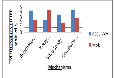
[image:4.595.320.546.257.412.2]A chart given below shows the different mechanisms which have different metric values on the basis of two parameters – Accuracy and MSE( Mean square error).
Fig-3: Plot in terms of parameters is given as under
Table-1: Comparison of various techniques associated with dental care
Title Technique Parameter Merits Demerits
A benchmark for examination of dental radiography
investigation algorithms[23]
Bitewing Radiography Tooth decay Detection
Challenge and
Cephalometric X-pillar Image Analysis
clinical determination, treatment and surgery of cavities
point by point quantitative assessment
Trust about programmed dental radiography
investigation
Tooth decay Detection in Panoramic Dental X-beam Images[23]
All encompassing dental X-beam pictures Stomatology, Teeth Segmentation, ROI, Jaws Partition
size of the teeth, the state of the mouth and teeth and the levels of commotion,
utilization of dynamic forms in the teeth division organize
Prerequisite of recognition of dental sicknesses, utilizing the depicted dental X-beam informational index
Discovery of proximal tooth decay in ordinary and computerized radiographs: an in vitro study[24]
contrast computerized pictures with customary film radiography in the determination of
proximal tooth decay
affectability,
specificity, positive prescient esteem (PPV) and negative prescient esteem (NPV) were altogether figured
the analytic precision of computerized pictures is great
Trust about programmed dental radiography
investigation
A Review of Dental Tooth decay Detection Technologies ( Jeffery
Tooth decay Detection & Assessment System (ICDAS) detection and
Tooth decay detection
technology Number methods are used of to detect tooth
© 2018, IRJET | Impact Factor value: 6.171 | ISO 9001:2008 Certified Journal | Page 2359 B. Price, 2015) assessment tool for dental
tooth decay, Radiography, Laser Fluorescence
decay
Outlining of a Computer Software for
Detection of
Approximal Tooth decay in Posterior Teeth[26]
digital radiography quantitative appraisal of the movement of carious injuries
precision to decide the profundity of carious sores
restricted capacity in distinguishing lacquer sores
Discovery and determination of the early tooth decay lesion[2]
Quantitative Light-prompted Fluorescence (QLF), DIAGNOdent (DD), Fiber-optic
Transillumination (FOTI) and Electrical Conductance (EC)
Sound, First Visual Change in lacquer, Distinct Visual Change in veneer, Localized lacquer breakdown Underlying dentine shadow Unmistakable pit with obvious dentine Extensive depression with noticeable dentine
detect early lesions amenable to prevention
Extra tooth decay identification
techniques ought to be utilized as a subordinate to clinical basic leadership and for tooth decay conclusion and treatment arranging in conjunction with tooth decay hazard evaluation
The utilization of
chomp wing
radiographs in the administration of dental tooth decay: logical and down to earth
considerations[27]
preventive care prompted (PCA) or agent mind exhorted (OCA), Posterior bitewing radiographs
detection of proximal, occlusal, secondary and root surface tooth decay
clinical anxieties about dentists' increasing
inability to detect and manage disease
Clinical research and epidemiology should be undertaken, and the inter-relationships between uses in these areas should be explored.
3. CONCLUSIONS
Electronic programmed dental radiography examination frameworks for clinical utilize spare time and manual expenses and maintain a strategic distance from issues caused by intra and between eyewitness varieties e.g. because of weariness, push or diverse levels of understanding. Computerized frameworks are ending up more accessible to dental practice. However these systems need to give pictures whose investigative regard is in any occasion com-story to the one of the consistent film radio-diagrams remembering the true objective to supplant the photos got with the radio-practical motion pictures. In our audit paper, we contemplated if dental specialists could determine teeth to have 95+% precision with the fundamental instruments of their eyes, tests and bitewing radio-diagrams, there would be no market interest for some other carries discovery advances. Currently accessible innovation and changes later on will upgrade exactness in recognition of tooth decay enhancing the oral wellbeing of general society.
REFERENCES
[1] P. L. Lin, P. Y. Huang, P. W. Huang, H. C. Hsu, and C. C. Chen, “Teeth segmentation of dental periapical
radiographs based on local singularity analysis,” Comput. Methods Programs Biomed., pp. 1–13, 2013.
[2] J. Gomez, “Detection and diagnosis of the early tooth decay lesion,” vol. 15, no. Suppl 1, pp. 1–7, 2015.
[3] M. A. Farooq, M. A. M. Azhar, and R. H. Raza, “Automatic Lesion Detection System (ALDS) for Skin Cancer Classification Using SVM and Neural Classifiers,” 2016 IEEE 16th Int. Conf. Bioinforma. Bioeng., pp. 301–308, 2016.
[4] V. Ponomaryov, “Computer-aided detection system based on PCA/SVM for diagnosis of breast cancer lesions,” 2015 Chil. Conf. Electr. Electron. Eng. Inf. Commun. Technol., pp. 429–436, 2015.
[5] I. Stanganelli, A. Brucale, L. Calori, R. Gori, A. Lovato, S. Magi, B. Kopf, R. Bacchilega, V. Rapisarda, A. Testori, P. A. Ascierto, E. Simeone, and M. Ferri, “Computer-aided Diagnosis of Melanocytic Lesions,” vol. 4582, pp. 4577–4582, 2005.
© 2018, IRJET | Impact Factor value: 6.171 | ISO 9001:2008 Certified Journal | Page 2360 [7] N. Situ, X. Yuan, N. Mullani, and G. Zouridakis,
“AUTOMATIC SEGMENTATION OF SKIN LESION IMAGES USING EVOLUTIONARY STRATEGY 2 . Engineering Technology Department , University of Houston,” pp. 1–4.
[8] D. Manju, M. Seetha, and K. V. Rao, “Comparison Study of Segmentation Techniques for Brain Tumour Detection,” vol. 2, pp. 261–269, 2013.
[9] P. L. Lin, P. W. Huang, Y. S. Cho, and C. H. Kuo, “An automatic and effective tooth isolation method for dental radiographs,” vol. 21, no. 1, pp. 126–136, 2013.
[10] “An Effective Shape Extraction Algorithm for Dental Radiographs using Contour Information,” vol. 8491, pp. 311–316, 2011.
[11] W. G. M. Geraets, C. Lindh, and H. Verheij, “Sparseness of the trabecular pattern on dental radiographs : visual assessment compared with semi-automated measurements,” vol. 85, no. August, pp. 455–460, 2012.
[12] V. B. Kumar, “Dermatological Disease Detection Using Image Processing and Machine Learning,” pp. 88–93, 2016.
[13] D. J. Cook, J. C. Augusto, and V. R. Jakkula, “Ambient intelligence : Technologies , applications , and opportunities,” Pervasive Mob. Comput., vol. 5, no. 4, pp. 277–298, 2009.
[14] D. Selvaraj, “MRI BRAIN IMAGE SEGMENTATION TECHNIQUES - A REVIEW,” vol. 4, no. 5, pp. 364–381, 2013.
[15] S. D. Khirade and A. B. Patil, “Plant Disease Detection Using Image Processing,” 2015 Int. Conf. Comput. Commun. Control Autom., pp. 768–771, 2015.
[16] M. Raghav and S. Raheja, “IMAGE DENOISING TECHNIQUES : LITERATURE REVIEW,” vol. 3, no. 5, 2014.
[17] A. Singh, “Analysis of Image Noise Removal Methodologies for High Density Impulse Noise,” vol. 3, no. 6, pp. 659–665, 2014.
[18] N. Singla, “A Comparative Study of Noising And Denoising Technique In Image Processing,” vol. 4, no. 3, pp. 38–42, 2016.
[19] S. Hsu, C. Han, M. Wen, Y. Wu, S. Member, and K. Fan, “Extraction of Visual Facial Features for Health Management,” pp. 1–11, 2014.
[20] J. C. Kavitha and A. Suruliandi, “Texture and color feature extraction for classification of melanoma using SVM,” 2016 Int. Conf. Comput. Technol. Intell. Data Eng. ICCTIDE 2016, 2016.
[21] I. Țăranu, “Data mining in healthcare: decision making and precision,” Database Syst. J., vol. 5, no. 4, pp. 33–40, 2015.
[22] B. A. Miah, “Detection of Lung Cancer from CT Image Using Image Processing and Neural Network,” no. May, pp. 21–23, 2015.
[23] S. Chang, M. Siao, T. Lai, B. Ibragimov, and T. Vrtovec, “A benchmark for comparison of dental radiography analysis,” vol. 31, pp. 63–76, 2016.
[24] M. I. Rockenbach, E. B. Veeck, and N. Pereira, “Detection of proximal tooth decay in conventional and digital radiographs : an in vitro study,” vol. 10, no. 4, pp. 115–120, 2008.
[25] A. P. Publication, “A Review of Dental Tooth decay Detection Technologies,” 2015.
[26] S. Valizadeh, M. Goodini, S. Ehsani, H. Mohseni, and F. Azimi, “Designing of a Computer Software for Detection of Approximal Tooth decay in Posterior Teeth,” vol. 12, no. 4, 2015.