Temporal integration of light flashes by the
human circadian system
Raymond P. Najjar, Jamie M. Zeitzer
J Clin Invest. 2016;
126(3)
:938-947.
https://doi.org/10.1172/JCI82306
.
BACKGROUND.
Beyond image formation, the light that is detected by retinal
photoreceptors influences subcortical functions, including circadian timing, sleep, and
arousal. The physiology of nonimage-forming (NIF) photoresponses in humans is not well
understood; therefore, the development of therapeutic interventions based on this
physiology, such as bright light therapy to treat chronobiological disorders, remains
challenging.
METHODS.
Thirty-nine participants were exposed to 60 minutes of either continuous light
(n = 8) or sequences of 2-millisecond light flashes (n = 31) with different interstimulus
intervals (ISIs; ranging from 2.5 to 240 seconds). Melatonin phase shift and suppression,
along with changes in alertness and sleepiness, were assessed.
RESULTS.
We determined that the human circadian system integrates flash sequences in
a nonlinear fashion with a linear rise to a peak response (ISI = 7.6 ± 0.53 seconds) and a
power function decrease following the peak of responsivity. At peak ISI, flashes were at
least 2-fold more effective in phase delaying the circadian system as compared with
exposure to equiluminous continuous light 3,800 times the duration. Flashes did not change
melatonin concentrations or alertness in an ISI-dependent manner.
CONCLUSION.
We have demonstrated that intermittent light is […]
Clinical Medicine
Neuroscience
Find the latest version:
Introduction
Mammals, including humans, have evolved to adapt their endoge-nous physiological and behavioral rhythms to be consistent with the Earth’s 24-hour light/dark cycle. Such endogenous circadian rhythms range from the behavioral sleep-wake and arousal cycles to the secretion pattern of hormones, such as melatonin, cortisol, and thyroid-stimulating hormone. Circadian rhythms are under the con-trol of a master oscillator located in the anterior hypothalamus just above the optic chiasm called the suprachiasmatic nucleus (SCN). The rhythms of the SCN have a period length that is close to, but not exactly, 24 hours and is entrained (synchronized) to the 24-hour light/dark cycle by light detected by ocular retinal photoreceptive elements (1–3). Inappropriate circadian entrainment is associated with alterations in thermoregulation, cardiovascular function, immune processes, sleep, vigilance, memory, and cognition (4–9).
In the mammalian retina, a small subset of ganglion cells con-tains the photopigment melanopsin and is intrinsically
photosen-sitive (10–12). These intrinsically photosenphotosen-sitive retinal ganglion cells (ipRGCs) endogenously transduce light but also integrate processed neural signals coming from the traditional rod and cone photoreceptors of the outer retina (13–15). Axons emerging from the ipRGCs project to nonimage-forming (NIF) centers in the mammalian brain to evoke subcortical changes that include, but are not limited to, the resetting of circadian timing (1, 10, 11, 16, 17), suppression of melatonin production (18), activation of the pupillary light reflex (14, 15), change in arousal levels (19, 20) and sleep drive (21), alleviation of seasonal depression (22, 23), and possibly the induction of migraine headaches (24). Given its NIF implications, light has been used for its considerable therapeutic potential in chronobiological disorders such as jet lag and shift work but has also been used to treat sleep and mood disorders as well as cognitive impairment via correction of circadian misalign-ment and an acute activating response (22, 23, 25–32).
Underlying these NIF responses to light, traditional rod and cones photoreceptors are thought to be implicated in the fast and transient responses, whereas melanopsin is hypothesized to have a predominant effect on the sustainability and persistence of a response (33, 34). To date, however, the integrative physiology
BACKGROUND. Beyond image formation, the light that is detected by retinal photoreceptors influences subcortical functions, including circadian timing, sleep, and arousal. The physiology of nonimage-forming (NIF) photoresponses in humans is not well understood; therefore, the development of therapeutic interventions based on this physiology, such as bright light therapy to treat chronobiological disorders, remains challenging.
METHODS. Thirty-nine participants were exposed to 60 minutes of either continuous light (n = 8) or sequences of 2-millisecond light flashes (n = 31) with different interstimulus intervals (ISIs; ranging from 2.5 to 240 seconds). Melatonin phase shift and suppression, along with changes in alertness and sleepiness, were assessed.
RESULTS. We determined that the human circadian system integrates flash sequences in a nonlinear fashion with a linear rise to a peak response (ISI = 7.6 ± 0.53 seconds) and a power function decrease following the peak of responsivity. At peak ISI, flashes were at least 2-fold more effective in phase delaying the circadian system as compared with exposure to equiluminous continuous light 3,800 times the duration. Flashes did not change melatonin concentrations or alertness in an ISI-dependent manner.
CONCLUSION. We have demonstrated that intermittent light is more effective than continuous light at eliciting circadian changes. These findings cast light on the phenomenology of photic integration and suggest a dichotomous retinohypothalamic network leading to circadian phase shifting and other NIF photoresponses. Further clinical trials are required to judge the practicality of light flash protocols.
TRIAL REGISTRATION. Clinicaltrials.gov NCT01119365.
FUNDING. National Heart, Lung, and Blood Institute (1R01HL108441-01A1) and Department of Veterans Affairs Sierra Pacific Mental Illness Research, Education, and Clinical Center.
Temporal integration of light flashes by the human
circadian system
Raymond P. Najjar and Jamie M. Zeitzer
Department of Psychiatry and Behavioral Sciences and Stanford Center for Sleep Sciences and Medicine, Stanford University, Stanford, California, USA. Mental Illness Research, Education and Clinical Center, Veterans Affairs Palo Alto Health Care System, Palo Alto, California, USA.
Conflict of interest: The authors have declared that no conflict of interest exists.
Submitted: April 10, 2015; Accepted: December 14, 2015.
light in a nonlinear fashion. Intermittent patterns of light tested thus far fail, however, to elicit greater changes in outcomes when compared with continuous light exposures. Shorter duration of light exposures in the order of microseconds to milliseconds have been tested in rodents (45–47). While a single 2-millisecond flash of light is insufficient to phase shift the murine circadian system, a sequence of flashes administered once per minute for 60 min-utes is sufficient to evoke phase delays (48). This study by Van Den Pol and colleagues, in addition to other published studies in rodents (45–47), established that the mammalian circadian sys-tem has the capacity to respond to a sequence of very brief, milli-second flashes of light.
[image:3.585.81.513.52.502.2]of NIF photoreception in humans remains incompletely under-stood, as it has wavelength (35–39), intensity (40), duration (41), and pattern (42–44) responses that are considerably distinct from the traditional perceptual image-forming responses, and most knowledge in this field has been imputed from studies of nonhu-man mammals. Understanding NIF characteristics and responses to light is crucial for the development and optimization of light therapy strategies. NIF responses to light in humans have mainly been tested using continuous or intermittent light exposures, ranging from the order of minutes up to more than 6 hours of bright light (41–43). Most of these studies that used intermittent light exposures imply that the circadian system is able to integrate
ity (Pittsburgh Sleep Quality Index, ref. 52, U = 142.5, Z = 0.65, P = 0.51, Mann-Whitney test) between the two groups.
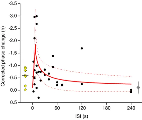
Robust circadian phase shift in response to flashes of light.
Within the group exposed to 1 hour of continuous light, cir-cadian timing was changed by –0.60 ± 0.34 hours (delay) (t = –5.04, df = 7, P < 0.01, paired t test). Within the group exposed to flashes, circadian timing changed by –2.99 to 0.30 hours in an ISI-dependent fashion (Figure 3). An initial linear rise to peak responses occurred when flashes were separated by 2.5 to 7.6 seconds of darkness, modeled with a standard linear fit using the formula y = m × x, such that m is the slope of the rise. Follow-ing the peak of responsivity of the system, change in circadian phase in response to the light flashes dropped following a non-linear, power function, as shown by y = y0 + A × xC, where y
0 is the
response of the system to a similar protocol without light admin-istration (set to –0.11 hours, as this change in circadian phase was observed in a separate cohort of subjects who took part in this protocol but were not exposed to light; ref. 44), A is a scaling factor, and c is the power coefficient describing the decay of the response. The slope of the initial rise (m) was calculated as 0.23 ± 0.07 h/s, the power constant (A) was 5.5 ± 6.0, and the power coefficient (c) was –0.67 ± 0.40.
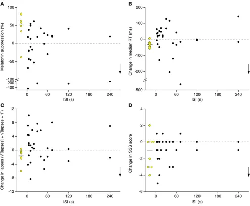
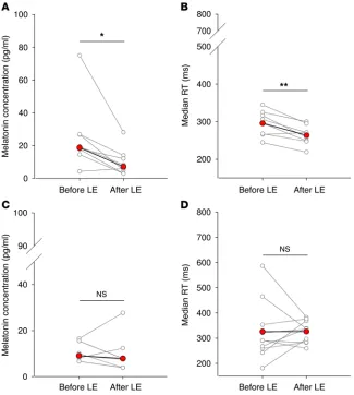
Flashes of light do not suppress melatonin in a dose-dependent manner. Continuous light suppressed melatonin secretion by 51%
± 40% (W = 35, Z = 2.31, P < 0.05, Wilcoxon signed-rank test) (Fig-ure 4A and Fig(Fig-ure 5A). Melatonin suppression was assessed in 28 of 31 participants in the group exposed to flashes, as melatonin concentrations in 3 participants were higher than the assay’s max-imum sensitivity, even after dilution of the saliva samples. The percentage of melatonin suppression fluctuated under the dif-ferent flash frequency conditions but did not vary systematically based upon ISI (Figure 4A). If we limited our analysis to the par-ticipants exposed to flashes who showed a similar corrected circa-dian phase shift as those exposed to continuous light (i.e., within the 95% CI of the circadian phase change elicited by continuous We have recently shown that a sequence of 60 ultrashort
2-millisecond flashes administered over 60 minutes can elicit a 45-minute circadian phase delay (44). Here, we compare the NIF effect of ultrashort 2-millisecond flashes to that of an equilu-minous continuous light 1,250 to 120,000 times the duration of the flashes, present a model of NIF temporal integration of light flashes in humans, and suggest an optimized and selective chrono-biological light therapy strategy.
Results
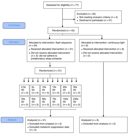
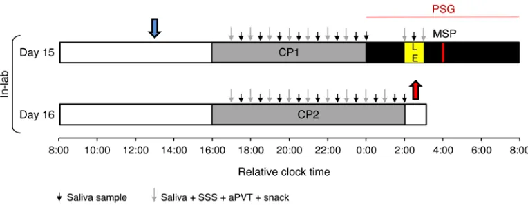
To examine the effects of different temporal patterns of light flashes on the human circadian timing system, we exposed 39 young healthy participants (aged 19 to 36 years) to either a con-tinuous 60-minute light exposure (n = 8) or a sequence of flashes of various frequencies from 0.004 to 0.4 Hz (interstimulus inter-vals [ISI] = 2.5 to 240 s) (n = 31) (Figure 1). Both continuous light and flashes were of equal illuminance (1,700–1,805 lux) and composed of similar broad-spectrum white light (Supplemental Figure 1 and Supplemental Table 1; supplemental material avail-able online with this article; doi:10.1172/JCI82306DS1). Light exposure was scheduled to be administered during the early biological night (Figure 2). Post-hoc analysis determined that light administration began 4.70 ± 0.92 hours after the onset of melatonin, a time during which light is expected to evoke delays in circadian timing (49). Acute changes in salivary melatonin concentrations, as well as objective and subjective alertness, were concomitantly measured (Figure 2).
Group differences. There was no difference in age (U = 104, Z =
–0.68, P = 0.50, Mann-Whitney test), sex distribution (χ2 = 2.39, P
= 0.12, χ2 test), phase angle of light exposure (t = –0.52, df = 37, P =
0.61, t test), integral of salivary melatonin between dim light mel-atonin onset (DLMO) and the onset of light exposure (t = –0.77, df = 37, P = 0.45, t test), morning or evening preference (Morn-ing-Eveningness Questionnaire, refs. 50, 51, U = 137.5, Z = 0.45, P = 0.65, Mann-Whitney test), or reported prior-month sleep
[image:4.585.99.481.54.203.2]–0.54, df = 4, P = 0.6; θ: t = –1.01, df = 4, P = 0.36; α: t = –1.11, df = 4
P = 0.33; paired t test) nor were there associations between these
measures and the ISI.
Discussion
This experiment demonstrates that, in direct contrast to image-forming photoreception, the human circadian timing system has the capacity to integrate light over a millisecond time scale. Fur-thermore, we have demonstrated that a specific temporal pattern of light has the capacity to increase the induction of circadian phase changes to a greater extent than that observed with con-tinuous light. As opposed to the response of the circadian timing system to light flashes, other NIF responses, including increased alertness and suppressed melatonin production, failed to show consistent responsivity to light flashes. This type of sequence of light flashes would be highly improbable to occur naturally and, as such, the response we observed is likely an unintended conse-quence of the circuitry of the system.
Previous studies in humans have contributed to the elucidation of the characteristics of NIF photoresponses, including wavelength sensitivity (33, 36, 38), timing (49, 54), intensity (40), and photic history (41). A limited number of studies have examined the effect of the temporal pattern of light. Previous studies examining the effect of continuous long- versus short-duration (minutes) inter-mittent light on the NIF system have implied that the effective-ness of light in changing circadian timing declines rapidly with the duration of the light pulse (42, 43, 55). We have previously shown that much of the effectiveness of light on changing the timing of the circadian pacemaker may, in fact, be mediated within the first few milliseconds of light exposure (44). Our findings suggest that within the NIF photoresponsive structure, the circadian system appears to integrate ultrashort 2-millisecond flashes of light over time in a nonlinear fashion. This temporal integration is such that 0.95 seconds of light delivered as discreet flashes evenly distrib-uted over an hour has the capacity to be more than 2-fold more effective on phase delaying the circadian system than an equilu-minous continuous light exposure 3,800 times the duration of the flashes. This is the first demonstration to our knowledge in any species that a sequence of ultrashort intermittent light flashes can be as effective, if not more effective, in changing the phase of the circadian timing system in comparison to a continuous white light of the same spectral composition and illuminance.
Circadian integration renders ultrashort flashes more efficient than continuous light. The responsivity of the circadian system to the flash
sequence could be explained by the electrophysiological properties of the ipRGCs, which have a sluggish off response, in that they con-tinue to fire for several seconds after cessation of a light stimulus (10, 56). When flashes are separated by a short ISI, the depolariza-tion of the ipRGCs would become sustained, similar to that evoked by continuous light exposure. The higher effectiveness of flashes on circadian photoreception could arise from the additional firing of cones, which indirectly project to ipRGCs, at light onset and offset, thus generating a continuous response boosted by superimposed multiple spiking responses at each light onset and offset. Flashes seem to offer an ideal stimulus that maximizes inputs from the visual system (cones), which is optimized to be maximally respon-sive to rapid temporal light fluctuations, and the more sluggish light: –0.36 to –0.83 h), we still observed that these individuals
(n = 7) did not exhibit a significant melatonin suppression after the different flash exposures (W = 12, Z = –0.25, P = 0.81, Wilcoxon signed-rank test) (Figure 5C). Melatonin suppression in this sub-group was significantly lower than that observed in the continuous light group (U = 46, Z = 2.03, P < 0.05, Mann-Whitney test).
Flashes of light do not change objective alertness and subjective sleepiness in a dose-dependent manner. Median reaction time (RT)
[image:5.585.45.280.55.253.2](t = 4.49, df = 7, P < 0.01, paired t test; Figure 4B and Figure 5B), number of lapses (W = 34, Z = 2.18, P < 0.05, Wilcoxon signed-rank test; Figure 4C), and shift in optimum RT (mean of the 10% fastest RT) (t = 3.62, df = 7, P < 0.01, paired t test) decreased under continuous light exposure. Continuous light did not decrease sub-jective sleepiness, as assessed by the Stanford Sleepiness Scale (SSS; ref. 53; W = 13.5, Z = 1.5, P = 0.19, Wilcoxon signed-rank test; Figure 4D). Median RTs, numbers of lapses, and SSS scores fluctuated under the different flash frequency conditions; how-ever, there was no consistent increase in alertness or decrease in sleepiness as a function of the ISI (Figure 4, B–D). Median RT (t = –0.02, df = 9, P = 0.98, paired t test; Figure 5D), number of lapses (W = 25.5, Z = –0.15, P = 0.87, Wilcoxon signed-rank test), and shift in optimum RT (W = 16, Z = –1.12, P = 0.27, Wilcoxon signed-rank test) did not change in participants exposed to flashes who showed a similar corrected circadian phase shift as those exposed to continuous light. There was no significant effect of continu-ous light on electroencephalographic correlates of arcontinu-ousal (Δ: t =
that rods have a substantive contribution in flashes subsequent to the first one, given that they are saturated at very low illuminances of light and regenerate at a much slower pace compared with cones.
Integration of light information in the retina, however, does not exclude the possibility of integration of photic drive by neurons in the SCN (61). As with the ipRGCs, SCN neurons exhibit persistent firing, even after cessation of a light stimulation (62), providing a substrate for temporal integration. Alternatively, some neurons in the SCN are activated by phases of darkness (61). While the role of these neurons and their implication in entrainment remain unclear, it is possible that the combination of the excitation by flashes of light with interspersed darkness is able to mediate the observed effects.
A circadian critical fusion frequency. When flashes were
sepa-rated by 2.5 to 7.6 seconds, circadian phase shift was reduced to a similar magnitude as the shift induced by continuous light. Thus, melanopsin-based system that is hypothesized to integrate light
[image:6.585.43.538.54.462.2]exposure of long durations so as to remove the vagaries of behav-ior (e.g., walking in a shaded area, weather) on light exposure and its effects on circadian timing. If the flashes are too far apart (e.g., ISI = 240 s), there may be insufficient drive on ipRGCs to evoke a change in circadian timing. Although a rapid dark regeneration of cone opsin is expected after short bleaches (57–59), if the flashes are too close together (e.g., 2.5–7.6 s), there may not be enough time for the bleached fragment of cone opsin to optimally regenerate; therefore, the loss of the superimposed boost in the response ren-ders these sequences similar to continuous light. A similar obser-vation has previously been noted in hamsters exposed to a train of 300-millisecond light pulses separated by 300 milliseconds of dark or 300 seconds of continuous light of equal total photon density (60). While it is a possibility that requires testing, we do not believe
the circadian system may “perceive” this frequency of stimulation the same as it would continuous light. This is akin to the phenom-enon of flicker fusion in image-forming vision, in which scotopic, rod-mediated vision has a critical fusion frequency (CFF) near 15 Hz, while cone-mediated vision reaches a CFF near 60 Hz (63). Based on our findings, we suggest that circadian CFF, after dark adaptation and under a broad band full retinal exposure to a pat-tern of 2-millisecond stimuli, occurs around 0.13 Hz (7.6 s ISI). As with CFF, we expect circadian CFF to vary across species (64).
A dichotomy in the NIF retinohypothalamic pathways. Given
the technical difficulties in assessing the effect of individual 2-millisecond light flashes on melatonin suppression or alert-ness changes, we cannot exclude that transient changes in these functions have occurred following each brief light exposure. Nev-ertheless, the absence of an ISI-dependent change in melatonin suppression and alertness over a 60-minute period under dif-ferent ISIs supports the idea that light-induced circadian phase
shifting is not subserved by the same neural pathways as either light-induced melato-nin suppression or light-induced changes in alertness. Light-induced changes in mela-tonin production, alertness, and phase shift are all thought to be communicated to the brain by the ipRGCs (16, 21, 65). While light- induced melatonin suppression is often used as a proxy for the effect of light on the circa-dian system, our data indicate that there are at least some circumstances in which this is inaccurate. It is possible that distinct sub-types of ipRGCs in the human retina (66, 67) may subserve different NIF functions. The M1 subtype of ipRGCs is implicated mainly in phase shifting the circadian system and has a faster response, a lower intrinsic threshold, and similar extrinsic input compared with other ipRGC subtypes (67). It is therefore possible that temporal integration is limited to M1 ipRGCs and that activation of these ipRGCs with light flashes is insufficient to induce light-induced melatonin suppression or alertness. An alternative possibility is that, as discussed above, the temporal integration of light is occurring at the level of the SCN. While the retinohypothalamic tract, which arises from the ipRGCs, projects to and con-veys light information to the SCN, it also projects to other brain nuclei, including the paraventricular nucleus of the hypothalamus (PVN) (68), pretectal area (16), lateral hypo-thalamic area (LHA) (69), and ventrolateral preoptic nucleus (VLPO) (68, 70). The PVN is involved in the control of melatonin secre-tion, while the other areas (pretectal area, LHA, VLPO) are all implicated in the regula-tion of sleep or wake. If temporal integraregula-tion in the SCN is required for the phase shifting response to flashes, despite whether similar integration occurs in the retina, it is not altogether unexpected that NIF responses, such as light-induced melatonin suppression and changes in alertness, which may not depend on the SCN, are not affected by the flashes in a dose-dependent manner.
Technical considerations. There was considerable variability
associated with responses to very short ISIs between the flashes of light. Some of this variability may be explained by difficulties in maintaining eyes open due to sleep inertia or anticipatory blinking, which is more likely to occur when the ISI is very short (e.g., every 2.5 s) rather than long (e.g., every 240 s). We did not record eyelid movement but did discuss with the participants the importance of remaining awake with their eyes open in the Ganzfeld dome. Light is able to penetrate through the closed eyelid in a wavelength- dependent fashion, such that approximately 3% to 14% of light is transmitted (71). We have recently shown that, given sufficient intensity, millisecond light flashes passing through the eyelid have the capacity to induce changes in circadian timing (72).
[image:7.585.44.369.55.417.2]less, if a participant had his eyes closed, the retinal illuminance of the stimulus would differ from the corneal measured illuminance and could contribute to the variability of the responses to the short ISI. Future studies are necessary to discover, and potentially remove, this source of variability in the response to short ISI.
It is worth mentioning that, even though we fit the observed phase shift as a function of ISI with a linear rise and exponential decay function, it is quite possible that other functions may be more appropriate for these data. This phenomenological fit, how-ever, was chosen as most suitable after multiple tests with various models, based on known retinal and neuronal physiology. Further studies on NIF photoreception are necessary to refine these mod-els and consecutively the fit of our data.
Flashes that are 2 milliseconds in duration, such as those used in this study, are faster than the pupil constriction response (73). While the pupil constricts after the flash occurs, we have found (data not shown) that there is a persistent pupillary constriction for 29.5 ± 3.24 seconds (n = 4) after an individual flash, thereby reducing the overall input to the retina, especially for flashes with an ISI under 15 seconds. Taking into account pupillary con-striction induced by both continuous and different sequences of light flashes, continuous light would still contain 850 to 18,000 times more light than the sequences of flashes used in this study. We could have, as many laboratories do, used an antimuscar-inic agent to dilate the pupils to avoid the pupillary light reflex and keep it from interfering with the amount of light striking the retina. Given the potential effect of antimuscarinics on the phys-iologic properties of retinal ganglion cells (74), we feel that the use of such agents would have created greater uncertainty in the interpretation of our results.
A final consideration is that the phase shift observed in our study under continuous light was smaller than that observed in previous studies (41, 49). This difference, however, is likely due to protocol dissimilarities in the intensity of the light exposure (49) and light history of the participants that could lead to a different sensitization of the photoreceptive system (41).
Ultrashort flashes revolutionize light therapy for chronobiolog-ical disorders. Whether for mood (23, 28, 75) or circadian
disor-ders (26), light has been shown to be an effective and safe, yet challenging therapy. Multiple modulations have been suggested for the chromaticity (76–78), intensity (79), and pattern (55, 80) of light exposures to render them optimal for clinical and per-sonal use. Here, we show that modulation of the temporal pat-tern of light at the millisecond time scale can be manipulated to isolate and optimize the effects of light on the circadian timing system. Using modern lighting technologies, such as ultra-bright light-emitting diodes, flashes can also be incorporated into por-table mask designs that could be controlled and timed via smart phone software to proactively phase shift and resynchronize jet-lagged travelers to their new time zone, help individuals who want to go to sleep earlier on a regular basis do so (e.g., adolescents), and work in conjunction with daytime light/dark exposure to aid adjusting shift workers. The efficiency of this light treatment is especially pronounced, as flashes have been shown to be able to phase delay the circadian system, even during sleep, when the cir-cadian system is most sensitive to light (54), while the eyelids are closed and without altering sleep (72). This type of light exposure,
therefore, opens new treatment possibilities for circadian disor-ders that will need to be tested in real-world clinical trials as well as possibilities for the alleviation of associated sleep, mood, cog-nitive, and metabolic declines.
Methods
Participants. Thirty-nine healthy adults (25 males and 14 females; 26.4 ± 5.06 years of age, mean ± SD) were empaneled from August 3, 2012, to August 12, 2015. Participants were enrolled if they reported mini-mal to no sleep disturbances (Pittsburgh Sleep Quality Index scores: 0–4; ref. 52), alcohol use (Alcohol Use Disorders Identification Test scores: 0–5; ref. 81), or depressive symptoms (Center for Epidemio-logic Studies–Depression scores: 0–19; ref. 82) and were of intermedi-ate chronotype (simplified version of Morning-Eveningness Question-naire scores: 11–26; refs. 50, 51).
Protocol design. Participants took part in a 16-day protocol. From day 1 to day 14 participants maintained a regular sleep time and wake time pattern at home. At-home sleep-wake patterns were monitored using actigraphs (Actiwatch, Philips, or Motionlogger, Ambulatory Monitor-ing, and self-reported sleep diaries, ref. 83). Participants entered the lab-oratory on day 15. The midpoint of sleep (MSP), defined as the midpoint of the average sleep period during the prior 14 days (day 1 to day 14), was calculated using the actigraphy data and used as the midpoint of an 8-hour sleep opportunity in the laboratory (e.g., a subject who habitually went to bed at 00:00 hours and woke at 08:00 hours would have a MSP of 04:00 hours and be scheduled to sleep in the lab from 00:00 hours until 08:00 hours). Participants whose bed time or wake time deviated by more than 30 minutes twice across the prior 14 days were not empan-eled in the study. All in-lab procedures were individually timed based on the participants’ MSP. Upon entry into the laboratory on day 15 until the conclusion of the study on day 16, subjects remained in a customize time isolation suite at the Veterans Affairs Palo Alto Health Care Sys-tem. Breakfast and lunch were provided to the subjects, and they had ad libitum use of an en suite bathroom. During waking hours and constant posture (CP), room lighting provided by standard overhead fluorescent lamps was dim (0.6–1.9 lux), so as to minimize the effects of background light on the circadian system or melatonin production (40). During the evenings of day 15 and day 16, subjects underwent a CP procedure (Fig-ure 2). The purpose of the CP was to hold constant or remove factors that might otherwise mask endogenous circadian rhythms (84). During the CP, instead of eating dinner, participants were given equicaloric liq-uid snacks (Ensure, Abbott Laboratories) every 60 minutes, such that the caloric intake that they would have received from their dinner was distributed over either 8 (CP1) or 10 (CP2) hours (85). During the CP, saliva samples were collected every 30 minutes. Alertness and sleepi-ness levels were monitored every 60 minutes (Figure 2).
Monitoring sleepiness and alertness. Subjective sleepiness was monitored by SSS (53). Objective alertness levels were monitored by testing RT over 10-minute segments via the auditory version of the psychomotor vigilance task using the PVT-192 (Ambulatory Moni-toring) (87). Auditory psychomotor vigilance task data were analyzed using the manufacturer’s software (React, Ambulatory Monitoring). Variables derived from the software included mean and median RT, number of lapses (nonresponse for 400 ms), and shifts in optimum RT (mean of the fastest 10% RTs).
Changes in brain electrophysiological activity were recorded dur-ing day 15 by electroencephalography (Siesta, Compumedics). Elec-troencephalography data derived from each cortical electrode (C3/4, O1/2) were referenced to an electrically neutral/reference auricular electrode. Data were transformed using a fast Fourier transform with a window length of 2 seconds, overlap of 0%, using a Hanning window type with a maximum frequency of 50 Hz, followed by Welch averag-ing with a mean averagaverag-ing type on 30 seconds for a threshold of 50% and feature extraction on both power (absolute IU2 and relative
per-centage) and frequency (mean Hz and peak Hz). Using this method for each 30-second bin, the absolute power in the different frequency spectra was calculated. Frequency spectra were grouped, as is typical in such analyses, into frequency bands: δ (0.5–4 Hz), θ (4–7.5 Hz), α (8–12.5 Hz), σ (12–14 Hz), β (14–29 Hz), and γ (30–40 Hz). Power spec-tra bands during the 10-minute auditory psychomotor vigilance task immediately prior to and at the end of the light exposure were ana-lyzed for electrophysiological correlates of arousal (Prana, PhiTools).
Data analysis. Phase shift, percentage melatonin suppression, and changes in the parameters of alertness and sleepiness (after light expo-sure – before light expoexpo-sure) were plotted against the ISI (e.g., Δϕ = f [ISI]; Figure 3). Data were fitted using models, as indicated in the Results. The number of lapses was square root transformed (√lapses + √[lapses + 1])
and plotted against ISI for a better clarity of the plot. CI was calculated such as CI = average ± 1.96 × σ/√n (where σ is the standard deviation
of the data and n is the number of data points). Negative change values for RT, number of lapses, and SSS indicate a faster RT, a decrease in the number or lapses, and a decrease in sleepiness, respectively. Negative percentage of melatonin suppression values indicate less melatonin suppression. Statistics, data fitting, and illustrations were done using OriginPro 8 SR2 (v8.0891, OriginLab). Figure 5 was done using Sigma-Plot 11.0 (Systat Software). All data are presented as mean ± SD.
Statistics. Group differences in age, morning or evening prefer-ence, and reported prior-month sleep quality were compared using a Mann-Whitney test. Sex distribution between the two groups was compared using a χ2 test. Phase angle of light exposure between
groups and melatonin phase change between nights in the group receiving continuous light were analyzed using a 2-tailed, paired t test after checking for normal distribution of the data. Nonnormally dis-tributed melatonin suppression data and discreet variables of lapses and changes in sleepiness assessed using the SSS were analyzed using the Wilcoxon signed-rank test. Normally distributed continuous data of median RT, shift in optimum RT, and electroencephalographic cor-relates of arousal were analyzed using a paired t test. All statistical tests used were 2 tailed.
Study approval. The study protocol was reviewed and approved by the Stanford University Institutional Review Board and conforms to the principles expressed in the Declaration of Helsinki. Subjects signed informed consent forms prior to any procedures.
receive either continuous light (n = 8, aged 27.4 ± 5.0 years) or one of twelve different light exposures that vary by the number of ultrashort 2-millisecond flashes administered during an hour (n = 31, aged 26.1 ± 5.1 years). Flash frequency within the 1-hour light exposure varied between 0.004 and 0.4 Hz (ISI = 2.5 to 240 s). Subjects were random-ized such that at least 2 participants were assigned to each of 12 ISI values (Figure 1 and Figure 3). Following an interim analysis, we added an extra 3 participants at an ISI of 10 seconds, 3 participants at an ISI of 5 seconds, and 1 participant at an ISI of 2.5 seconds. Randomization was done by J.M. Zeitzer through a random sequence generator algo-rithm, and participants were assigned to experimental groups by study personnel. All data assessments were done blind to condition. Flashes were delivered using the xenon flash unit of a ColorDome (Diagnosys). Continuous light was delivered using a broad-spectrum 400 W blue metal halide bulb (Eye Hortilux) with an adapted ballast and a cus-tom-built light box fitted with ultraviolet light filtration (Rosco 03114 2024), neutral density filters (Rosco 3402), and diffusers (Rosco 117). Illuminance of both light exposures was matched to be around 1,800 lux using the ILT1700 radiometer (International Light Technologies). Extensive characterization of the light sources is presented in Supple-mental Table 1 and SuppleSupple-mental Figure 1.
Circadian phase shift and melatonin suppression. Circadian phase was assessed on days 15 and 16 through examination of the endoge-nous melatonin secretion profile. Saliva (at least 1 ml) was collected every 30 minutes by placing an absorbing swab into the mouth and moving it around slowly over 5 minutes. Next, the swab was put into a tube (Salivettes, Sarstedt). Samples were centrifuged after collection and then frozen and stored at –80°C until assayed. Salivary melatonin was assayed in duplicate according to the manufacturer’s instructions using the Bühlmann direct saliva Melatonin ELISA Kits (Alpco) based on the Kennaway G280 anti-melatonin antibody. All samples from a single subject were assayed on the same ELISA plate.
Onset of melatonin secretion was calculated as the time at which salivary melatonin concentrations exceeded a subject-specific dynamic threshold (Supplemental Figure 2). The dynamic threshold was calculated as the mean of the first 3 daytime melatonin concentra-tions plus twice the standard deviation of these values (86). DLMO was estimated for each participant on days 15 (DLMO15) and 16 (DLMO16). Circadian phase shift (Δϕ) elicited by the light exposure was calcu-lated as follows: Δϕ = DLMO15 – DLMO16 (Supplemental Figure 2). Phase delays are represented by convention negative numbers, and phase advances are represented positive numbers. The phase angle (θ) between DLMO15 and the onset of the experimental light exposure was also calculated to ensure that the participants were exposed to light at a similar circadian phase (Supplemental Figure 2). Average phase angle of light exposure (4.70 hours) of participants in both groups was plotted on the phase response curve to 1 hour of light, adapted from St Hilaire et al. (49) to obtain an average phase shift (–1.23 hours). Indi-vidual deviations from the average phase angle were used to concom-itantly obtain a percentage of change from average phase shift. The percentage of change obtained was then applied to each participant’s phase shift to obtain a corrected phase shift that takes into account the differences in circadian timing of the light exposure.
the Department of Veterans Affairs Sierra Pacific Mental Illness Research, Education, and Clinical Center.
Address correspondence to: Jamie M. Zeitzer, Psychiatry and Behav-ioral Sciences, Stanford University, VA Palo Alto Health Care Sys-tem, 3801 Miranda Avenue (151Y), Palo Alto, California 94304, USA. Phone: 650.493.5000, ext. 62410; E-mail: [email protected]. Raymond P. Najjar’s present address is: Visual Neurosciences Research Group, Singapore Eye Research Institute, Singapore.
Author contributions
JMZ designed research. RPN performed research and melatonin assays. RPN and JMZ wrote the paper and analyzed the data.
Acknowledgments
We would like to thank Yvonne Quevedo, Ban Ku, and Cheng-Ann Wang for recruiting participants and conducting the experimental sessions and Chun-Ping (Phoebe) Liao for assisting with the mel-atonin ELISA assays. This research was supported by the National Heart, Lung, and Blood Institute (1R01HL108441-01A1) and
1. Czeisler CA, Wright KP Jr. Influence of light on circadian rhythmicity in humans. In: Turek FW, Zee PC, eds. Regulation of Sleep and Circadian Rhythms. New York, New York, USA: Marcel Dekker; 1999:149–180.
2. Duffy JF, Kronauer RE, Czeisler CA. Phase-shift-ing human circadian rhythms: influence of sleep timing, social contact and light exposure. J Phys-iol. 1996;495(pt 1):289–297.
3. Skene DJ, Lockley SW, Thapan K, Arendt J. Effects of light on human circadian rhythms. Reprod Nutr Dev. 1999;39(3):295–304. 4. Dijk DJ, Czeisler CA. Contribution of the
circa-dian pacemaker and the sleep homeostat to sleep propensity, sleep structure, electroencephalo-graphic slow waves, and sleep spindle activity in humans. J Neurosci. 1995;15(5 pt 1):3526–3538. 5. Spiegel K, Sheridan JF, Van Cauter E. Effect of sleep deprivation on response to immunization. JAMA. 2002;288(12):1471–1472.
6. Weibel L, Brandenberger G. Disturbances in hormonal profiles of night workers during their usual sleep and work times. J Biol Rhythms. 1998;13(3):202–208.
7. Wright KP Jr, Hull JT, Czeisler CA. Relation-ship between alertness, performance, and body temperature in humans. Am J Physiol. 2002;283(6):R1370–R1377.
8. Young ME, Bray MS. Potential role for peripheral circadian clock dyssynchrony in the pathogen-esis of cardiovascular dysfunction. Sleep Med. 2007;8(6):656–667.
9. Roenneberg T, Allebrandt KV, Merrow M, Vetter C. Social jetlag and obesity. Curr Biol. 2012;22(10):939–943.
10. Berson DM, Dunn FA, Takao M. Phototransduc-tion by retinal ganglion cells that set the circa-dian clock. Science. 2002;295(5557):1070–1073. 11. Provencio I, et al. A novel human opsin in the
inner retina. J Neurosci. 2000;20(2):600–605. 12. Provencio I, Jiang G, De Grip WJ, Hayes WP,
Rollag MD. Melanopsin: An opsin in melano-phores, brain, and eye. Proc Natl Acad Sci U S A. 1998;95(1):340–345.
13. Belenky MA, Smeraski CA, Provencio I, Sollars PJ, Pickard GE. Melanopsin retinal ganglion cells receive bipolar and amacrine cell synapses. J Comp Neurol. 2003;460(3):380–393. 14. Lucas RJ, et al. Diminished pupillary light reflex
at high irradiances in melanopsin-knockout mice. Science. 2003;299(5604):245–247. 15. Hattar S, et al. Melanopsin and rod-cone
photoreceptive systems account for all major accessory visual functions in mice. Nature.
2003;424(6944):76–81.
16. Hattar S, et al. Central projections of melanopsin-expressing retinal ganglion cells in the mouse. J Comp Neurol. 2006;497(3):326–349. 17. Panda S, et al. Melanopsin (Opn4) requirement
for normal light-induced circadian phase shift-ing. Science. 2002;298(5601):2213–2216. 18. Lewy AJ, Wehr TA, Goodwin FK, Newsome DA,
Markey SP. Light suppresses melatonin secretion in humans. Science. 1980;210(4475):1267–1269. 19. Badia P, Myers B, Boecker M, Culpepper J, Harsh
JR. Bright light effects on body temperature, alertness, EEG and behavior. Physiol Behav. 1991;50(3):583–588.
20. Cajochen C, Zeitzer JM, Czeisler CA, Dijk DJ. Dose-response relationship for light intensity and ocular and electroencephalographic cor-relates of human alertness. Behav Brain Res. 2000;115(1):75–83.
21. Altimus CM, et al. Rods-cones and melanopsin detect light and dark to modulate sleep indepen-dent of image formation. Proc Natl Acad Sci U S A. 2008;105(50):19998–20003.
22. Kripke DF. Light treatment for nonseasonal depression: speed, efficacy, and combined treat-ment. J Affect Disord. 1998;49(2):109–117. 23. Wirz-Justice A, van der Velde P, Bucher A, Nil R.
Comparison of light treatment with citalopram in winter depression: a longitudinal single case study. Int Clin Psychopharmacol. 1992;7(2):109–116. 24. Noseda R, et al. A neural mechanism for
exac-erbation of headache by light. Nat Neurosci. 2010;13(2):239–245.
25. Anderson JL, Glod CA, Dai J, Cao Y, Lockley SW. Lux vs. wavelength in light treatment of Sea-sonal Affective Disorder. Acta Psychiatr Scand. 2009;120(3):203–212.
26. Eastman CI, Gazda CJ, Burgess HJ, Crowley SJ, Fogg LF. Advancing circadian rhythms before eastward flight: a strategy to prevent or reduce jet lag. Sleep. 2005;28(1):33–44.
27. Glickman G, Byrne B, Pineda C, Hauck WW, Brainard GC. Light therapy for seasonal affective disorder with blue narrow-band light-emitting diodes (LEDs). Biol Psychiatry. 2006;59(6):502–507. 28. Kripke DF, Mullaney DJ, Klauber MR, Risch SC,
Gillin JC. Controlled trial of bright light for non-seasonal major depressive disorders. Biol Psychi-atry. 1992;31(2):119–134.
29. Lack L, Wright H. The effect of evening bright light in delaying the circadian rhythms and lengthening the sleep of early morning awaken-ing insomniacs. Sleep. 1993;16(5):436–443. 30. Parry BL, et al. Neuroendocrine effects of light
therapy in late luteal phase dysphoric disorder. Biol Psychiatry. 1994;36(6):356–364.
31. Wirz-Justice A, et al. A randomized, double-blind, placebo-controlled study of light therapy for antepartum depression. J Clin Psychiatry. 2011;72(7):986–993.
32. Wright KP Jr, Hull JT, Hughes RJ, Ronda JM, Czeisler CA. Sleep and wakefulness out of phase with internal biological time impairs learning in humans. J Cogn Neurosci. 2006;18(4):508–521. 33. Gooley JJ, et al. Spectral responses of the human
circadian system depend on the irradiance and duration of exposure to light. Sci Transl Med. 2010;2(31):31ra33.
34. Gooley JJ, et al. Melanopsin and rod-cone photoreceptors play different roles in mediat-ing pupillary light responses durmediat-ing exposure to continuous light in humans. J Neurosci. 2012;32(41):14242–14253.
35. Bailes HJ, Lucas RJ. Human melanopsin forms a pigment maximally sensitive to blue light (λmax ≈ 479 nm) supporting activation of G(q/11) and G(i/o) signalling cascades. Proc Biol Sci. 2013;280(1759):20122987.
36. Brainard GC, et al. Action spectrum for mel-atonin regulation in humans: evidence for a novel circadian photoreceptor. J Neurosci. 2001;21(16):6405–6412.
37. Najjar RP, et al. Aging of non-visual spectral sen-sitivity to light in humans: compensatory mecha-nisms? PLoS One. 2014;9(1):e85837.
38. Thapan K, Arendt J, Skene DJ. An action spec-trum for melatonin suppression: evidence for a novel non-rod, non-cone photoreceptor system in humans. J Physiol. 2001;535(pt 1):261–267. 39. Zaidi FH, et al. Short-wavelength light sensitivity
of circadian, pupillary, and visual awareness in humans lacking an outer retina. Curr Biol. 2007;17(24):2122–2128.
40. Zeitzer JM, Dijk DJ, Kronauer R, Brown E, Czeisler CA. Sensitivity of the human circadian pacemaker to nocturnal light: melatonin phase resetting and suppression. J Physiol. 2000;526(pt 3):695–702. 41. Chang A-M, et al. Human responses to
bright light of different durations. J Physiol. 2012;590(13):3103–3112.
42. Gronfier C, Wright KP Jr, Kronauer RE, Jewett ME, Czeisler CA. Efficacy of a single sequence of intermittent bright light pulses for delay-ing circadian phase in humans. Am J Physiol. 2004;287(1):E174–E181.
44. Zeitzer JM, Ruby NF, Fisicaro RA, Heller HC. Response of the human circadian system to millisecond flashes of light. PLoS One. 2011;6(7):e22078.
45. Arvanitogiannis A, Amir S. Resetting the rat cir-cadian clock by ultra-short light flashes. Neurosci Lett. 1999;261(3):159–162.
46. Nelson DE, Takahashi JS. Sensitivity and inte-gration in a visual pathway for circadian entrain-ment in the hamster (Mesocricetus auratus). J Physiol. 1991;439:115–145.
47. Vidal L, Morin LP. Absence of normal photic integration in the circadian visual system: response to millisecond light flashes. J Neurosci. 2007;27(13):3375–3382.
48. Van Den Pol AN, Cao V, Heller HC. Circadian system of mice integrates brief light stimuli. Am J Physiol. 1998;275(2 pt 2):R654–R657.
49. St Hilaire MA, et al. Human phase response curve to a 1 h pulse of bright white light. J Physiol. 2012;590(13):3035–3045.
50. Horne JA, Ostberg O. A self-assessment question-naire to determine morningness-eveningness in human circadian rhythms. Int J Chronobiol. 1976;4(2):97–110.
51. Reite M, Weissberg MP, Ruddy J. Circadian rhythm-based sleep complaints. In: Clinical Manual for Evaluation and Treatment of Sleep Disorders. Washington, DC, USA: American Psy-chiatric Pub; 2008:97–121.
52. Buysse DJ, Reynolds CF 3rd, Monk TH, Berman SR, Kupfer DJ. The Pittsburgh Sleep Quality Index: a new instrument for psychiatric practice and research. Psychiatry Res. 1989;28(2):193–213. 53. Hoddes E, Zarcone V, Smythe H, Phillips
R, Dement WC. Quantification of sleep-iness: a new approach. Psychophysiology. 1973;10(4):431–436.
54. Khalsa SBS, Jewett ME, Cajochen C, Czeisler CA. A phase response curve to single bright light pulses in human subjects. J Physiol. 2003;549(pt 3):945–952.
55. Gronfier C, Wright KP, Kronauer RE, Czeisler CA. Entrainment of the human circadian pace-maker to longer-than-24-h days. Proc Natl Acad Sci U S A. 2007;104(21):9081–9086.
56. Emanuel AJ, Do MTH. Melanopsin tristability for sustained and broadband phototransduction. Neuron. 2015;85(5):1043–1055.
57. Hollins M, Alpern M. Dark adaptation and visual pigment regeneration in human cones. J Gen Physiol. 1973;62(4):430–447.
58. Rushton WAH, Henry GH. Bleaching and regen-eration of cone pigments in man. Vision Res. 1968;8(6):617–631.
59. Coolen ACC, van Norren D. Kinetics of human
cone photopigments explained with a Rush-ton-Henry model. Biol Cybern. 1988;58(2):123–128. 60. Nelson DE, Takahashi JS. Integration and
saturation within the circadian photic entrain-ment pathway of hamsters. Am J Physiol. 1999;277(5):R1351–R1361.
61. Groos GA, Mason R. The visual properties of rat and cat suprachiasmatic neurones. J Comp Phys-iol. 1980;135(4):349–356.
62. Drouyer E, Rieux C, Hut RA, Cooper HM. Responses of suprachiasmatic nucleus neurons to light and dark adaptation: relative contribu-tions of melanopsin and rod-cone inputs. J Neuro-sci. 2007;27(36):9623–9631.
63. Hecht S, Shlaer S. Intermittent stimulation by light. J Gen Physiol. 1936;19(6):965–977. 64. Jarvis JR, Prescott NB, Wathes CM. A
mechanis-tic inter-species comparison of flicker sensitivity. Vision Res. 2003;43(16):1723–1734.
65. Güler AD, et al. Melanopsin cells are the principal conduits for rod-cone input to non-image-form-ing vision. Nature. 2008;453(7191):102–105. 66. Schmidt TM, Chen S-K, Hattar S. Intrinsically
photosensitive retinal ganglion cells: many subtypes, diverse functions. Trends Neurosci. 2011;34(11):572–580.
67. Zhao X, Stafford BK, Godin AL, King WM, Wong KY. Photoresponse diversity among the five types of intrinsically photosensitive retinal ganglion cells. J Physiol. 2014;592(7):1619–1636. 68. Levine JD, Weiss ML, Rosenwasser AM, Miselis
RR. Retinohypothalamic tract in the female albino rat: a study using horseradish peroxidase conjugated to cholera toxin. J Comp Neurol. 1991;306(2):344–360.
69. Johnson RF, Morin LP, Moore RY. Retinohy-pothalamic projections in the hamster and rat demonstrated using cholera toxin. Brain Res. 1988;462(2):301–312.
70. Lu J, Shiromani P, Saper CB. Retinal input to the sleep-active ventrolateral preoptic nucleus in the rat. Neuroscience. 1999;93(1):209–214. 71. Robinson J, Bayliss SC, Fielder AR. Transmission
of light across the adult and neonatal eyelid in vivo. Vision Res. 1991;31(10):1837–1840. 72. Zeitzer JM, Fisicaro RA, Ruby NF, Heller HC.
Mil-lisecond flashes of light phase delay the human circadian clock during sleep. J Biol Rhythms. 2014;29(5):370–376.
73. Feinberg R, Podolak E. Latency Of Pupillary Reflex To Light Stimulation And Its Relationship To Aging. Washington, DC, USA: Federal Aviation Agency, Office of Aviation Medicine, Georgetown Clini-cal Research Institute; 1965.
74. Strang CE, Renna JM, Amthor FR, Keyser KT. Muscarinic acetylcholine receptor
local-ization and activation effects on ganglion response properties. Invest Ophthalmol Vis Sci. 2010;51(5):2778–2789.
75. Lam RW, et al. The Can-SAD study: a random-ized controlled trial of the effectiveness of light therapy and fluoxetine in patients with winter seasonal affective disorder. Am J Psychiatry. 2006;163(5):805–812.
76. Najjar RP, et al. Chronic artificial blue-enriched white light is an effective countermeasure to delayed circadian phase and neurobehavioral decrements. PLoS One. 2014;9(7):e102827. 77. Vetter C, Juda M, Lang D, Wojtysiak A,
Roen-neberg T. Blue-enriched office light competes with natural light as a zeitgeber. Scand J Work Environ Health. 2011;37(5):437–445. 78. Viola AU, James LM, Schlangen LJM, Dijk DJ.
Blue-enriched white light in the workplace improves self-reported alertness, performance and sleep quality. Scand J Work Environ Health. 2008;34(4):297–306.
79. Van Someren EJ, et al. Bright light therapy: improved sensitivity to its effects on rest-activity rhythms in Alzheimer patients by application of nonparametric methods. Chronobiol Int. 1999;16(4):505–518.
80. Revell VL, et al. Advancing human circadian rhythms with afternoon melatonin and morning intermittent bright light. J Clin Endocrinol Metab. 2006;91(1):54–59.
81. Babor TF, Higgins-Biddle JC, Saunders JB, Mont-eiro MG. AUDIT: The Alcohol Use Disorders Iden-tification Test: guidelines for use in primary health care. 2nd ed. Geneva, Switzerland: World Health Organization. Department of Mental Health and Substance Dependence; 2001.
82. Radloff LS. The CES-D scale a self-report depres-sion scale for research in the general population. Appl Psychol Meas. 1977;1(3):385–401. 83. Carney CE, et al. The consensus sleep diary:
standardizing prospective sleep self-monitoring. Sleep. 2012;35(2):287–302.
84. Duffy JF, Dijk D-J. Getting through to circadian oscillators: why use constant routines? J Biol Rhythms. 2002;17(1):4–13.
85. Mifflin MD, et al. A new predictive equation for resting energy expenditure in healthy individu-als. Am J Clin Nutr. 1990;51(2):241–247. 86. Voultsios A, Kennaway DJ, Dawson D. Salivary
melatonin as a circadian phase marker: valida-tion and comparison to plasma melatonin. J Biol Rhythms. 1997;12(5):457–466.