Transverse maxillary deficiency in adults still poses a challenge in orthodontics. Maxillary con-striction is clinically manifested as a narrow palate and a high palatal vault, a complete unilateral or bilateral crossbite and dark buccal corridors. The dysfunction secondary to malocclusion may also be associated with impaired nasal patency leading to open-mouth breathing and all its sequelae [1]. It is difficult to estimate the prevalence of trans-verse maxillary deficiency in the adult population. According to Bailey [2], approximately 30% of all
adult patients requiring orthodontic surgery pres-ent with maxillary constriction. Midpalatal suture expansion, associated with expanding the maxil-lary bone base, increasing arch widths and restor-ing proper interarch relationships is a well-estab-lished treatment of severe maxillary constriction. The first reports on midpalatal suture expansion date back to the 19th century, when Talma and Le-foulon used a palatal expander with a C-shaped spring as the main source of applied force. Then, in 1860, Angella reported in Dental Cosmos a case
CLINICAL CASES
Rafał Nowak
A, D–F, Ewa Zawiślak
B–DAn Analysis of Early Changes to the Craniofacial Skeleton
Induced by Transpalatal Distraction in a Patient
with Maxillary Constriction, Based on Computed
Tomography Scans – Case Report
Analiza wczesnych zmian w szkielecie czaszki twarzowej
po leczeniu zwężenia szczęk metodą dystrakcji przezpodniebiennej
na podstawie tomografii komputerowej – opis przypadku
Department of Maxillofacial Surgery, Wroclaw Medical University, Wrocław, Poland
A – koncepcja i projekt badania, B – gromadzenie i/lub zestawianie danych, C – analiza i interpretacja danych, D – napisanie artykułu, E – krytyczne zrecenzowanie artykułu, F – zatwierdzenie ostatecznej wersji artykułu
Abstract
Transpalatal distraction is a recognized surgical treatment of transverse maxillary deficiency in adult patients. In skeletally mature individuals, rapid maxillary expansion (RME) is ineffective or leads to dental and periodontal complications. Transpalatal distraction works on the principle of distraction osteogenesis, increasing the maxillary base width and its transverse dimensions. Skeletal expansion of the maxilla will lead to an increase in the volume of the nasal cavity with proper conversion of the nasal breathing track. The increase in width of the maxilla will also overcome the unsightly dark corridors during a smile. The clinical manifestation of the maxillary expansion during active distraction treatment is the emergence of diastema between the maxillary central incisors. Currently available diagnostic imaging techniques such as computed tomography (CT) and 3D image reconstruction of cra-niofacial bone structure enable precise analysis of active treatment.
The paper presents the case of a 29-year-old woman with maxillary constriction and skeletal class III malocclusion, treated with transpalatal distraction. The changes in maxillofacial complex were quantified based on the selected axial computed tomography scans taken before and after the end of active treatment. Furthermore, a 3D recon-struction of the craniofacial skeleton was performed so as to obtain a realistic picture of TPD-induced changes (Dent. Med. Probl. 2016, 53, 1, 134–141).
Key words: computed tomography, maxillary constriction, transpalatal distraction, crossbite.
Słowa kluczowe: tomografia komputerowa, zwężenie szczęki, dystrakcja przezpodniebienna, zgryz krzyżowy.
Dent. Med. Probl. 2016, 53, 1, 134–141
pansion, SARME – surgically assisted rapid max-illary expansion) and surgical methods (TPD – transpalatal distraction, LFI – E – segmented Le Fort I osteotomy with expansion) [4].
The treatment method should be chosen based on patient age and their skeletal maturity. The midpalatal suture in patients with primary den-tition and early mixed denden-tition is composed of connective tissue and the midface skeleton does not show resistance to orthodontic expanders. In older patients, with late mixed dentition, the ex-pansion is typically possible with the use of a Hy-rax appliance as the midpalatal suture closes due to mineralization.
In skeletally mature individuals, rapid max-illary expansion (RME) is ineffective or leads to dental and periodontal complications [5–8]. In or-der to avoid treatment failure in adult patients, sur-gically assisted methods were introduced [9, 10].
The main goal of surgically assisted maxillary expansion is to reduce or eliminate the bone re-sistance of the maxilla. The surgery includes an incision created within the maxilla from the pir-iform aperture, through its anterior surface and zygomaticoalveolar crest to its insertion into the sphenoid bone, which is along the LeFort I oste-otomy line. As an essential part of the procedure, the midpalatal suture is surgically separated [3]. The force-generating appliance is either a tooth-anchored expander (SARME, surgically assist-ed rapid palatal expansion) or a bone-anchorassist-ed transpalatal distractor (TPD, transpalatal distrac-tion) [4, 9]. The transpalatal distractor applies force directly onto the palatine bone, which helps avoid the adverse effects of expander force applied onto teeth in the palatal expansion (SARME) method.
Midpalatal suture expansion alters the struc-ture and physiology of the facial skeleton. The low-ering of the palatal vault increases the volume of the nasal cavity, which improves facial features and the patency of the upper respiratory tract [11, 12].
In young individuals, the center of rotation of maxillary bones, as they expand, is thought to be located within the frontonasal suture. However, with age it lowers towards the midpalatal suture
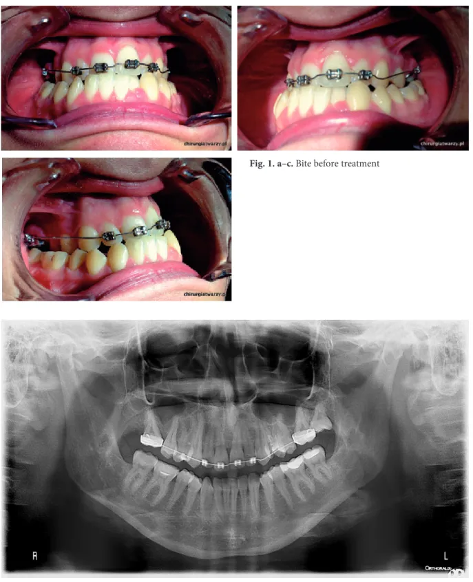
etal class III malocclusion and transverse max-illary deficiency (Fig. 1 a–c). The complex orth-odontic and surgical treatment plan assumed transpalatal distraction (TPD) as the first stage of treatment, followed by deficit compensation and a second procedure of bimaxillary osteotomy (BIMAX). The preoperative diagnostic imaging included plain craniofacial computed tomography. Computed tomography of the craniofacial bones makes it possible to appreciate all of the significant anatomical structures, which may show individu-al variability, and their relationships, as well as to plan the surgical intervention.
The surgery was performed under general an-esthesia. The Le Fort I osteotomy was performed from the symmetrical approaches in the oral ves-tibule and maxillary labial frenum, including the bilateral separation of the maxilla from the ptery-goid processes of the sphenoid and surgical mobili-zation of the midpalatal suture. After checking the symmetry and mobility of the palatal leaves, the Uni-Smile® transpalatal distractor (Titamed,
Bel-gium) size 16 was fixed on the hard palate between the second premolars. The abutment plates were lo-cated horizontally at 1.0 cm from the gingival mar-gin and perpendicularly to the skeletal line of the midpalatal suture. Intraoperative distractor activa-tion yielded a 1.0 mm wide diastema. The wound was closed using 4-0 absorbable sutures. The medi-cal treatment involved peri- and postoperative pre-ventive antibiotic therapy, using analgesics, antie-dema agents and decongestant nasal drops.
A standard distractive treatment protocol was followed including the latency, distraction, and retention phases. The surgery was followed by a 6-day latency phase. The distractor did not ex-pand spontaneously owing to the blocking screw being placed and tightened intraoperatively. Dur-ing the distraction phase, the patient activated the distractor herself at home, twice daily (AM/PM) by a quarter turn, which corresponds to 0.5 mm ex-pansion per day. The duration of the active treat-ment phase depends on the transverse maxillary deficiency. In this particular case, the distraction phase lasted for 15 days. The total number of
acti-vations was 29 (each being a quarter turn), which corresponds to the overall expansion of 7.25 mm.
Directly after completion of the active treat-ment, the follow-up, non-enhanced craniofacial CT scan and a posteroanterior (P-A) cephalomet-ric radiograph were performed. The most notice-able treatment-induced change was a diastema as a clinical manifestation of midpalatal suture ex-pansion (Fig. 4 a–c).
Treatment Outcome
Evaluation
Treatment outcomes were quantified based on the measurements of the axial tomograms taken before (time point T1) and after the active treat-ment (time point T2). The axial tomograms se-lected for the analysis were taken at the level of
Fig. 1. a–c. Bite before treatment
the incisive foramen and the anterior nasal spine. The measurement reference points were chosen based on the criterion of a sharp image of cer-tain anatomical structures on the tomograms, which enabled measurement reproducibility. The five transverse measurements (L1, L2, L3, L4, L5) were in the axial plane at the level of the incisive foramen (Fig. 7 a–b). The measurements quanti-fied skeletal changes in the transverse maxillary
dimension, topographically related to the follow-ing tooth groups: first medial incisors, canines, first and second premolars as well as first molars. On the computed tomography scans at the level of the nasal spine, three distances were marked (W1, W2, W3), corresponding to the increase of transverse maxillary dimension at the respective teeth group levels: canines, second premolars and first molars (Table 1, Figure 8 a–b). The selected
computed tomography scans were analyzed us-ing OsiriX® software (Pixemo, Switzerland). The
analysis was performed twice – at baseline, before
treatment commencement (T1) and after the ac-tive treatment (T2). Additionally, the evaluation of treatment outcomes and changes within the
mid-Fig. 5. Panoramic x-ray after treatment
Table 1. The analysis of changes in transverse maxillary dimension before and after the active treatment based on computed tomography scans – see the article for the legend
L1 L2 L3 L4 L5 W1 W2 W3
T1 10.35 32.58 42.44 44.90 52.98 27.95 43.00 51.51 T2 20.58 40.76 49.81 52.73 59.63 34.89 49.46 56.61 ∆T +10.23 +8.18 +7.37 +7.83 +6.65 +6.94 +6.46 +5.1
Fig. 6. Frontal cephalometry x-ray
face skeleton induced by the transpalatal distrac-tion included qualitative assessment based on 3D craniofacial models. The 3D model of the facial skeleton was generated based on craniofacial com-puted tomography using OsiriX software and sub-sequently saved as a jpg file (Fig. 9 a–b).
Results and Discussion
The analysis of the craniofacial skeletal chang-es based on computed tomography scans is one of the assessment methods applicable to surgi-cal treatment outcomes [15–17]. Quantification of treatment outcomes based on x-ray images may be
difficult due to the inability to achieve reproduc-ible projection and the presence of artefacts. The accurate analysis of anatomical structures and the choice of measurement reference points on the CT scans enable precise monitoring of both quantita-tive and qualitaquantita-tive aspects of maxillary expan-sion.
The results of transversal analysis of axial pro-jections generated at the level of the incisive fora-men and the anterior nasal spine were shown in Table 1.
The measurements at the level of the incisive foramen revealed the largest maxillary expansion near the medial incisors (+ 10.23 mm), whereas the lowest value (+ 6.65 mm) was observed near
Fig. 7. CT scan – measurements on the level of incisive foramen: a) before treatment, b) after treatment
the first molars. The measurements at the level of the anterior nasal spine revealed the largest max-illary expansion near the canines (+ 6.94 mm). The expansion gradually decreased posteriorly, amounting to 5.1 mm near the first molars. These findings correlate with the views of other authors, who observed the largest expansion of the midpal-atal suture within its anterior segment and suggest that the center of rotation is situated within the frontonasal suture, resembling an unfolding fan in the sagittal plane and a pyramid in the axial plane [11–13].
Furthermore, we determined the ratio of dis-tractor screw expansion to the increase of maxil-lary transversal dimension (skeletal change incre-ments) at the predefined measurement reference points, using the following formula:
Z = [∆T]/7.25
where [∆T] stands for changes of dimensions at the following measurement points: L1, L2, L3, L4, L5, W1, W2 and W3, respectively; whereas 7.25 is a constant expressed in millimeters and stands for the distractor screw expansion.
Measurement results are shown in Table 2. Our measurements indicate that the most sig-nificant skeletal change takes place within the
an-terior maxilla near the medial incisors (L1), where the ratio of distractor screw expansion to max-illary transverse dimension increase is approx-imately 1:1.5 (exactly 1.41). This ratio decreases posteriorly, reaching almost 1:1 (exactly 0.97) near the first molars (L5). On the average, at the lev-el of the incisive foramen, each 1.00 mm expan-sion of the distractor screw yielded an increase of maxillary transverse dimension by 1.41 mm and 0.97 mm near the upper incisors and first molars, respectively. On the average, at the level of the an-terior nasal spine, each 1.00 mm expansion of the distractor screw yielded an increase of maxillary transverse dimension by 0.95 mm and 0.70 mm near the canines (W1) and first molars (W3), re-spectively.
We believe this is very valuable information, which after a statistical analysis in a larger patient sample, may help develop predictable treatment plans and determine patients needs for maxillary expansion after the initial surgical intervention.
The 3D models yield the most realistic picture of changes to the craniofacial skeleton induced by transpalatal distraction.
The 3D visualization provides an interesting qualitative supplement as a three-dimensional re-construction of the computed tomography scans.
Fig. 9. 3D CT reconstruction: a) before treatment, b) after treatment
Table 2. Analysis of changes in transverse maxillary dimension with the increasing length of the distractor
L1 L2 L3 L4 L5 W1 W2 W3
T2–T1 [∆T] 10.23 8.18 7.37 7.83 6.65 6.94 6.46 5.1
verse maxillary deficiency. J. Oral Maxillofac. Surg. 1997, 55, 728–731.
[3] Koudstaal M.J., Poort L.J., Van der Wal K.G.H., Wolvius E.B., Prahl-Andersen B., Schulten A.J.M.: Sur-gically assisted rapid maxillary expansion (SARME): a review of the literature. Int. J. Oral Maxillofac. Surg. 2005, 34, 709–714.
[4] Mommaerts M.Y.: Transpalatal distraction as a method of maxillary expansion. Br. J. Oral Maxillofac. Surg. 1999, 37, 268–272.
[5] Pinto P.X., Mommaerts M.Y., Wreakes G., Jacobs W.V.J.A.: Immediate postexpansion changes following the use of the transpalatal distractor. J. Oral Maxillofac. Surg. 2001, 59, 994–1000.
[6] Matteini C., Mommaerts M.Y.: Posterior transpalatal distraction with pterygoid disjunction: A short-term mod-el study. Am. J. Orthod. Dentofacial Orthop. 2001, 120, 498–502.
[7] Baysal A., Karadede I., Hekimoglu S., Ucar F., Ozer T., Veli I., Uysal T.: Evaluation of root resorption fol-lowing rapid maxillary expansion using cone-beam computed tomography. Angle Orthod. 2012, 82, 488–494. [8] Boryor A., Hohmanna A., Wunderlich A., Geiger M., Kilic F., Sander M., Sander C., Bockers T.,
Sander F.G.: In vitro results of rapid maxillary expansion on adults compared with finite element simulations. J. Biomech. 2010, 43, 1237–1242.
[9] Gautam P., Valiathan A., Adhikaric R.: Stress and displacement patterns in the craniofacial skeleton with rap-id maxillary expansion: A finite element method study. Am. J. Orthod. Dentofacial Orthop. 2007, 132, 5, e1-5.e11. [10] Chrcanovic B.R., Custódio A.L.: Orthodontic or surgically assisted rapid maxillary expansion. Oral Maxillofac.
Surg. 2009, 13, 123–137.
[11] Biedziak B.: Orthodontic palatal expansion – assessment widening of maxilla and nasal cavity in computed to-mography scans. Czas Stomatol. 2009, 62, 912–921 [in Polish].
[12] Seeberger R., Kater W., Schulte-Geers M., Davids R., Freier K., Thiele O.: Changes after surgically-assist-ed maxillary expansion (SARME) to the dentoalveolar, palatal and nasal structures by using tooth-borne distrac-tion devices. Br. J. Oral Maxillofac Surg. 2011, 49, 381–385.
[13] Kamińska I.: Orthodontic effect of palatal expansion. Ann. Pomeranian Med. Univ. Szczecin, 2008, 54, 94–105 [in Polish]. [14] Koudstaal M.J., Smeets J.B.J., Kleinrensink G.J., Schulten A.J.M., Van der Wal K.G.H.: Relapse and sta-bility of surgically assisted rapid maxillary expansion: an anatomic biomechanical study. J. Oral Maxillofac. Surg. 2009, 67, 10–14.
[15] Goldenberg D.C., Goldenberg F.C., Alonso N., Gebrin E.S., Amaral T.S., Scanavini M.A., Ferreira M.C.: Hyrax appliance opening and pattern of skeletal maxillary expansion after surgically assisted rapid palatal expansion: a computed tomography evaluation. Oral Surg. Oral Med. Oral Pathol. Oral Radiol. Endod. 2008, 106, 812–819. [16] Pereira M.D., Prado G.P.R., Abramoff M.M.F., Aloise A.C., Ferreira L.M.: Classification of midpalatal
su-ture opening after surgically assisted rapid maxillary expansion using computed tomography. Oral Surg. Oral Med. Oral Pathol. Oral Radiol. Endod. 2010, 110, 41–45.
[17] Strzecki A., Miechowicz S., Pawłowska L.: Analysis of dental and skeletal changes after rapid maxillary expan-sion – CBCT and 3D digital model – case report. J. Stoma. 2014, 67, 99–113.
Address for correspondence:
Rafał Nowak
Department of Maxillofacial Surgery Wroclaw Medical University Borowska 213
50-556 Wrocław Poland
E-mail: [email protected] Conflict of Interest: None declared Received: 9.11.2015
Revised: 30.11.2015 Accepted: 5.12.2015