EXPLORATION OF THE ROLE OF CIB1 IN CELL SURVIVAL AND TUMOR GROWTH
Justin Layne Black
A dissertation submitted to the faculty at the University of North Carolina at Chapel Hill in partial fulfillment of the requirements for the degree of Doctor of Philosophy in the Department of
Biochemistry and Biophysics in the School of Medicine.
Chapel Hill 2015
Approved by: Leslie V Parise Wolfgang Bergmeier
iii
ABSTRACT
Justin Layne Black: Exploration of the role of CIB1 in cell survival and tumor growth (Under the direction of Leslie V. Parise)
CIB1 is an intracellular protein with diverse functions in cancer cell biology. Here I explore two important functions of CIB1: 1) The role of CIB1 in cell survival and tumor growth in triple negative breast cancer; and 2) The interaction between CIB1 and α-integrin cytoplasmic tails and the role of this interaction in cell biology.
iv
v
ACKNOWLEDGMENTS
The work presented in this dissertation represents only a portion of countless hours of effort I have spent studying and researching the protein CIB1 through the lenses of biochemistry, cell biology, genetics, and cancer biology. While this exercise has often pushed me to my intellectual limits, I would be remiss to provide any impression that this work is the product of my personal exertion alone. Consequently, I gratefully take this small space to acknowledge and publicly express my gratitude to several of the individuals who have contributed in a variety of ways to this dissertation. Each of these people have exemplified not only intellectual expertise, but also personal kindness. I have been changed for the better through my associations with these distinguished people.
I have been fortunate to work in a fantastic lab under the mentorship of Leslie Parise. I will always be grateful that Leslie gave me the freedom to pursue an unconventional project and non-traditional path. I owe a debt of gratitude to Tina Leisner and Thomas Freeman, who taught me everything I know about cell biology and biochemistry, as well as the other members of the Parise Lab. It has been a privilege to work with a team of kind and thoughtful people.
I also thank my excellent thesis committee, who have annually provided helpful criticism of my work and have always been optimistic and encouraging. In addition, I thank each member of my committee for taking time to provide occasional one-on-one mentoring.
vi
to Corbin Jones and Dominik Reinhold for assistance with design and analysis of RNAseq experiments. A special thanks goes to Howard Fried who taught me molecular biology and served as a sounding board through much of the early months of my thesis project. Thank you to Tom Stewart for patient instruction of basic principles of statistics. I also want to thank the following people for their assistance in experimental design and data collection: Bob Bagnell and Vicky Madden of the UNC Microscopy Services Laboratory; Ash Tripathy of the Macromolecular Interactions Facility; and Charlene Santos and Mark Ross from the Animal Studies Core Facility.
I would also like to thank the team at Carolina Kickstart: Don Rose, Andy Kant, John Sheridan, and Jason Doherty. It is safe to say that without Carolina Kickstart my thesis project would have turned out very differently, and my experience at UNC would have been far less rewarding.
Finally, three special thanks to the people who made the effort worthwhile. First I thank my wife, Melissa, for everything. She is my best friend and has been my greatest advocate every step of the way. She inspires me to work harder and reach higher than I could on my own. Second, I thank my three kids: Andrew, Isaac, and Lydia. All three were born during graduate school (Andrew and Isaac – Sep 2010; Lydia – June 2013). Their smiles make going home every night even more enjoyable. Third, I thank my mom and dad, who have always supported and encouraged me to pursue my dreams, regardless of the path, and my brothers (Logan, Brandon, Keaton, Nathan, and Dallin). I have been blessed with an amazing family.
vii
TABLE OF CONTENTS
Abstract... iii
Acknowledgments ... v
List of Figures ... x
List of Tables ... xii
List of Abbreviations ... xiii
Chapter 1: An introduction ... 1
1.1 Cancer Introduction ... 1
1.1.1 Triple negative breast cancer ... 3
1.1.2 PI3K-AKT and RAS-RAF-MEK-ERK cancer signaling pathways... 3
1.1.3 Challenges of inhibiting PI3K and RAS pathways clinically ... 7
1.1.4 Oncogene and non-oncogene addiction ... 9
1.2 CIB1 Introduction ...10
1.2.1 CIB1 structure ...11
1.2.2 CIB1 Binding partners ...12
1.2.3 CIB1 in integrin αIIbβ3 signaling and function ...16
1.2.4 CIB1 in cell migration ...19
1.2.5 CIB1 in calcium signaling ...19
1.2.6 CIB1 myristoylation ...20
1.2.7 CIB1 in cell survival and proliferation ...21
1.2.8 CIB1 in Cancer ...24
viii
2.1 Introduction ...26
2.2 Results ...27
2.2.1 CIB1 depletion induces cell death in a TNBC cell line panel ...27
2.2.2 CIB1 depletion from MDA-MB-468 TNBC cells decreases proliferation and increases cell death ...36
2.2.3 CIB1 is required for MDA-MB-468 xenograft tumor growth ...40
2.2.4 PAK1 activation partially rescues cells from CIB1 depletion ...44
2.2.5 CIB1 depletion induces genetic programs that reduce proliferation and survival ...46
2.2.6 CIB1 mRNA expression does not correlate with TNBC prognosis ...51
2.3 Discussion ...53
2.4 Methods ...57
CHAPTER 3: CIB1 BINDS α-INTEGRIN CYTOPLASMIC TAILS IN VITRO AND IN CELLS ...61
3.1 Introduction ...61
3.1.1 Integrin activation and signaling is regulated by cytoplasmic binding proteins. ...61
3.1.2 CIB1 may modulate integrin-dependent cell function and signaling. ...61
3.1.3 Exploring CIB1-integrin binding in vitro and in cells ...62
3.2 Results ...62
3.2.1 CIB1 binds α-integrin tails via a conserved region spanning the transmembrane and cytoplasmic tail domains of the integrin ...62
3.2.2 CIB1 can access integrin αV membrane proximal residues in the presence of a physiologically relevant lipid bilayer ...65
3.2.3 Integrin mutants may affect CIB1-integrin function in cells ...73
3.2.4 CIB1 binding to integrin αV may affect cell signaling and proliferation ...76
3.2.5 CIB1 binds to different integrin α subunits via distinct residues in the CIB1 hydrophobic pocket ...78
ix
3.4 Methods ...86
Future directions ...93
Conclusion ...95
Appendix A ...97
Appendix B ... 106
Appendix C ... 117
x
LIST OF FIGURES
Figure 1-1. Cancer genesis. ... 2 Figure 1-2. PTEN inhibits PI3K activation of PDK1 via dephosphoryaltion
of PIP3 ... 6 Figure 1-3. Network of CIB1 binding partners ...15 Figure 1-4. Integrin inside-out and outside-in signaling and potential roles
of CIB1 ...18 Figure 1-5. Role of CIB1 in activation of AKT and ERK signaling pathways ...23 Figure 2-1. CIB1 depletion induces cell death in a panel of TNBC cell lines ...30 Figure 2-2. CIB1 depletion reduces cell proliferation in a panel of TNBC
cell lines ...33 Figure 2-3. Effect of CIB1 depletion on cell death in non-TNBC cell lines ...35 Figure 2-4. CIB1 depletion decreases TNBC cell proliferation and increases
cell death in vitro and in vivo ...39 Figure 2-5. CIB1 depletion shrinks TNBC tumors in vivo ...42 Figure 2-6. CIB1 depletion decreases pERK and pAKT and increases DNA
damage in vivo ...43 Figure 2-7. Expression of constitutively active PAK1 partially rescues CIB1
depletion-induced cell death ...45 Figure 2-8. CIB1 depletion results in differential expression of 812 genes ...50 Figure 2-9. CIB1 expression is not prognostic for TNBC patient survival ...52 Figure 2-10. Proposed mechanism of CIB1 regulation of TNBC cell survival
and potential role of CIB1 in non-oncogene addiction...56 Figure 3-1. CIB1 binding to integrin αV cytoplasmic tail peptides is disrupted
by alanine substitutions in the transmembrane region of the
putative CIB1 binding domain ...64 Figure 3-2. Nanodisc structure and synthesis ...66 Figure 3-3. CIB1 binding to membrane-embedded α-integrin subunits is
disrupted by mutations in the C-terminal portion of the integrin
xi
Figure 3-5. Cleavage and purification of MBP-αIIb and purification of
nanodiscs ...72 Figure 3-6. Integrins αV and α5 co-immunoprecipitate with CIB1. ...74 Figure 3-7. Integrin αV mutations alter CIB1-αV binding in cells ...75 Figure 3-8. Integrin αV mutants may affect cell proliferation and cell
signaling ...77 Figure 3-9. CIB1 makes unique contacts with different α-integrin subunits ...79 Figure 3-10. Alanine substitutions in the CIB1 hydrophobic pocket disrupt
xii
LIST OF TABLES
Table 1-1. Commonly mutated cancer genes ... 4
Table 1-2. Table of CIB1 binding partners ...14
Table 2-1. PTEN status in TNBC cell line panel ...34
Table 2-2. Cell lines and cell culture conditions used in TNBC Panel ...57
xiii
LIST OF ABBREVIATIONS
7-AAD 7-Aminoactinomycin D
AKT Protein Kinase B
ASK1 Apoptosis signal-regulating kinase 1
BRAF V-Raf murine sarcoma viral oncogene homolog B1 CHO Chinese hamster ovary cells
CIB1 Calcium and integrin binding protein 1
CnB Calcineurin B
CTRL Control
DMD Discrete molecular dynamics
DNA-PKcs DNA-Protein Kinase catalytic subunit
Dox Doxycycline
EGFR Epidermal growth factor receptor EMT Epithelial to mesenchymal transition ERK Mitogen-activated protein kinase 1 FAK Focal adhesion kinase
FDA Food and drug administration H&E Hematoxylin and eosin
HER2 Human epidermal growth factor receptor 2 HPLC High pressure liquid chromatography HUVEC Human umbilical vein endothelial cells ITC Isothermal titration calorimetry
Kd Equilibrium dissociation constant
xiv MEF Mouse embryonic fibroblast
MEK Mitogen-activated protein kinase kinase kinase 1 MSP1 Membrane scaffold protein
MW Molecular weight
NMR Nuclear magnetic resonance
NRAS Neuroblastoma ras viral oncogene homolog
pERK phosphorylated ERK
PAK1 p21 activated kinase 1 pAKT phosphorylated AKT
PARP1 Poly-ADP ribose polymerase PBS Phosphate-buffered saline
PDB Protein databank
PDK1 3-phophoinositide-dependent protein kinase 1 PI3K Phosphatidylinositol-3-kinase
PIK3CA Phosphatidylinositol-3-kinase catalytic subunit PIP2 Phosphatidylinositol-4,5-bisphosphate
PIP3 Phosphatidylinositol-3,4,5-triphosphate
PS Phosphatidylserine
PTEN Phosphatase and Tensin homolog deleted on chromosome Ten PVDF Polyvinylidene diflouride
RNAi RNA interference RNAseq RNA sequencing
ROS Reactive oxygen species RTK Receptor tyrosine kinase
SCR Scrambled
xv SEM Standard error of the mean shRNA small hairpin RNA
SK1 Sphingosine Kinase 1 TBS Tris-buffered saline
TEV Tobacco etch virus protease
Thr Threonine
TNBC Triple Negative Breast Cancer
TUNEL Terminal deoxynucleotidyl transferase dUTP nick end labeling
1
CHAPTER 1: AN INTRODUCTION
1.1 Cancer Introduction
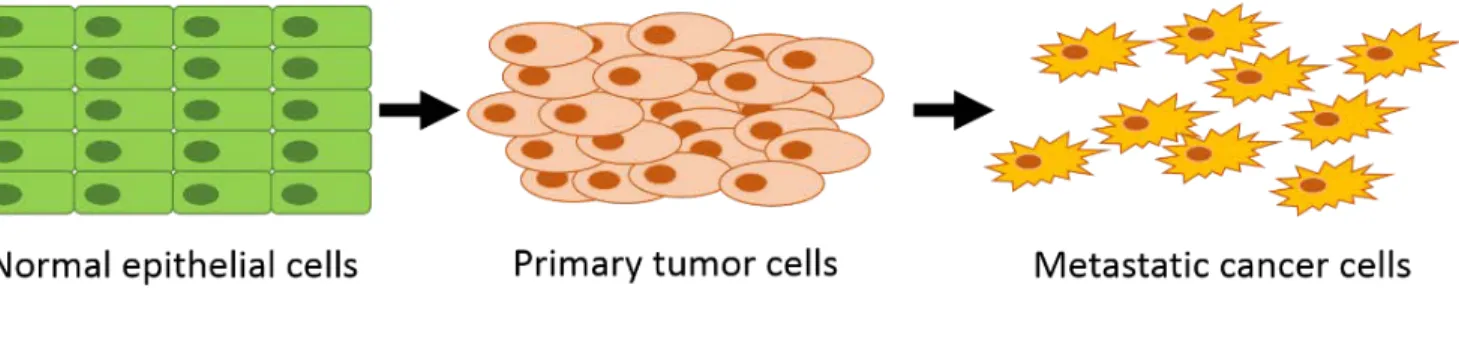
Cancer is the second leading cause of death worldwide, with over 12 million new diagnoses and more than 7 million deaths attributed to cancer in 2008 (1). Cancer is a heterogeneous group of diseases in which normal cells become transformed and exhibit uncontrolled growth (2). At the molecular level, cancer is driven by genetic mutations resulting in activation of oncogenes or inactivation of tumor suppressor genes (the concept of oncogenes and tumor suppressor genes has been reviewed extensively – a few helpful and relevant references may be found here (2-7)). These events lead to unchecked cell proliferation and survival resulting in tumor formation and, eventually, metastasis to foreign tissue sites and patient death (Figure 1-1) (8,9). Cancer therapy presents significant challenges for physicians, and there is an unmet need for targeted treatments with improved efficacy and safety profiles.
2
3 1.1.1 Triple negative breast cancer
Breast cancer is diagnosed in more than 230,000 women each year in the United States (10), and is the leading cause of cancer-related deaths among women worldwide (1). Approximately 15 percent of breast cancers are a subtype called triple negative breast cancer (TNBC), defined by lack of expression of three receptors: estrogen receptor (ER); progesterone receptor, and human epidermal growth factor receptor 2 (HER2) (11). TNBC is an aggressive cancer subtype with generally poor prognosis. Targeted treatments are used for ER-positive (e.g. Tamoxifen) and HER2-positive (e.g. Herceptin) breast cancers. These targeted treatments improve overall survival for patients with these sub-types (12). However, no targeted therapies are currently approved for TNBC (13). Although TNBC is sensitive to chemotherapy, the overall prognosis in TNBC is worse than non-TNBC sub-types due to high rates of relapse. TNBC patients also have a shorter overall survival rate relative to other breast cancer subtypes (14). Ongoing research has explored novel targets for TNBC therapy, and several targeted agents have progressed into clinical trials. However, there remains an unmet need for new targeted therapeutics with improved safety and efficacy to improve TNBC patient outcomes.
1.1.2 PI3K-AKT and RAS-RAF-MEK-ERK cancer signaling pathways
4
genes, often result in unrestrained activation of oncogenic signaling pathways. Several commonly mutated genes resulting in aberrant oncogenic signaling include PIK3CA, BRAF, KRAS, PTEN, and NRAS (see Table 1-1) (16). These particular mutations result in aberrant activation of PI3K-AKT and RAS-RAF-MEK-ERK, two commonly activated oncogenic signaling pathways in TNBC that are pertinent to the data presented herein (17). Cross-talk between these pathways enables cancer cells to compensate for inhibition of one pathway by activating the other pathway (17,18).
Table 1-1. Commonly mutated cancer genes. Selected genes from a list of the most commonly mutated cancer genes. See Lawrence et al. for complete list (16).
Aberrant activation of the RAS-RAF-MEK-ERK signaling pathway leads to unrestrained cell survival and proliferation. The RAS oncogene is one of the most commonly mutated genes in cancer (reportedly mutated in ~30% of all cancers) and the RAS signaling pathways have frequently been cited as targets for cancer therapy (19-21). Despite significant effort, scientists and drug developers have been unsuccessful at developing small molecule inhibitors of RAS, although recent advances in siRNA delivery technology may offer an effective alternative approach (22). However, RAS mutations are rare in TNBC (23). Several studies, including the
Gene name Protein name
Oncogene/ Tumor Suppressor
PIK3CA
Phosphatidylinositol 4,5-bisphosphate 3-kinase catalytic
subunit alpha Oncogene
BRAF V-Raf murine sarcoma viral oncogene homolog B1 Oncogene
KRAS V-KI-Ras2 Kirsten rat sarcoma viral oncogene homolog Oncogene
PTEN Phosphatase and tensin homolog Tumor Suppressor
5
Cancer Genome Atlas study, reported RAS mutations in less than two percent of all breast cancers (24-26). In addition to RAS mutations, RAF is commonly mutated in cancer and also contributes to activation of MEK-ERK signaling and cell proliferation (27).
6
7
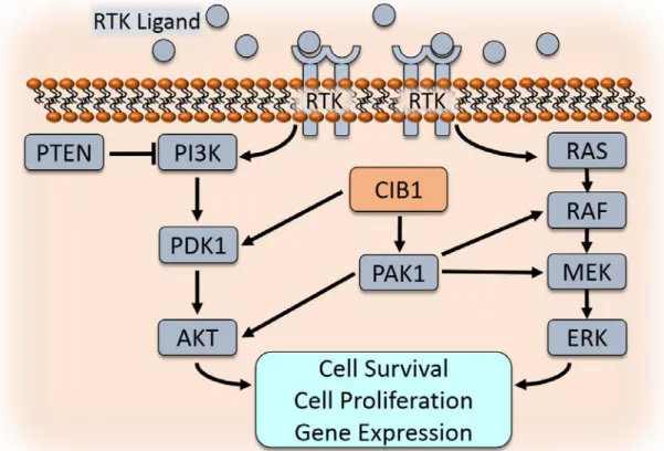
Due to the clinical relevance and the high rate of mutation in the PI3K-AKT and RAS-RAF-MEK-ERK signaling pathways, extensive work has been done to characterize these genes/pathways. Here I briefly summarize some of the relevant elements of these two important signaling cascades. Both RAS and PI3K signaling are stimulated by receptor tyrosine kinase (RTK) activation (28,37). For example, epidermal growth factor receptors (EGFR) are bound by extracellular epidermal growth factor (EGF), stimulating a conformational change and activation of EGFR. Active EGFR phosphorylates and activates its substrates, such as PI3K. PI3K stimulates activation of PDK1 via PIP3, leading to AKT activation as well as downstream activities including cell survival, cell proliferation, and gene expression (see Figure 1-5). PI3K activity is inhibited by the phosphatase PTEN. Cells harboring PTEN mutations or PTEN deficiency exhibit activated PI3K signaling, leading to uncontrolled cell survival and growth.
1.1.3 Challenges of inhibiting PI3K and RAS pathways clinically
Inhibitors targeting PI3K, AKT, RAF, and MEK have been tested in clinical studies, with unexceptional efficacy and safety profiles. Here I will briefly review these pathway inhibitors and some of the challenges inhibiting their development. RAF inhibitors have proven to be effective only in BRAF-mutant cancers (e.g. BRAFV600E-mutant melanoma) but not in cancers with active RAS-RAF-MEK-ERK driven by other mutations (38). Similarly, MEK inhibitors have had limited efficacy in clinical trials when used as single-agent therapies (39). At least thirteen individual MEK inhibitors have been tested clinically, but only one (Trametinib, GlaxoSmithKline) has shown clinical efficacy and received FDA approval. Like RAF inhibitors, Trametinib is indicated only for patients with BRAF-mutated melanoma (40,41).
8
Sciences) has received FDA approval for several indications in leukemia and lymphoma (43,44). PI3K inhibitors are likely to be most effective when used in combination with other inhibitors (i.e. chemotherapy, RTK inhibitors, and MEK inhibitors) (45,46). AKT inhibitors are also being developed and have demonstrated promising efficacy, but also have some safety concerns including skin rashes and hyperglycemia, and combination approaches are being explored (45-48). Due to the limitations of PI3K and AKT inhibitors in clinical studies, novel isoform-specific inhibitors, as well as inhibitor combinations, are being explored for clinical efficacy and safety.
9
explore alternative inhibitors, inhibitor combinations, and novel targets to safely and effectively inhibit these pathways for therapeutic benefit.
1.1.4 Oncogene and non-oncogene addiction
Many cancers in which PI3K-AKT and RAS-RAF-MEK-ERK pathways are activated may become entirely dependent on these pathways for cell survival and proliferation. The terms “oncogene addiction” and “tumor suppressor gene hypersensitivity” describe this dependency of cancer cells on particular genetic mutations or activation of oncogenic pathways to maintain the malignant phenotype (4,53). This addiction to a particular gene/protein provides a promising strategy for cancer treatment, as it suggests that targeted inhibition of a specific oncogene may be sufficient to impair cancer cell proliferation and induce cell death. Oncogene addiction results in a critical weakness that can be exploited to improve cancer therapy (53). For example, HER2-positive breast cancer is a breast cancer subtype driven by amplification of the RTK HER2, making it attractive for oncogene-targeted therapy. In 1998, the FDA approved trastuzumab (trade name Herceptin, Genentech), a monoclonal antibody targeted to the HER2 receptor, for the treatment of HER2+ breast cancer. Trastuzumab in combination with chemotherapy significantly reduces tumor size and improves overall patient survival compared to chemotherapy alone (54-56). Thus, targeting a specific oncogene is an effective and clinically-relevant method to treat cancer. Additional oncogenes and targeted oncogene inhibitors are being researched and developed as targeted cancer therapeutics.
10
is not mutated and unable to induce transformation, has begun to yield additional targeted treatment options (58,59). A key characteristic of non-oncogene addiction is the differential response in cancer versus normal cells. Cancer cells may become particularly reliant on, or addicted to, a non-oncogene to support proliferation and survival, whereas normal cells may tolerate loss of the same gene. For example, BRCA2-mutant cancers have a defect in double-strand DNA break repair and consequently are particularly sensitive to DNA damage inducing agents (e.g. cisplatin) (60,61). Interestingly, BRCA2 mutant cancer cells are also heavily reliant on single-strand DNA repair mechanisms. Inhibition of Poly-ADP-ribose polymerase (PARP1), a DNA single strand repair enzyme, has emerged as a viable treatment option in BRCA2-mutant cancers (62). PARP1 is not an oncogene, PARP1-/- mice are viable (63), and in the absence of genotoxic stress, PARP1 is not required for normal cell survival (64,65). Therefore, the dependence of BRCA2 mutant tumors on PARP1-mediated DNA repair is an example of non-oncogene addiction. PARP1 inhibitors are lethal in BRCA2 mutant cancer cells (62), and have been developed and successfully utilized in the clinic (66,67). Olaparib (Lynparza) received FDA approval in 2014 for patients with advanced ovarian cancer bearing BRCA mutations, and ongoing clinical trials are exploring the benefit of olaparib in BRCA-mutant breast cancer. These results confirm the clinical relevance of targeting non-oncogene addiction for therapeutic benefit.
New genes and proteins that function via oncogene and non-oncogene addiction to promote cancer cell survival and proliferation are being characterized. Identification of new targets leads to development of improved therapeutics against these novel targets, resulting in better outcomes for patients. There is a significant unmet need to identify novel therapeutic targets in TNBC and other cancers driven by PI3K-AKT and RAS-RAF-MEK-ERK signaling. This dissertation presents compelling evidence for CIB1 as a novel therapeutic target in TNBC.
11
CIB1 was discovered in 1997 as a binding partner of the integrin αIIb cytoplasmic tail (68). Over the past 18 years, CIB1 structure and function have been explored in cardiovascular disease, cancer, and other diseases. Here I will introduce CIB1 by reviewing CIB1 structure, binding partners, cell biology, and relevant diseases.
1.2.1 CIB1 structure
CIB1 is a 191 amino acid (22kDa) calcium-binding protein with an N-terminal myristoylation site (68-70). The structure of CIB1 was determined by multiple studies, utilizing techniques including NMR (71-74), circular dichroism (69), and X-ray crystallography (75,76). The current consensus CIB1 structure is primarily influenced by the crystal structure solved by Gentry et al (75), and NMR studies performed by Hans Vogel and colleagues (71). CIB1 is composed of 10 α-helices including 4 EF-hands, helix-loop-helix structures capable of divalent cation binding, which form the core structure of CIB1 (75). Two of these EF-hand domains bind divalent cation. Calcium binds tightly to EF-III (1.9 µM) and EF-IV (0.5 µM) (71,75). Mg2+ binds EF-III as well, but with lower affinity than Ca2+. Two additional auxiliary Ca2+-binding sites exist on the surface of the N-terminal domain and contribute to CIB1 folding. The N-terminal and C-terminal α-helices do not participate in the EF-hand structure. The C-terminal α-helix (H10) of CIB1 lays in a hydrophobic binding pocket and is proposed to undergo displacement to enable CIB1 interaction with integrin αIIb and other binding partners (71,72).
12
function. In summary, CIB1 is a small, divalent cation binding protein with a hydrophobic binding pocket capable of interacting with multiple binding partners.
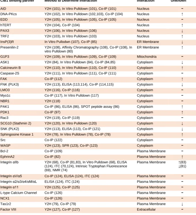
1.2.2 CIB1 Binding partners
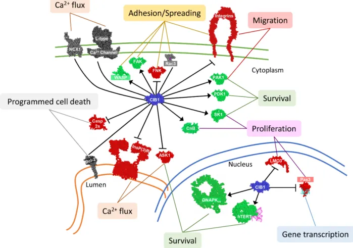
CIB1 was discovered as a binding partner of the integrin αIIb cytoplasmic tail, and has since been shown to bind many other proteins. CIB1 reportedly binds to at least 41 partners, making it a promiscuous and functionally diverse protein (see Table 1-2). Binding partners include nuclear, cytoplasmic, and transmembrane proteins (see Figure 1-3). It has been proposed that CIB1 is diffusely expressed throughout the cytoplasm and nucleus in normal resting cells, but that CIB1 can localize to a particular subcellular area or binding partner upon cellular activation or stimulation, such as fluctuations in intracellular Ca2+ concentration (70,78,79,81-85). Further work is necessary to fully characterize CIB1 interactions, including cell type specificity, and responses to signals and stimuli.
The interaction of CIB1 with several serine/threonine kinases, PAK1 and PDK1, as well as the aforementioned interaction between CIB1 and integrin cytoplasmic tails, are of particular interest to this discussion. CIB1-integrin binding will be introduced below, and discussed in detail in Chapter 3.
RAS-RAF-13
MEK-ERK signaling (87-91). Interestingly, PAK1 may also facilitate MEK-ERK pathway activation independent of its kinase activity by serving as a scaffold to facilitate RAF-MEK binding and activation (92). This scaffolding function was previously observed in PI3K-PDK1-AKT signaling, where PAK1 functions as a scaffold between PDK1 and AKT to facilitate AKT Threonine (Thr) 308 phosphorylation (93-95). It is probable that CIB1 regulates AKT and ERK signaling via its interaction with PAK1 (96).
CIB1 also binds and activates PDK1 (3-phophoinositide-dependent protein kinase 1), the enzyme that links PI3K and AKT signaling (97). PI3K generates PIP3, and PIP3 binding to PDK1 is necessary for PDK1 activity. Therefore, localization of PDK1 to the cell membrane is a prerequisite for PDK1 activation of its substrates, including phosphorylation of AKT at Thr 308 (98-100). Zhao et al discovered CIB1 as a PDK1 binding partner, and proposed that CIB1 shuttles PDK1 to the cell membrane, thereby facilitating PDK1-AKT interaction and AKT phosphorylation (97). Importantly, CIB1-PDK1 binding was demonstrated to be important for cancer cell survival and inhibition of stress-induced apoptosis (97).
14
Table 1-2. Table of CIB1 binding partners. A comprehensive list of CIB1 binding partners reported in the literature, including techniques used to detect the interaction, the cellular location of the interacting protein, and the role of CIB1 binding (e.g. activation).
CIB1 binding partner Method to Determine Interaction
Location of Interaction CIB1 effect (↑) Activates (↓) Inhibits (−) Interacts/ Unknown
AID Y2H (101), In Vitro Pulldown (101), Co-IP (101) Nucleus − DNA-PKcs Y2H (102), In Vitro Pulldown (102,103), Co-IP (104) Nucleus ↑ EDD Y2H (105), In Vitro Pulldown (105), Co-IP (105) Nucleus − hTERT Y2H (104), Co-IP (104) Nucleus ↑ PAX3 Y2H (106), In Vitro Pulldown (106) Nucleus ↓ TRF2 Y2H (103), In Vitro Pulldown (103) Nucleus ↑ InsP(3)R In Vitro Pullodwn (107), Co-IP (85) ER Membrane ↓ Presenilin-2 Y2H (108), Affinity Chromatography (108), Co-IP (108), In
vitro Pulldown (80)
ER Membrane ↑
G1P3 Y2H (109), In Vitro Pulldown (109), Co-IP (109) Mitochondria − ASK1 Y2H (84), In Vitro Pulldown (84), Co-IP (84,85) Cytoplasm ↓ Calcineurin B Y2H (110), In Vitro Pulldown (110), Co-IP (110) Cytoplasm ↑ Caspase-2S Y2H (111), In Vitro Pulldown (111), Co-IP (111) Cytoplasm − FAK Co-IP (112) Cytoplasm ↑ FNK (PLK3) Y2H (113), ELISA (113,114), Co-IP (114,115) Cytoplasm ↓ LMO3 Y2H (116), Co-IP (116) Cytoplasm − Myo1c Co-IP (117), In Vitro Pulldown (117) Cytoplasm − NBR1 Y2H (118) Cytoplasm − PAK1 Co-IP (86), ELISA (86), SPOT peptide assay (86) Cytoplasm ↑ PDK1 Co-IP (97) Cytoplasm ↑ Rac3 Y2H (119), Co-IP (119) Cytoplasm − SCG10 (Stathmin 2) Y2H (120), In Vitro Pulldown (120) Cytoplasm ↓ SNK (PLK2) Y2H (113), ELISA (113), Co-IP (121) Cytoplasm ↓ Sphingosine Kinase 1 Y2H (78), In Vitro Pulldown (78), Co-IP (78) Cytoplasm ↑ Src Co-IP (122) Cytoplasm − WASP Y2H (123), SPR (123), Co-IP (123) Cytoplasm − Bcl-2 Co-IP (109) Plasma Membrane − EphrinA2 Co-IP (82) Plasma Membrane ↑ Integrin αIIb Y2H (68), Co-IP (81,83), In Vitro Pulldown (68), ELISA
(124), ITC (70,124), Intrinsic Tryptophan Fluorescence (69), NMR (74)
Plasma Membrane ↑(83) ↓(81)
15
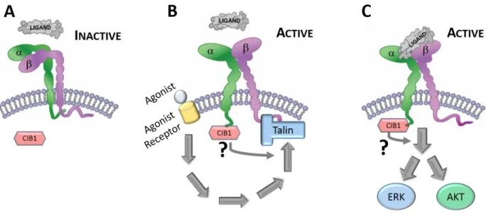
16 1.2.3 CIB1 in integrin αIIbβ3 signaling and function
Following identification of CIB1 as a binding partner for the platelet-specific integrin αIIb cytoplasmic tail, early studies from our lab and others focused on understanding the molecular and cellular consequences of this interaction. Platelets are anucleate circulating blood cells that become “activated” and contribute to thrombus/clot formation in response to vascular injury. As the major adhesion receptor on platelets and megakaryocytes (platelet precursor cells), integrin αIIbβ3 plays a critical role in the process of platelet-platelet adhesion (aggregation) and spreading. It was hypothesized that CIB1 may play an important role in platelet function by either regulating αIIbβ3 activation (inside-out signaling) or αIIbβ3 ligand binding and downstream signaling (outside-in signaling) (see Figure 1-4). Because platelets are not amenable to direct genetic manipulation, many of the early studies exploring CIB1 function utilized primary megakaryocytes and megakaryocyte-like cell lines to understand how CIB1 may regulate αIIbβ3 outside-in and/or inside-out signaling events.
Yuan, et al explored the role of CIB1 in αIIbβ3 inside-out signaling, and reported that overexpression of CIB1 in megakaryocytes completely inhibits agonist-induced fibrinogen binding, while overexpression of a CIB1 mutant that does not bind to the αIIb tail fails to suppress αIIbβ3 activation (81). In addition, RNAi depletion of CIB1 enhances agonist-induced αIIbβ3 activation, indicating that CIB1 is a negative regulator of αIIbβ3 activation (81). Taken together, the data from this study suggest that CIB1 is an inhibitor of integrin αIIbβ3 inside-out signaling.
17
complementary studies in Chinese Hamster Ovary (CHO) cells overexpressing integrin αIIbβ3. CIB1 overexpression in these cells resulted in increased cell spreading and cell migration on fibrinogen, reportedly through the interaction between CIB1 with Rac3 (119,122). Cumulatively, these studies suggest that CIB1 may promote integrin αIIbβ3 dependent outside-in signaling.
We and others used CIB1 knockout (CIB1-/-) mice to study the role of CIB1 in platelet function, with somewhat differing results from different groups. Naik, et al reported that CIB1-/- mice have impaired hemostasis (extended tail bleeding time). Because they found that CIB1-/- platelets are also defective in spreading on fibrinogen (the ligand for αIIbβ3) compared to WT platelets (130), and that blocking CIB1 with anti-CIB1 antibody decreased platelet spreading (83), they concluded that CIB1 is required for outside-in signaling. However, Denofrio, et al found no defects in CIB1-/- mouse hemostasis or platelet spreading (131). While much is known about the molecular interaction between CIB1 and integrin αIIb, further studies are required to elucidate the specific role of CIB1 in inside-out versus outside-in signaling, and to definitively determine whether CIB1 is necessary for hemostasis regulation.
18
19 1.2.4 CIB1 in cell migration
The role of CIB1 in cell migration has been explored in multiple contexts, and an interesting dichotomy has emerged with some studies concluding that CIB1 inhibits cell migration, and contradictory reports suggesting that CIB1 promotes cell migration. Several studies concluded that CIB1 expression resulted in increased cell migration on fibronectin in CHO, T47D, HUVEC, primary mouse endothelial cells, and megakaryocytes (122,128,133,134). One proposed mechanism through which CIB1 could promote cell migration is through activation of kinases ERK and PI3K (122). In contrast to these reports, Leisner, et al published that CIB1 inhibits cell migration on fibronectin in fibroblast cells via interaction with PAK1. CIB1 binds to PAK1 via the NH2-terminal autoinhibitory domain, thereby activating PAK1 and stimulating PAK1-dependent LIMK/cofilin signaling resulting in decreased actin polymerization, and consequently decreased migration (135). Although multiple studies have observed CIB1-dependent effects on cell migration, different studies have reported different outcomes on cell migration. It is possible that these discrepancies are due to varying cell types and/or fibronectin concentrations used. Nonetheless, these studies indicate that CIB1 plays an important role in regulating cell migration via activation of cellular signaling pathways.
1.2.5 CIB1 in calcium signaling
20
either Ca2+ or Mg2+. Although cation-dependent structural differences in CIB1 allude to functional consequences such as altered ligand binding, no significant difference in CIB1 binding to αIIb CT has been observed between Ca2+-CIB1 and Mg2+-CIB1 (70,74).
Several studies have explored how CIB1 regulates Ca2+ signaling, as well as how intracellular Ca2+ concentrations affect CIB1 function. Interaction of CIB1 with the inositol 1,4,5 triphosphate receptor (InsP3R) Ca2+ release channel was found to inhibit Ca2+ release from the endoplasmic reticulum (79). In separate studies, ASK1 was shown to bind competitively to CIB1 and decrease CIB1-InsP3R binding, resulting in increased Ca2+ release, and ROS-induced cell death (85). Interestingly, increased Ca2+ decreases CIB1-ASK1 binding, suggesting a potential feedback mechanism by which CIB1 may regulate, and be regulated by, intracellular Ca2+ release (84). This interplay between CIB1 and intracellular Ca2+ has potentially significant functional consequences. CIB1 binding to ASK1 inhibits ASK1 kinase activity, interferes with ASK1 binding to additional binding partners (e.g. TRAF2), and mitigates ROS-induced cell death in SH-SY5Y neuroblastoma cells (84). These results indicate an important role for CIB1 in the relationship between ASK1 and InsP3R in Ca2+-dependent signaling and maintenance of cell viability. Further studies are necessary to better understand the cellular consequences of Ca2+ concentration on CIB1 interactions and function. Although many details remain unknown, these results suggest that changes in intracellular divalent cation concentrations and binding to CIB1 are likely to influence CIB1 function.
1.2.6 CIB1 myristoylation
21
is an essential cellular process with implications in cancer and infectious diseases (137). Interfering with CIB1 myristoylation via mutations or protein fusions at the CIB1 N-terminus interferes with the cellular functions of CIB1 (78,79,97,108,110). Several publications concluded that CIB1 myristoylation is important to direct CIB1 to the membrane (79) and to facilitate shuttling of CIB1 binding partners, such as sphingosine kinase 1 (SK1) (78) and calcineurin B (CnB) (110), to the membrane. Some myristoylated proteins such as recoverin and visinin-like protein 1 (VILIP-1) behave as Ca2+/myristoyl switches, where the protein undergoes a conformational shift upon binding to Ca2+, leading to the exposure of the buried N-terminal myristoyl moiety to the intracellular environment (138,139). While no structural evidence exists that CIB1 behaves in such a manner, Jarman, et al reported that CIB1 functions as a Ca2+/myristoyl switch and shuttles SK1 to the cell membrane in an agonist- and Ca2+-dependent manner. CIB1 interacts with the calmodulin binding site of SK1 and is required for SK1-dependent generation of the anti-apoptotic signaling molecule sphingosine-1-phosphate to promote SK1-dependent cell survival (78). The authors proposed an interesting model, whereby cytoplasmic CIB1 binds to Mg2+ under basal conditions which prevents the interaction of CIB1 and SK1. Upon increased intracellular Ca2+, the myristoyl group is released, enabling CIB1 to shuttle various binding partners to the membrane. Despite some controversy over whether CIB1 functions as a Ca2+/myristoyl switch per se (79,80), these findings suggest a mechanism by which CIB1 can regulate binding partner function at the cell membrane. Further studies are necessary to determine whether CIB1 in fact functions as a Ca2+/myristoyl switch and to further define how CIB1 myristoylation affects cell biology and disease.
1.2.7 CIB1 in cell survival and proliferation
22
and cancer, as well as other diseases. The first reported observation of a CIB1-dependent effect on cell proliferation came in 2006, when Yuan, et al reported that MEFs derived from CIB1-/- embryos proliferated at a slower rate than WT MEFs (140). In subsequent studies, endothelial cells derived from CIB1-/- mice also showed reduced proliferation rates compared to WT controls, providing additional evidence for a compelling link between CIB1 and proliferation (133). Recently, several independent reports have suggested that CIB1 promotes cancer cell proliferation and survival by regulating oncogenic signaling pathways (78,84,85,96,141,142). Leisner et al. explored one potential mechanism through which CIB1 promotes proliferation and survival and found that CIB1 promotes the activation of ERK and AKT, essential components of two pro-proliferation and pro-survival signaling pathways frequently activated in breast cancer (96,143). The Ras-Raf-MEK-ERK and PI3K-AKT pathways are frequently activated in cancer cells (144). RNAi knockdown of CIB1 in MDA-MB-468 breast cancer cells and SK-N-SH neuroblastoma cells led to decreased ERK and AKT phosphorylation and induced translocation of GAPDH to the nucleus. Nuclear GAPDH either induces or correlates with several additional events, including the activation of histone variant γH2AX and checkpoint protein CHK1. These events are markers of, or directly lead to, DNA damage, dysregulation of cell cycle checkpoints, and induction of non-apoptotic cell death (96). It is likely that CIB1 promotes ERK and AKT activation via its interactions with PAK1 and PDK1 (see Figure 1-5). Additional studies provide evidence that CIB1 promotes ERK and AKT activation and signaling, supporting the mechanism proposed by Leisner, et al (82,122,135).
23
24 1.2.8 CIB1 in Cancer
Cancer is a heterogeneous disease caused by oncogene activation and/or tumor suppressor inactivation. Cancer cells become reliant on cell signaling pathways to promote cell survival and proliferation (53). For example, many basal-like breast tumors exhibit activated ERK and AKT signaling pathways (28,145,146). In addition, cancer cells may become reliant on non-oncogenes via non-oncogene addiction, a description of non-mutated non-overexpressed genes/proteins that are essential for cancer cell survival but dispensable for normal cells (58).
In addition to internal factors driving tumor cell survival and proliferation, growing tumors depend on a steady blood supply to provide nutrients and oxygen (147). As tumors grow, they stimulate new blood vessel growth, a process termed tumor-induced angiogenesis. Angiogenesis inhibitors have been researched and used clinically to prevent tumor growth and induce tumor regression (e.g. bevacizumab) (148,149). The role of CIB1 in tumor-induced angiogenesis has been explored using molecular, cellular, and in vivo models.
25
a reduced blood supply (150). These data suggest that CIB1 is critical for both tumor cell survival, as well as tumor-induced angiogenesis. CIB1 should be further interrogated in specific cancer subtypes to determine whether it might be a novel therapeutic target.
26
CHAPTER 2: CIB1 DEPLETION IMPAIRS CELL SURVIVAL AND TUMOR GROWTH IN
TRIPLE NEGATIVE BREAST CANCER
2.1 Introduction
Breast cancer is diagnosed in over 230,000 people each year in the United States (10). Approximately 16% of all new breast cancer diagnoses are triple negative breast cancer (TNBC), a subtype of breast cancer that lacks expression of estrogen receptor, progesterone receptor, and human epidermal growth factor receptor 2 (11). Many breast cancer therapies target one of these three receptors and are therefore ineffective for the treatment of TNBC.
In breast cancer, and other cancers, cell survival and cell proliferation are driven by oncogenic signaling pathways. A majority of TNBC cases are basal-like, and typically exhibit constitutively activated RAF-MEK-ERK and PI3K-AKT signaling pathways (11,151). Dual inhibition of both ERK and AKT signaling pathways has been identified as a promising approach to treat TNBC (151,152). However, preclinical and clinical studies have suggested that combined inhibition of both PI3K and MEK may improve efficacy at the expense of increased toxicity (17,45,51). New targeted therapies with enhanced efficacy and safety are necessary to improve patient outcomes (14,153).
27
serine/threonine kinase PAK1 (86,122). We recently showed that CIB1 depletion in two cancer cell lines (SK-N-SH neuroblastoma and MDA-MB-468 TNBC) disrupted both AKT and ERK signaling, resulting in the induction of a DNA damage response and a unique mechanism of non-apoptotic cell death (96).
Because of our initial observation that CIB1 is essential for MDA-MB-468 TNBC growth and survival in vitro, we hypothesized that CIB1 may have a broader role in TNBC and in tumor growth in vivo. Here we present evidence that CIB1 is necessary for proliferation and survival in TNBC cell lines with elevated AKT activation and/or low PTEN expression. We further demonstrate that CIB1 depletion results in dramatic TNBC tumor shrinkage in vivo. To gain further insight into the effects of CIB1 depletion, we present RNA sequence (RNAseq) analysis revealing that CIB1 depletion induces genetic programs that correlate with decreased proliferation and survival, and cell differentiation. We show that high CIB1 expression is not associated with susceptibility to CIB1 depletion or with TNBC patient prognosis. Taken together, these findings are consistent with the emerging theory of non-oncogene addiction, where a subset of TNBCs appear to be reliant on a non-oncogenic protein, CIB1, for cell survival and tumor growth. Our results further suggest that CIB1 may be a novel target for TNBC therapy.
2.2 Results
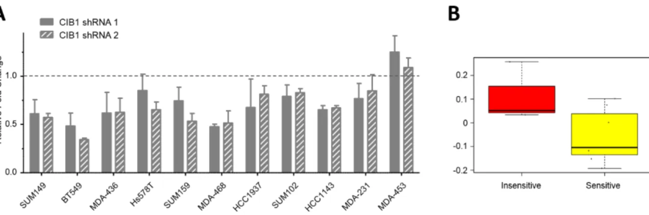
2.2.1 CIB1 depletion induces cell death in a TNBC cell line panel
28
significant, HCC1143 (Figure 2-1A, P=0.08), did exhibit a significant decrease in proliferation rate (Figure 2-2A, P<0.003). Ultimately, we observed some response in either cell viability, cell proliferation, or both, in nine out of eleven TNBC cell lines.
30
31
Therefore, we compared activated (phosphorylated) ERK (pERK) and AKT (pAKT) levels in CIB1-depleted versus control cells in the TNBC cell line panel (Figure 2-1B). We first noted that CIB1 depletion resulted in decreased pERK and pAKT in most cell lines. Interestingly, we observed that CIB1 depletion increased cell death in all eight cell lines that have relatively high basal levels of pAKT, and noted that seven out of these eight cell lines also had elevated pERK. However, pERK is also elevated in two out three cell lines that did not respond to CIB1 depletion and, therefore, ERK phosphorylation status does not appear to be a good predictor of sensitivity to CIB1 depletion.
32
may be a therapeutic approach to induce TNBC cell death regardless of CIB1 expression levels, particularly in cells with high basal levels of pAKT and/or low levels of PTEN.
33
34
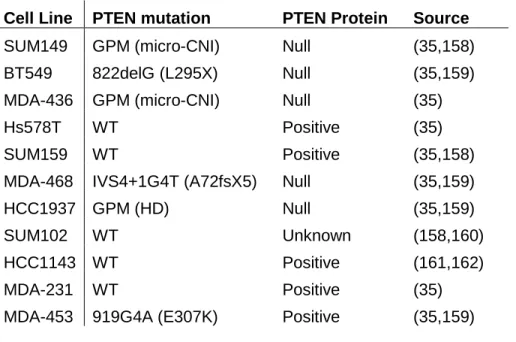
Cell Line PTEN mutation PTEN Protein Source
SUM149 GPM (micro-CNI) Null (35,158) BT549 822delG (L295X) Null (35,159) MDA-436 GPM (micro-CNI) Null (35)
Hs578T WT Positive (35)
SUM159 WT Positive (35,158)
MDA-468 IVS4+1G4T (A72fsX5) Null (35,159)
HCC1937 GPM (HD) Null (35,159)
SUM102 WT Unknown (158,160)
HCC1143 WT Positive (161,162)
MDA-231 WT Positive (35)
MDA-453 919G4A (E307K) Positive (35,159)
35
36
2.2.2 CIB1 depletion from MDA-MB-468 TNBC cells decreases proliferation and increases cell death
Data presented here and elsewhere demonstrate that CIB1 depletion increased cell death of MDA-MB-468 (MDA-468) cells (Figure 2-1) (96), but not in non-cancerous cells (133,163). While these data suggest that CIB1 may be a promising target for TNBC therapy, we sought in vivo validation. We utilized a doxycycline-inducible shRNA system to regulate CIB1 expression in MDA-468 tumor xenografts. MDA-468 cells were engineered to express either CIB1 shRNA (MDA-468-CIB1shRNA) or control (scrambled) shRNA (MDA-468-SCRshRNA) in response to the antibiotic doxycycline (Dox). MDA-468-CIB1shRNA cells treated with doxycycline showed significant depletion of CIB1 by Western blot (Figure 2-4C). Consistent with previous findings (96), CIB1 depletion decreased phosphorylation of ERK and AKT and increased phosphorylation of the DNA damage marker, γH2AX (Figure 2-4C).
37
39
Figure 2-4. CIB1 depletion decreases TNBC cell proliferation and increases cell death in
40
2.2.3 CIB1 is required for MDA-MB-468 xenograft tumor growth
To test whether CIB1 was necessary for TNBC tumor growth and survival in vivo we used a xenograft model and injected MDA-468-CIB1shRNA and MDA-468-SCRshRNA cells subcutaneously into the flanks of immunocompromised mice. Once tumors reached a volume of approximately 100 mm3, mice were randomized into groups receiving sucrose, or sucrose plus doxycycline, and tumor volume was monitored for 5 weeks. We observed a rapid arrest of tumor growth followed by a drastic decrease in tumor volume in CIB1-depleted tumors (Figure 2-4B). In contrast, control tumors continued to grow steadily throughout the treatment period. After 5 weeks, CIB1-depleted tumors were not visible compared to control tumors, which were visibly bulging from the flanks of the mice. Upon completion of the study, tumors were resected and weighed. The average mass of CIB1-depleted tumors was significantly smaller than control tumors (Figure 2-5C).
42
43
Figure 2-6. CIB1 depletion decreases pERK and pAKT and increases DNA damage in vivo.
44
2.2.4 PAK1 activation partially rescues cells from CIB1 depletion
45
Figure 2-7. Expression of constitutively active PAK1 partially rescues CIB1
46
2.2.5 CIB1 depletion induces genetic programs that reduce proliferation and survival
47
differential gene expression is a downstream cellular response to overcome the negative effects of CIB1 depletion, rather than a direct effect of loss of CIB1. Further experiments are required to follow up on this interesting observation.
Because CIB1 depletion induces MDA-468 cell death, we next examined the RNAseq data for differential expression of genes involved in cell survival and cell death. We identified 99 differentially expressed genes that were positively associated with increased cell death (several of these genes are listed in Figure 2-8C-D). For example, we observed a Log2 fold change of +4.1 for the gene DKK3 (Dickkopf-3). DKK3 is a secreted glycoprotein that potently inhibits Wnt signaling pathways (168), and has been characterized as a tumor suppressor. Interestingly, DKK3 expression is suppressed in breast cancer (169). Adenovirus-mediated delivery of the DKK3 gene to xenograft tumors resulted in tumor regression and induction of cell death in prostate, testicular, and breast cancer models (170-172). Taken together, these results suggest that CIB1 depletion-mediated upregulation of DKK3 may have therapeutic benefit in killing cancer cells and inducing tumor regression, and support the tumor regression data presented herein (See Figures 2-5 and 2-6).
48
another example of a cancer drug target gene downregulated upon CIB1 depletion. Tissue factor is normally considered in the context of the coagulation cascade, but has recently been shown to promote cancer progression, including breast cancer (178,179). Treatment with tissue factor inhibitor ixolaris reduced tumor growth and metastasis in mouse tumor models (180,181). Therefore, CIB1 depletion-induced knockdown of tissue factor provides another beneficial effect of targeting CIB1 in TNBC. These examples (glutathione-s-transferase and tissue factor) of CIB1 depletion-induced downregulated genes suggest that inhibition or knockdown of CIB1 may simultaneously inhibit multiple cancer drug targets.
50
51
2.2.6 CIB1 mRNA expression does not correlate with TNBC prognosis
52
53
2.3 Discussion
TNBC is a breast cancer subtype with generally poor prognosis and no available targeted treatment options (14). Two oncogenic pathways, RAF-MEK-ERK and PI3K-AKT, are aberrantly active in the majority of TNBC (151). Because CIB1 promotes both of these signaling pathways (96), we hypothesized that CIB1 might be essential to TNBC cell survival. The data presented here provide evidence that CIB1 depletion impairs cell survival in a majority of TNBC cell lines and shrinks TNBC xenograft tumors, suggesting that CIB1 may have a broad role in TNBC survival and tumor growth. Furthermore, dependence on CIB1 expression is associated with active AKT and/or low PTEN expression. PTEN mutation or deletion is significantly associated with incidence of basal-like breast cancer in mice and humans (35,185). These data suggest that CIB1 inhibition may be an effective therapeutic option for TNBC patients with PTEN-deficient tumors.
54
ERK activity, or via its interaction with SK1 or ASK1. Alternatively, perhaps the observed effects of CIB1 depletion rely on simultaneous aberration of a combination of these pathways. Further work should be done to explore potential synergism between these cell signaling pathways in the induction of cell death in response to CIB1 depletion.
Because CIB1 is essential for TNBC survival and tumor growth, we asked whether CIB1 expression is prognostic of TNBC patient survival. Recently CIB1 expression was reported to be relatively higher in hepatocellular carcinoma tumor center compared to non-tumorous liver tissues from 100 patient samples (141), as well as in breast cancer tissue compared to matched non-cancerous breast tissue from nine patient samples (114). We found no association between CIB1 mRNA expression and patient relapse-free survival in both TNBC and ER-negative breast cancer. In contrast to previous reports, our study used gene expression data from thousands of breast cancer patients across four established datasets (157,182-184). While the data presented here suggest that CIB1 expression is not prognostic in TNBC, it is possible that CIB1 does have prognostic implications in other types of cancer. Our results indicate that CIB1 expression is not predictive of TNBC patient prognosis, and further suggest that CIB1 overexpression does not promote tumorigenesis per se.
non-55
overexpressed gene/protein that is nonetheless essential to maintain oncogenic signaling pathways (58,59). For example, ATM-deficient tumor cells display non-oncogene addiction to the enzyme DNA-dependent protein kinase catalytic subunit (DNA-PKcs), and DNA-PKcs has been identified as a potential drug target in ATM-defective malignancies (191). Based on this example, our data suggest that PTEN-defective TNBC tumors may display non-oncogene addiction to CIB1, implicating CIB1 as a novel drug target in TNBC.
56
Figure 2-10. Proposed mechanism of CIB1 regulation of TNBC cell survival and potential
57
2.4 Methods
Cell lines and cell culture – Cell lines and cell culture conditions are listed in Table 2-2.
Cell Line Media
BT549 RPMI+10%FBS+10ug/mL Insulin HCC1143 RPMI+10%FBS
HCC1937 RPMI+10%FBS
Hs578T DMEM+10%FBS+10ug/mL Insulin MDA-231 DMEM+10%FBS
MDA-436 DMEM+10%FBS+10ug/mL Insulin MDA-453 DMEM+10%FBS
MDA-468 DMEM+10%FBS
SUM102 DMEM/F12+5%FBS+10ug/mL Insulin+1ug/mL Hydrocortisone SUM149 DMEM/F12+5%FBS+10ug/mL Insulin+1ug/mL Hydrocortisone SUM159 DMEM/F12+5%FBS+10ug/mL Insulin+1ug/mL Hydrocortisone ZR-75-1 RPMI+10%FBS+10ug/mL Insulin
SKBR3 McCoy’s5a+10%FBS
ME16C MEBM (MEGM Bullet Kit: MEGM+BPE+hEGF+Hydrosortisone+Insulin All cell lines were grown at 37ᵒC in 5% CO2 humidified air.
Table 2-2. Cell lines and cell culture conditions used in TNBC Panel. Table used with permission from (155).
58
RNAseq analysis – MDA-468_SCRshRNA and MDA-468_CIB1shRNA cells were treated with doxycycline for <96 hours. After removing dead cells, RNA was isolated from viable cells (RNeasy kit, Qiagen, Venlo, Netherlands), and cDNA generated (QuantiTect Reverse Transcription kit, Qiagen). cDNA was sequenced at the UNC High Throughput Sequencing Facility on an Illumina HiSeq 2000 (Illumina, San Diego, CA). Differential gene expression analysis was performed using DESeq2 (192) and differentially expressed genes were selected based on Log2 fold change ≥ ± 2 and Benjamini-Hochberg adjusted p-value < 0.05. Lists of differentially expressed genes are displayed in Appendices A (upregulated genes) and B (downregulated genes). Differentially expressed genes were analyzed using Ingenuity Pathway Analysis (Qiagen). The median-centered gene expression dataset and methods from Prat et al (156) were used for Significance Analysis of Microarrays on the CIB1 KD sensitive versus insensitive cell lines, and for the identification of cell line Differentiation Scores; both of these analyses were performed with R version 3.1. To identify other gene signatures with similar profiles in human breast tumors (182), 10,508 gene signatures were retrieved from the GSEA database and via manual curation, each signature score was identified for each tumor by taking the average value of all signature genes within the median-centered gene set, then Pearson Correlation Values were obtained in Excel contrasting the CIB1 KD signature with all signatures.
Colony formation assay – MDA-468-control and -CIB1shRNA cells were treated ± 1 μg/ml Dox for 48 hours prior to plating at a density of 2000 cells/well. Cells were allowed to grow 9 days in the absence or presence of Dox, with media changes every 4 days. Cells were stained with crystal violet (0.05% w/v in 4% formaldehyde) (Sigma, St. Louis, MO) and colonies counted using ImageJ software.
RNA interference – Cells were transduced with either control shRNA
(ACCGCTCTTCACACAGATCCTCTTCAAGAGAGAGGATCTGTGTGAAGAGCTTTTTC), CIB1
59
(ACCGTGCCCTTCGAGCAGATTCTTCAAGAGAGAATCTGCTCGAAGGGCACTTTTTC), or CIB1 shRNA 2, (CAGCCTTAGCTTTGAGGACTTCTCGAGAAGTCCTCAAAGCTAAGGCTG). For inducible RNAi experiments, MDA-468 cells were transduced with either inducible control shRNA (GCTACACTATCGAGCAATTTTGGATCCAAAATTGCTCGATAGTGTAGC) or inducible CIB1 shRNA (GGCTTAGTGCGTCTGAGATTTGGATCCAAATCTCAGACGCACTAAGCC) using the pLV-H1-TetO-Puro lentiviral plasmid (Biosettia, San Diego, CA). Lentiviral particles were prepared as described previously (96).
Cell proliferation and cell death assays – MDA-468-CIB1shRNA and MDA-468-SCRshRNA cells were plated at 3x10⁵ cells per 10cm dish, treated ± Doxycycline, and allowed to grow for 3, 5, or 7 days. Cells were harvested by trypsinization and counted using a hemacytometer. Cell viability was determined by trypan blue exclusion.
Flow cytometry – Cells were grown in the presence of Doxycycline for 5 days. MDA-468-CIB1shRNA and MDA-468-SCRshRNA cells were detached using 2 mM EDTA and 5x10⁵ cells were stained with FITC Annexin V (BD Biosciences), 7-AAD (eBioscience), or the combination of FITC Annexin V and 7-AAD according to the BD FITC Annexin V Apoptosis Detection Kit protocol. Data was collected from 104 cells using a FACS Canto flow cytometer (BD), then analyzed using FACS Diva software (BD).
Histology and microscopy – Fixed tumors were paraffin embedded, slides were cut and H&E staining completed by the Lineberger Comprehensive Cancer Center Animal Histopathology Core Lab. TUNEL assay was performed according to manufacturer’s protocol (Promega). Microscopy was performed using an Olympus BX61 Wide Field Microscope at the UNC Microscopy Services Laboratory.
60
Tumors were ranked from lowest to highest based on CIB1 expression levels and divided into two equal groups based on CIB1 gene expression.
Constitutively active PAK1 expression – Constitutively active PAK1 (caPAK1) harboring activating mutations L107F and T423E was cloned into the pCDH-EF1-MCS-T2A-Puro vector (System Biosciences, Mountain View, CA). Lentiviral particles were produced as described previously (96). MDA-468 cells were transduced with caPAK1, then after 48 hours cells were transduced with either CIB1 shRNA or Control shRNA. Cells were harvested 96 hours after shRNA treatment and counted. Cell viability was determined by trypan blue exclusion.
Western blotting – Cell and tumor lysates were prepared using CHAPS lysis buffer (20 mM HEPES, 150 mM NaCl, 5% v/v glycerol, 10 mM CHAPS, 0.1 mM CaCl2, 0.05 mM MgCl2, 20 mM NaF, 10 mM β-Glycerophosphate, 0.1 mM Sodium Pervanadate, 1.25 mg/mL N-Ethylmalemide, and Protease Inhibitor Cocktail III (BioVision). Protein concentration of tumor lysates was determined using BCA Assay (Thermo Scientific), equal amounts of total protein were separated by SDS-PAGE, transferred to PVDF, and incubated with indicated primary antibodies overnight at 4oC, and visualization was performed using ECL2 (Pierce). The following antibodies were used: CIB1 chicken polyclonal antibody was produced as described previously (86); antibodies against pAKT473 (9271), pERK (9101), total AKT (4691), and γH2Ax (9718) were obtained from Cell Signaling Technology (Danvers, MA); ERK polyclonal 94), PTEN 9145) and PAK1 (sc-882) antibodies were purchased from Santa Cruz Biotechnology (Dallas, TX); Rac monoclonal antibody was purchased from EMD Millipore (Billerica, MA).
61
CHAPTER 3: CIB1 BINDS α-INTEGRIN CYTOPLASMIC TAILS IN VITRO AND IN CELLS
3.1 Introduction
Integrins are transmembrane cell adhesion receptors that form a critical link between the cellular cytoskeleton and extracellular matrix, and also have important roles in cell adhesion, migration, and signal transduction (194). Normal integrin function is essential in development, immune response, hemostasis, and cancer (194-198). Therefore, integrin structure, function, and binding partners have been studied intensively toward increased understanding of relevant biological processes and diseases.
3.1.1 Integrin activation and signaling is regulated by cytoplasmic binding proteins.
Integrin-dependent signaling pathways are precisely regulated to maintain physiological behavior. Integrins are heterodimeric receptors composed of an α subunit and a β subunit, and have a large extracellular domain, a single transmembrane domain, and a short cytoplasmic tail. Transmission of signals through the cell membrane via integrins is regulated by the binding of cytoplasmic proteins to the integrin tails. For example, kindlin and talin are cytoplasmic proteins that bind to integrin β-subunit cytoplasmic tails and have important roles in integrin activation (199-201).
3.1.2 CIB1 may modulate integrin-dependent cell function and signaling.
62
(70,108,111,118). CIB1 binding to αIIb regulated integrin αIIb agonist-induced signaling and function (81). Additional studies revealed that CIB1 bound to αIIb in a highly conserved domain spanning the transmembrane and cytoplasmic domains of the integrin (69,70). Because CIB1 binds a conserved region of αIIb and is ubiquitously expressed, we hypothesized that CIB1 may bind additional integrins and have a broader role in integrin biology.
3.1.3 Exploring CIB1-integrin binding in vitro and in cells
The binding of CIB1 to α-integrin cytoplasmic tail peptides was tested by several in vitro binding assays. The Kd for CIB1 binding to 8 different integrin peptides was determined by isothermal titration calorimetry, with a maximum Kd of 23.5μM (αM) and a minimum Kd of 0.9μM (α2), and the interactions were further tested by competitive binding assays to measure the ability of various α-integrin peptides to compete with αIIb for CIB1 binding (124). These results demonstrated that CIB1 is capable of binding to multiple α-integrin subunits. I asked whether CIB1-integrin binding is physiologically relevant, and explored this question through biochemical and cell-based assays. Here I present evidence that CIB1 interacts with specific residues in the transmembrane domain of the integrin cytoplasmic tail, and that CIB1 can access these residues in the presence of a cell membrane. I present additional data to support the functional consequences of CIB1 binding to integrin in cells. Finally, I discuss the relevance of these findings to cell biology and to broadly understanding CIB1 function in human health and disease.
3.2 Results
3.2.1 CIB1 binds α-integrin tails via a conserved region spanning the transmembrane and cytoplasmic tail domains of the integrin
63
64
Figure 3-1. CIB1 binding to integrin αV cytoplasmic tail peptides is disrupted by alanine
65
3.2.2 CIB1 can access integrin αV membrane proximal residues in the presence of a physiologically relevant lipid bilayer
Because CIB1 binds to αV at a region spanning the cytoplasmic and transmembrane domains, it is possible that the cell membrane physically impedes CIB1 from accessing its αV binding domain inside a cell. We hypothesized that CIB1 can access its binding site on the integrin, even when the integrin is embedded in a lipid bilayer membrane. To test this hypothesis we embedded integrin αV polypeptides into nanodiscs, soluble lipid bilayer discs, and tested CIB1 binding to integrin αV via pulldown assays (Figure 3-2).
66
67
Figure 3-3. CIB1 binding to membrane-embedded α-integrin subunits is disrupted by
68
Although preliminary results from CIB1-nanodisc binding assays were encouraging, we encountered technical hurdles that impeded further experimentation and data collection. These technical challenges stem from the goal to reduce non-specific binding, a challenge inherent in binding assays. Several attempts were made to reduce non-specific binding of CIB1 to empty nanodiscs or free protein in these assays, to enable accurate measurement of CIB1 binding to membrane-embedded integrin. I will outline here several challenges, respective troubleshooting steps, and outcomes.
69
70
Testing for differential binding of CIB1 to WT or mutant integrin peptides embedded in nanodiscs introduced additional challenges and troubleshooting. The nanodiscs consisted of purified proteins MSP1 (membrane scaffold protein) and integrin peptides linked to MBP (maltose binding protein) organized with lipids into nanodisc complexes. Integrins were linked to MBP to facilitate expression and purification from E. coli. During nanodisc preparation, a mixed population of products was produced including integrin nanodiscs, empty nanodiscs, and free protein in solution (Figure 3-4). Because both MSP1 and MBP had a hexahistidine tag at the N-terminus of the protein, it was impossible to distinguish between the different populations during preparation, purification, and binding assays. This mixed population made experimental results difficult to interpret. To overcome these challenges, I attempted to cleave the MBP from purified MBP-integrin using TEV (Tobacco Etch Virus) protease followed by a negative selection using amylose resin to bind His-MBP. While this technique reduced the amount of MBP-integrin in solution, the cleavage reaction and purification did not reach 100% efficiency, as evidenced by the signal from integrin, MBP and MBP-integrin in the flow through, wash, and bead samples (Figure 3-5A).
71
(data not shown), approximately 7x the predicted molecular weight of MBP-integrin, which differs from the molecular weight observed by gel (Figure 3-5). Therefore, size exclusion chromatography was was unable to effectively separate empty nanodiscs, MBP-integrin nanodiscs, and free MBP-integrin in solution. Despite multiple approaches, a homogenous population of integrin-embedded nanodiscs was never achieved and the results of binding assays were inconclusive.
72
73
3.2.3 Integrin mutants may affect CIB1-integrin function in cells
CIB1 regulates signaling through pathways that are also activated downstream of ligand binding to integrins (196). For example, our lab recently found that CIB1 is an upstream regulator of both ERK and AKT signaling pathways (96). However, it is unknown whether αv-dependent activation of these same pathways requires CIB1 or whether it is a parallel event, independent of CIB1. CIB1 is known to regulate activation of the related integrin αIIbβ3 (81), but the effect of CIB1 on αvβ3 activation or downstream signaling has never been studied.
In addition to its interaction with integrin αIIb (69,70,81), we demonstrated that CIB1 co-immunoprecipitates with integrins αV and α5 from whole cell lysates (Figure 3-6) (124). Since I found that introduction of amino acid substitutions in the CIB1 binding domain on the integrin disrupted CIB1-integrin binding in vitro, I asked whether these mutations to the full-length integrin had any effect on CIB1-integrin binding and function in cells. I hypothesized that cells expressing mutant integrin αV 4A would have impaired signaling and integrin-mediated adhesion, compared to cells expressing αV WT.
74
75
76
3.2.4 CIB1 binding to integrin αV may affect cell signaling and proliferation
Integrin αVβ3 binds to the extracellular matrix component vitronectin. To test whether disruption of CIB1-αV binding affects cell adhesion, I plated M21L-αV WT and M21L-αV 4A cells on vitronectin and measured cell adhesion using a centrifugation-based adhesion assay. I found no difference in M21L cell adhesion to vitronectin after 30, 60, or 90 minutes between M21L-αV WT and M21L-αV 4A cells (Figure 3-8A).
In addition to cell adhesion, integrins transmit signals from the extracellular environment to the inside of the cell and thereby promote cellular processes including cell proliferation. To determine whether CIB1-αV binding affects cell proliferation, I plated equal numbers of M21L-αV WT/2A/4A and then measured total cell number after three days of growth. While the results show that there is not a statistically significant difference in cell proliferation between the cell types, the results trend towards a decrease in proliferation in cells expressing αV 2A, and trend toward an increase in proliferation in cells expressing αV 4A (Figure 3-8B).
77
78
3.2.5 CIB1 binds to different integrin α subunits via distinct residues in the CIB1 hydrophobic pocket
CIB1 interacts with α-integrin subunits via a hydrophobic binding pocket (71,72). The C-terminus of CIB1 forms an α-helix that, in the absence of a binding partner, lays in the hydrophobic pocket (75). The CIB1 structure based on a homology model of calcineurin B, an EF-hand containing protein relative of CIB1, demonstrates how the C-terminal helix may undergo a conformational shift to enable integrin binding to the CIB1 hydrophobic pocket (207). I utilized this structure to explore CIB1 binding to α-integrin peptides.
79