Viral metagenomics, protein structure, and reverse genetics: Key strategies
for investigating coronaviruses
Bryan A. Johnson
a, Rachel L. Graham
b, Vineet D. Menachery
a,⁎aDepartment of Microbiology and Immunology, University of Texas Medical Branch, Galveston, TX, USA bDepartment of Epidemiology, University of North Carolina at Chapel Hill, Chapel Hill, NC, USA
A B S T R A C T
Viral metagenomics, modeling of protein structure, and manipulation of viral genetics are key approaches that have laid the foundations of our understanding of coronavirus biology. In this review, we discuss the major advances each method has provided and discuss how future studies should leverage these strategies synergis-tically to answer novel questions.
1. Introduction
The Severe Acute Respiratory Syndrome (SARS) epidemic
fi
rst
emerged in southern China in late 2002 and rapidly spread world-wide,
causing 8096 con
fi
rmed cases in 27 countries and resulting in 774
deaths (
World Health Organization, 2004
). De
fi
ned as a novel
cor-onavirus (CoV) (
Drosten et al., 2003; Ksiazek et al., 2003; Peiris et al.,
2003a; Rota et al., 2003
), SARS-CoV was generally agreed to have
originated in bats and highlighted the risk posed by viruses emerging
from zoonotic sources (
Guan et al., 2003; Lau et al., 2005
;
Li et al.,
2005b
;
Tang et al., 2006
;
Tu et al., 2004
). A decade later, Middle East
Respiratory Syndrome CoV (MERS-CoV) was identi
fi
ed as the causative
agent of another ongoing outbreak (
Zaki et al., 2012
). In the
fi
ve years
since, signi
fi
cant progress has been made in understanding the origins,
biology, and emergence potential of CoVs. These studies have been
aided by advancements in three critical research areas: viral
metage-nomics, structural modeling studies, and reverse genetics. In this
re-view, we detail novel insights recently de
fi
ned by advances in each
approach and discuss their impacts on our understanding of CoV
in-fection and emergence. We also consider how these strategies can be
integrated to better prepare for the next emergent CoV strain.
2. Exploring an unknown frontier
Viral metagenomics has greatly expanded the scope and
under-standing of CoVs. Before 2002, the CoV family consisted of a relatively
modest number of viruses infecting the airway or the fecal-oral tracts.
With the only known human CoV strains causing mild disease, the
fa-mily had not been considered a signi
fi
cant threat to human public
health. In 2002, SARS initially presented itself as an atypical
pneumonia for which no known causal agent could be determined
(
Peiris et al., 2003b
). Eventually, the novel CoV was isolated from
in-fected patients and sequenced, demonstrating SARS to be caused by a
genetically distinct CoV of unknown origin (
Peiris et al., 2003a; Rota
et al., 2003
). With the earliest cases of SARS occurring in food service
workers handling exotic animals, initial studies focused on surveying
animals in live markets for the presence of SARS-CoV progenitors using
traditional viral discovery methods, including seropositivity studies,
isolation in culture, visualization of virions using electron microscopy
(
Guan et al., 2003; Tu et al., 2004
). Such viral discovery e
ff
orts led to
the identi
fi
cation of a SARS-CoV strain in a Himalayan palm civet that
shared 99.8% nucleotide identity with the epidemic strain (
Guan et al.,
2003
). Later, the observation that neither farmed nor wild civets
har-bored SARS antibodies outside of live animal markets led to the
in-vestigation of other zoonotic sources for SARS-CoV (
Poon et al., 2005;
Tu et al., 2004
). Studies quickly identi
fi
ed SARS progenitors circulating
in bats belonging to the
Rhinolophus
genus (commonly referred to as
horseshoe bats). Full genome comparisons determined that these
pro-genitor strains had similar genome organization to and a high
nucleo-tide sequence identity (88
–
92%) with SARS-CoV, suggesting that the
epidemic strain emerged from these bat CoV populations (
Lau et al.,
2005
;
Li et al., 2005b
;
Ren et al., 2006
;
Tang et al., 2006
). Together,
these studies established the classic model of SARS-CoV emergence,
whereby civets initially infected with SARS-CoV served as intermediate
hosts, leading to the generation of an adapted strain capable of human
infection.
The discovery of the progenitor SARS-like CoV strains circulating in
Chinese bat populations led to a global e
ff
ort to identify and de
fi
ne the
phylogenetic relationships of the Coronaviridae family. Over the past
15 years, dozens of animal populations have been surveyed, and the
⁎Correspondence to: University of Texas Medical Branch, 301 University Blvd., Route 610, Galveston, TX 77555-0610, USA.
E-mailaddress:[email protected](V.D.Menachery).
https://doi.org/10.1016/j.virol.2017.12.009
Received 29 September 2017; Received in revised form 4 December 2017; Accepted 11 December 2017
Available online 24 December 2017
full-length genomes of numerous novel CoVs have been identi
fi
ed,
greatly expanding the CoV family tree (
Drexler et al., 2014
). In contrast
to the identi
fi
cation of SARS-CoV which relied on traditional discovery
methods, the expansion of the CoV family tree has largely occurred
through viral metagenomics, that is the through the direct examination
of CoV genetic material obtained from environmental samples
(
Edwards and Rohwer, 2005; Simmonds et al., 2017
). Many of these
studies have relied on PCR based assays targeting conserved CoV
se-quences such as the RdRp gene (
Drexler et al., 2014
); the advent of
inexpensive high-throughput deep sequencing methods has and will
likely increasingly be exploited to study CoV populations (
Alagaili
et al., 2014; Anthony et al., 2013; Briese et al., 2014; Cotten et al.,
2013a, 2013b, 2014; Donaldson et al., 2010
). Currently, the
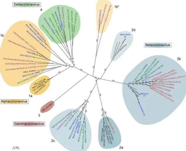
Cor-onaviridae family is divided into 4 unique clades designated the Alpha-,
Beta-, Gamma-, and Deltacoronaviruses (referred to hereafter by their
historical designation as Groups 1
–
4, respectively) (
Fig. 1
) and include
viruses known to infect humans, bats, other mammals, and several
avian species. Among these, only CoVs designated in black were
iden-ti
fi
ed prior to the emergence of SARS-CoV. During the outbreak
(2002
–
2005, red) and its immediate aftermath (2006
–
2011, green), a
number of SARS-CoV-related and progenitor strains were identi
fi
ed and
formed the core of the new group 2b branch. Similarly, identi
fi
cation of
novel bat CoV sequences, including HKU4, HKU5, and HKU9-CoVs,
populated newly formed groups. Likewise, the discovery of both
HCoV-NL63 and HCoV-HKU1, which cause minor diseases in humans,
expanded upon CoV groups that already existed. Together, viral
me-tagenomic studies in the wake of the SARS-CoV epidemic provided the
fi
rst robust look into the existing CoV phylogeny and provided a
fra-mework for understanding the sources of CoV emergence.
This expanded phylogeny born from metagenomic studies of
SARS-CoVs allowed for the rapid identi
fi
cation of the MERS-CoV as a group
2C CoV. Similar in sequence to HKU4 and HKU5-CoV, this novel human
CoV was quickly distinguished from SARS-CoV; HKU4 and HKU5 were
used as reagents to verify and characterize the new strain (
Agnihothram
et al., 2014a, 2014b
). Further study led to the determination that
HKU4, but not HKU5, could bind the MERS-CoV receptor, human
di-peptidyl peptidase 4 (DPP4) (
Wang et al., 2014
). Just as SARS-CoV
infection was traced to food service workers handling exotic animals,
the observation that many MERS patients had been in contact with
dromedary camels led to the search for MERS-CoV progenitors in camel
populations (
Azhar et al., 2014; Reusken et al., 2013
). Camel herds
throughout the Middle East were found to have MERS-CoV neutralizing
antibodies and to harbor CoVs with nearly identical sequence to
MERS-CoV (
Azhar et al., 2014; Haagmans et al., 2014; Hemida et al., 2014;
Reusken et al., 2013
). Examination of historical serum samples
sug-gested that MERS-CoV had been present in dromedaries since the 1980s
and likely originated in Eastern Africa, traveling to Saudi Arabia via the
camel trade (
Muller et al., 2014
). Together, the emergence of
MERS-CoV highlighted the utility of expanding the MERS-CoV phylogeny through
viral metagenomic studies.
therapeutic treatment. The CoV S protein is subdivided into an S1
do-main, primarily responsible for receptor binding, and an S2 dodo-main,
which is responsible for the fusion of the viral and cellular membranes
(
Lu et al., 2015
). In 2005, the crystal structure of the SARS-CoV
re-ceptor-binding domain (RBD) bound to human ACE2 indicated the
presence of two subdomains, a core domain and the receptor-binding
motif (RBM). Interacting with the N-terminal lobe of human ACE2, 14
RBM residues interact with 18 residues on human ACE2 to promote
binding (
Li et al., 2005a
). Subsequent studies demonstrated that the
primary barriers to host transition are di
ff
erences in ACE2 sequences
between species, necessitating changes in both topology and charge
within the RBM (
Li, 2008; Li et al., 2005c; Qu et al., 2005; Wu et al.,
2012
). Importantly, ine
ffi
cient binding to both bat and mouse ACE2 by
the epidemic SARS-CoV strains suggested that mutations were required
for emergence (
Frieman et al., 2012; Hou et al., 2010; Li, 2013; Roberts
et al., 2007
). Aided by the solved SARS-CoV S structure, epitope
map-ping of previously known SARS-CoV monoclonal neutralizing
anti-bodies (mAbs) revealed that they bound primarily to the RBD, likely
disrupting interaction with ACE2 (
Cao et al., 2010; He et al., 2006,
2005b; Lu et al., 2004
). In concordance, vaccine experiments with the
SARS-CoV RBD induced neutralizing antibodies and protected against
viral challenge (
Du et al., 2007; He et al., 2005a, 2004; Zakhartchouk
et al., 2007
). In addition, mAbs targeting the RBD were also developed
for prophylaxis; however, these mAbs were susceptible to escape
mu-tations and lacked neutralization capacity against zoonotic SARS-CoV
strains, thus limiting their value (
Rockx et al., 2008, 2010; Sui et al.,
2014
). Coupled with the identi
fi
cation of additional SARS like-CoVs
circulating in bat populations (
Lau et al., 2005
;
Li et al., 2005b
;
Ren
et al., 2008
;
Tang et al., 2006
), the initial therapeutics developed to
target the SARS-CoV RBD are unlikely to protect against newly
emer-gent infections. However, these structural studies have provided
im-portant insights that have informed both studies of CoV emergence and
therapeutic treatments against future emergent strains.
As was the case for viral metagenomics, structural studies
ex-amining SARS-CoV S provided a blueprint for investigations into the
MERS-CoV S. The MERS-CoV RBD was rapidly identi
fi
ed, and its crystal
structure was solved (
Chen et al., 2013; Lu et al., 2013; Wang et al.,
2013
). The MERS-CoV RBD can also be divided into two subdomains:
an RBM that binds the MERS-CoV receptor, DPP4, and a core domain
with remarkable structural similarity to the SARS-CoV core domain.
Eighteen amino acids within the RBM interact with 13 residues on
DPP4 to promote binding (
Lu et al., 2013; Wang et al., 2013
). While
di
ff
erences in host sequences are again a barrier, DPP4 is relatively
conserved among mammals (
Barlan et al., 2014; Falzarano et al., 2014;
Müller et al., 2012; Raj et al., 2014; van Doremalen et al., 2014
).
Common small animal models are a notable exception, with mice, rats,
hamsters, and ferrets all encoding DPP4 proteins that cannot support
infection, constituting a signi
fi
cant barrier to MERS-CoV research
(
Barlan et al., 2014; Coleman et al., 2014; de Wit et al., 2013; van
Doremalen et al., 2014
). Like SARS-CoV, the MERS-CoV RBD is also a
strong immunogen; several vaccine studies have demonstrated its
po-tential to induce neutralizing antibodies (
Du et al., 2013a, 2013b; Mou
et al., 2013
). Together, these observations illustrate how structural
prediction can be utilized for the development of vaccines and
neu-tralizing antibodies.
Recently, advances in structural studies have produced a wealth of
new CoV S structures (
Kirchdoerfer et al., 2016; Pallesen et al., 2017;
Walls et al., 2016a, 2016b; Yuan et al., 2017
). While previous e
ff
orts
had been made to explore CoV S proteins, these studies had elucidated
only portions of S, including the post-fusion core and RBDs bound to
receptors. While cryo-EM studies of SARS-CoV virions provided insights
into the S glycoprotein, the lack of high-resolution CoV S trimer
structures had limited progress in understanding entry and infection
(
Beniac et al., 2007; Neuman et al., 2006
). Several groups recently
overcame this barrier by fusing trimerization motifs into the CoV S
(
Kirchdoerfer et al., 2016; Pallesen et al., 2017; Walls et al., 2016a,
While
a
number
of
studies
had
provided
evidence
for
SARS-CoV's
origin
in
bats,
viral
metagenomic
surveys
continued
to
add
detail
to
the
CoV
tree.
Despite
having
high
sequence
identity,
potential
SARS-CoV
progenitors,
such
as
HKU3,
had
<
70%
amino
acid
identity
within
the
S1
domain
of
the
spike
(S)
protein
(
Lau
et
al.,
2005
;
Li
et
al.,
2005b
;
Ren
et
al.,
2006
;
Tang
et
al.,
2006
)
and
could
not
bind
either
the
civet
or
the
human
receptor,
angiotensin-converting
enzyme
2
(ACE2)
(
Lu
et
al.,
2015;
Ren
et
al.,
2008
).
However,
recent
surveys
in
East
Asia
further
expanded
the
CoV
tree
and
identi
fi
ed
several
new
group
2B
CoVs
with
higher
sequence
identity
to
SARS-CoV
S1.
Four
such
virus
sequence
clusters
have
been
identi
fi
ed:
SHC014,
LYRa11,
WIV1,
and
WIV16,
with
82.4%,
84.4%,
86.5%,
and
95.4%
amino
acid
identity
with
SARS-CoV
S1,
respectively
(
Ge
et
al.,
2013;
He
et
al.,
2014;
Yang
et
al.,
2015
).
Importantly,
three
CoVs
(SHC014,
WIV1,
and
WIV16)
have
been
shown
experimentally
to
bind
civet
and
human
ACE2,
suggesting
that
the
epidemic
strain
SARS-CoV
may
have
emerged
from
one
of
the
quasi-species
pools
in
the
SARS-like
CoV
population
(
Ge
et
al.,
2013;
Yang
et
al.,
2015
).
Similarly,
more
distantly
related
MERS-like
CoVs
have
been
found
in
bats
throughout
Africa,
which
possibly
spread
MERS-CoV
progenitor
virus(es)
to
camels
in
the
region
(
Annan
et
al.,
2013;
Corman
et
al.,
2014;
Ithete
et
al.,
2013
).
Two
recent
studies
discovered
the
MERS-like
CoVs
NeoCoV
and
PREDICT/PDF-2180;
each
share
>
85%
nucleotide
identity
with
MERS-CoV
(
Anthony
et
al.,
2017;
Corman
et
al.,
2014
)
but
have
low
sequence
identity
within
MERS
S1.
Neither
virus
has
been
shown
to
utilize
human
DPP4
as
a
receptor,
suggesting
that
they
are
not
likely
to
emerge
in
humans.
Even
the
ori-gins
of
other
human
CoVs
have
recently
been
linked
to
bats;
several
CoV
sequences
were
isolated
from
Kenyan
bats
and
found
to
be
closely
related
to
HCoV-NL63
and
HCoV-229E
(
Tao
et
al.,
2017
).
Together,
the
continuation
of
viral
metagenomic
surveys
provides
a
critical
resource
to
quickly
place
novel
strains
and
to
de
fi
ne
the
origins
of
emergent
viruses.
Since
the
emergence
of
SARS-CoV
in
2002,
viral
metagenomics
has
provided
an
indispensable
tool
for
the
study
of
CoVs.
Examinations
of
CoVs
currently
circulating
in
animal
populations
using
culture
in-dependent
sequencing
techniques
have
allowed
the
fi
eld
to
trace
the
zoonotic
origins
of
human
CoVs
to
progenitor
strains
circulating
in
bats,
providing
insights
into
their
emergence.
Viral
metagenomics
has
also
greatly
expanded
our
understanding
of
the
diversity
of
the
Coronaviridae
family
exempli
fi
ed
by
the
rapid
classi
fi
cation
of
MERS-CoV
within
the
existing
group
2C
clade.
This
designation
permitted
application
of
reagents
against
similar
group
2C
CoVs
and
had
im-plications
for
characterization
and
treatment.
Importantly,
the
ex-istence
of
SARS
like-
and
MERS
like-CoVs
circulating
in
zoonotic
po-pulations
indicates
a
continued
threat
for
the
emergence
and
reemergence
of
CoVs.
Using
viral
metagenomic
techniques,
surveillance
can
possibly
identify
and
help
predict
the
next
emergent
CoV
strains.
3.
Coronavirus
spike
structure:
examining
host
tropism
and
epitope
discovery
Studying
protein
structure
has
long
been
useful
for
understanding
biological
functions
and
developing
therapeutics
against
emergent
viruses,
including
in
fl
uenza,
Ebola,
and
Zika
(
Kang
et
al.,
2017;
Saphire,
2013;
Wu
and
Wilson,
2017
).
Notably,
CoV
research
pioneered
these
types
of
studies
in
the
context
of
an
emerging
virus
outbreak,
with
structural
models
of
the
SARS-CoV
Spike
(S)
protein
providing
insight
into
its
transition
to
new
hosts
and
its
neutralization
(
Li,
2013;
Lu
et
al.,
2015
).
Recently,
advances
in
structural
biology
and
cryo-electron
mi-croscopy
(cryo-EM)
have
permitted
the
recovery
of
CoV
S
proteins
in
their
trimeric
conformation
(
Kirchdoerfer
et
al.,
2016;
Pallesen
et
al.,
2017;
Walls
et
al.,
2016a,
2016b;
Yuan
et
al.,
2017
).
The
resulting
ef-forts
have
not
only
improved
our
understanding
of
CoV
biology
but
have
also
opened
novel
avenues
for
therapeutic
treatments.
2016b; Yuan et al., 2017
); the resulting chimeric proteins had increased
stability, permitting the
fi
rst characterizations of the CoV S in trimeric
form. These initial studies provided novel insights into S proteolytic
cleavage, viral fusion/entry, and conservation with other viral entry
proteins. Subsequently, these studies spurred a wealth of new data and
analysis.
Solving of the CoV S trimer provides a model upon which to base
new hypotheses and analyses. The initial SARS-CoV S trimer structure
indicated that the position of the SARS-CoV RBD within the S trimer
may be dynamic, with the RBD shu
ffl
ing between and an exposed
“
standing
”
state and a
“
lying
”
conformation, where the RBD is buried
between N-terminal domains within the trimer (
Yuan et al., 2017
)
(
Fig. 2
). With receptor binding predicted to occur only in the
“
standing
”
position, neutralizing antibodies that bind the N-terminal domain
(NTD) and prevent conformational switching may prove uniquely
ef-fective; however, a lack of conservation across the S1 NTD of group 2B
viruses suggests this domain may be a poor target (
Fig. 2
D). In contrast,
the structure of the S trimer suggested that the SARS-CoV fusion peptide
(FP) and heptad repeat 1 (HR1) regions of S2 are exposed at the surface
of the S trimer, indicating the opportunity to identify neutralization
epitopes on a highly conserved region of the S protein (
Yuan et al.,
2017
). The MERS-CoV S trimer was also solved recently by two separate
groups. Like SARS-CoV, the MERS-CoV RBD also alternates between
standing and lying states and has exposed residues in the FP and HR1
domains in S2 (
Pallesen et al., 2017; Yuan et al., 2017
). Importantly, a
neutralizing antibody targeting the S2 region was thoroughly
char-acterized and has potential as a prophylaxis agent (
Pallesen et al.,
2017
). In addition, studies examining the HCoV-NL63 S trimer suggest
that CoVs may utilize glycosylation to prevent recognition of
immunogenic epitopes by the host (
Walls et al., 2016b
). With similar
glycosylation sites found on the SARS and MERS-CoV S trimers, these
structural studies highlight areas on the S protein where antibody
binding may be inhibited through glycan shielding (
Yuan et al., 2017
).
Together, these structural data provide critical insights for the
devel-opment of therapeutics against both current and potentially emergent
CoVs.
Overall, structural studies examining CoV S glycoproteins have
provided invaluable insights into both protein function and therapeutic
design. The structures of the SARS- and MERS-CoV RBDs de
fi
ned key
correlates impacting host tropism and immunogenic features for
vac-cine and therapeutic studies. Moving forward, the solution of the
tri-meric forms of CoV S proteins creates an opportunity to better
under-stand and model viral entry and fusion. Importantly, observations of S
topology and glycosylation can be exploited in the development of
universal therapeutics and vaccines. Together, the application of
fi
nd-ings from structural studies has the potential to help identify, mitigate,
or potentially prevent the next CoV outbreak.
3.1. Deriving insights by manipulating viral genetics
The use of reverse genetic systems (RGSs) to produce infectious
particles and manipulate the genetic composition of the virus is an
in-dispensable tool in virology (
Perez, 2017
). For CoVs, several systems
have been developed (
Almazán et al., 2014
) and used to assess CoV
protein function, design therapeutics, and evaluate the emergence
po-tential of novel CoVs. Together, these RGS platforms have been key in
characterizing and understanding CoVs infection in the context of
re-cent outbreaks.
For many years, several barriers, including CoVs
’
large size
(~30 Kb) and the existence of toxic elements within the genome
pro-moted genetic instability, limiting the development of CoV RGSs. While
several other robust systems exist (
Almazán et al., 2014
), our
labora-tories have primarily utilized a sub-cloning strategy originally
devel-oped for transmissible gastroenteritis virus (TGEV) and subsequently
deployed for other CoVs, including SARS-CoV and MERS-CoV (
Beall
et al., 2016; Becker et al., 2008; Scobey et al., 2013; Yount et al., 2000,
2003, 2002
). Brie
fl
y, the full-length CoV genome is divided into cDNAs
and cloned into separate plasmids with class IIG or IIS restriction sites
added to each end. Fragments are then directionally assembled into a
full-length cDNA CoV genome by in vitro ligation. The CoV genome is
subsequently transcribed, and full-length RNA is electroporated into
cells to produce viable viruses (
Almazán et al., 2014
). Importantly,
fragments can be strategically divided within toxic and unstable
ele-ments, breaking up these sequences to achieve stable propagation of the
sub-clones. The use of smaller plasmids for propagation and targeted
mutagenesis also limits the accumulation of undesired mutations during
bacterial expansion and maintains
fi
delity with the source CoV
se-quence. Together, this and other RGSs provide critical tools needed to
understand CoV infection and pathogenesis.
With the development of RGSs, mutations could be easily made in
the context of emergent CoVs utilizing traditional cloning methods. For
instance, reporter strains for both SARS-CoV and MERS-CoV were
quickly generated by replacing
“
accessory
”
ORFs with reporter genes,
including GFP, RFP, and luciferase (
Scobey et al., 2013; Sims et al.,
2005; Yount et al., 2006
). Similarly, RGSs have been key in the creation
of mouse-adapted CoVs. Following in vivo passage, reverse genetics was
used to reintroduce adaptation mutations into the SARS-CoV and
MERS-CoV clones. These studies preserved a more uniform virus
po-pulation and permitted the evaluation of the roles of individual
muta-tions in mouse adaptation (
Cockrell et al., 2016; Day et al., 2009;
Frieman et al., 2012; Roberts et al., 2007
). Reverse genetics has also
been useful for the analysis of viral protein function, identifying key
roles in viral antagonism of interferon (IFN) responses, in
fl
ammation,
and host processes (
Snijder et al., 2016
). For example, ablation of
nonstructural protein 16 (nsp16) function by replacing key residues at
initial studies justi
fi
ed further examinations and characterizations of
full-length SHC014-CoV and WIV1-CoV and indicated that mutations in
the viral backbones are also required for emergence and pathogenesis.
Together, these two strategies leverage reverse genetics to create
chi-meric coronaviruses, bypassing the limitations of species speci
fi
c
cul-ture systems to analyze the emergence potential of zoonotic CoVs.
As demonstrated above, RGSs are an indispensable tool for the
characterization of both human and zoonotic CoVs. Future studies will
continue to exploit the utility of reverse genetics to investigate viral
protein function and to identify CoV therapeutic candidates, including
live-attenuated vaccine candidates based on viral protein inactivation
or deletion. Additionally, the practice of creating chimeric viruses
consisting of viable portions from established CoVs in conjunction with
zoonotic sequences can greatly enhance the utility of metagenomic
studies. Construction of these chimeras could also prove useful in
vaccine and therapeutic development, as a candidate's e
ffi
cacy against
zoonotic strains may predict its utility against future emergent viruses.
However, reverse genetic studies involving the creation of these
chi-meras raise biosafety concerns, particularly in light of the recent pause
on gain of function studies associated with in
fl
uenza and coronaviruses.
While risks of research of this nature should not be taken lightly (
Weiss
et al., 2015
), it is important to take into consideration that the
ex-periments described above have provided invaluable information
re-garding zoonosis (
Racaniello, 2016
). Manipulation of viral pathogens
using reverse genetics systems is a proven strategy for the
character-ization of pathogens and the development of therapeutics and the
de-bate about its future role in biomedical research should be discussed in
an evidence based fashion (
Casadevall and Imperiale, 2014
). Future
studies need to be designed with oversight and discussion from the
scienti
fi
c community and should seek to strike a balance between the
utility of the information gained with the potential risks involved.
4. Concluding remarks
Since the emergence of SARS-CoV, metagenomics, structural, and
reverse genetics studies have been critical research approaches in the
study of CoVs. Investigators have utilized viral metagenomics to de
fi
ne
the evolutionary histories of many human CoV strains and has been
instrumental in identifying numerous zoonotic CoVs circulating in
an-imal populations. Researchers have built structural models of the S
protein that have provided molecular explanations for host tropism and
have identi
fi
ed epitope candidates for therapeutic development.
Creation of reverse genetics systems by members of the
fi
eld has been
critical for the manipulation of viral sequences, for enhancing our
un-derstanding of CoV protein function and host adaptation, and for
de-veloping live-attenuated vaccine platforms. Together, studies utilizing
these strategies have informed our current understanding of CoV
emergence, pathogenesis, and treatment.
While these strategies have been used individually, future work can
take advantage of their complementary nature. For instance, recent
fi
ndings with MERS-CoV indicate its exploitation of
α
2,6-linked sialic
acids acts as a secondary receptor through the N-terminal domain
(NTD) of S1, which is structurally conserved (
Li et al., 2017
) but
di-vergent in sequence among CoVs (
Fig. 2
D) (
Li et al., 2017; Walls et al.,
2016a, 2016b; Yuan et al., 2017
). Reverse genetics can be used to create
mutants and chimeras to determine the e
ff
ect sialic acid binding has on
host tropism and to determine if this function of the NTD is conserved
across similar zoonotic CoVs strains. Similarly, creating chimeric
viruses through reverse genetics has already proven vital in the study of
zoonotic viruses (
Agnihothram et al., 2014b; Becker et al., 2008;
Menachery et al., 2015, 2016; Sheahan et al., 2008
); e
ff
orts to explore
changes in the structure of proteins from zoonotic strains relative to the
established strains may reveal structural requirements necessary for
emergence. Reverse genetic and structural studies, by identifying
con-served features associated with emergence and pathogenesis, can help
identify which CoVs identi
fi
ed in animal populations are likely to pose a
Fig. 3.Dual approaches to leverage reverse genetics.Utilizing coronavirus molecular clones, two strategies have been employed to explore the emergence and pathogenic potential of sequences derived from zoonotic populations. A) Replacing the wild-type spike proteins, this strategy explores the capacity of the spike proteins within the context of a viral backbone known to be capable of replication. These studies provide insights into the potential of spike proteins to mediate infection of human cells and cause in vivo disease and aid in examinations of the broad efficacy of therapeutics directed against CoV spike proteins. B) Utilizing portions or whole spike proteins of replication-competent CoVs, this approach examines the capacity of the viral backbone in mediating infection and pa-thogenesis. These studies provide insights into whether the backbone has the capacity to infect and cause disease if paired with receptor binding/entry. This approach can also evaluate the efficacy of therapeutics targeting portions of the CoV genome other than spike. Both approaches have been used to examine bat viruses currently circulating in animal populations around the world.
its
active
sites
sensitized
murine
hepatitis
virus
(MHV)
and
SARS-CoV
to
the
Type
I
IFN
response,
attenuated
viral
replication
in
vivo,
and
portended
nsp16
mutants
as
a
potential
live-attenuated
vaccine
plat-form
(
Menachery
et
al.,
2017,
2014;
Züst
et
al.,
2011
).
Similarly,
RGSs
have
been
used
to
develop
and
characterize
two
other
live-attenuated
vaccine
strategies
via
the
deletion
of
SARS-CoV
E
or
the
inactivation
of
the
exonuclease
(ExoN)
activity
encoded
within
nsp14
(
DeDiego
et
al.,
2007;
Fett
et
al.,
2013;
Graham
et
al.,
2012;
Netland
et
al.,
2010
).
Together,
RGSs
have
been
key
in
characterizing
CoV
infection
and
de
fi
ning
viral
protein
function
in
the
context
of
infection.
Scobey, T.D., Gralinski, L.E., Denison, M.R., Zambon, M., Baric, R.S., 2014a. Evaluation of serologic and antigenic relationships between middle eastern re-spiratory syndrome coronavirus and other coronaviruses to develop vaccine plat-forms for the rapid response to emerging coronaviruses. J. Infect. Dis. 209, 995–1006. Agnihothram, S., Yount, B.L., Donaldson, E.F., Huynh, J., Menachery, V.D., Gralinski,
L.E., Graham, R.L., Becker, M.M., Tomar, S., Scobey, T.D., Osswald, H.L., Whitmore, A., Gopal, R., Ghosh, A.K., Mesecar, A., Zambon, M., Heise, M., Denison, M.R., Baric, R.S., 2014b. A mouse model for Betacoronavirus subgroup 2c using a bat coronavirus strain HKU5 variant. MBio 5, e00047–00014.
Alagaili, A.N., Briese, T., Mishra, N., Kapoor, V., Sameroff, S.C., Burbelo, P.D., de Wit, E., Munster, V.J., Hensley, L.E., Zalmout, I.S., Kapoor, A., Epstein, J.H., Karesh, W.B., Daszak, P., Mohammed, O.B., Lipkin, W.I., 2014. Middle East respiratory syndrome coronavirus infection in dromedary camels in Saudi Arabia. MBio 5, e00884–00814. Almazán, F., Sola, I., Zuñiga, S., Marquez-Jurado, S., Morales, L., Becares, M., Enjuanes, L., 2014. Reprint of: coronavirus reverse genetic systems: infectious clones and re-plicons. Virus Res. 194, 67–75.
Annan, A., Baldwin, H.J., Corman, V.M., Klose, S.M., Owusu, M., Nkrumah, E.E., Badu, E.K., Anti, P., Agbenyega, O., Meyer, B., Oppong, S., Sarkodie, Y.A., Kalko, E.K., Lina, P.H., Godlevska, E.V., Reusken, C., Seebens, A., Gloza-Rausch, F., Vallo, P., Tschapka, M., Drosten, C., Drexler, J.F., 2013. Human betacoronavirus 2c EMC/2012-related viruses in bats, Ghana and Europe. Emerg. Infect. Dis. 19, 456–459.
Anthony, S.J., Gilardi, K., Menachery, V.D., Goldstein, T., Ssebide, B., Mbabazi, R., Navarrete-Macias, I., Liang, E., Wells, H., Hicks, A., Petrosov, A., Byarugaba, D.K., Debbink, K., Dinnon, K.H., Scobey, T., Randell, S.H., Yount, B.L., Cranfield, M., Johnson, C.K., Baric, R.S., Lipkin, W.I., Mazet, J.A., 2017. Further evidence for bats as the evolutionary source of middle east respiratory syndrome coronavirus. MBio 8. Anthony, S.J., Ojeda-Flores, R., Rico-Chávez, O., Navarrete-Macias, I.,
Zambrana-Torrelio, C.M., Rostal, M.K., Epstein, J.H., Tipps, T., Liang, E., Sanchez-Leon, M., Sotomayor-Bonilla, J., Aguirre, A.A., Ávila-Flores, R., Medellín, R.A., Goldstein, T., Suzán, G., Daszak, P., Lipkin, W.I., 2013. Coronaviruses in bats from Mexico. J. Gen. Virol. 94, 1028–1038.
Aylor, D.L., Valdar, W., Foulds-Mathes, W., Buus, R.J., Verdugo, R.A., Baric, R.S., Ferris, M.T., Frelinger, J.A., Heise, M., Frieman, M.B., Gralinski, L.E., Bell, T.A., Didion, J.D., Hua, K., Nehrenberg, D.L., Powell, C.L., Steigerwalt, J., Xie, Y., Kelada, S.N., Collins, F.S., Yang, I.V., Schwartz, D.A., Branstetter, L.A., Chesler, E.J., Miller, D.R., Spence, J., Liu, E.Y., McMillan, L., Sarkar, A., Wang, J., Wang, W., Zhang, Q., Broman, K.W., Korstanje, R., Durrant, C., Mott, R., Iraqi, F.A., Pomp, D., Threadgill, D., de Villena, F.P., Churchill, G.A., 2011. Genetic analysis of complex traits in the emerging Collaborative Cross. Genome Res. 21, 1213–1222.
Azhar, E.I., El-Kafrawy, S.A., Farraj, S.A., Hassan, A.M., Al-Saeed, M.S., Hashem, A.M., Madani, T.A., 2014. Evidence for camel-to-human transmission of MERS coronavirus. N. Engl. J. Med. 370, 2499–2505.
Barlan, A., Zhao, J., Sarkar, M.K., Li, K., McCray, P.B., Perlman, S., Gallagher, T., 2014. Receptor variation and susceptibility to Middle East respiratory syndrome
coronavirus infection. J. Virol. 88, 4953–4961.
Beall, A., Yount, B., Lin, C.M., Hou, Y., Wang, Q., Saif, L., Baric, R., 2016.
Characterization of a pathogenic Full-Length cDNA clone and transmission model for porcine epidemic diarrhea virus strain PC22A. MBio 7, e01451–01415.
Becker, M.M., Graham, R.L., Donaldson, E.F., Rockx, B., Sims, A.C., Sheahan, T., Pickles, R.J., Corti, D., Johnston, R.E., Baric, R.S., Denison, M.R., 2008. Synthetic re-combinant bat SARS-like coronavirus is infectious in cultured cells and in mice. Proc. Natl. Acad. Sci. USA 105, 19944–19949.
Beniac, D.R., deVarennes, S.L., Andonov, A., He, R., Booth, T.F., 2007. Conformational reorganization of the SARS coronavirus spike following receptor binding: implica-tions for membrane fusion. PLoS One 2, e1082.
Briese, T., Mishra, N., Jain, K., Zalmout, I.S., Jabado, O.J., Karesh, W.B., Daszak, P., Mohammed, O.B., Alagaili, A.N., Lipkin, W.I., 2014. Middle East respiratory syn-drome coronavirus quasispecies that include homologues of human isolates revealed through whole-genome analysis and virus cultured from dromedary camels in Saudi Arabia. MBio 5, e01146–01114.
Cao, Z., Liu, L., Du, L., Zhang, C., Jiang, S., Li, T., He, Y., 2010. Potent and persistent antibody responses against the receptor-binding domain of SARS-CoV spike protein in recovered patients. Virol. J. 7, 299.
Casadevall, A., Imperiale, M.J., 2014. Risks and benefits of gain-of-function experiments with pathogens of pandemic potential, such as influenza virus: a call for a science-based discussion. MBio 5, e01730–01714.
Chen, Y., Rajashankar, K.R., Yang, Y., Agnihothram, S.S., Liu, C., Lin, Y.L., Baric, R.S., Li, F., 2013. Crystal structure of the receptor-binding domain from newly emerged Middle East respiratory syndrome coronavirus. J. Virol. 87, 10777–10783. Cockrell, A.S., Yount, B.L., Scobey, T., Jensen, K., Douglas, M., Beall, A., Tang, X.C.,
Marasco, W.A., Heise, M.T., Baric, R.S., 2016. A mouse model for MERS coronavirus-induced acute respiratory distress syndrome. Nat. Microbiol. 2, 16226.
Coleman, C.M., Matthews, K.L., Goicochea, L., Frieman, M.B., 2014. Wild-type and innate immune-deficient mice are not susceptible to the Middle East respiratory syndrome coronavirus. J. Gen. Virol. 95, 408–412.
Corman, V.M., Ithete, N.L., Richards, L.R., Schoeman, M.C., Preiser, W., Drosten, C., Drexler, J.F., 2014. Rooting the phylogenetic tree of middle East respiratory syn-drome coronavirus by characterization of a conspecific virus from an African bat. J. Virol. 88, 11297–11303.
Cotten, M., Lam, T.T., Watson, S.J., Palser, A.L., Petrova, V., Grant, P., Pybus, O.G., Rambaut, A., Guan, Y., Pillay, D., Kellam, P., Nastouli, E., 2013a. Full-genome deep sequencing and phylogenetic analysis of novel human betacoronavirus. Emerg. Infect. Dis. 19, 736–742B.
Cotten, M., Watson, S.J., Kellam, P., Rabeeah, A.A., Makhdoom, H.Q., Assiri, A., Al-Tawfiq, J.A., Alhakeem, R.F., Madani, H., AlRabiah, F.A., Al Hajjar, S., Al-nassir, W.N., Albarrak, A., Flemban, H., Balkhy, H.H., Alsubaie, S., Palser, A.L., Gall, A., Bashford-Rogers, R., Rambaut, A., Zumla, A.I., Memish, Z.A., 2013b. Transmission and evolution of the Middle East respiratory syndrome coronavirus in Saudi Arabia: a descriptive genomic study. Lancet 382, 1993–2002.
Cotten, M., Watson, S.J., Zumla, A.I., Makhdoom, H.Q., Palser, A.L., Ong, S.H., Al Rabeeah, A.A., Alhakeem, R.F., Assiri, A., Al-Tawfiq, J.A., Albarrak, A., Barry, M., Shibl, A., Alrabiah, F.A., Hajjar, S., Balkhy, H.H., Flemban, H., Rambaut, A., Kellam, P., Memish, Z.A., 2014. Spread, circulation, and evolution of the Middle East re-spiratory syndrome coronavirus. MBio 5.
Day, C.W., Baric, R., Cai, S.X., Frieman, M., Kumaki, Y., Morrey, J.D., Smee, D.F., Barnard, D.L., 2009. A new mouse-adapted strain of SARS-CoV as a lethal model for evaluating antiviral agents in vitro and in vivo. Virology 395, 210–222.
de Wit, E., Prescott, J., Baseler, L., Bushmaker, T., Thomas, T., Lackemeyer, M.G., Martellaro, C., Milne-Price, S., Haddock, E., Haagmans, B.L., Feldmann, H., Munster, V.J., 2013. The middle East respiratory syndrome coronavirus (MERS-CoV) does not replicate in Syrian hamsters. PLoS One 8, e69127.
DeDiego, M.L., Alvarez, E., Almazán, F., Rejas, M.T., Lamirande, E., Roberts, A., Shieh, W.J., Zaki, S.R., Subbarao, K., Enjuanes, L., 2007. A severe acute respiratory syn-drome coronavirus that lacks the E gene is attenuated in vitro and in vivo. J. Virol. 81, 1701–1713.
Donaldson, E.F., Haskew, A.N., Gates, J.E., Huynh, J., Moore, C.J., Frieman, M.B., 2010. Metagenomic analysis of the viromes of three North American bat species: viral di-versity among different bat species that share a common habitat. J. Virol. 84, 13004–13018.
Drexler, J.F., Corman, V.M., Drosten, C., 2014. Ecology, evolution and classification of bat coronaviruses in the aftermath of SARS. Antivir. Res. 101, 45–56.
Drosten, C., Gunther, S., Preiser, W., van der Werf, S., Brodt, H.R., Becker, S., Rabenau, H., Panning, M., Kolesnikova, L., Fouchier, R.A., Berger, A., Burguiere, A.M., Cinatl, J., Eickmann, M., Escriou, N., Grywna, K., Kramme, S., Manuguerra, J.C., Muller, S., Rickerts, V., Sturmer, M., Vieth, S., Klenk, H.D., Osterhaus, A.D., Schmitz, H., Doerr, H.W., 2003. Identification of a novel coronavirus in patients with severe acute re-spiratory syndrome. N. Engl. J. Med. 348, 1967–1976.
Du, L., Kou, Z., Ma, C., Tao, X., Wang, L., Zhao, G., Chen, Y., Yu, F., Tseng, C.T., Zhou, Y., Jiang, S., 2013a. A truncated receptor-binding domain of MERS-CoV spike protein potently inhibits MERS-CoV infection and induces strong neutralizing antibody re-sponses: implication for developing therapeutics and vaccines. PLoS One 8, e81587. Du, L., Zhao, G., He, Y., Guo, Y., Zheng, B.J., Jiang, S., Zhou, Y., 2007. Receptor-binding
domain of SARS-CoV spike protein induces long-term protective immunity in an animal model. Vaccine 25, 2832–2838.
Du, L., Zhao, G., Kou, Z., Ma, C., Sun, S., Poon, V.K., Lu, L., Wang, L., Debnath, A.K., Zheng, B.J., Zhou, Y., Jiang, S., 2013b. Identification of a receptor-binding domain in the S protein of the novel human coronavirus Middle East respiratory syndrome coronavirus as an essential target for vaccine development. J. Virol. 87, 9939–9942. Edwards, R.A., Rohwer, F., 2005. Viral metagenomics. Nat. Rev. Microbiol. 3, 504–510. Falzarano, D., de Wit, E., Feldmann, F., Rasmussen, A.L., Okumura, A., Peng, X., Thomas,
public
health
risk
and
merit
further
study.
Additionally,
insights
from
these
strategies
can
be
applied
to
existing
experimental
systems.
For
example,
variation
in
host
response
to
infection
is
currently
a
major
area
of
research
(
Aylor
et
al.,
2011;
Menachery
and
Baric,
2013
)
and
models
of
host
genetic
diversity,
such
as
collaborative
cross
(CC)
mouse
panel,
have
identi
fi
ed
genetic
loci
that
modulate
SARS-CoV
disease
outcome
(
Gralinski
et
al.,
2015,
2017;
Xiong
et
al.,
2014
).
Applying
insights
from
viral
metagenomics,
structure,
and
reverse
genetics,
o
ff
er
the
opportunity
to
utilize
the
CC
to
identify
host
genes
that
contribute
to
emergence
of
zoonotic
CoVs.
Similarly,
host-pathogen
interactions
may
in
fl
uence
and
modulate
both
viral
sequence
and
structure;
reverse
genetic
systems
can
be
utilized
to
con
fi
rm
these
hypotheses.
Together,
the
synergistic
use
of
metagenomics,
structural
biology,
and
reverse
genetic
systems
has
signi
fi
cant
potential
to
identify
the
molecular
de-terminates
of
CoV
infection
and
pathogenesis.
Integration
of
these
three
strategies
can
help
characterize
pre-emergent
CoV
populations,
al-lowing
to
the
fi
eld
to
make
predictions
about
which
zoonotic
CoVs
are
likely
to
emerge,
prepare
for
future
outbreaks,
and
will
facilitate
the
development
of
therapeutic
strategies
against
CoV
infection.
Acknowledgements
Research
in
this
manuscript
was
supported
by
grants
from
the
National
Institute
of
Allergy
&
Infectious
Disease
and
the
National
Institute
of
Aging
of
the
NIH
under
awards
U19
AI109761
(RLG),
U19AI100625
(VDM),
and
R00AG049092
(VDM).
The
content
is
solely
the
responsibility
of
the
authors
and
does
not
necessarily
represent
the
o
ffi
cial
views
of
the
NIH.
References
M.J., van Doremalen, N., Haddock, E., Nagy, L., LaCasse, R., Liu, T., Zhu, J., McLellan, J.S., Scott, D.P., Katze, M.G., Feldmann, H., Munster, V.J., 2014. Infection with MERS-CoV causes lethal pneumonia in the common marmoset. PLoS Pathog. 10, e1004250.
Fett, C., DeDiego, M.L., Regla-Nava, J.A., Enjuanes, L., Perlman, S., 2013. Complete protection against severe acute respiratory syndrome coronavirus-mediated lethal respiratory disease in aged mice by immunization with a mouse-adapted virus lacking E protein. J. Virol. 87, 6551–6559.
Frieman, M., Yount, B., Agnihothram, S., Page, C., Donaldson, E., Roberts, A., Vogel, L., Woodruff, B., Scorpio, D., Subbarao, K., Baric, R.S., 2012. Molecular determinants of severe acute respiratory syndrome coronavirus pathogenesis and virulence in young and aged mouse models of human disease. J. Virol. 86, 884–897.
Ge, X.Y., Li, J.L., Yang, X.L., Chmura, A.A., Zhu, G., Epstein, J.H., Mazet, J.K., Hu, B., Zhang, W., Peng, C., Zhang, Y.J., Luo, C.M., Tan, B., Wang, N., Zhu, Y., Crameri, G., Zhang, S.Y., Wang, L.F., Daszak, P., Shi, Z.L., 2013. Isolation and characterization of a bat SARS-like coronavirus that uses the ACE2 receptor. Nature 503, 535–538. Graham, R.L., Becker, M.M., Eckerle, L.D., Bolles, M., Denison, M.R., Baric, R.S., 2012. A
live, impaired-fidelity coronavirus vaccine protects in an aged, immunocompromised mouse model of lethal disease. Nat. Med. 18, 1820–1826.
Gralinski, L.E., Ferris, M.T., Aylor, D.L., Whitmore, A.C., Green, R., Frieman, M.B., Deming, D., Menachery, V.D., Miller, D.R., Buus, R.J., Bell, T.A., Churchill, G.A., Threadgill, D.W., Katze, M.G., McMillan, L., Valdar, W., Heise, M.T., Pardo-Manuel de Villena, F., Baric, R.S., 2015. Genome Wide Identification of SARS-CoV Susceptibility Loci Using the Collaborative Cross. PLoS Genet. 11, e1005504. Gralinski, L.E., Menachery, V.D., Morgan, A.P., Totura, A.L., Beall, A., Kocher, J., Plante,
J., Harrison-Shostak, D.C., Schäfer, A., Pardo-Manuel de Villena, F., Ferris, M.T., Baric, R.S., 2017. Allelic variation in the toll-like receptor adaptor protein Ticam2 Contributes to SARS-coronavirus pathogenesis in mice. G3 7, 1653–1663. Guan, Y., Zheng, B.J., He, Y.Q., Liu, X.L., Zhuang, Z.X., Cheung, C.L., Luo, S.W., Li, P.H.,
Zhang, L.J., Guan, Y.J., Butt, K.M., Wong, K.L., Chan, K.W., Lim, W., Shortridge, K.F., Yuen, K.Y., Peiris, J.S., Poon, L.L., 2003. Isolation and characterization of viruses related to the SARS coronavirus from animals in southern China. Science 302, 276–278.
Haagmans, B.L., Al Dhahiry, S.H., Reusken, C.B., Raj, V.S., Galiano, M., Myers, R., Godeke, G.J., Jonges, M., Farag, E., Diab, A., Ghobashy, H., Alhajri, F., Al-Thani, M., Al-Marri, S.A., Al Romaihi, H.E., Al Khal, A., Bermingham, A., Osterhaus, A.D., AlHajri, M.M., Koopmans, M.P., 2014. Middle East respiratory syndrome coronavirus in dromedary camels: an outbreak investigation. Lancet Infect. Dis. 14, 140–145. He, B., Zhang, Y., Xu, L., Yang, W., Yang, F., Feng, Y., Xia, L., Zhou, J., Zhen, W., Feng, Y.,
Guo, H., Zhang, H., Tu, C., 2014. Identification of diverse alphacoronaviruses and genomic characterization of a novel severe acute respiratory syndrome-like cor-onavirus from bats in China. J. Virol. 88, 7070–7082.
He, Y., Li, J., Heck, S., Lustigman, S., Jiang, S., 2006. Antigenic and immunogenic characterization of recombinant baculovirus-expressed severe acute respiratory syndrome coronavirus spike protein: implication for vaccine design. J. Virol. 80, 5757–5767.
He, Y., Lu, H., Siddiqui, P., Zhou, Y., Jiang, S., 2005a. Receptor-binding domain of severe acute respiratory syndrome coronavirus spike protein contains multiple conforma-tion-dependent epitopes that induce highly potent neutralizing antibodies. J. Immunol. 174, 4908–4915.
He, Y., Zhou, Y., Liu, S., Kou, Z., Li, W., Farzan, M., Jiang, S., 2004. Receptor-binding domain of SARS-CoV spike protein induces highly potent neutralizing antibodies: implication for developing subunit vaccine. Biochem. Biophys. Res. Commun. 324, 773–781.
He, Y., Zhu, Q., Liu, S., Zhou, Y., Yang, B., Li, J., Jiang, S., 2005b. Identification of a critical neutralization determinant of severe acute respiratory syndrome (SARS)-as-sociated coronavirus: importance for designing SARS vaccines. Virology 334, 74–82. Hemida, M.G., Chu, D.K., Poon, L.L., Perera, R.A., Alhammadi, M.A., Ng, H.Y., Siu, L.Y., Guan, Y., Alnaeem, A., Peiris, M., 2014. MERS coronavirus in dromedary camel herd, Saudi Arabia. Emerg. Infect. Dis. 20, 1231–1234.
Hou, Y., Peng, C., Yu, M., Li, Y., Han, Z., Li, F., Wang, L.F., Shi, Z., 2010. Angiotensin-converting enzyme 2 (ACE2) proteins of different bat species confer variable sus-ceptibility to SARS-CoV entry. Arch. Virol. 155, 1563–1569.
Ithete, N.L., Stoffberg, S., Corman, V.M., Cottontail, V.M., Richards, L.R., Schoeman, M.C., Drosten, C., Drexler, J.F., Preiser, W., 2013. Close relative of human Middle East respiratory syndrome coronavirus in bat, South Africa. Emerg. Infect. Dis. 19, 1697–1699.
Kang, C., Keller, T.H., Luo, D., 2017. Zika virus protease: an antiviral drug target. Trends Microbiol.
Kirchdoerfer, R.N., Cottrell, C.A., Wang, N., Pallesen, J., Yassine, H.M., Turner, H.L., Corbett, K.S., Graham, B.S., McLellan, J.S., Ward, A.B., 2016. Pre-fusion structure of a human coronavirus spike protein. Nature 531, 118–121.
Ksiazek, T.G., Erdman, D., Goldsmith, C.S., Zaki, S.R., Peret, T., Emery, S., Tong, S., Urbani, C., Comer, J.A., Lim, W., Rollin, P.E., Dowell, S.F., Ling, A.E., Humphrey, C.D., Shieh, W.J., Guarner, J., Paddock, C.D., Rota, P., Fields, B., DeRisi, J., Yang, J.Y., Cox, N., Hughes, J.M., LeDuc, J.W., Bellini, W.J., Anderson, L.J., Group, S.W., 2003. A novel coronavirus associated with severe acute respiratory syndrome. N. Engl. J. Med. 348, 1953–1966.
Lau, S.K., Woo, P.C., Li, K.S., Huang, Y., Tsoi, H.W., Wong, B.H., Wong, S.S., Leung, S.Y., Chan, K.H., Yuen, K.Y., 2005. Severe acute respiratory syndrome coronavirus-like virus in Chinese horseshoe bats. Proc. Natl. Acad. Sci. USA 102, 14040–14045. Li, F., 2008. Structural analysis of major species barriers between humans and palm civets
for severe acute respiratory syndrome coronavirus infections. J. Virol. 82, 6984–6991.
Li, F., 2013. Receptor recognition and cross-species infections of SARS coronavirus. Antivir. Res. 100, 246–254.
Li, F., Li, W., Farzan, M., Harrison, S.C., 2005a. Structure of SARS coronavirus spike receptor-binding domain complexed with receptor. Science 309, 1864–1868. Li, W., Hulswit, R.J.G., Widjaja, I., Raj, V.S., McBride, R., Peng, W., Widagdo, W.,
Tortorici, M.A., van Dieren, B., Lang, Y., van Lent, J.W.M., Paulson, J.C., de Haan, C.A.M., de Groot, R.J., van Kuppeveld, F.J.M., Haagmans, B.L., Bosch, B.J., 2017. Identification of sialic acid-binding function for the Middle East respiratory syndrome coronavirus spike glycoprotein. Proc. Natl. Acad. Sci. USA.
Li, W., Shi, Z., Yu, M., Ren, W., Smith, C., Epstein, J.H., Wang, H., Crameri, G., Hu, Z., Zhang, H., Zhang, J., McEachern, J., Field, H., Daszak, P., Eaton, B.T., Zhang, S., Wang, L.F., 2005a. Bats are natural reservoirs of SARS-like coronaviruses. Science 310, 676–679.
Li, W., Zhang, C., Sui, J., Kuhn, J.H., Moore, M.J., Luo, S., Wong, S.K., Huang, I.C., Xu, K., Vasilieva, N., Murakami, A., He, Y., Marasco, W.A., Guan, Y., Choe, H., Farzan, M., 2005b. Receptor and viral determinants of SARS-coronavirus adaptation to human ACE2. EMBO J. 24, 1634–1643.
Lu, G., Hu, Y., Wang, Q., Qi, J., Gao, F., Li, Y., Zhang, Y., Zhang, W., Yuan, Y., Bao, J., Zhang, B., Shi, Y., Yan, J., Gao, G.F., 2013. Molecular basis of binding between novel human coronavirus MERS-CoV and its receptor CD26. Nature 500, 227–231. Lu, G., Wang, Q., Gao, G.F., 2015. Bat-to-human: spike features determining 'host jump' of
coronaviruses SARS-CoV, MERS-CoV, and beyond. Trends Microbiol. 23, 468–478. Lu, L., Manopo, I., Leung, B.P., Chng, H.H., Ling, A.E., Chee, L.L., Ooi, E.E., Chan, S.W.,
Kwang, J., 2004. Immunological characterization of the spike protein of the severe acute respiratory syndrome coronavirus. J. Clin. Microbiol. 42, 1570–1576. Menachery, V.D., Baric, R.S., 2013. Bugs in the system. Immunol. Rev. 255, 256–274. Menachery, V.D., Gralinski, L.E., Mitchell, H.D., Dinnon, K.H., Leist, S.R., Yount, B.L., Graham, R.L., McAnarney, E.T., Stratton, K.G., Cockrell, A.S., Debbink, K., Sims, A.C., Waters, K.M., Baric, R.S., 2017. Middle east respiratory syndrome coronavirus non-structural protein 16 is necessary for interferon resistance and viral pathogenesis. mSphere 2.
Menachery, V.D., Yount, B.L., Josset, L., Gralinski, L.E., Scobey, T., Agnihothram, S., Katze, M.G., Baric, R.S., 2014. Attenuation and restoration of severe acute respiratory syndrome coronavirus mutant lacking 2′-o-methyltransferase activity. J. Virol. 88, 4251–4264.
Menachery, V.D., Yount Jr., B.L., Debbink, K., Agnihothram, S., Gralinski, L.E., Plante, J.A., Graham, R.L., Scobey, T., Ge, X.Y., Donaldson, E.F., Randell, S.H., Lanzavecchia, A., Marasco, W.A., Shi, Z.L., Baric, R.S., 2015. A SARS-like cluster of circulating bat coronaviruses shows potential for human emergence. Nat. Med. 21, 1508–1513. Menachery, V.D., Yount Jr., B.L., Sims, A.C., Debbink, K., Agnihothram, S.S., Gralinski,
L.E., Graham, R.L., Scobey, T., Plante, J.A., Royal, S.R., Swanstrom, J., Sheahan, T.P., Pickles, R.J., Corti, D., Randell, S.H., Lanzavecchia, A., Marasco, W.A., Baric, R.S., 2016. SARS-like WIV1-CoV poised for human emergence. Proc. Natl. Acad. Sci. USA 113, 3048–3053.
Mou, H., Raj, V.S., van Kuppeveld, F.J., Rottier, P.J., Haagmans, B.L., Bosch, B.J., 2013. The receptor binding domain of the new middle East respiratory syndrome cor-onavirus maps to a 231-residue region in the spike protein that efficiently elicits neutralizing antibodies. J. Virol. 87, 9379–9383.
Muller, M.A., Corman, V.M., Jores, J., Meyer, B., Younan, M., Liljander, A., Bosch, B.J., Lattwein, E., Hilali, M., Musa, B.E., Bornstein, S., Drosten, C., 2014. MERS cor-onavirus neutralizing antibodies in camels, Eastern Africa, 1983–1997. Emerg. Infect. Dis. 20, 2093–2095.
Müller, M.A., Raj, V.S., Muth, D., Meyer, B., Kallies, S., Smits, S.L., Wollny, R., Bestebroer, T.M., Specht, S., Suliman, T., Zimmermann, K., Binger, T., Eckerle, I., Tschapka, M., Zaki, A.M., Osterhaus, A.D., Fouchier, R.A., Haagmans, B.L., Drosten, C., 2012. Human coronavirus EMC does not require the SARS-coronavirus receptor and maintains broad replicative capability in mammalian cell lines. MBio 3.
Netland, J., DeDiego, M.L., Zhao, J., Fett, C., Alvarez, E., Nieto-Torres, J.L., Enjuanes, L., Perlman, S., 2010. Immunization with an attenuated severe acute respiratory syn-drome coronavirus deleted in E protein protects against lethal respiratory disease. Virology 399, 120–128.
Neuman, B.W., Adair, B.D., Yoshioka, C., Quispe, J.D., Orca, G., Kuhn, P., Milligan, R.A., Yeager, M., Buchmeier, M.J., 2006. Supramolecular architecture of severe acute re-spiratory syndrome coronavirus revealed by electron cryomicroscopy. J. Virol. 80, 7918–7928.
Pallesen, J., Wang, N., Corbett, K.S., Wrapp, D., Kirchdoerfer, R.N., Turner, H.L., Cottrell, C.A., Becker, M.M., Wang, L., Shi, W., Kong, W.P., Andres, E.L., Kettenbach, A.N., Denison, M.R., Chappell, J.D., Graham, B.S., Ward, A.B., McLellan, J.S., 2017. Immunogenicity and structures of a rationally designed prefusion MERS-CoV spike antigen. Proc. Natl. Acad. Sci. USA 114, E7348–E7357.
Peiris, J.S., Lai, S.T., Poon, L.L., Guan, Y., Yam, L.Y., Lim, W., Nicholls, J., Yee, W.K., Yan, W.W., Cheung, M.T., Cheng, V.C., Chan, K.H., Tsang, D.N., Yung, R.W., Ng, T.K., Yuen, K.Y., group, Ss, 2003a. Coronavirus as a possible cause of severe acute re-spiratory syndrome. Lancet 361, 1319–1325.
Peiris, J.S., Yuen, K.Y., Osterhaus, A.D., Stöhr, K., 2003b. The severe acute respiratory syndrome. N. Engl. J. Med. 349, 2431–2441.
Perez, D., 2017. Reverse Genetics of RNA Viruses. Springer Science+Business Media, New York, NY.
Poon, L.L., Chu, D.K., Chan, K.H., Wong, O.K., Ellis, T.M., Leung, Y.H., Lau, S.K., Woo, P.C., Suen, K.Y., Yuen, K.Y., Guan, Y., Peiris, J.S., 2005. Identification of a novel coronavirus in bats. J. Virol. 79, 2001–2009.
Qu, X.X., Hao, P., Song, X.J., Jiang, S.M., Liu, Y.X., Wang, P.G., Rao, X., Song, H.D., Wang, S.Y., Zuo, Y., Zheng, A.H., Luo, M., Wang, H.L., Deng, F., Wang, H.Z., Hu, Z.H., Ding, M.X., Zhao, G.P., Deng, H.K., 2005. Identification of two critical amino acid residues of the severe acute respiratory syndrome coronavirus spike protein for its variation in zoonotic tropism transition via a double substitution strategy. J. Biol. Chem. 280, 29588–29595.