IDENTIFICATION OF NOVEL REGULATORY AND TARGET PROTEINS IN THE P53 PATHWAY: APC2 AND PFK2
Yizhou Joseph He
A dissertation submitted to the faculty of the University of North Carolina at Chapel Hill in partial fulfillment of the requirements for the degree of Doctor of Philosophy in the Curriculum in
Genetics and Molecular Biology in the School of Medicine.
Chapel Hill 2014
iii ABSTRACT
Yizhou Joseph He: IDENTIFICATION OF NOVEL REGULATORY AND TARGET PROTEINS IN THE P53 PATHWAY: APC2 AND PFK2
(Under the direction of Yanping Zhang)
The Mdm2 proto-oncoprotein is the primary negative regulator for the tumor suppressor p53. While it is believed that Mdm2 degradation is regulated via its own E3 ubiquitin ligase activity, recent development of knock-in mouse models demonstrate that in vivo Mdm2 E3 ligase function is dispensable for the degradation of Mdm2 itself. Here, we show that the anaphase promoting complex/cyclosome (APC/C) is an E3 ubiquitin ligase for Mdm2
degradation. We demonstrate that APC2, a scaffold subunit of APC/C, binds to Mdm2 and is required for Mdm2 polyubiquitination and proteasomal degradation. Downregulation of APC2 by RNAi results in transcription-independent accumulation of Mdm2 and attenuation of stress-induced p53 stabilization, leading to decreased senescence and increased cell survival.
Furthermore, APC2 expression is frequently downregulated in human cancers and in tumor cell lines, and often correlates with Mdm2 overexpression. Our study shows the regulation of Mdm2 by APC/C E3 ubiquitin ligase, modifying our understanding of Mdm2 degradation in vivo, and providing important therapeutic implications for tumors with Mdm2 overexpression.
iv
show that the suppression of PFK2 is specific to nucleotide shortage. Decreased expression of PFK2 resulted in a decrease in the rate of glycolysis and an increase in PPP activity, leading to an increased nucleotide pool and improved DNA damage repair efficiency. Importantly,
v PREFACE
Chapter 2 is adapted from a research article accepted for publication in Cell Cycle. The concept of the project was developed by me and Yanping Zhang. Tae-Hyung performed the half-life assay, Laura Tollini performed the MG-132 analysis, and Yoko Itahana performed the in vitro ubiquitin ligase assay. The rest of the experiment was performed by me. The manuscript was written by myself and Laura Tollini and finalized by Yanping Zhang.
Yizhou He, Laura Tollini, Tae-Hyung Kim, Yoko Itahana, and Yanping Zhang. The anaphase-promoting complex/cyclosome is an E3 ubiquitin ligase for Mdm2. Cell Cycle 2014; 13:17 - 16; PMID: 24804778;.
Chapter 3 is adapted from a research article currently in preparation. The concept of the project was developed by me and Yanping Zhang. All experiment was performed by me. The manuscript was written by myself and Patrick Leslie and finalized by Yanping Zhang.
vi
TABLE OF CONTENTS
LIST OF FIGURES ... xi
LIST OF ABBREVIATIONS AND SYMBOLS ... xiii
1. CHAPTER 1. INTRODUCTION ... 1
The discovery of the tumor suppressor gene p53 ... 1
The discovery of Mdm2 as the major negative regulator of p53 ... 3
p53 is a stress activated transcription factor ... 4
Regulating Mdm2 to modulate p53 function ... 6
Regulation of Mdm2 degradation ... 8
The Mdm2-p53 pathway is altered in human cancer ... 10
p53 plays a role in regulation of cell cycle arrest and apoptosis ... 12
p53 and Mdm2 play a role in mitotic regulation ... 13
Anaphase Promoting Complex and Spindle Assembly Checkpoint ... 15
p53 plays a role in glucose metabolism ... 18
Glycolysis, Nucleotide Shortage and Cancer ... 20
2. CHAPTER 2. IDENTIFICATION APC2 AS E3 UBIQUITIN LIGASE FOR MDM2 ... 25
INTRODUCTION ... 25
RESULTS ... 27
Mdm2 E3 ubiquitin ligase function is dispensable for Mdm2 self-degradation under physiological conditions ... 27
Mdm2 interacts with APC2 ... 29
APC2 is important for mdm2 degradation ... 30
APC2 is important for stress induced p53 stabilization and activation ... 31
APC2 downregulation correlates with cancer development ... 32
DISCUSSION ... 34
EXPERIMENTAL PROCEDURE ... 36
Cell culture, transfection... 36
vii
DNA plasmids, siRNA and lenti-viral based shRNA ... 36
SDS-Page and western blotting ... 36
IP-Western blotting ... 37
Half-life assay ... 37
Beta-Gal staining ... 37
Luciferase assay ... 38
Antibodies ... 38
In vitro ubiquitination assay ... 38
In vivo ubiquitination assay ... 38
FIGURES ... 40
Figure 2-1. Mdm2 E3 ubiquitin ligase function is dispensable for Mdm2 autodegradation under physiological conditions. ... 40
Figure 2-2. Mdm2 E3 dead mutant still degraded in proteasome dependent manner. ... 41
Figure 2-3. Cell lysate contains E3 ubiquitin ligase activity for Mdm2 polyubiquitination in vitro. ... 42
Figure 2-4. Mdm2 oscillates in a p53 independent manner in Serum Starvation synchronized cells. ... 43
Figure 2-5. Mdm2 oscillates in a p53 independent manner in Double Thymidine Block synchronized cells. ... 44
Figure 2-6. Mdm2 interacts with APC2 and Cullin 1. ... 45
Figure 2-7. Mdm2 interacts with APC2. ... 46
Figure 2-8. Mdm2 interacts with Fzr1. ... 47
Figure 2-9. APC2 Knocking Down led to extended Mdm2 half-life. ... 48
Figure 2-10. APC2 Knocking Down leads to Mdm2 accumulation. ... 49
Figure 2-11. APC2 is important for Mdm2 degradation. ... 50
Figure 2-12. APC2 knocking down leads to reduced cell death and senescence. ... 51
Figure 2-13. APC2 knocking down leads to attenuated p53 accumulation. ... 52
Figure 2-14. APC2 is down regulated in Cancer cell lines expressing high level of Mdm2. ... 53
Figure 2-15. APC2 gene copy is lost in multiple cancers. ... 54
viii
3. CHAPTER 3. IDENTIFICATION PFK2 AS P53 SUPPRESSION TARGET
GENES ... 56
INTRODUCTION ... 56
RESULTS ... 59
Identification of PFK2 as a p53 transcriptional suppression target ... 59
Nucleotide shortage leads to p53-dependent suppression of PFK2 ... 62
PFK2 suppression leads to increased DNA damage repair and cell survival ... 63
PFK2 suppression leads to decreased glycolytic flux and increased PPP activity ... 64
PFK2 suppression leads to increased PPP activity and nucleotide production ... 64
PFK2 depletion-mediated increase in DNA damage repair rate and cell survival requires nucleotide production from the PPP ... 65
DISCUSSION ... 67
MATERIALS AND METHODS ... 70
Chromatin immunoprecipitation assay ... 70
Measurement of fructose-2, 6-bisphosphate levels ... 70
Measurement of cellular nucleotide levels ... 70
Cell culture and reagents ... 70
Plasmids and adenovirus ... 71
Microarray analysis ... 71
Immunofluorescence assays ... 71
Antibodies ... 72
DNA damage repair efficiency assay ... 72
Lentivirus-based shRNA and siRNA treatment ... 72
FIGURES ... 74
Figure 3-1. Cellular PFK2 mRNA and protein expression is suppressed upon p53 activation. ... 74
Figure 3-2. UV and Nutlin-3 induced p53 activation lead to PFK2 suppression in p53 dependent manner. ... 75
Figure 3-3. p53 binds to the first PFK2 intron and suppresses PFK2 transcription. ... 76
Figure 3-4. Not All stress activates p53 lead to PFK2 suppression. ... 77
Figure 3-5. Nucleotide shortage activates p53 and suppresses PFK2 expression. ... 78
ix
Figure 3-7. p53 promote DNA damage repair depend on its ability to suppress
PFK2 suppression. ... 80
Figure 3-8. PFK2 suppression leads to increased cell survival. ... 81
Figure 3-9. PFK2 suppression leads to decreased glycolytic flux. ... 82
Figure 3-10. PFK2 suppression leads to increased PPP activity and nucleotide production. ... 83
Figure 3-11. PFK2 suppression leads to increased nucleotide production. ... 84
Figure 3-12. PFK2 suppression promotes DNA damage repair and cell survival in a PPP-dependent manner. ... 85
Figure 3-13. Nucleotide supply is a rate limiting factor for UV induced DNA damage repair and cell survival. ... 86
Figure 3-14. PFK2 suppression promotes DNA damage repair and cell survival in a nucleotide-dependent manner. ... 87
4. CHAPTER 4. SUMMARY AND FUTURE DIRECTIONS ... 88
Mdm2 protein level is cell cycle regulated by APC/C ... 88
APC/C may mediate the tetraploidy checkpoint response to Mdm2-p53 ... 90
Mdm2 may promote Metaphase/Anaphase transition ... 92
Implications of APC/C mediated Mdm2 degradation in cancer diagnosis and treatment ... 95
In response to nucleotide shortage, p53 suppresses glycolysis to promote nucleotide synthesis ... 97
Potential use of nucleoside and glucose supplements in sunburn treatment ... 98
RB-E2F pathway contributes to tumor initiation by inducing nucleotide shortage ... 99
From nucleotide shortage to nucleotide overproduction in cancer development ... 101
APC/C and Cul1-betaTRCP may regulate the nucleotide shortage response through the Mdm2-p53-PFK2 pathway ... 103
FIGURES ... 106
Figure 4-1. Prometaphase arrest leads to decrease of Mdm2 protein level in proteasome dependent manner... 106
Figure 4-2. Mdm2 overexpression leads to irregular nuclear shape. ... 107
Figure 4-3. Mdm2 knock down lead to increased G2/M and round up cell independent of p53. ... 108
x
Figure 4-5. p53 knock out lead to tetraploidy and p53 Mdm2 double knock out
lead to aneuploidy. ... 110 Figure 4-6. p53’s function in tetraploidy control, cell cycle arrest, apoptosis,
xi
LIST OF FIGURES
Figure 2-1. Mdm2 E3 ubiquitin ligase function is dispensable for Mdm2
autodegradation under physiological conditions. ... 40
Figure 2-2. Mdm2 E3 dead mutant still degraded in proteasome dependent manner. ... 41
Figure 2-3. Cell lysate contains E3 ubiquitin ligase activity for Mdm2 polyubiquitination in vitro. ... 42
Figure 2-4. Mdm2 oscillates in a p53 independent manner in serum starvation synchronized cells. ... 43
Figure 2-5. Mdm2 oscillates in a p53 independent manner in double thymidine block synchronized cells. ... 44
Figure 2-6. Mdm2 interacts with APC2 and Cullin 1. ... 45
Figure 2-7. Mdm2 interacts with APC2. ... 46
Figure 2-8. Mdm2 interacts with Fzr1. ... 47
Figure 2-9. APC2 Knock down lead to extended Mdm2 half-life. ... 48
Figure 2-10. APC2 Knock down leads to Mdm2 accumulation. ... 49
Figure 2-11. APC2 is important for Mdm2 degradation. ... 50
Figure 2-12. APC2 knock down leads to reduced cell death and senescence. ... 51
Figure 2-13. APC2 knock down leads to attenuated p53 accumulation. ... 52
Figure 2-14. APC2 is down regulated in cancer cell lines expressing high level of Mdm2. ... 53
Figure 2-15. APC2 gene copy is lost in multiple cancers. ... 54
Figure 2-16. APC2 is under-expressed in multiple cancers. ... 55
Figure 3-1. Cellular PFK2 mRNA and protein expression is suppressed upon p53 activation. ... 74
Figure 3-2. UV and Nutlin-3 induced p53 activation lead to PFK2 suppression in p53 dependent manner. ... 75
Figure 3-3. p53 binds to the first PFK2 intron and suppresses PFK2 transcription. ... 76
Figure 3-4. Not all stress activates p53 to lead to PFK2 suppression. ... 77
xii
Figure 3-6. PFK2 suppression leads to increased DNA damage repair. ... 79 Figure 3-7. p53 promotion of DNA damage repair depends on its ability to suppress
PFK2 expression. ... 80 Figure 3-8. PFK2 suppression leads to increased cell survival. ... 81 Figure 3-9. PFK2 suppression leads to decreased glycolytic flux. ... 82 Figure 3-10. PFK2 suppression leads to decreased glycolytic flux and increased
PPP activity. ... 83 Figure 3-11. PFK2 suppression leads to increased nucleotide production. ... 84 Figure 3-12. PFK2 suppression promotes DNA damage repair and cell survival in a
PPP-dependent manner. ... 85 Figure 3-13. Nucleotide supply is a rate limiting factor for UV induced DNA damage
repair and cell survival. ... 86 Figure 3-14. PFK2 suppression promotes DNA damage repair and cell survival in a
nucleotide-dependent manner. ... 87 Figure 4-1. Prometaphase arrest leads to the decrease of Mdm2 protein levels in a
proteasome dependent manner. ... 106 Figure 4-2. Mdm2 overexpression leads to irregular nuclear shape. ... 107 Figure 4-3. Mdm2 knock down leads to increased G2/M and rounded up cell
morphology independent of p53. ... 108 Figure 4-4. Mdm2 knock down lead to increased mitotic metaphase independent of
p53. ... 109 Figure 4-5. p53 knock out leads to tetraploidy and p53, Mdm2 double knock out
leads to aneuploidy. ... 110 Figure 4-6. Function of p53 in tetraploidy control, cell cycle arrest, apoptosis,
xiii
LIST OF ABBREVIATIONS AND SYMBOLS
4-OHT 4-Hydroxytamoxifen
5-FU -
Act D actinomycin D
APC/C anaphase promoting complex/cyclosome
ATP adenosine triphosphate
ARF alternative reading frame
cDNA complementary deoxyribonucleic acid
ChIP chromatin immunoprecipitation
CMV cytomegalovirus
DAPI 4’,6-diamidino-2-phenylindole
DNA deoxyribonucleic acid
dNDP nucleoside-diphosphate
dNTP deoxynucleotide triphosphates
DTT dithiothreitol
FBS fetal bovine serum
Fzr1 fizzy related 1
G6P Glucose 6-phosphate
G6PD Glucose-6-phosphate dehydrogenase
GFP green fluorescent protein
IgG immunoglobulin G
IP immunoprecipitation
kDa kiloDalton
mRNA messenger ribonucleic acid
xiv
MEFs mouse embryo fibroblasts
NAD nicotinamide adenine dinucleotide
NADP nicotinamide adenine dinucleotide phosphate
NDP Nucleoside-diphosphate
PBS phosphate buffered saline
PCR polymerase chain reaction
PFK2 Phosphofructokinase 2 (PFKFB3)
PFKFB3 6-phosphofructo-2-kinase/fructose-2,6-biphosphatase 3
PMSF phenylmethanesulfonylflouride
PPP Pentose Phosphate Pathway
preRCs Pre-replication complexs
qRT-PCR quantitative real time polymerase chain reaction
RING really interesting new gene
RNA ribonucleic acid
ROS reactive oxygen species
rRNA ribosomal ribonucleic acid
SAM significance analysis of microarrays
SDS-PAGE sodium dodecyl sulfate polyacrylamide gel electrophoresis shRNA short hairpin ribonucleic acid
siRNA small interfering ribonucleic acid
SV-40 simian vacuolating virus 40
TCGA The Cancer Genome Atlas
UV Ultraviolet
1
1. CHAPTER 1. INTRODUCTION The discovery of the tumor suppressor gene p53
The tumor suppressor gene TP53 (p53 hereafter) was initially identified as an oncogene for three very good reasons: 1. p53 was first discovered via co-immunoprecipitation with the oncogenic viral SV-40 large T-antigen(Chang, Simmons et al. 1979; Kress, May et al. 1979; Lane and Crawford 1979; Linzer and Levine 1979). 2. p53 cDNA cloned from cancer lines was found to be capable of transforming both primary cells (Jenkins, Rudge et al. 1984) and normal embryonic fibroblasts in cooperation with the activated form of the known oncogene Ha-RAS (Eliyahu, Raz et al. 1984; Parada, Land et al. 1984). 3. p53 inhibition by anti-sense RNAi or antibody micro-injection was found to inhibit cell cycle progression and proliferation (Mercer, Nelson et al. 1982; Mercer, Avignolo et al. 1984; Reich and Levine 1984; Shohat, Greenberg et al. 1987). At the time, this evidence collectively pointed towards a role for p53 as an oncogene capable of promoting malignant transformation.
It was later revealed that oncogenic viral SV-40 large T-antigen interaction with p53 inhibits p 3’s t m s pp ess f n t n (Radna, Caton et al. 1989; Hara, Tsurui et al. 1991; Lin and Simmons 1991; Olson and Levine 1994). The transformation activity of p53 cDNA cloned from cancer lines were the result of mutant p53, rather than wild-type p53 (Eliyahu, Goldfinger et al. 1988; Finlay, Hinds et al. 1988; Hinds, Finlay et al. 1989). Finally, cell lines demonstrating slower proliferation after p53 knockdown were observed to contain a dominate negative mutant p53, and micro-injection of p53 antibody led to activation of p53 transcriptional activity instead of inhibition of p53 (Hupp and Lane 1994; Hupp and Lane 1994; Hupp, Sparks et al. 1995).
2
transformed foci, suggesting that p53 is actually a tumor suppressor gene (Eliyahu, Michalovitz et al. 1989; Finlay, Hinds et al. 1989). Genetic analysis of colorectal cancer reveals a very high rate of heterozygous loss of the short arm of chromosome 17, which carries the p53 gene (Vogelstein, Fearon et al. 1988). Loss of 17p is a very frequent feature in many of the major forms of human cancers—including breast cancer (Mackay, Steel et al. 1988), astrocytoma (James, Carlbom et al. 1989), small cell lung carcinoma (Yokota, Wada et al. 1987) and chronic myeloid leukemia (Borgstrom, Vuopio et al. 1982). PCR analysis and sequencing of the
remaining p53 allele shows that it often contains a point mutation (Hollstein, Hergenhahn et al. 1999). Similar observations have been made in the case of lung cancer and many other cancer types (Takahashi, Nau et al. 1989). As of today, we understand that the p53 gene itself is
mutated in approximately 50% of all human tumors (Vousden and Lu 2002; Vazquez, Bond et al. 2008).
Considering the high mutation rate of p53 in human cancers and the fact that cells with wild type p53 were originally considered to be p53 negative cells, it is not surprising that the initial cloning and experimentation made use of mutant p53. Because p53 is a short lived protein, wild-type p53 protein is almost undetectable in conventional immunochemical and
immunohistochemical assays. In contrast, mutant p53 is often more stable and high expression frequently correlates with tumor development (Lane and Benchimol 1990). Cells with wild type p53 were therefore believed to be negative for p53 in early studies, further contributing to the identification of p53 as an oncogene.
3
day they are proved to point out a good direction of future study and bring us one step closer to a comprehensive understanding of these pathways and systems.
The discovery of Mdm2 as the major negative regulator of p53
Murine Double Minute 2 (Mdm2) was originally identified from purified acentric
chromosomes, also known as double minutes, in a spontaneously transformed mouse 3T3-DM cell line together with Mdm1 and Mdm3 (Cahilly-Snyder, Yang-Feng et al. 1987). These
amplified double minutes often contain genes conferring a selective growth advantage to cells. Mdm2, but not Mdm1 and Mdm3, was identified to have tumorigenic potential when ectopically overexpressed (Fakharzadeh, Trusko et al. 1991). Later, Mdm2 was demonstrated to bind to p53 and block p53 mediated transactivation (Cahilly-Snyder, Yang-Feng et al. 1987; Finlay, Hinds et al. 1989; Fakharzadeh, Trusko et al. 1991; Momand, Zambetti et al. 1992; Oliner, Kinzler et al. 1992; Buolamwini, Addo et al. 2005). Human Mdm2 gene was mapped to
chromosome 12q13-14, and was shown to be amplified in a subset of soft tissue sarcomas and osteosarcomas (Oliner, Kinzler et al. 1992). As of today, Mdm2 is found to be mutated or overexpressed in up to 50% of tumors (Vousden and Lu 2002; Vazquez, Bond et al. 2008).
In addition to binding to p53 to block p53 mediated transactivation through its N-terminus p53 binding domain, the C-N-terminus of Mdm2 was found to contain intrinsic E3
ubiquitin ligase activity, which promotes the ubiquitination and degradation of p53 (Haupt, Maya et al. 1997; Honda, Tanaka et al. 1997; Deshaies and Joazeiro 2009). On the other hand, Mdm2 is a transcriptional target of p53 and its transcription is directly promoted by p53, forming a regulatory feedback loop between Mdm2 and p53 to maintain cellular homeostasis (Barak, Juven et al. 1993; Wu, Bayle et al. 1993).
4
Moreover, this mono-ubiquitination is generally agreed to be required to at least initiate p53 polyubiquitination and subsequent degradation. Mice generated by homologous recombination to carry a null allele for Mdm2 died during development, just prior to embryo implantation (Jones, Roe et al. 1995; Montes de Oca Luna, Wagner et al. 1995). This embryonic lethal phenotype could be rescued by concomitant deletion of p53, suggesting a role for Mdm2-directed p53 inhibition during murine development (Jones, Roe et al. 1995; Montes de Oca Luna, Wagner et al. 1995). Furthermore, induction of p53 using a conditional hypomorphic allele of Mdm2 in mice increased radio-sensitivity in a fraction of tissues where increased apoptosis was observed suggest reduced expression of Mdm2 result in insufficient suppression of p53 and lead to p53 activation (Mendrysa, McElwee et al. 2003). Collectively this work helped to establish Mdm2 as the major negative regulator of p53 in vivo.
p53 is a stress activated transcription factor
The tumor suppressor p53 is a transcription factor that is activated in response to a wide variety of stressors, including ribosomal stress, oncogene activation, DNA damage, nutrient stress, oxidative stress, ribonucleotide depletion, as well as many others (Levine, Hu et al. 2006). Once activated, p53 protein levels increase enhancing its ability to bind to p53-responsive DNA sequence elements in the genome. The p53 binding consensus DNA
5
The functions of the p53-response genes fall into several categories: cell cycle arrest and senescence (p21 (el-Deiry, Tokino et al. 1993), 14-3-3 sigma (Hermeking, Lengauer et al. 1997), GADD-45 (Carrier, Smith et al. 1994; Zhan, Bae et al. 1994), PIG-3 (Flatt, Polyak et al. 2000), CDC25C(Krause, Haugwitz et al. 2001)), apoptosis (Fas (Owen-Schaub, Zhang et al. 1995), killer/DR5 (Wu, Burns et al. 1997), APAF-1 (Moroni, Hickman et al. 2001), bax (Miyashita, Krajewski et al. 1994; Selvakumaran, Lin et al. 1994), noxa (Oda, Ohki et al. 2000) and PUMA (Nakano and Vousden 2001), PERP (Attardi, Reczek et al. 2000), scotin (Bourdon, Renzing et al. 2002), P53AIP1 (Oda, Arakawa et al. 2000), BID (Sax, Fei et al. 2002), Caspase6
(MacLachlan and El-Deiry 2002), BIRC5 (Hoffman, Biade et al. 2002)), DNA damage repair (P53R2 (Nakano, Balint et al. 2000), DDB2 (Hwang, Ford et al. 1999), XPC (Adimoolam and Ford 2002),REDD1 (Ellisen, Ramsayer et al. 2002)), and glucose metabolism (TIGAR (Bensaad, Tsuruta et al. 2006), PGAM (Ruiz-Lozano, Hixon et al. 1999), HexoKinase (Mathupala, Heese et al. 1997), AIF (Ohiro, Garkavtsev et al. 2002), GLS2 (Suzuki, Tanaka et al. 2010), SCO2 (Matoba, Kang et al. 2006)). A number of p53-responsive or p53-regulated genes produce secreted proteins such as IGF-BP3 (Buckbinder, Talbott et al. 1995), PAI (Kunz, Pebler et al. 1995) maspin (Zou, Gao et al. 2000), thrombospondin (Dameron, Volpert et al. 1994), and TSAP-6(Amzallag, Passer et al. 2004). Finally, a large number of p53-responsive genes (p21, WIP-1, SIAH-1, PTEN, TSC-2, IGF-BP-3, cyclin G, p73delta N, Mdm2, COP-1 and Pirh-2) initiate positive or negative feedback loops with the p53 stress response pathway (Harris and Levine 2005).
6
recruitment of transcription co-factors, as well as differences in the DNA sequences of response elements could play a role (Levine, Hu et al. 2006).
Regulating Mdm2 to modulate p53 function
Most stress signals lead to post translational modification and subsequently inhibit the interaction of Mdm2 with p53, thereby resulting in p53 stabilization, transactivation, and the induction of downstream genetic programs to resolve the crisis cause by the stress (Meek 2009). For instance, DNA damage activates the telangiectasia mutated (ATM) and
ataxia-telangiectasia and Rad-3 related (ATR) protein kinases. Activation of ATM and ATR promotes CHK1 or CHK2 phosphorylation and activation, which in turn results in phosphorylation of p53 and Mdm2 (Maya, Balass et al. 2001; Shiloh 2003; Gannon, Woda et al. 2012). p53 activation as a result of the ATM and ATR pathways promotes induction of genes involved in cell cycle arrest and DNA repair. Cells from ataxia-telangiectasia patients that harbor a mutation in ATM often demonstrate defective double-strand break repair, defective cell cycle control, and enhanced sensitivity to ionizing radiation (Shiloh 2003).
7
phosphorylation of p53 Ser18 is not required for p53-dependent tumor suppression (Armata, Garlick et al. 2007). Other mouse knock-in studies have also shown that phosphorylation at Ser23 (Ser20 in human) may not be essential for p53 response to DNA damage (Wu, Earle et al. 2002). Together, these studies call into question the necessity of p53 post-translational
modification for its tumor suppressor and DNA damage function, and indicate the existence of other mechanisms and pathways for modulating the p53 response.
In addition to DNA damage , nucleolar stress (also known as ribosomal stress) and oncogenic stress also lead to p53 activation through Mdm2. RPL5 was first reported to bind to Mdm2 in a 5S rRNA-RPL5-Mdm2-p53 ribonucleoprotein complex, but at the time, the
significance of such an interaction was not understood (Marechal, Elenbaas et al. 1994). Nearly a decade later, ribosomal proteins RPL5, RPL11, and RPL23 were all reported to bind to Mdm2, blocking the E3 ubiquitin ligase function of Mdm2, and promoting p53 accumulation (Lohrum, Ludwig et al. 2003; Zhang, Wolf et al. 2003; Bhat, Itahana et al. 2004; Dai and Lu 2004; Dai, Zeng et al. 2004; Jin, Itahana et al. 2004). Later studies provided evidence supporting the roles of additional ribosomal proteins, including RPS7 (Chen, Zhang et al. 2007; Zhu, Poyurovsky et al. 2009), RPL26 (Ofir-Rosenfeld, Boggs et al. 2008), and RPS3 (Yadavilli, Mayo et al. 2009), as Mdm2 binding partners. A number of reports investigating RP-Mdm2 binding have use “n e st ess” s the upstream signal responsible for induction of the RP-Mdm2-p53 esp nse. “Nucleolar st ess” may refer to any stress that leads to perturbations of ribosome biogenesis and the subsequent breakdown of nucleolar structure, resulting in activation of p53. These observations have therefore led to the hypothesis that the nucleolus functions as a central stress response regulator for p53 activation (Rubbi and Milner 2003).
ARF, a tumor suppressor transcribed from an alternate reading frame of CDKN2A, has been demonstrated to bind to Mdm2 and inhibit Mdm2-mediated p53 ubiquitination and
8
tumor development (Kamijo, Zindy et al. 1997). Mutation or epigenetic silencing of the CDKN2A locus encoding ARF is a common occurrence in mouse-derived tumors (Eischen, Weber et al. 1999; Schmitt, McCurrach et al. 1999). However, the mechanisms through which oncogenic signals act to trigger ARF activation remain unclear.
Mdm2 is also reported to be modified through many different types of posttranslational modifications such as ubiquitination, sumoylation, and phosphorylation (Meek and Knippschild 2003). The amino terminus of Mdm2 contains two clusters of phosphorylation sites that may be modified by AKT (Ser166, Ser186), cyclinA-CDK1/ 2 (Thr219), c-Abl (Tyr294), and CK2 (Ser269) (Hay and Meek 2000). DNA damage is known to activate ATM kinase-induced phosphorylation of Mdm2 at Ser395 (Maya, Balass et al. 2001). Mdm2 phosphorylation inhibits Mdm2 directed destabilization of p53. An Mdm2 Y394F mutant that prevented c-Abl-dependent phosphorylation of Mdm2 at Tyr394 was shown to increase Mdm2 activity and subsequently down-regulate p53 transactivation, supporting the hypothesis that Mdm2 phosphorylation can also block Mdm2 from binding to p53, which results in increased p53 stabilization and activity (Goldberg, Vogt Sionov et al. 2002).
Altogether, it becomes clear that Mdm2 is the major negative regulator of p53 stability and activity and is itself subject to complex regulatory mechanisms. Differences in
post-translational modifications or interaction with various inhibitory proteins in response to different sources of stress signal lead to difference in p53 activation and the appropriate downstream genetic programs.
Regulation of Mdm2 degradation
9
proteasome-mediated degradation (Fang, Jensen et al. 2000; Honda and Yasuda 2000). However, recent studies characterizing Mdm2C462A/C462Aknock-in mice have challenged the Mdm2 autoubiquitination dogma (Itahana, Mao et al. 2007; Clegg, Itahana et al. 2008). In
Mdm2C462A/C462A mouse embryonic fibroblasts (MEFs) the mutant Mdm2C462A protein is degraded
as rapidly as wild type Mdm2, while p53 degradation is blocked, indicating that Mdm2 E3 ligase activity is not required for its own degradation when endogenously expressed (Itahana, Mao et al. 2007; Clegg, Itahana et al. 2008).This suggests that other E3 ubiquitin ligases likely exist to regulate Mdm2 stability.
In light of the in vivo data, the potential for an outside E3 ubiquitin ligase to function in Mdm2 eg t n h s been m e exp ess y st d ed. The C n1/β-TRCP E3 ubiquitin ligase complex was identified to interact with Mdm2 and this interaction was demonstrated to lead to poly-ubiquitination and degradation of Mdm2 (Inuzuka, Tseng et al. 2010). However, the eg t n f Mdm2:C n1/β-TRCP interaction suggests that this interaction only occurs following DNA damage (Inuzuka, Tseng et al. 2010). Furthermore, knock down of Cullin1-βTRCP does not block p53 activation; instead, it affects the regulation of Mdm2 and p53 during the recovery of cells to following exposure to stress (Inuzuka, Tseng et al. 2010). More recently, NEDD4-1 was biochemically identified to function as an E3 ligase and contribute to the
regulation of Mdm2 protein stability in cells (Xu, Fan et al. 2014), however, NEDD4-1 catalyzes the formation of K63-type polyubiquitin chains on Mdm2 that are distinct from the K48-type polyubiquitin chains typically required for proteasomal degradation (Xu, Fan et al. 2014).
10 The Mdm2-p53 pathway is altered in human cancer
Inactivation of the p53 tumor suppression pathway is frequently observed in human cancer. Generally, p53 is estimated to be mutated in more than 50% of cancer cases (Vousden and Lu 2002; Vazquez, Bond et al. 2008), but the specific rate of direct p53 mutation varies between tumor types with the highest frequency observed in cancer of the colon and lung (60-65%) and the lowest in leukemias (10%) (Soussi 1996). While inactivation of p53 alone is generally insufficient to cause tumorigenesis, the combination of p53 loss with oncogene activation or loss of a second tumor suppressor will promote cellular transformation (Hahn and Weinberg 2002). p53 loss or mutation is thought to lead to accumulation of genetic lesions and often observed in benign tumors that become metastatic. When left unchecked through
inefficient DNA repair, lack of apoptosis and senescence, selection and maintenance of growth promoting mutations can promote genetic instability and ultimately malignant potential (Soussi 1996).
Approximately 90% of point mutations in p53 occur in the DNA binding domain, with about 20% of these point mutation occurring on “h tsp t” d ns (17 , 24 , 248, 249, and 273) (Hainaut, Soussi et al. 1997). Missense mutations, or insertions/deletions, lead to expression of mutant p53 protein 90% of the time, and nonsense mutations leading to the absence of protein in the remaining 10%. DNA binding activity of these point mutants often correlates with
suppression of cell growth, indicating that p53 transactivation of its downstream target genes is an essential component to tumor suppression (Ory, Legros et al. 1994; Rolley, Butcher et al. 1995).
11
implying that Mdm2 overexpression is sufficient to inactivate the p53 pathway (Momand, Jung et al. 1998).
In multiple studies, Mdm2 protein overexpression, independent of gene amplification, has been observed, which suggests that assessment of gene amplification alone greatly
underestimates the occurrence of Mdm2 protein overexpression in human cancer. For example, despite the observation that only about 16% of glioblastomas are reported to contain Mdm2 gene amplification, around 60% of glioblastomas demonstrate Mdm2 protein over-expression (Ghimenti, Fiano et al. 2003). Another study found no Mdm2 gene amplification in Childhood Acute Lymphoblastic Leukemia but detected Mdm2 protein overexpression in tumors with wild type p53 (Zhou, Yeager et al. 1995). A similar study in melanoma found that 27% of in situ, and 56% of invasive primary and metastatic melanomas contain Mdm2 overexpression, however, only 1% of in situ and none of the metastatic cases contained Mdm2 gene amplification (Polsky, Bastian et al. 2001). These studies suggest that the majority of Mdm2 protein overexpression is due to misregulation of Mdm2 at the post-transcriptional level.
As a small protein inhibitor of Mdm2, ARF blocks Mdm2-mediated inhibition of p53, thereby promoting p53 dependent growth inhibition (Sherr 2001). ARF is often lost or mutated in many cancers, including melanoma where p53 mutation is rare. The Ink4a-ARF locus often undergoes genetic modification to disable one or both components, p16Ink4a and p14ARF; Germline mutations that alter both p14ARF and p16INK4A occur in proximately 40% of familial
melanomas (Rizos, Darmanian et al. 2001). Furthermore, both p14ARF and p16INK4A are altered
in approximately 40% of sporadic melanomas, and p14ARF is exclusively targeted in an
additional 11% (Rizos, Puig et al. 2001); one point mutation observed in melanoma, located in exon 2 of the Ink4a-ARF locus, is shared by both p16INK4A and p14ARF. Strikingly, some of these
mutants alter the cellular localization of p14ARF, which can affect Mdm2:ARF interaction, and
12
p53 plays a role in regulation of cell cycle arrest and apoptosis
Soon after the discovery of p53 as an SV40 large T antigen binding protein, it was found that T-antigen binding inhibits both p53 and the tumor suppressor RB simultaneously (Hara, Tsurui et al. 1991; Nevins 2001; Sherr and McCormick 2002). The RB-E2F and MDM2-p53 pathways were soon discovered to be disabled in most, if not all, human tumors (Nevins 2001). Tumorigenic viral strains including SV40 (Bryan and Reddel 1994), adenovirus, and human papillomavirus (HPV) use large T antigen, E1A/E1B (Jones 1990; Debbas and White 1993), and E6/E7(Vousden 1990; Moody and Laimins 2010; Jiang and Yue 2014), respectively, to inhibit the RB-E2F pathway and the Mdm2-p53 pathway simultaneously, suggesting that either of these two pathways may be sufficient for suppressing tumor development. Although both the RB pathway and p53 pathway play important roles in regulating cell cycle arrest and apoptosis (Polager and Ginsberg 2009; Udayakumar, Shareef et al. 2010), the role of p53 in cell cycle arrest and apoptosis has been more extensively studied.
Following upstream activation and protein stabilization, p53 directs downstream events to regulate the cell cycle. One of the most well characterized target genes of p53 is p21 (WAF1, Cip-1) (el-Deiry, Tokino et al. 1993). Multiple p53 binding sites were identified near the p21 transcription start site, making p21 a high affinity target for p53. After induction, p21 binds to multiple cyclin-CDK complexes to block their kinase activity on RB. The result is inhibition of the G1/S transition of the cell cycle, preventing DNA replication from initiation (Xiong, Hannon et al. 1993).
p53 is reported to directly induce transcription of Bax, Noxa, and Puma. Bax was the first pro-apoptotic gene shown to be directly transcriptionally induced by p53 and through
13
cytochrome c release (Nakano and Vousden 2001). In addition, p53 induces transcription of APAF-1, a critical component of the apoptosome, and caspase-6, an executioner caspase that acts downstream of the activated apoptosome (Kannan, Kaminski et al. 2001; MacLachlan and El-Deiry 2002) to facilitate the formation and function of the apoptosome.
Although the role of p53 in cell cycle arrest and apoptosis is evident, and connection of cell cycle arrest and cancer seems to be obvious, recent studies using mouse models have suggested that the p53-mediated upregulation of genes that promote cell cycle arrest,
senescence, and apoptosis is dispensable for its tumor suppression activity (Li, Kon et al. 2012; Valente, Gray et al. 2013), implicating other functions of p53 as the key to its tumor suppressor function.
p53 and Mdm2 play a role in mitotic regulation
Tetraploidy, characterized as cells carrying precisely twice the normal number of
14
tetraploid cell does not routinely end up in aneuploidy (Lengauer, Kinzler et al. 1997;
Stukenberg 2004). An increase in tetraploid and polyploid tumor cells is specifically correlated with loss of p53 function in multiple mouse and human cancer models (Ramel, Sanchez et al. 1995; Galipeau, Cowan et al. 1996). p53-/- mice rapidly develop thymic lymphoma, which are
usually composed of aneuploid cells (Donehower, Harvey et al. 1992; Jacks, Remington et al. 1994). Furthermore, lymphomas and sarcomas from p53+/- mice with loss of heterozygosity of
wild type p53 allele, exhibit more chromosomal instability than tumors that retain the wild type p53 allele (Venkatachalam, Shi et al. 1998). Additionally, cells from p53-/- animals (Fukasawa,
Choi et al. 1996), as well as cells deficient in p53 downstream transcriptional targets such as p21 (Mantel, Braun et al. 1999) or Gadd45 (Hollander, Sheikh et al. 1999), accumulate aberrant chromosomal numbers, even before demonstration of a malignant phenotype (Fukasawa, Wiener et al. 1997).
It is clear that p53 plays a critical role in preventing the proliferation of tetraploid cells. However, the mechanism through which p53 is able to sense tetraploid or mitotic slippage remains unclear. Recent studies suggest that one possible way that p53 may sense tetraploid or mitotic slippage is through its major negative regulator Mdm2. Two different p53 wild-type cancer cell lines (U2OS and HCT116) treated with Nutlin-3, a Mdm2 inhibitor that disrupt Mdm2-p53 interaction, for 24 hours accumulated 2N and 4N DNA content, suggestive of G1 and G2 phase cell cycle arrest (Shen, Moran et al. 2008). However, upon removal of Nutlin-3, 4N cells entered S phase and re-replicated their DNA, indicating that the previously observed G2 arrest is the result of mitotic slippage induced tetraploid G1 arrest. Despite the wild type p53 status of these cells, inhibition of Mdm2-p53 interaction is sufficient to induce mitotic slippage, and restoration of Mdm2-p53 interaction afterwards clearly bypasses the tetraploid checkpoint function of p53. A separate study also shows elevated Mdm2 expression leads to increase polyploid and aneuploidy, correlating with age, in mice (Lushnikova, Bouska et al. 2011).
15
(Lundgren, Montes de Oca Luna et al. 1997). Together these results indicate that upstream signals from Mdm2 may regulate the p53 response in the tetraploidy and aneuploidy checkpoint, yet how Mdm2 is regulated during mitosis remains unknown.
Anaphase Promoting Complex and Spindle Assembly Checkpoint
The anaphase-promoting complex or cyclosome (APC/C) is a multi-subunit E3 ubiquitin ligase complex that controls the degradation of many proteins. The protein levels of most of these proteins oscillate through the cell cycle, controlling multiple cell cycle transitions, including the metaphase-anaphase transition and mitotic exit (Peters 2006; Thornton and Toczyski 2006; Yu 2007). Binding of APC/C to one of its two co-activators, Cdc20 or Fzr1, activates its ubiquitin ligase activity, and contributes to its substrate recognition and specificity. Cdc20 is the mitotic activator of APC/C while Fzr1 mainly interacts with APC/C in telophase and G1 (Yu 2007). Securin and mitotic cyclins are the major substrates of APC/C-Cdc20 during metaphase-anaphase transition; degradation of securin and cyclin B will lead to the activation of separase, which cleaves the cohesin complex and triggers the separation of two sister-chromatids. Cyclin B degradation also leads to inactivation of Cdk1 and promotes mitotic exit (Kim and Yu 2011). Inactivation of CDK1 activity reduces inhibitory phosphorylation of Cdh1 (Lukas, Sorensen et al. 1999; Keck, Summers et al. 2007; Lau, Inuzuka et al. 2013), and leads to activation of APC/C-Fzr1 activity (Harper, Burton et al. 2002; Peters 2006).
The major role of APC/C-Cdc20 is to promote the metaphase to anaphase transition, while APC/C-Cdh1 plays an important role in promoting mitotic exit by mediating the
16
Cdc25A (Donzelli, Squatrito et al. 2002), Skp2 (Bashir, Dorrello et al. 2004; Wei, Ayad et al. 2004) and Cks1 (Bashir, Dorrello et al. 2004). In addition, APC/C-Fzr1 may control the G1/S transition by controlling the destruction of the replication regulators Geminin (McGarry and Kirschner 1998) and Cdc6 (Petersen, Wagener et al. 2000). APC/C also promotes degradation of its own E2, UbcH10 (Rape and Kirschner 2004), which lead to the stabilization of Cyclin A and inactivation of APC/C-Fzr1. Therefore, APC/C is not only a mitotic E3 ubiquitin ligase, but also a key regulator in governing the length of the G1 phase, in part by complex with its co-activator Fzr1 to direct the timely loading of Pre-replication complexes (preRCs) at the origins of DNA replication in S phase (Zhang, Wan et al. 2014).
Maintenance of genome integrity is critical for cell division, the precise process through which two sets of chromosome separate into two daughter cells. Cells replicate their
chromosomes in S phase; cohesion serves to tether the sister chromosomes together. In pro-meta phase, the spindle checkpoint monitors the microtubule attachment to two opposing kinetochores of all pairs of sister chromatids (Bharadwaj and Yu 2004; Musacchio and Salmon 2007). During this process, a single unattached kinetochore is sufficient to activate the spindle checkpoint, block APC/C-Cdc20 activation, and therefore block anaphase transition. Only after all pairs of sister kinetochores are properly captured by spindle microtubules and are under tension, is APC/C-Cdc20 activated, leading to the degradation of securin and cyclin B, activation of separase, removal of sister-chromatid cohesion, and sister-chromatid separation (Zhang, Wan et al. 2014).
The core components of the mitotic spindle checkpoint include the Mitotic Arrest
17
2007). The Mitotic Checkpoint Complex (MCC) contains Mad2, BubR1, Bub3, and Cdc20, and inhibits APC/C-Cdc20 synergistically in vivo (Sudakin, Chan et al. 2001; Yu 2002). Depletion of either Mad2 or BubR1 from human cells shortens the mitotic duration between nuclear envelope breakdown and anaphase onset.
The mitotic checkpoint often referred to as the spindle assembly checkpoint, delays completion of mitosis until all chromosomes have been properly aligned and separated. Altered expression of mitotic checkpoint components has been documented in many human cancers including leukemia, breast, colorectal, ovarian and lung (Kops, Weaver et al. 2005), suggesting the importance of their function in tumor suppression. Despite this protective mechanism, cells that are exposed to a spindle damaging agent such as nocodazole, eventually exit from mitosis (“m t t s pp ge” “m t t t st phe”) without undergoing cytokinesis (Castedo, Perfettini et al. 2004). Normal cells stably arrest in the subsequent G1 phase with 4N DNA content, in contrast, cells that lack functional p53 enter the S phase and initiate DNA replication regardless of an abnormal number of chromosomes, resulting in tetraploidy and aneuploidy (Hirano and Kurimura 1974; Cross, Sanchez et al. 1995; Minn, Boise et al. 1996; Khan and Wahl 1998; Lanni and Jacks 1998; Casenghi, Mangiacasale et al. 1999; Stewart, Leach et al. 1999; Borel, Lohez et al. 2002).
The mitotic checkpoint obtains upstream signals from spindle assembly progression, and controls its downstream target APC/C to regulate mitotic progression accordingly. Mitotic
slippage terminates mitotic progression in the middle of mitosis leaving duplicated
18 p53 plays a role in glucose metabolism
Recent studies indicate that p53 plays an important role in suppressing glucose
consumption and glycolysis at multiple levels. For example, p53 reduces the expression of the glucose transporter gene GLUT3 through the inhibition of the IKK/nuclear factor-κB p thw y, which stimulates GLUT3 expression (Kawauchi, Araki et al. 2008). p53 also transcriptionally suppresses glucose transporter GLUT1 and GLUT4 (Schwartzenberg-Bar-Yoseph, Armoni et al. 2004), by direct binding to its promoter region.
The committing step of glycolysis is catalyzed by phosphofructokinase 1 (PFK1), which converts fructose 6-phosphate to fructose 1,6-bisphosphate. PFK1 is inhibited by ATP and citrate and activated by AMP (Sola-Penna, Da Silva et al. 2010). The feedback inhibition of PFK1 can be overridden by fructose 2,6-bisphosphate, a metabolite whose levels are controlled by the bi-functional enzyme 6-phosphofructo-2-kinase/fructose-2,6-bisphosphatase
(PFK2/FBPase). p53 induces the expression of the TP53-induced glycolysis and apoptosis regulator (TIGAR), which exhibits bisphosphatase activity and is able to reduce levels of
fructose 2,6-bisphosphate (Bensaad, Tsuruta et al. 2006). Through up-regulation of TIGAR p53 can slow down the glycolytic rate. TIGAR can also be expressed independently of p53 in a number of tumor cells and plays a role in tumorigenesis (Cheung, Athineos et al. 2013).
Phosphoglycerate mutase (PGM) acts at the middle stage of glycolysis, converting 3-phosphoglycerate to 2-3-phosphoglycerate. p53 promotes the degradation of PGM proteins through an undefined mechanism (Kondoh, Lleonart et al. 2005) in a cell type-specific manner. For example, while p53 reduces PGM protein in embryonic fibroblast cells, it transcriptionally activates PGM expression in muscle cells (Ruiz-Lozano, Hixon et al. 1999).
19
glyceraldehyde 3-phosphate and fed into an intermediate step of glycolysis. R5P is required for de novo synthesis of nucleotides and many other metabolites including NAD(P)+ and ATP
(Wamelink, Struys et al. 2008). NADPH provides reducing power for multiple anti-oxidant systems, and supplies electrons for reductive biosynthesis of lipids, deoxynucleotides and cholesterol (Riganti, Gazzano et al. 2012; Stanton 2012). Glucose-6-phosphate dehydrogenase (G6PD) catalyzes the rate limiting step of PPP; p53 is observed to inhibit G6PD activity in various cell lines and mouse tissues (Jiang, Du et al. 2011). p53 deficient cells show increased PPP flux, NADPH production and glucose consumption in the absence of stress, which
suggests that p53 inactivation contributes to increased glucose consumption and nucleotide production through a hyperactive G6PD. Interestingly, p53 binds to the G6PD protein and converts it from an active dimer to an inactive monomer (Jiang, Du et al. 2011). Moreover, p53 can inactivate G6PD at a sub-stoichiometric ratio through transient interaction, suggesting a “ t yt ” function of p53 in promoting protein conformational changes (Jiang, Du et al. 2011). More importantly, only the cytoplasmic transcriptional inactivate form of the p53 monomer can inactivate G6PD (Jiang, Du et al. 2011). p53 activation leads to the formation of p53 tetramers, nuclear localization, and loss of its ability to inactivate G6PD.
p53-mediated induction of TIGAR, and perhaps also inactivation of PGM, lead to accumulation of glucose-6-phosphate, which upon p53 activation indirectly raises the PPP flux. Although p53 seems to be able to both promote and inhibit PPP (Jiang, Du et al. 2011), p53-mediated inhibition of G6PD and the induction of TIGAR differ temporally. Upon activation, p53 promotes PPP through induction of TIGAR and inactivation of PGM, resulting in increased glucose consumption and nucleotide production; the mechanism for this action remains
20 Glycolysis, Nucleotide Shortage and Cancer
Otto Warburg was the first to demonstrate that glucose consumption in tumor cells is greatly elevated compared with normal cells, and thus this phenomenon is referred to as the Warburg effect. Interestingly, tumor cells demonstrate elevated aerobic glycolysis even in the presence of adequate oxygen and generate lactate rapidly (Warburg 1956). It was believed that altered glucose metabolism is the major cause of the cancer development, however, the link between glucose metabolism and genome instability or cancer initiation has not been expressly demonstrated. The Warburg effect is also observed in rapidly dividing normal cells, indicating that it likely represents a metabolic adaptation to support cell proliferation, rather than a defect of tumor cells. Since glycolysis produces less ATP from glucose, but provides more of the building blocks required for cell proliferation, cancer cells (and normal proliferating cells) have been proposed to be in need of an activated glycolysis pathway, despite the presence of oxygen, to proliferate (Lopez-Lazaro 2008). Today, mutations in oncogenes and tumor suppressor
genes are known to be responsible for malignant transformation, and the Warburg effect is now considered to be the consequence of rapid growth characteristic of cancer cells rather than a cause of cancer (Bertram 2000).
Despite the fact that during development normal cells can sustain the metabolic needs of fast growth, for these cells to achieve a high glycolytic metabolism and become tumorigenic they must acquire addition mutations (IDH1, IDH2, SDH, FH, PKM2, PFK2 etc.) in the
metabolic pathway. The diversity of mechanisms affected by these mutations suggests that high glycolytic metabolism is not due to the natural adaption of fast growth in cancer cells, but
instead, contributes to cancer development in a way not yet understood.
21
cell to respond to oxidative stress and nucleotide shortage. Although research studies suggest that oxidative stress can induce DNA damage, direct evidence supporting a link between oxidative stresses induced DNA damage and cancer development is still missing. On the other hand, H2O2 is known to cause oxidative stress but has been demonstrated to be safe for teeth
whiting and wound treatment, rather than acting as a carcinogen that could cause skin cancer. Furthermore, antioxidant treatments have not been demonstrated to have an effect in cancer prevention or treatment (Pais and Dumitrascu 2013). Oxidative stress is elevated in
neurodegenerative diseases including Lou Gehrig's disease (Motor Neuron Disease or Amyotrophic Lateral Sclerosis), Parkinson's disease, Alzheimer's disease, and Huntington's disease.(Nunomura, Castellani et al. 2006; Boskovic, Vovk et al. 2011; Patel and Chu 2011). Furthermore, cumulative oxidative stress with disrupted mitochondrial respiration and
mitochondrial damage is correlated to these neurodegenerative diseases (Ramalingam and Kim 2012). Surprisingly, all three diseases demonstrate decreased risk of multiple cancers; while Parkinson's disease correlates to a lower rate of other cancers, and increased risk of melanoma is observed (Kerr 2002; Zanetti, Rosso et al. 2007; Bajaj, Driver et al. 2010; Garber 2010; Devine, Plun-Favreau et al. 2011; Lai, Liao et al. 2013). These clinical data argue against the role of oxidative stress in promoting cancer development.
22
response to nucleotide shortage (Linke, Clarkin et al. 1996), but how p53 alters the
transcriptional landscape to generate more nucleotides and relieve nucleotide shortage-induced DNA damage remains poorly understood.
Unlike chemical induced DNA damage, nucleotide shortage induced replication stress and consequent DNA damage is native spontaneous cellular stress. This may explain why human and mice live in climate controlled environment still get cancer without exposure to carcinogen. Nucleotide shortage during DNA replication will lead to increased mismatch and strand breakage, together resulting in infidelity in DNA replication and genome instability. Nucleotide shortage can happen following exposure to natural stressors such as UV induced DNA damage, premature activation of DNA replication, interrupted glucose supply, and others.
UV irradiation from the sun is the major source of DNA damage for most people in their daily lives. Ultraviolet radiation causes sunburns and increases the risk of three types of skin cancer: melanoma, basal-cell carcinoma, and squamous cell carcinoma (Section on and Balk 2011). UV-induced direct DNA damage leads to increased dNTP consumption in DNA damage repair and nucleotide shortage (also referred to as “deoxynucleoside triphosphate pools
mb n e”). Nucleotide shortage can lead to replication stress and indirect DNA damage, which can lead to tumorigenesis.
Premature activation of DNA replication can also lead to nucleotide shortage. Abnormal activation of E2F leads to premature initiation of DNA replication even in the presence of an insufficient pool of nucleotides. Inhibition of the RB pathway results in a nucleotide shortage. The RB pathways are disabled in most, if not all, human tumors (Nevins 2001), suggesting that nucleotide shortage is a common feature in cancer development.
Although, tetraploidy and aneuploidy are common features of cancers, the detailed mechanism connecting tetraploidy and aneuploidy to tumorigenesis is incompletely understood. The extra chromosome copy in tetraploid cells leads to a great increase in the need for
23
and can lead to increased risk of nucleotide shortage during DNA replication, and as a consequence result in genome instability.
De novo synthesis of cellular nucleotides is dependent on glucose supply, making
prolonged interruption of glucose supply another cause of nucleotide shortage. A causal
relationship between malignant cancers and thrombosis has been known since the 19th century (Sutherland, Weitz et al. 2003), and the association between cancer and excessive blood coagulation has since attracted much attention. Because blood coagulation and cancer development are both hard to detect until very late, it is difficult to determine whether cancer causes blood coagulation or if blood coagulation contributes to cancer development. It is easy to understand how malignant cancers can block blood vessels and cause blood coagulation, therefore it is widely accepted that metastatic cancer cells cause blood coagulation, and consequently ischemia or stroke (Sutherland, Weitz et al. 2003). However, blood coagulation can effectively reduce the regional glucose supply and lead to nucleotide shortage, thereby potentially increasing the risk of cancer initiation.
People with diabetes are at significantly higher risk for multiple type cancer. In fact, type 2 diabetes and cancer share many common risk factors, however, the potential biologic links between diabetes and cancer is poorly understood (Giovannucci, Harlan et al. 2010). Although diabetic patients have high blood glucose, glucose cannot enter the cell due to the lack of insulin or insulin resistance. Lack of cellular glucose can block nucleotide synthesis and lead to nucleotide shortage induced cancer initiation. Consistent with this idea, increased DNA double-strand breaks and p53 activity is observed in Type 2 diabetes and congenital hyperinsulinism (Tornovsky-Babeay, Dadon et al. 2014), indicative of nucleotide shortage induced DNA damage response.
24
25
2. CHAPTER 2. IDENTIFICATION APC2 AS E3 UBIQUITIN LIGASE FOR MDM2
INTRODUCTION
Mdm2 is well characterized as the major negative regulator of the tumor suppressor p53 (Momand, Zambetti et al. 1992; Oliner, Pietenpol et al. 1993; Manfredi 2010). The C-terminus of Mdm2 has an intrinsic E3 ubiquitin ligase activity, which promotes the ubiquitination and
degradation of p53 (Haupt, Maya et al. 1997; Honda, Tanaka et al. 1997; Kubbutat, Jones et al. 1997; Brooks and Gu 2006). Mdm2 is a transcriptional target of p53, forming a regulatory feedback loop between Mdm2 and p53 to maintain cellular homeostasis (Barak, Juven et al. 1993; Juven, Barak et al. 1993; Perry, Piette et al. 1993; Honda, Tanaka et al. 1997; Lahav 2008). The importance of Mdm2 in proper p53 regulation makes the understanding of its own regulation of critical concern. In order for the Mdm2-p53 feedback loop to function properly, tight regulation of Mdm2 degradation is essential. Previous in vitro and overexpression studies have demonstrated that Mdm2 regulates its own degradation by autoubiquitination, targeting itself for proteasome-mediated degradation (Fang, Jensen et al. 2000; Honda and Yasuda 2000). However, recent studies of Mdm2C462A/C462Aknock-in mice have challenged the Mdm2
autoubiquitination dogma. In Mdm2C462A/C462A mouse embryonic fibroblasts (MEFs) the mutant Mdm2C462A protein is degraded as rapidly as the wild type Mdm2 while p53 degradation is
blocked, indicating that Mdm2 E3 ligase activity is not required for its own degradation when endogenously expressed (Itahana, Mao et al. 2007; Clegg, Itahana et al. 2008), suggesting that other E3 ubiquitin ligases regulate Mdm2 stability.
26
complex was identified to interact with Mdm2 and this interaction was demonstrated to lead to poly-ubiquitination and degradation of Mdm2 (Inuzuka, Tseng et al. 2010). However, the eg t n f Mdm2:C n1/β-TRCP interaction suggests that this interaction only occurs following DNA damage. Furthermore, knocking down Cullin1-βTRCP d d n t b k p 3 activation; instead, it affected the regulation of Mdm2 and p53 during the recovery of cells to basal conditions following exposure to stress (Inuzuka, Tseng et al. 2010). More recently, NEDD4-1 was biochemically identified to contribute to the regulation of Mdm2 protein stability in cells by functioning as an E3 ligase (Xu, Fan et al. 2014). However, NEDD4-1 catalyzes the formation of K63-type polyubiquitin chains on Mdm2 that are distinct from the K48-type polyubiquitin chains typically required for proteasomal degradation. Notably, K63-type
polyubiquitination by NEDD4-1 competes with K48-type polyubiquitination on Mdm2 in cells. As a result, NEDD4-1-mediated ubiquitination stabilizes Mdm2. Our study was designed to identify E3 ubiquitin ligases responsible for the regulation of Mdm2 stability under physiological
27 RESULTS
Mdm2 E3 ubiquitin ligase function is dispensable for Mdm2 self-degradation under physiological conditions
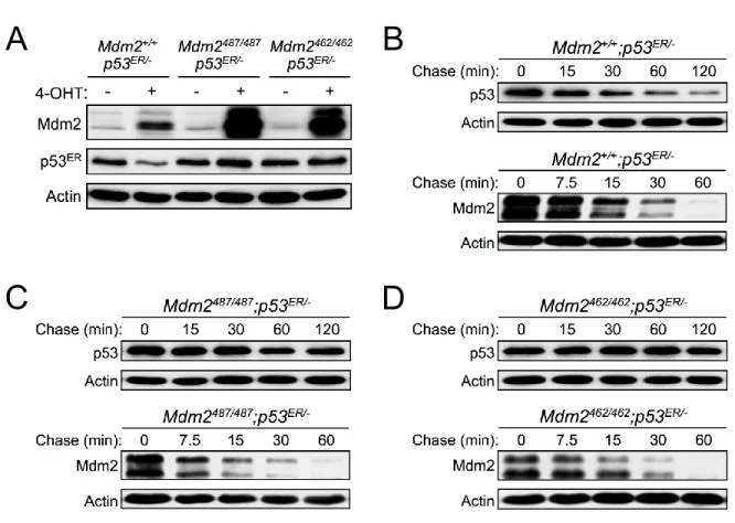
To examine the role of Mdm2 E3 ligase function in Mdm2 self-degradation in vivo, we compared degradation dynamics of Mdm2 in MEFs expressing wild type (WT) Mdm2 or the E3 ligase inactive mutant Mdm2. We generated two Mdm2 knock-in mouse models with inactivated Mdm2 E3 ubiquitin ligase activity towards p53: the Mdm2Y487A (corresponding to human
Mdm2Y489A) and Mdm2C462A (corresponding to human Mdm2C464A) (Itahana, Mao et al. 2007).
Both of the mutated Mdm2 genes are under control of native promoter and hence results in physiological levels of Mdm2 expression. While both the Mdm2Y487A and Mdm2C462A proteins
display disrupted E3 ubiquitin ligase activity for p53, the mechanism of disruption in each of these mutants is unique. The Mdm2Y487A mutation located to the C-terminal of the RING domain, in a region previously demonstrated to be critical for Mdm2 E3 ubiquitin ligase activity towards p53, however, this mutation retains an intact RING domain (Uldrijan, Pannekoek et al. 2007). In contrast, the Mdm2C462A mutation affects one of the four critical cysteine residues responsible for maintaining the RING domain structure; disruption of the Mdm2 RING domain structure results in the loss of E3 ubiquitin ligase activity (Uldrijan, Pannekoek et al. 2007). Because the Mdm2C462A mutation results in early embryonic lethality in homozygous mice, we crossed Mdm2C462A/+ mice with mice expressing inducible p53ERTAM (p53ERhereafter); the
inducible nature of the p53ER fusion protein allows for generation of mice and MEFs with inactive p53 (Martins, Brown-Swigart et al. 2006). The addition of 4-Hydroxytamoxifen (4-OHT) activates p53ER to allow for transactivation of Mdm2 and the study of Mdm2 dynamics in the otherwise lethal Mdm2C462A background. In the presence of 4-OHT, the half-life of p53ER was
28
Mdm2C462A/C462A;p53ER/- MEFs (Figures 2-1C, 2-1D). These results support previous findings
and indicate that in vivo the Mdm2Y487A and Mdm2C462A mutant proteins have indeed lost E3
ubiquitin ligase activity for p53 degradation.
To evaluate the importance of Mdm2 E3 ligase function in the degradation of Mdm2 itself, we assessed the half-life of Mdm2 in Mdm2+/+;p53ER/-, Mdm2Y487A/Y487A;p53ER/- and
Mdm2C462A/C462A;p53ER/- MEFs. Despite disrupting p53 degradation, Mdm2Y487A andMdm2C462A
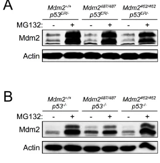
protein itself was degraded normally when compared with WT Mdm2 (Figures 2-1B, 2-1C, 2-1D). Furthermore, treatment with the proteasome inhibitor MG132 led to similar stabilization of WT Mdm2, Mdm2Y487A, and Mdm2C462A (Figure 2-2A),suggesting that each of these proteins is
degraded, at a similar rate, in a proteasome dependent manner. To eliminate the possible aberrant effect of p53ER on transcriptional regulation of Mdm2, Mdm2+/+;p53-/-,
Mdm2Y487A/Y487A;p53-/-, and Mdm2C462A/C462A;p53-/- mice were generated, and MEFs were isolated
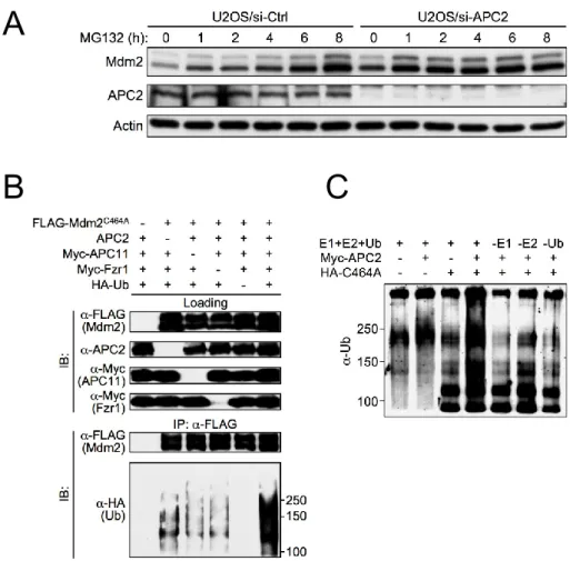
and similarly subjected to MG132 treatment. As observed in the p53ER/- genetic background, in the absence of p53, a similar level of Mdm2 stabilization was observed regardless of the E3 ligase status (Figure 2-2B). These results suggest that while Mdm2 E3 ligase function is necessary for the degradation of p53, under physiological conditions, it is dispensable for its own degradation through the proteasome-dependent pathway.
In light of the normal, proteasome-dependent Mdm2 degradation dynamics observed in Mdm2 E3 inactive MEFs, we postulated that alternative E3 ubiquitin ligase(s) is responsible for Mdm2 degradation under physiological conditions. To test this hypothesis, we used an in vitro ubiquitin ligase assay to assess the E3 ubiquitin ligase activity of HeLa cell lysate towards bacterial purified Mdm2 protein. Consistent with previous studies (Fang, Jensen et al. 2000), and similar to most other RING finger domain-containing proteins that confer E3 ligase activity (Deshaies and Joazeiro 2009), purified WT Mdm2 protein demonstrated intrinsic
autoubiquitination activity in the in vitro ubiquitin ligase assay; in contrast, Mdm2C464A mutant
29
following addition of HeLa cell lysate to the Mdm2C464A protein reaction samples, strong
polyubiquitin chain formation was observed (Figure 2-3A). The ability for Mdm2C464A to be
polyubiquitinated in the presence of HeLa cell lysate suggests that an E3 ubiquitin ligase(s) capable of polyubiquitinating Mdm2 is present in the lysate. To rule out the possibility that WT Mdm2 present in the HeLa lysate is responsible for the observed polyubiquitination of
Mdm2C464A, we assessed polyubiquitin chain formation in cell lysate isolated from WT,
Mdm2+/+;p53-/- and Mdm2-/-;p53-/- MEFs. Polyubiquitin chain formation in WT, Mdm2+/+;p53-/- and Mdm2-/-;p53-/- MEF cell lysate occurs at similar levels (Figure 2-3B), indicating that a component in the MEF cell lysates, independent of both Mdm2 and p53, is capable of promoting Mdm2 polyubiquitination.
We then tested if this polyubiquitin chain formation is dependent on E1 and E2 supplied in vitro by individually removing each of these components. The polyubiquitin chain formation is
dependent on E2 supplied in vitro, but it seems the HeLa cell lysate contained sufficient E1 ubiquitin activating enzyme to support the in vitro ubiquitination reaction (Figure 2-3C).
Furthermore, we found that the E3 ubiquitin ligase activity from HeLa cell lysate is diminished in the presence of SDS, providing further evidence that an enzymatic activity in the HeLa lysate is essential for the observed polyubiquitin chain formation (Figure 2-3D).
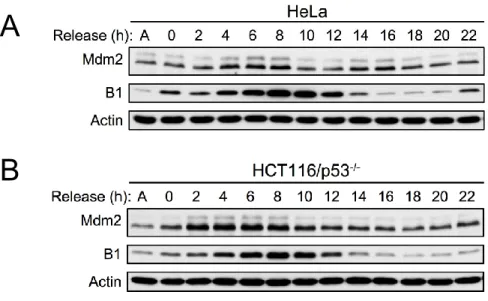
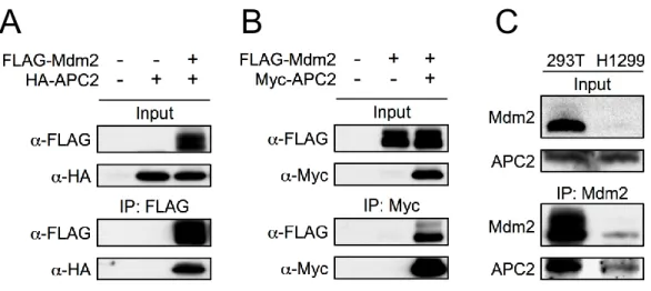
Mdm2 interacts with APC2
Previous studies have indicated that Mdm2 protein levels oscillate with the cell cycle (Gu, Ying et al. 2003; Inuzuka, Tseng et al. 2010). Accordingly, we also found that in WT and p53 -/-MEFs synchronized by serum starvation Mdm2 protein levels oscillated in a manner correlated to cyclin B1 oscillations (Figures 2-4A, 2-4B). Similar results were also observed in U2OS cells synchronized with serum starvation (Figure 2-4C), and HeLa and HCT116/p53-/- cells
30
E3 ubiquitin ligases, in a manner similar to cyclin B1. Furthermore, the observed oscillation of Mdm2 in p53 null cells suggests that this manner of Mdm2 regulation is p53-independent.
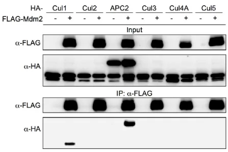
To test the possibility that a Cullin family E3 ubiquitin ligase is responsible for the ubiquitination and degradation of Mdm2, we first determined the binding affinity of Cullin family E3 ubiquitin ligases with Mdm2 via immunoprecipitation. As shown in Figure 2-6A, Mdm2
interacted with Cullin1 when co-overexpressed, providing additional support for previous studies characterizing Cullin1:Mdm2 binding (Inuzuka, Tseng et al. 2010). Additionally, we observed Mdm2 interaction with APC2, a scaffold subunit of APC/C, which is a multi-subunit E3 ubiquitin ligase complex that controls the degradation of many proteins involved in cell cycle regulation (Figure 2C). Most APC/C substrates oscillate their protein levels through the cell cycle, including the metaphase-anaphase transition and mitotic exit (Peters 2006; Thornton, Ng et al. 2006; Yu 2007). We confirmed APC2:Mdm2 interaction with reciprocal immunoprecipitation of ectopically expressed proteins (Figures 2-7A, 2-7B), as well as with immunoprecipitation of endogenous Mdm2 in 293T and H1299 cells. 293T and H1299 cells express different levels of Mdm2, which allowed for correlation of the levels of Mdm2 with the levels of APC2 being
co-immunoprecipitated (Figure 2-7C).
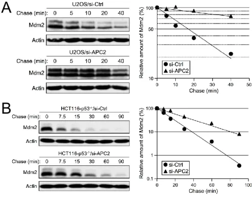
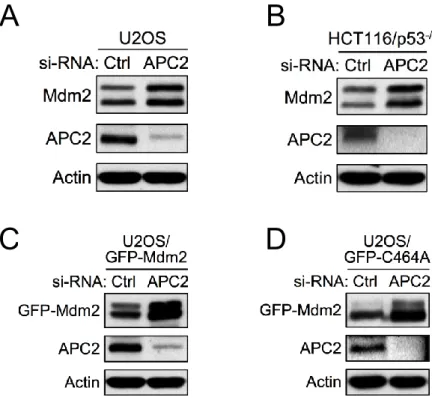
APC2 is important for mdm2 degradation
To test if APC/C is a physiological E3 ubiquitin ligase for Mdm2, we knocked down APC2 in human tumor cell lines and examined the levels of Mdm2. Knockdown of APC2 by siRNA in both U2OS (p53 positive) and HCT116/p53-/- (p53 negative) cells led to accumulation
31
Mdm2C464A mutant under the control of the CMV promoter. As shown in Figure 2-10C,
knockdown of APC2 by siRNA led to a strong accumulation of ectopic GFP-tagged Mdm2, indicating that the accumulation of Mdm2 is independent of transcriptional regulation. More importantly, knockdown of APC2 by siRNA also led to a strong accumulation of ectopic GFP-tagged Mdm2C464A, which suggests that the Mdm2 accumulation observed following
downregulation of APC2 is independent of Mdm2 E3 ligase activity (Figure 2-10D). Disruption of proteasome-mediate degradation by MG132 treatment resulted in an accumulation of Mdm2 in U2OS cells transfected with a control knockdown siRNA (siCtrl), but did not further stabilize Mdm2 in cells with knocked down APC2 (siAPC2), which had higher levels of Mdm2 prior to MG132 treatment (Figure 2-11A). Together, these results indicate that APC2 is important for Mdm2 degradation in a manner independent of p53 status and Mdm2 transcriptional regulation.
APC2 is important for stress induced p53 stabilization and activation
Because Mdm2 is the primary regulator for p53, mechanisms functioning to regulate Mdm2 will inevitably affect p53 function as well. To investigate the functional connections of APC2 with p53 activity, we examined the effect of downregulation of APC2 on p53 induced growth inhibition. We generated MEF cells with stable knockdown of APC2 in both WT and p53 -/- genetic backgrounds. Stable knockdown of APC2 in the WT, but not p53-/- background, corresponded to a higher number of cells displaying flattened and enlarged morphology, indicative of senescence. Assessment of -galactosidase activity was utilized to determine whether the flattened and enlarged MEF cells were indeed senescent. MEFs with APC2
knocked down exhibited significantly weaker activity of senescence-associated -galactosidase
32
transcription using a p21-luciferase assay. As shown in Figure 2-12B, p53-dependent activation of the p21 promoter was greatly reduced by knockdown of APC2.
To investigate the role of APC2 in the p53 stress response, we knocked down APC2 by shRNA in WT MEFs and examined cell survival following exposure to ultraviolet (UV) radiation. While APC2 knockdown did not affect cell survival under unstressed conditions, it increased cell survival upon UV induced DNA damage (Figure 2-12C). This increase in cell survival was attenuated when Mdm2-p53 interaction was disrupted by Nutlin-3 (Figure 2-12D), suggesting that the effect of APC2 on cell survival following UV radiation is p53-dependent.
To determine whether APC2 might regulate stress-induced p53 expression, siRNA was used to knock down APC2 in U2OS cells, followed by treatment with genotoxic agents to challenge the cells. Indeed, downregulation of APC2 substantially reduced the level of p53 induced by the DNA damaging agents, as observed following treatment with UV (Figure 2-13A) and doxorubicin (Figure 2-13B), as well as in response to ribosomal stress, as induced by actinomycin D (Figure 2-13C). These results indicate that APC2 plays an important role in p53 stabilization in response to genotoxic stress.
APC2 downregulation correlates with cancer development
33
ovarian, lung, thyroid, liver, and renal cancer tissues in The Cancer Genome Atlas (TCGA) also revealed that APC2 gene copy number was decreased compared to the gene copy number observed in normal tissues (Figure 2-16A). The decreased level of APC2 mRNA and gene copy number in a variety of cancer types suggests that APC2 may be clinically relevant in the