Non-adherence to the rule of 3 does not increase the risk
of adverse events in esophageal dilation
Karina V. Grooteman,1,2,3Louis M. Wong Kee Song,2Frank P. Vleggaar,3Peter D. Siersema,1,3 Todd H. Baron2,4
Nijmegen, the Netherlands; Rochester, Minnesota, USA; Utrecht, the Netherlands; Chapel Hill, North Carolina, USA
Background and Aims:Although the rule of 3 is recommended to minimize the risk of perforation when esoph-ageal dilation is performed using bougie dilators, there are no data to validate its use. Our aim was to investigate the association between the rule of 3 and adverse events (AEs) in esophageal dilation.
Methods:A retrospective chart review in patients who underwent esophageal bougie or balloon dilation between December 1991 and February 2013 at a tertiary hospital was performed. Data collection included patient demo-graphics, stricture and procedural characteristics, AEs, and follow-up. Univariate logistic regression models were used to assess the risk of AEs and perforations.
Results:A total of 297 patients (median age, 63 years; 60% men) underwent 2216 esophageal bougie or balloon dilations. Major AEs occurred in 22 (1%) dilation sessions, including 11 (0.5%) perforations, 4 (0.2%) fistulas, 3 (0.1%) hospitalizations for pain management, 2 (0.09%) clinically significant hemorrhages, 1 (0.04%) fever, and 1 (0.04%) tracheoesophageal voice prosthesis leak. Mean duration of treatment was 43.2 months (standard deviation, 47.7 months). Most strictures were benign (n Z275; 93%) and complex in nature (nZ198; 67%). Non-adherence to the rule of 3 occurred in 190 (13%) dilations with bougie dilators. Non-adherence was not asso-ciated with a higher rate of major AEs (1/190, 0.5% vs 15/953, 1.6%;PZ.18) and perforations (0/190, 0% vs 7/952, 0.7%;PZ.18). Gender, complex strictures, location of the stricture, type of dilator, and additional interventions were also not associated with major AEs or perforations. However, malignant strictures were associated with an increased risk of major AEs (odds ratio, 3.5; 95% confidence interval, 1.1-12.0) and perforations (odds ratio, 8.3; 95% confidence interval, 2.2-31.9).
Conclusions:Non-adherence to the rule of 3 does not appear to increase the risk of AEs, particularly perforation, after esophageal dilation using bougie dilators. Caution is needed with the dilation of malignant strictures, as there is an increased risk of perforations and AEs. However, large prospective studies are needed to verify the results of this study. (Gastrointest Endosc 2017;85:332-7.)
INTRODUCTION
Esophageal dilation is used to treat strictures of the esophagus from a variety of benign and malignant disor-ders. In the past, peptic strictures were the most frequent reason for dilation; however, there has been a decrease in
this entity with the introduction of proton pump inhibitors. Nowadays, esophageal strictures from malignancy, post-surgery, caustic ingestion, radiation therapy, photody-namic therapy (PDT), wide mucosectomy, eosinophilic esophagitis, and Schatzki rings are increasingly encoun-tered.1-3 The aims of dilation are to alleviate dysphagia,
Abbreviations: AE, adverse event; ASGE, American Society for Gastroin-testinal Endoscopy; PDT, photodynamic therapy; TTS, through-the-scope.
DISCLOSURE:All authors disclosed no financial relationships relevant to this publication.
Copyrightª2017 by the American Society for Gastrointestinal Endoscopy 0016-5107/$36.00
http://dx.doi.org/10.1016/j.gie.2016.07.062
Received February 4, 2015. Accepted July 29, 2016.
Current affiliations: Department of Gastroenterology and Hepatology, Radboud University Medical Center, Nijmegen, the Netherlands (1), Division of Gastroenterology and Hepatology, Mayo Clinic, Rochester, Minnesota, USA (2), Department of Gastroenterology and Hepatology, University Medical Center Utrecht, Utrecht, the Netherlands (3), Division of Gastroenterology and Hepatology, University of North Carolina, Chapel Hill, North Carolina, USA (4).
reduce the risk of pulmonary aspiration, maintain oral nutrition, and facilitate passage of endoscopes.4 Caustic, radiation-induced, post-surgical, and PDT-induced stric-tures are more likely to remain refractory to dilation.5 Adverse events (AEs) of dilation include perforation, bleeding, pulmonary aspiration, and fistula formation. Perforation occurs in 0.1% to 2.6% of procedures and is the most feared AE with a mortality rate as high as 20%.6,7Risk factors for dilation-induced perforation include eosinophilic esophagitis, complex strictures, and malignant stenoses.6,8
Balloon and bougie dilators are used to perform dilation. As opposed to radial forces incurred by balloon dilators, bougie dilators also generate longitudinal forces. This dif-ference has never been proven to have any effect on the rate of AEs or clinical outcome between Savary (bougie) and through-the-scope (TTS) balloon dilators.9-12 Theoret-ically, more dilation sessions are needed with bougie dila-tors because the rule of 3 may limit the number of sessions. The rule of 3 states that no more than 3 dilators of progressively increasing diameter should be passed after moderate resistance is encountered. This rule is applicable to bougienage alone and was intended to preclude over-aggressive dilation and consequent perforation. Although the rule of 3 makes common sense, it is not evidence based. One study suggests that dilation using larger incre-mental diameters may be safe for simple strictures.13
This single-center retrospective study assesses over 2200 esophageal dilations and the association between the rule of 3 and AEs during esophageal bougienage.
PATIENTS AND METHODS
A retrospective chart review was performed of patients who underwent esophageal dilation between December 1991 and February 2013 at Mayo Clinic, Rochester, Minne-sota. The following data were extracted: demographics, indi-cation for dilation, stricture characteristics, technique of endoscopic dilation, adherence to the rule of 3, AEs, need for additional treatment, and post-treatment outcomes.
The definition of the rule of 3 for bougie dilators was taken from the American Society for Gastrointestinal Endoscopy (ASGE) guideline on esophageal dilation: after moderate resistance is encountered, no more than 3 consecutive dilators in increments of 1 mm should be passed in a single session.14 Consequently, when the degree of resistance was not reported, adherence to the rule of 3 was scored as “unknown.” This rule is not applicable for balloon dilation, as there is no degree of resistance felt during the dilation. We subjectively defined adherence to the rule of 3 for balloon dilation when the difference in size between the first and last balloon was 3 mm. AEs were defined by the ASGE consensus for endoscopic AEs.15 AEs after dilation in another facility and those due to an esophageal stent
(eg, stent migration) or progression of disease were not included. Pneumatic balloon dilation in patients with achalasia were excluded. Strictures were defined as simple or complex using previous definitions.14 Complex strictures are asymmetric, with a diameter 12 mm, or inability to pass an endoscope. The study was approved by the Institutional Review Board, and all patients gave written informed consent for the endoscopic procedures. Sample size was calculated based on the assumption that the AE prevalence would be 3% with non-adherence and 1% with adherence to the rule of 3. With a power of 90% and a significance level of .05, the target sample size was 2000 dilations. Descriptive statistics were reported as median or mean and range for continuous variables. The
c2
test and the Fisher exact test were used for categorical variables and the Student t test was used for continuous variables to sub-analyze the value of the rule of 3 in dila-tions with bougie dilators. Univariate logistic regression models were used to model the risk of AEs and perfora-tions. Odds ratios (ORs) and 95% confidence intervals (95% CI) were used to describe the associations. A P value .05 was considered statistically significant. The analyses were performed using JMP software, version 9 (SAS Institute Inc., Cary, NC).
RESULTS
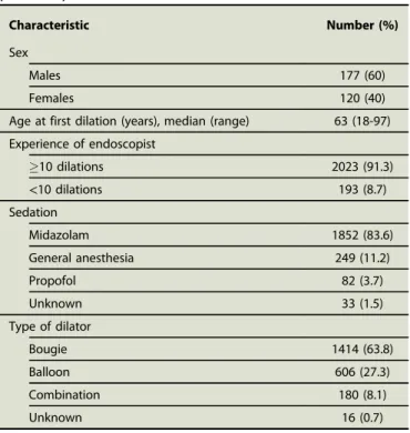
Of 1306 eligible patients, 297 patients (60% men) were randomly selected for analysis (Table 1). The stricture characteristics are listed in Table 2. The median age at TABLE 1. Characteristics of the patients (n[297) and procedures (n[2216)
Characteristic Number (%)
Sex
Males 177 (60)
Females 120 (40)
Age at first dilation (years), median (range) 63 (18-97)
Experience of endoscopist
10 dilations 2023 (91.3)
<10 dilations 193 (8.7)
Sedation
Midazolam 1852 (83.6)
General anesthesia 249 (11.2)
Propofol 82 (3.7)
Unknown 33 (1.5)
Type of dilator
Bougie 1414 (63.8)
Balloon 606 (27.3)
Combination 180 (8.1)
time of first dilation was 63 years (range, 18-97 years). Forty-seven (16%) patients had undergone previous dila-tions at other facilities. All diladila-tions were performed by or with a staff physician who dictated the endoscopy report. Mean duration of treatment was 43.2 months (stan-dard deviation, 47.7 months). Most of the strictures were benign (93%) and complex (67%) in nature. Only 53 (18%) strictures were ulcerative at the time of dilation. The most common stricture location was the proximal esophagus, and the more common causes were radiation and post-surgery.
A total of 2216 dilation sessions were performed. A major AE occurred in 22 (1.0%) of these procedures: 11 (0.5%) perforations, 4 (0.2%)fistulas, 3 (0.1%) hospitaliza-tions for pain management, 2 (0.09%) clinically significant bleedings, 1 (0.04%) fever, and 1 (0.04%) tracheoesopha-geal voice prosthesis leak. Table 3 shows the association of major AEs for 8 potential risk factors. Malignant structures were found to be associated with major AEs (OR, 3.5; 95% CI, 1.1-12.0). All other factors, including location of the stricture, complex stricture, type of dilator, non-accordance to the rule of 3, dilation of more than 3 mm, and additional procedures performed, were not associated with major AEs.
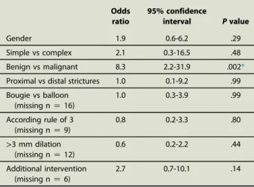
Perforations occurred in 11 (0.5%) of all dilation proced-ures. One perforation occurred before dilation was performed when the trachea was cannulated instead of the esophagus. Table 4 shows the association of the potential risk factors for perforation. Only malignant structures were associated with the occurrence of perforation (OR, 8.3; 95% CI, 2.2-31.9); all the other factors were not found to be associated with perforation.
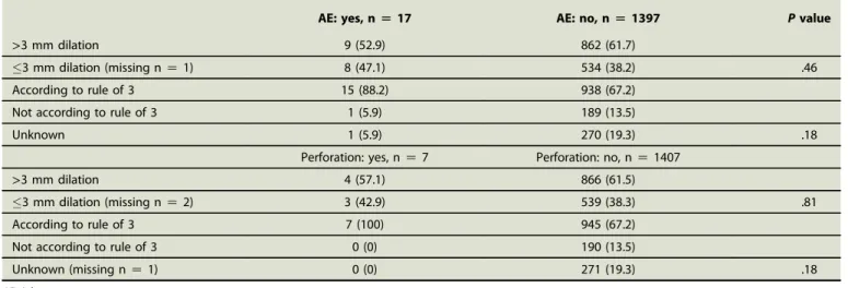
A sub-analysis for the rule of 3 and dilations greater than 3 mm for bougie dilators was performed. There was no dif-ference in incidence of AEs (1/190, 0.5% vs 15/953, 1.6%, P Z .18) or perforations (0/190, 0% vs 7/952, 0.7%, P Z .18) in dilation sessions whether the rule of 3 was adhered to or not (Table 5). Neither was a difference in AEs or perforations found for dilations greater than 3 mm. The rates of perforation according to the cause of stric-ture are shown inFigure 1. A higher rate of perforations was seen in patients with strictures of unknown cause, TABLE 2. Stricture characteristics
Characteristic Number (%)
Benign/malignant
Benign 275 (93)
Malignant 22 (7)
Simple/complex
Simple 99 (33)
Complex 198 (67)
Cause
Radiation 64 (22)
Post-surgery 59 (20)
Schatzki ring 22 (7)
Malignant 21 (7)
Unknown 19 (6)
Peptic 17 (6)
Post-endotherapy 17 (6)
Cricopharyngeal bar 17 (6)
Caustic 9 (3)
Other 52 (17)
Location
Proximal 138 (47)
Mid 47 (16)
Distal 74 (25)
Anastomosis 22 (7)
Unspecified 16 (5)
TABLE 3. Identification of variables associated with major AEs
Odds ratio
95% confidence
interval Pvalue
Gender 1.1 0.5-2.6 .85
Simple vs complex 0.95 0.3-2.8 .92
Benign vs malignant 3.5 1.1-12.0 .047*
Proximal vs distal strictures 0.7 0.2-3.4 .70
Bougie vs balloon (missing nZ16)
0.4 0.1-1.4 .15
According rule of 3 (missing nZ9)
0.5 0.2-1.5 .20
>3 mm dilation (missing nZ12)
0.7 0.3-1.6 .38
Additional intervention (missing nZ6)
1.2 0.5-2.9 .74
*Statistically significant.
TABLE 4. Identification of variables associated with perforations
Odds ratio
95% confidence
interval Pvalue
Gender 1.9 0.6-6.2 .29
Simple vs complex 2.1 0.3-16.5 .48
Benign vs malignant 8.3 2.2-31.9 .002*
Proximal vs distal strictures 1.0 0.1-9.2 .99
Bougie vs balloon (missing nZ16)
1.0 0.3-3.9 .99
According rule of 3 (missing nZ9)
0.8 0.2-3.3 .80
>3 mm dilation (missing nZ12)
0.6 0.2-2.2 .44
Additional intervention (missing nZ6)
2.7 0.7-10.1 .14
radiation-induced strictures, and post-surgery strictures. Three perforations occurred in 2 patients with strictures of unknown cause. These patients had multiple dilations with multiple biopsy specimens taken. These dilations took place after 2000. The specimens did not result in a diagnosis, which makes it unlikely that these 2 patients had eosinophilic esophagitis. The percentage of perfora-tions was equally distributed to the number of the dilation session (Supplementary Fig. 1, available online at www. giejournal.org). The risk of perforation did not differ among the dilation sessions.
At the time of analysis, 98 patients (33%) were deceased. Median age at the time of death was 72 years (range, 29-100 years). The cause of death was unknown in 14 patients (14%). Mortality was not related to dilation in most cases (81%), but 5 (5%) died within 1 month of the dilation procedure for which the exact cause of death
is unknown. In 4 of these patients, death was not related to the initial dilation, because no additional procedures were performed and no AEs were reported during or after the dilation. One patient was a 71-year-old woman with esophageal cancer. Her dilation was complicated by a perforation for which a covered esophageal stent was placed; she was hospitalized for 5 days and died 2.5 weeks after discharge.
DISCUSSION
This study shows that non-adherence to the rule of 3 is not associated with a higher rate of AEs or perforations after esophageal dilation. The only factor that was associ-ated with an increased risk of AEs and perforations was a malignant stricture, which is in accordance with the TABLE 5. Sub-analysis of bougie dilators for adverse events and perforations (%)
AE: yes, n[17 AE: no, n[1397 Pvalue
>3 mm dilation 9 (52.9) 862 (61.7)
3 mm dilation (missing nZ1) 8 (47.1) 534 (38.2) .46
According to rule of 3 15 (88.2) 938 (67.2)
Not according to rule of 3 1 (5.9) 189 (13.5)
Unknown 1 (5.9) 270 (19.3) .18
Perforation: yes, nZ7 Perforation: no, nZ1407
>3 mm dilation 4 (57.1) 866 (61.5)
3 mm dilation (missing nZ2) 3 (42.9) 539 (38.3) .81
According to rule of 3 7 (100) 945 (67.2)
Not according to rule of 3 0 (0) 190 (13.5)
Unknown (missing nZ1) 0 (0) 271 (19.3) .18
AE, Adverse event.
4.5 4 3.5 3 2.5 2 1.5 1 0.5 0
malignancy (n=74)unknown (n=112)
radiation-induced (n=679)
post-surgery (n=609)other causes (n=749)
% perforations
literature.9,14 Complex strictures are considered to be a risk factor for perforation, but this is mostly based on a retrospective study in which all perforations occurred after Maloney dilation and half of the strictures were malignant. Complex strictures were overrepresented in this study, but we did notfind an association between complex strictures and AEs. Overall, the importance of complex strictures as a risk factor for AEs might be overestimated. A trend was seen whereby an additional esophageal therapeutic intervention was performed during the same session. In patients undergoing multiple dilations, the perforation rate did not differ between dilation sessions.
Strengths of this study include the large number of dila-tions performed, the investigation of other variables in addition to the rule of 3, and the preponderance of com-plex strictures. Although another study suggested that it was safe to dilate more than 3 mm in one session, most of the patients in that study had simple strictures.13 In our study, most of the patients had complex strictures. We found that dilation to>3 mm in one session was not associated with more AEs, particularly perforation. Similar to the other studies, we did not find a difference in the rate of AEs or perforations between bougie and balloon dilators.9-12 The perforation rate found in our study (0.5%) is comparable with those reported in the literature. However, most patients included in the randomized studies comparing balloon and bougie dilation had benign strictures with lower AE rates.10,12
Our study is limited by its retrospective nature. We cannot exclude the possibility that AEs were not reported to our center after the patient was discharged from our institution. This could have led to an underestimation of the AE rate. In addition, application of objective measures to the rule of 3, which is subjective and based on tactile sensation, is fraught with difficulties. Moreover, there could be reporting bias by endoscopists if the rule of 3 was not precisely followed, as emphasized frequently in guidelines. Thus, there may be underreporting of lack of adherence. Furthermore, there are different definitions of the rule in different guidelines.4,14 We cannot exclude eosinophilic esophagitis as a confounder in this study. However, we do not think this had a large impact on our study, because biopsy specimens of strictures of unknown cause did not indicate a diagnosis of eosinophilic esopha-gitis after this became a recognized entity. Finally, it is important to address the inclusion of balloon dilations. In clinical practice, many physicians also use the rule of 3 in balloon dilation. We hypothesized that if the rule of 3 added value in decreasing AEs, it would apply to both bougie and balloon dilation. However, if this hypothesis is incorrect, the inclusion of balloon dilations decreases the power of the study.
Based on these results, we believe that the rule of 3 does not need to be used when esophageal dilation is per-formed with bougie dilators. However, it still serves as a useful conservative guide, but might need to be updated
to current practice. In our experience, initial dilation is started at a diameter of 1 to 2 mm larger than the esti-mated diameter of the stricture. The latter is based on whether a normal caliber (depending on type, 9-10 mm) or small caliber (4-5 mm) endoscope is able to pass the stricture. Dilation is continued until “firm” resistance is felt in the case of bougie dilation and/or the amount of blood at the end of the dilator is more than“slight.”The same applies for balloon dilation, but the degree of resis-tance is assessed by moving the endoscope and the inflated balloon back and forth within the stricture at each diameter. At the end of dilation, endoscopy is performed to confirm the presence of a tear and exclude major bleeding or a perforation.
As an alternative to the rule of 3, it is proposed to perform a prospective study investigating a “rule of 6,” where 3 consecutive bougie dilations are performed with incre-ments of 2 mm each. Our study suggests that this may be a reasonable alternative to the rule of 3 for most patients. Nonetheless, it is important to emphasize that caution is needed when performing dilation of malignant strictures. The same applies for other specific diseases such as eosin-ophilic esophagitis.8 Unfortunately, these patients were not well represented in our study population. These findings make clear that the proposed “rule of 6”should not be applied to every patient, and exceptions for patients with specific risk factors for AEs must be made.
In conclusion, adherence to the rule of 3 does not appear to reduce the risk for AEs, including esophageal perforation, after esophageal dilation with bougie dilators. Malignancy of the stricture is associated with an increased risk of AEs and perforations. Prospective trials would be useful to support thesefindings.
REFERENCES
1.Tucker LE. Esophageal stricture: results of dilation of 300 patients. Mo Med 1992;89:668-70.
2.Raymondi R, Pereira-Lima JC, Valves A, et al. Endoscopic dilation of benign esophageal strictures without fluoroscopy: experience of 2750 procedures. Hepatogastroenterology 2008;55:1342-8.
3.Siersema PD. Treatment options for esophageal strictures. Nat Clin Pract Gastroenterol Hepatol 2008;5:142-52.
4.Riley SA, Attwood SE. Guidelines on the use of oesophageal dilatation in clinical practice. Gut 2004;53(Suppl 1):i1-6.
5.Lew RJ, Kochman ML. A review of endoscopic methods of esophageal dilation. J Clin Gastroenterol 2002;35:117-26.
6.Quine MA, Bell GD, McCloy RF, et al. Prospective audit of perforation rates following upper gastrointestinal endoscopy in two regions of England. Br J Surg 1995;82:530-3.
7.Piotet E, Escher A, Monnier P. Esophageal and pharyngeal strictures: report on 1,862 endoscopic dilatations using the Savary-Gilliard technique. Eur Arch Otorhinolaryngol 2008;265:357.
8.Dellon ES, Gibbs WB, Rubinas TC, et al. Esophageal dilation in eosinophilic esophagitis: safety and predictors of clinical response and complications. Gastrointest Endosc 2010;71:706-12.
10.Scolapio JS, Pasha TM, Gostout CJ, et al. A randomized prospective study comparing rigid to balloon dilators for benign esophageal stric-tures and rings. Gastrointest Endosc 1999;50:13-7.
11.Yamamoto H, Hughes RW Jr, Schroeder KW, et al. Treatment of benign esophageal stricture by Eder-Puestow or balloon dilators: a compari-son between randomized and prospective nonrandomized trials. Mayo Clin Proc 1992;67:228-36.
12.Saeed ZA, Winchester CB, Ferro PS, et al. Prospective randomized comparison of polyvinyl bougies and through-the-scope balloons for
dilation of peptic strictures of the esophagus. Gastrointest Endosc 1995;41:189-95.
13.Kozarek RA, Patterson DJ, Ball TJ, et al. Esophageal dilation can be done safely using selective fluoroscopy and single dilating sessions. J Clin Gastroenterol 1995;20:184-8.
14.Standards of Practice Committee; Egan JV, Baron TH, Adler DG, et al. Esophageal dilation. Gastrointest Endosc 2006;63:755-60.
1 2 3 4 5 6 7 8 9 10
other(11-107)
1.6 1.4 1.2 1 0.8 0.6 0.4 0.2 0
% perforations