A COMPARISON OF INTRAORAL IMAGE QUALITY, ERROR RATES, OPERATOR AND PATIENT DOSIMETRY BETWEEN A HAND-HELD DEVICE AND
WALL-MOUNTED X-RAY SOURCES
Billy James Phillips
A thesis submitted to the faculty at the University of North Carolina at Chapel Hill in partial fulfillment of the requirements for the degree of Master of Science in Oral Maxillofacial
Radiology, School of Dentistry.
Chapel Hill 2014
Approved by:
Enrique Platin
John B. Ludlow
ii
iii ABSTRACT
Billy James Phillips: A Comparison of Intraoral Image Quality, Error Rates, Operator and Patient Dosimetry between a Hand-Held Device and Wall-Mounted X-Ray Sources
(Under the direction of Enrique Platin)
Objectives: To compare handheld and wall-mounted x-ray sources during Full Mouth
Examination (FMX).
Methods: One operator simulated exposure of 10 FMX on a RANDO phantom for each of
seven studies: three handheld and four wall-mounted. Optical Stimulated Luminescence (OSL)
dosimetry was utilized to record dose. Effective dose was calculated using 2007 International
Commission on Radiologic Protection (ICRP) tissue weights. Differences due to technique were
evaluated using Analysis of Variance (ANOVA). A cohort of 75 dental students exposed one
FMX utilizing each device. Observers were calibrated and blinded to assess technique errors and
Line Pair (LP) resolution.
Results: Mean FMX dose was significantly less for handheld 36µSv than for wall-mounted
devices 98µSv (p=0.0217). Mean operator exposures were indistinguishable from ambient
background levels (<2 µ Gy/study). Mean total technique error was not different between devices
(p = 0.29). Mean LP resolution was significantly higher for the handheld device, (p < 0.01).
Conclusion: Operator, patient dose, and mean sum of total errors were not different. LP
resolution was significantly higher for the handheld device during FMX simulation.
iv
TABLE OF CONTENTS
LIST OF TABLES…...v
LIST OF FIGURES……….………..vi
LIST OF ABBREVIATIONS……….………..vii
INTRODUCTION………..……….1
REVIEW OF LITERATURE………..………3
REFERENCES………...……….5
INTRODUCTION………..……….7
MATERIALS & METHODS………10
RESULTS………..17
DISCUSSION………25
v
LIST OF TABLES
Table 1. Technical specifications for the handheld and wall mounted x-ray devices……...……14
Table 2. Effective dose ANOVA table and Tukey test ……….19
Table 3. ANOVA full model……….19
Table 4. Linear mixed model………..….………..20
Table 5. Descriptive statistics ……….………..……22
Table 6. Inter-observer Kappa values………...……….23
vi
LIST OF FIGURES
Figure 1. First dental x-ray ...8
Figure 2. Left: Nomad Pro® (Aribex, Orem, UT) handheld device. Right: Planmeca Intra® (Planmeca USA Inc., Roselle, IL) wall-mounted x-ray source……….….10
Figure 3A. PSP exposure latitude wall mounted x-ray device………...……….…….14
Figure 3B. PSP latitude handheld x-ray device………..………...15
Figure 3C. Normalized and optimal exposures (Sec) for wall mounted and handheld x-ray device………..………...15
Figure 4A. Hand-held device LP resolution ………...………...…...16
Figure 4B. Wall-mounted device LP resolution ………..………..………...16
Figure 5. Mean dose per exposure (Sec) mrad ………..…………...17
Figure 6. Tissue specific equivalent dose comparisons ………..………..20
Figure 7. Mean error per FMX………..21
vii
LIST OF ABBREVIATIONS
ALARA As Low As Reasonably Achievable
ANOVA Analysis of Variance
ASE Agreement Standard Error
CBCT Cone Beam Computed Tomography
cc Cone Cut
cm Centimeter
CT Computerized Tomography
DDS Doctor of Dental Surgery
DPI Dots per Inch
DXTTR Dental X-Ray Trainer
EPR Electronic Patient Record
EUS Effective Dose per individual, United States of America population
FDA Food and Drug Administration
FMX Full Mouth Survey examinations
ha Horizontal Angulation
ICRP International Commission on Radiologic Protection
id Image Distortion
JAD-RADTM Dental X-ray Shield
kVp Kilovoltage
LP Line Pair
mA Milliamperage
viii
mR Millirads
NCRP National Council of Radiation Protection and Measurement
OSL Optical Stimulated Luminescence
PA Periapical
PID Positioning Indicating Device
pp Packet Placement
PSP Photostimuable Plates
SAS Statistical Analysis System
UNC University of North Carolina at Chapel Hill
µSv Micro Sieverts
1
INTRODUCTION:
There has been no independent research concerning the use of handheld x-ray devices in
academic dental curriculums. Furthermore, there is widely varied state regulation of their use in
the private practice of dentistry. The North Carolina Department of Health and Human Services,
Protection Section, mandates facilities planning to utilize handheld dental x-ray units to receive
an approval for rule exemptions from the Radiation Protection Section prior to use of the unit.
“Exemptions will not be granted for routine dental x-rays where permanently installed units are a
viable option.” Applicable rules for exemption are outlined in The North Carolina Regulations
for Protection against Radiation (15A NCAC 11).
Similar, restrictions on the use of handheld devices apply with the military Dental Corps.
Department of the Navy policy 6600 Ser M3C/AT – 17215, 09/22/2008: Appropriate Use of
Hand-Held X-Ray Units for Oral and Maxillofacial Radiography restricts the use of handheld
x-ray devices forensic, combat, humanitarian, and emergency operations where access to
traditional fixed x-ray sources are not available.
Such restriction and policy while well intentioned by state regulatory radiation protection
agencies and the military are not evidence based. Such restriction and policy decisions require
current research into the occupational and patient risk to exposure to ionizing radiation from
2
The safety of the handheld x-ray device is not the only question current research must
investigate. Even if proven safe the diagnostic quality of intraoral images acquired by these
devices must also be evaluated. The As Low As Reasonably Achievable (ALARA) principle
recommends that the diagnostic task be matched to the imaging modality selected by the
practitioner and that patient benefit outweigh any risk to exposure to ionizing radiation. It does
not matter if the handheld x-ray device is safe if image quality is not adequate diagnostically.
The primary aim of this study was to evaluate the utility of a handheld x-ray device
within a Dental School environment. Study null hypotheses include: 1) There was no statistical
difference in image quality error between the Aribex, Nomad Pro and the Planmeca Intra
wall-mounted sources. 2) There was no statistical difference in recorded personnel dosimetry between
the Aribex, Nomad Pro and Planmeca devices.
This study was the first independent research to compare operator and patient dosimetry,
image quality, and technique error rates in a Dental School curriculum between the Aribex,
Nomad Pro® (Aribex, Orem, UT) and Planmeca Intra® (Planmeca USA Inc., Roselle, IL). The
research protocol was designed to simulate a worst-case clinical scenario to evaluate the research
questions. A cohort of 2nd year dental students without prior clinical radiology training was
identified. The operators utilized no lead apron shielding during image acquisition with the
handheld x-ray source. The integrated internal and external shielding of the handheld device
provided the only operator shielding to scatter radiation. Operators were required to complete an
online training module on the use of the Nomad Pro and, successfully complete a test. Round
3
REVIEW OF LITERATURE:
The history of dentistry and the use and efficacy of diagnostic imaging are well
documented. The dynamic history and technology associated with the clinical practice of
dentistry is ever evolving and spans advancements in restorative materials, armamentarium, and
the diagnostic sciences. The potential for risk to the clinical practitioner and patient is always
present and mandates continued extensive investigation to evaluate the risk to benefit ratio in
treatment planning and clinical decision-making. There is specific biological risk in diagnostic
imaging and exposure to ionizing radiation.
The risk, diagnostic efficacy, and benefits associated with the materials and equipment
utilized to acquire diagnostic imaging is well documented in the published literature. Richards
evaluated occupational exposure and compared patient exposure reported in the published
literaturewith then current techniques and rightly acknowledges risk will never reach zero.1-8
Our knowledge of the biological risk, deterministic and stochastic effects, of exposure to
ionizing radiation is presented in additional research by Wall9, and White10. Radiation protection
recommendationsand guidelines are outlined by the 2007 Recommendations of the International
Commission of Radiological Protection11 and the National Research Council12.
In 2006, Americans were exposed to more than seven times as much ionizing radiation
from diagnostic imaging compared to recorded figures from the early 1980s, and diagnostic
imaging constituted nearly half the total radiation exposure of the U.S. population from all
sources. The average effective dose per individual in the U.S. population (EUS) from all sources
has increased by a factor of 1.7 to 6.2 mSv as a result of the growth in utilization of x-ray
4
Computerized Tomography (CT) first introduced in 1972 has become integral in medical
diagnostic imaging. The expanding clinical application of CT alone has directly led to an annual
10% increase in CT procedures over the past twenty years. In 2006, 62 million procedures were
reported that is, 207 CT examinations per 1000 population, with an average dose per
examination of 7 mSv. Currently, medical CT represents 50% of all medical exposure and 24%
of the exposure from natural and man-made sources combined13. In addition, approximately 18
million nuclear medicine procedures were reported in the US in 2006 or, 60 per 1000 population,
double the procedures reported in the early 1980s. CT, fluoroscopy and nuclear medicine
procedures contributed 2.7 mSv to EUS in 2006, accounting for 90% of all medical diagnostic
imaging procedures13.
The documented explosion of EUS directly attributed to diagnostic imaging stands in stark
contrast to the significant historical reduction in exposure to ionizing radiation in dental intraoral
radiography. To address this growth in high dose medical and dental imaging clinicians must
consider all factors: risk benefit ratios, appropriate selection criteria, imaging techniques, specific
diagnostic task, and the impact of ordered diagnostic imaging on treatment planning and patient
outcomes. Along with clinical justification for diagnostic imaging considerations must also be
given to occupational risk to personnel from exposure to ionizing radiation. NCRP Report No.
160 documents diagnostic exposure is an integral and growing piece of total exposure EUS, and
5
REFERENCES:
1. Richards. Albert G., Sources of x-radiation in the dental office. Dent Radior Photogr 37(3):51-68, 1964.
2. Richards, Albert G, Colquitt, Wayne N. Reduction in dental x-ray exposures during the past 60 years. JADA 103:713-718, 1981.
3. Nolan, W. E. Radiation hazards to the patient from oral roentgenography. JADA 47(6): 681-684, 1953.
4. Budowsky, J., and others. Radiation exposure to the head and abdomen during oral roentgenography. JADA 52(5):555-559, 1956.
5. Bailey, N. A. Patient exposure to ionizing radiation in dental radiography. Radiol 69:42-45,1957.
6. Blackman, S., and Greening, J. R. Radiation hazards in dental radiography. Br Dent J 102:167-172, 1957.
7. Bjarngard, B., and others. Radiation doses in oral radiology. Odontology Rev 10(4):355-366, 1959.
8. Alcox, R. W., and Jameson, W.R. Patient exposures from intraoral radiographic examinations. JADA 88(3):588-579, 1974.
9. Wall, B.F. Book Review: Ionizing Radiation Exposure of the Population of the United States: NCRP Report No. 160. Radiation Protection Dosimetry. 136(2):136-138, 2009.
10. White S. C. Assessment of radiation risk from dental radiography. Dentomaxillofac Radiol 121:118-126, 1992.
11. Valentin J. The 2007 Recommendations of the International Commission on Radiological Protection. Publication 103. Ann ICRP 2007;37:1-332.
12. National Research Council. Health effects of exposure to low levels of ionizing radiation. BEIR V. Washington D.C. National Academy Press, 1990.
13. NCRP Report No. 160. Ionizing Radiation Exposure of the Population of the United States. National Council of Radiation Protection & Measurements. Mar., 2009.
6
15. Kleir, D.J., Hicks, M.J., Flaitz, C.M. A comparison of Ultraspeed and Ektaspeed dental x-ray film; In vitro study of radiographic appearance of interproximal lesions. Oral surg. Oral med. Oral Pathol. 63:381-385, 1987.
16. Goren A.D., Bonvento, M., Biernacki J., Colosi D.C. Radiation exposure with the NOMAD® portable X-ray System. Dentomaxillofac Radiol 37:109-112, 2008.
17. Webber R. L., Benton P. A., Ryge G. Diagnostic variations in radiographs. Oral Surg. Oral Med. Oral Pathol. 26:800-9, Dec. 1968.
18. Suleiman O.H., Spelic D.C., Conway B., Hart J.C., Boyce P.R., Antonsen, R. G. Jr. Radiographic trends of dental offices and dental schools. JADA 130(7):1104-1110, 1999.
19. ADA council on Scientific Affairs. The use of dental radiographs update and recommendations. JADA 137(9):1304-12, 2006.
20. Bailey E., Gray J., Ludlow J. Image Quality and Radiation Dose for Intraoral Radiography: Hand-Held (Nomad®) Battery Powered vs. Wall-Mount X-ray Systems. 41st Annual Conference on Radiation Control Conference of Radiation Control Program Directors. May, 2009.
21. Huda W, Rill LN, Benn DK, Pettigrew JC. Comparison of a Photostimulable phosphor system with film for dental radiology. Oral Surg. Oral Med. Oral Pathol. 83:725-31,
1997.
22. Borg E, Attaelmanan A, Grondahl HG. Image plate systems differ in physical performance. Oral Surg. Oral Med. Oral Pathol. 89:118-24, 2000.
23. Hildebolt CF, Couture RA, Whiting BR. Dental Photostimulable Phosphor Radiography. Dental Clinics of North America, 44:2, 2000. 20. Gibbs S. J. Council on Dental Materials, Instruments, & Equipment. Biological effects of radiation from Dental radiography. JADA 105275-281, 1982.
24. Mourshed F.A.,A study of intraoral radiographic errors made by dental students. Oral Surg Oral Med Oral Pathol 32(5):824-828, 1971.
25. Ilkay, P., Meryem T.A., Evaluation of Radiographic Errors Made by Undergraduate Dental Students in Periapical Radiography. NYSDJ Aug/Sep, 45- 48, 2009.
26. Crandell, D.E. Cause and Frequency of Intra-oral X-ray Errors by Dental and Hygiene Students. J. Dent. Educ. 22:189-196, 1958.
7
INTRODUCTION:
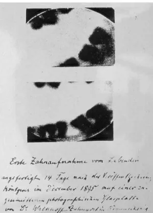
Since the first dental x-ray taken by Otto Walkoff of himself (Fig 1) in 1896, exposure to
x-rays from diagnostic dental radiographic procedures has been of great concern. The dental
profession has historically sought through technological advancement to reduce both
occupational and patient exposure to ionizing radiation. Richards1 evaluated occupational
exposure and reported environmental controls and operator protections to reduce dose based on
techniques and equipment available in the 1960’s. A subsequent study by Richards2 in 1981
compared patient exposure to dose reported in the published literature with then current
techniques.3 – 7 Nolan3 had demonstrated a range of total exposure with 25 films per full-mouth
examination at 35 to 315 R, averaging 12.6 R/film. By 1974 Alcox8 reported an average patient
exposure of 271 mR/film. In 1981 in a comparison study, Richards2 reported patient exposure of
170 mR/film and 143 mR/film respectively with and without backscatter. Richards2 dramatically
illustrated the significant historical reduction in patient exposure with the development of
increasingly faster film sensitivities. He reported a reduction in patient dose, by a factor of 0.02
with the introduction of Ektaspeed film when compared to the very slow Regular film available
in the 1920’s. All of these studies document the dental professions repeated success in reducing
both occupational and patient exposure to ionizing radiation through improved technique,
8
Fig 1: First dental x-ray. © Google: https://www.google.com/search?q=first+dental+x+ray.
Further reduction may yet be achieved with continued advances in technology. In his
conclusions, Richards2 acknowledged the risk will never reach zero but that the risk involved in
dental diagnostic imaging was acceptable. Such advances will always require rigorous evaluation
to achieve the principle of ALARA.
While the impetus for the development of handheld devices may not have been dose
reduction it could be one of the currently available tools to further reduce patient exposure to
ionizing radiation during intraoral diagnostic imaging. The utility of handheld x-ray units for
intraoral radiography has been demonstrated in field, forensic, humanitarian, and surgical
applications. The safety of this technology has been reported in controlled experiments as well as
9
cleared for sale by the Food and Drug Administration (FDA). The state of North Carolina
currently requires an exemption for use of handheld devices, and is contemplating the approval
of handheld devices within general dental practice. The current policyof the United States Navy
on the use of handheld x-ray devices in the practice of military dentistry restricts their use as a
substitute when traditional wall-mounted units are available; citing fixed unit advantages: higher
operating potentials, beam quality, shorter exposure times, higher operation tempo, rigid
mounting, remote activation, and reduced operator exposure. Navy policy delegates the
appropriate use of handheld x-ray devices to emergency and forensic use outside the dental
clinic, deployment, and humanitarian operations. 11
This study was the first independent study to evaluate the safety and efficacy of handheld
devices in a cohort of students within an educational environment. The potential patient and
operator dose impact of substituting handheld devices in an educational institutional setting has
not been reported. The objective of this study was to compare image quality, error rates,
operator and patient dosimetry during Full Mouth Survey examinations (FMX) between the
Nomad Pro® (Aribex, Orem, UT) handheld device and the Planmeca Intra® (Planmeca USA
10
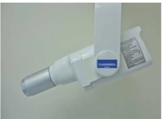
Fig 2. Left: Nomad Pro® (Aribex, Orem, UT) handheld device. Right: Planmeca Intra®
(Planmeca USA Inc., Roselle, IL) wall-mounted x-ray source.
MATERIALS & METHODS:
One operator exposed seven imaging studies, a total of 180 images per study,
simulating 10 FMX’s on a RANDO phantom (The Phantom Laboratory, Salem, NY). Three
studies were completed using the handheld device and four were completed using the
wall-mounted x-ray source. Photostimuable Phosphor Plates (PSP) receptor settings for an average
adult were used. The wall-mounted source and the hand-held device each incorporated a 6 cm
diameter open cone. Technical specifications for the handheld and wall mounted x-ray device are
presented in Table 1. Optical Stimulated Luminescence (OSL) dosimetry was utilized to record
operator and phantom dose (Nanodot, Landauer Inc., Glenwood, IL). Three dosimeters per area
were attached to thyroid, waist and trigger hand areas of the operator prior to each study.
Dosimeters were placed within the phantom in twenty-four predetermined anatomic locations
(Ludlow et al 2008). Dosimeters were read with a MicroStar reader (Landauer Inc., Glenwood,
11
A cohort study of seventy-five 2nd year dental students at the University of North
Carolina School of Dentistry DDS program was assigned to pre-clinical radiology to acquire
intraoral images. The use of the Aribex, Nomad® Pro handheld device was integrated as part of
their preclinical laboratory curriculum. Each student completed a standard 18 image FMX
examination (14 periapicals and 4 horizontal bitewings) using the Aribex, Nomad Pro®
(Aribex, Orem, UT) handheld device and an additional FMX series using the Planmeca
Intra® (Planmeca USA Inc., Roselle, IL) wall mounted x-ray source. A training module was
viewed by each student prior to the use of the handheld device. A test over this material was
taken following training. Test scores were recorded. A pass rate of 100% was required. Exam
questions were indexed to the training material for easy reference by the student if there was an
incorrect response to a training question.
Radiographic training mannequins (DXTTR, Rinn Corp) were used for patient
simulation. Five DXTTR’s were selected and labeled for sole use throughout the duration of the
study. Each DXTTR was examined and refurbished prior to the beginning of the study with
complete mounted maxillary and mandibular adult dentitions, including third molars. The same
DXTTR was used by the student for both handheld and wall mounted image acquisition. Images
produced with the Aribex, Nomad® Pro handheld device and wall mounted x-ray source utilized
the Rinn Extension Cone Paralleling (XCP) receptor-holding device (Rinn, corporation). PSP
receptors were used for image acquisition (Gendex Dental Systems, Des Plaines, IL). The images
were scanned with a Scanora unit at 300 dots per inch and processed for viewing utilizing
VixWin software (Gendex Dental Systems, Hatfield, PA). The original FMX examinations were
12
Following completion of each FMX sequence, students acquired two additional images of
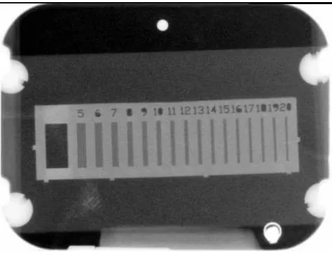
an LP test phantom held in a jig utilizing each x-ray device. The jig consisted of a 16-group Line
pair per millimeter (LP) test tool with a range of resolutions from 5 to 20 line pairs per
millimeter (Model 07-555, Nuclear Associates, Division of Victoreen, Carle Place, NY), and a
JAD-RADTM collimator attachment (JADRAD Dental Diagnostics, Farmington, CT). The test
tool and JAD-RADTM were secured to a positioning platform. The jig was placed on a tripod
allowing easy transport and positioning between imaging stations. Users were instructed to align
and center the central ray with the JAD-RADTM face of the jig. Receptors were positioned in the
apparatus parallel to the LP tool plane at a distance of 3cm from the LP tool and imaged. The jig
is designed with a Plexiglas holder to standardize the position of the receptor from the LP tool.
Images were viewed in a randomized order by three masked observers to determine the number
of line pairs resolved. In order to move FMX and LP images from the student EPR account into a
research module and de-identify the images, the FMX and LP images were exported and
re-labeled utilizing a random number generator in groups of 10 FMX and LP images to 15 shadow
charts.
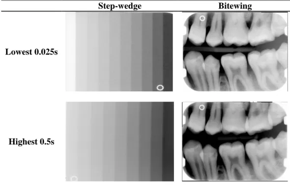
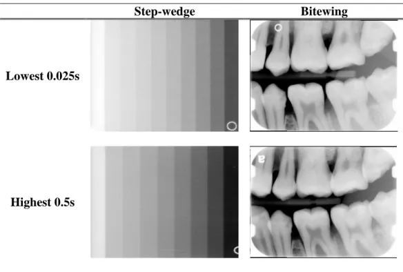
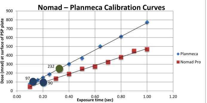
Optimal exposure and exposure latitude for PSP receptors was evaluated for each device
by assessing step-wedge images, Fig 3. Three OSL dosimeters were exposed at each exposure
time and the mean absorbed dose was calculated. Based on mean absorbed dose a cofactor was
derived to normalize dose for the beam energy for each device, Fig 4. Normalized effective dose
was compared between handheld and wall mounted x-ray exposure settings.
Three experienced observers (A, B, and C) were calibrated per the UNC, Division of
13
FMX and LP was scored for the presence of imaging technique errors to include: receptor
placement, vertical angulation, horizontal angulation, image distortion, presence of a cone cut,
double image, the number of line pairs resolved, and required image re-takes utilizing the clinical
Radiographic Analysis Form of the University of North Carolina, School of Dentistry. Following
a two week wash-out period, the observers re-evaluated a randomly selected sample (10%) of the
original 150 FMX and LP images. A questionnaire was designed to evaluate the student’s
perceived ease of use for the Aribex, Nomad® Pro handheld device and wall mounted x-ray
sources. Students completed the forms at the end of each imaging sequence.
Statistical analysis was completed utilizing SAS 9.2 (SAS Institute, Cary, NC).
Operator exposures and phantom doses were evaluated separately using ANOVA. A paired t-test
analysis of wall-mounted and hand-held data was used for the outcome variable mean sum total
error rate. Mean LP resolution was assessed using ANOVA. A linear mixed model was used for
each of the outcome variables, x-ray source, receptor position, and observer as modeled factors.
Kappa and McNemar intra and inter-observer agreement and discordance were calculated.
Survey data was scored using a Likert scale response and analyzed with descriptive statistics.
A general linear model with correlated errors (Diggle et al. 2002) was fit to the triple
repeated design where each student used both devices and took x-rays of the anterior, posterior,
and bitewing locations using each device. The covariance matrix was assumed to be of direct
product form (Galecki, 1994) with unstructured covariance matrices specified for device,
location, and observer. Pairwise interactions were included in the initial model and removed if
not statistically significant using the Wald statistic. The Kenward-Roger degrees-of-freedom
14
no differences between locations. P-values <0.05 are considered statistically significant. This is
the first step to evaluate and introduce handheld x-ray devices within a Dental School
curriculum.
Table 1. Technical specifications for the handheld and wall mounted x-ray devices.
Nomad Pro Wall Mounted
Kilovoltage (kVp) 60 (constant potential) 70 (constant potential)
mA 2.5 8
Clinical exposure time sec: PSP
Anterior PA 0.16 0.20
Posterior PA 0.19 0.32
Bitewing 0.20 0.32
PID diameter (cm) 6 6
Source-to-skin distance (cm) 20 30
Focal spot size (mm) 0.4 0.7
Exposure activation On unit Remote
Fig 3 A. PSP exposure latitude wall mounted x-ray device.
Step-wedge Bitewing
Lowest 0.025s
15
Figure 3B. PSP latitude handheld x-ray device.
Step-wedge Bitewing
Lowest 0.025s
Highest 0.5s
Figure 3 C. Normalized and optimal exposures (Sec) for wall mounted and handheld x-ray
devices.
16
Figure 4A. Hand-held device LP resolution.
Figure 5. Mean dose per exposure (Sec) mrad.
Nomad programmed setting and Planmeca technique chart settings for PBW
Nomad programmed setting and Planmeca adjusted for equivalent
ANOVA indicated a statistical significant difference among wall mounted a
device effective dose (µSv), (p < 0.01
differences between the wall mounted and handheld doses, the
wall-mounted doses. Tukey HSD test indicated no statistical significant difference between
normalized wall mounted and handheld doses.
less for the Nomad Pro 36µSv (8.4) than for wa
normalized dose for the wall-mou
were indistinguishable from ambient background levels (<2 µGy/study) and were not different
for handheld and wall mounted sources
FMX imaging simulation, Table 3. 97 232 90 0 100 200 300 400 500 600 700 800 900
0.00 0.20 0.40
D o se ( m ra d ) a t su rf a ce o f P S P p la te
Nomad –
17Mean dose per exposure (Sec) mrad.
Nomad programmed setting and Planmeca technique chart settings for PBW
Nomad programmed setting and Planmeca adjusted for equivalent exposure
RESULTS:
indicated a statistical significant difference among wall mounted a
, (p < 0.01). Tukey HSD tests indicated statistical significant
differences between the wall mounted and handheld doses, the wall mounted and
Tukey HSD test indicated no statistical significant difference between
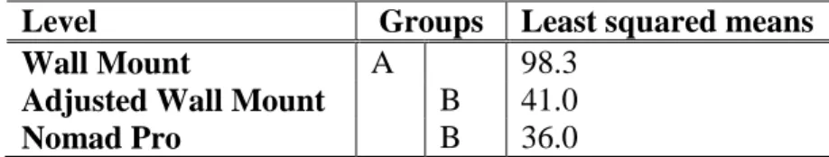
ll mounted and handheld doses. Mean (SD) FMX effective dose was significantly
less for the Nomad Pro 36µSv (8.4) than for wall-mount 98µSv (14.3) (p=0.0217). The
mounted source was 41µSv, Table 2. Mean operator exposures
were indistinguishable from ambient background levels (<2 µGy/study) and were not different
handheld and wall mounted sources (p=0.2624) or dosimeter location (p=0.6815) during
Table 3. Total effective patient dose was reduced 12% with the
0.40 0.60 0.80 1.00 1.20
Exposure time (sec)
– Planmeca Calibration Curves
indicated a statistical significant difference among wall mounted and handheld
). Tukey HSD tests indicated statistical significant
wall mounted and normalized
Tukey HSD test indicated no statistical significant difference between
dose was significantly
mount 98µSv (14.3) (p=0.0217). The
Mean operator exposures
were indistinguishable from ambient background levels (<2 µGy/study) and were not different
(p=0.2624) or dosimeter location (p=0.6815) during
Total effective patient dose was reduced 12% with the 1.20
Planmeca
18
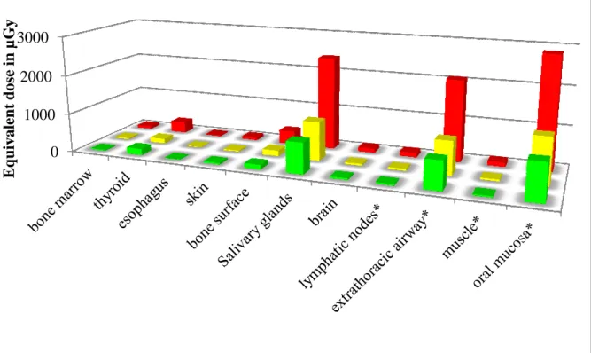
handheld device during FMX simulation. A comparison of tissue equivalent dose is
presented, Fig 5.
For image analysis a linear mixed model was separately used for each outcome.
Unstructured covariance structure was assumed for the six repeated measurements. Pairwise
interaction between device and observer was included in the model, and removed if not
statistically significant. The least squared means were then calculated from each final model.
Significant level was p = 0.05.
ANOVA indicated no statistical significant difference in mean (SD) total technique error
between devices adjusting for observers, (p = 0.29). A statistical significant interaction between
device and observer, (p < 0.01) was observed. Mean sum total error rate by location: posterior
periapical, anterior periapical, and bitewing are presented in, Table 5.
For mean LP resolution, there was not statistically significant interaction between
observer and device indicating that the pattern of responses for the observers was similar for the
two devices (P>.05). There was a statistically significant difference among observer mean LP
resolution adjusting for device (p < 0.01) as well as a significant difference between devices
adjusting for observer (p < 0.01), Table 4. Higher LP resolution favoring the handheld x-ray
source was demonstrated, Table 5.
Weighted Kappa using sum total error and mean LP resolution was utilized to evaluate
inter-observer reliability. The weighting scheme was linear with no proportionality.
Inter-observer weighted Kappa values were moderate in evaluation of packet placement (pp) and
19
line pair LP resolution was substantial between observer A and C. Between all observers
weighted Kappa values range fair to substantial in cone cut (cc) errors, and image distortion (id)
errors, Table 6. Intra-observer theoretically in almost all previous research is better than
inter-observer reliability. Given the experience level of observers and that there was no
expectation of learning during the wash-out period it was elected not to pursue
intra-observer agreement.
Operator satisfaction with the handheld device during FMX simulation was
favorable. Considering ease of use of the handheld device 97% of the operators were
satisfied compared to only 50% satisfied with the wall mounted device. 87% of the
operators would recommend the handheld device to colleagues and 99% of the operators were
satisfied with their overall experience with the handheld device, Table 7.
Table 2. Effective Dose ANOVA Table and Tukey Test
Source df F Ratio Prob > F
Device 2 34.1298 0.0005*
Level Groups Least squared means
Wall Mount A 98.3
Adjusted Wall Mount B 41.0
Nomad Pro B 36.0
Table 3. ANOVA Full Model
Source Nparm df Sum of Squares F Ratio Prob > F Device 1 1 0.3112963 1.2815 0.2624
Study 3 3 1.0980815 1.5068 0.2227
Figure 6. Tissue specific equivalent dose comparisons
Nomad Pro
Table 4. Linear mixed model.
Outcome Factor
err device observer device*observer err_bw device observer device*observer LP device observer device*observer 0 1000 2000 3000 E q u ival en t d os e i n µ G y 20
fic equivalent dose comparisons
Wall Unit Normalized Exposure Wall Unit
DF F P
1 / 74 1.10 0.2977 2 / 74 37.96 <.0001 2 / 74 5.66 0.0052 1 / 74 1.52 0.2221 2 / 74 20.27 <.0001
(removed)
1 / 74 35.12 <.0001 2 / 74 58.45 <.0001
(removed)
21
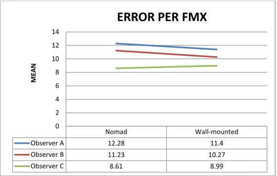
Figure 7. Mean error per FMX.
Figure 8. Mean LP resolution.
Nomad Wall-mounted
Observer A 12.28 11.4
Observer B 11.23 10.27
Observer C 8.61 8.99
0 2 4 6 8 10 12 14 M E A N
ERROR PER FMX
Nomad Wall-mounted
Observer A 6.05 5.58
Observer B 6.55 6.31
Observer C 6.17 5.75
22
Table 5. Descriptive statistics.
Variable Label device observer N Missing Q1 Median Q3 Mean Std
err
sum of (err1-err18)
Nomad
A 75 0 8 13 16 12.28 5.45
B 75 0 7 11 16 11.23 5.60
C 75 0 6 8 11 8.61 3.86
Wall Mounted
A 75 0 9 12 14 11.40 4.51
B 75 0 8 10 12 10.27 3.86
C 75 0 7 9 11 8.99 3.42
err_ant anterior - sum of technique errors Nomad
A 75 0 2 3 5 3.40 2.19
B 75 0 0 2 4 2.40 2.30
C 75 0 1 2 3 2.24 1.79
Wall Mounted
A 75 0 2 3 4 2.93 1.92
B 75 0 0 2 2 1.53 1.67
C 75 0 0 1 2 1.56 1.51
err_post
posterior - sum of technique
errors
Nomad
A 75 0 4 6 8 5.99 3.19
B 75 0 3 6 9 5.92 3.39
C 75 0 3 4 6 4.37 2.22
Wall Mounted
A 75 0 5 6 8 6.08 2.93
B 75 0 4 6 8 6.21 2.52
C 75 0 4 5 7 5.47 2.53
err_bw
bitewing - sum of technique
errors
Nomad
A 75 0 2 3 4 2.89 1.78
B 75 0 2 2 4 2.91 1.87
C 75 0 1 2 3 2.00 1.46
Wall Mounted
A 75 0 1 2 3 2.39 1.56
B 75 0 1 2 4 2.52 1.87
C 75 0 1 2 3 1.96 1.44
LP line pair resolution
Nomad
A 75 0 6 6 6 6.05 0.82
B 75 0 6 7 7 6.55 0.81
C 75 0 6 6 7 6.17 0.55
Wall Mounted
A 74 1 5 6 6 5.58 0.60
B 75 0 6 6 7 6.31 0.49
23
Table 6. Inter-observer Kappa values.
Observer pp (Packet placement)
ha (Horizontal angulation)
va (Vertical angulation)
id (Image Distortion)
LP (line pair resolution)
Weighted Kappa
ASE Weigh ted Kappa
ASE Weighte d Kappa
AS E
Weighte d Kappa
AS E
Weighte d Kappa
AS E
A B 0.51 0.14 0.44 0.14 0 0 0.61 0.18 0.23 0.19
A C 0.03 0.09 0.56 0.13 0 0 0.35 0.20 0.69 0.19
24
Table 7. Operator satisfaction
Variable Label Category* Freq Pct (%)
Q1 Aribex, Nomad® Pro handheld device is easy to use.
1 1 1
3 1 1
4 23 30
5 51 67
Q2 Conventional wall-mounted x-ray source is easy to use.
1 1 1
2 8 11
3 29 38
4 31 41
5 7 9
Q3 Aribex, Nomad® Pro handheld device is much better than conventional wall-mounted x-ray source.
1 1 1
2 8 11
3 16 21
4 31 41
5 20 26
Q4 I would use the Aribex, Nomad® Pro handheld device in the future.
1 1 1
3 3 4
4 34 45
5 38 50
Q5 I would purchase the Aribex, Nomad® Pro handheld device for my practice.
0 1 1
1 1 1
2 3 4
3 19 25
4 26 34
5 26 34
Q6 I would recommend the Aribex, Nomad® Pro handheld device to colleagues.
1 1 1
3 9 12
4 40 53
5 26 34
Q7 The CD/ROM on-line training module
adequately prepared me to complete the clinical procedure.
missing 1 1
0 2 3
1 1 1
3 4 5
4 42 55
5 26 34
Q8 Overall, how satisfied are you with your experience with the Aribex, Nomad® Pro handheld device.
3 1 1
4 35 46
5 40 53
25
DISCUSSION:
The significant differences noted in patient effective dose and LP resolution between
devices were primarily due to unit technical specifications and technique protocols: kVp, mA,
exposure, and focal spot size. The calculated mAs was 0.5 and 2.56 for the handheld and
wall-mounted devices respectively. Dose reduction was attributed to the optimization of exposure
factors while LP resolution was primarily due to smaller focal spot size with the handheld unit.
A limitation within the methodology however is noted with scanning acquired images at 300 dots
per inch (DPI). At the lower 300 DPI scanner setting, versus 600 DPI, mean average LP
resolution observed approaches the theoretical threshold for resolution. Scan setting at 600 DPI
may demonstrate different LP resolution results. Further research in image quality with the
Nomad Pro is needed to fully evaluate actual threshold for LP resolution with a handheld device.
More rigorous observer calibration may be beneficial in future comparison image quality
studies assessing handheld and wall mounted x-ray sources. For sum total technique error,
Observer A reported the highest number of average errors and Observer C the lowest for both
devices. Both Observer A and B reported slightly lower average sum total errors for the
wall-mounted x-ray source while Observer C noted no difference, Observer difference does not
follow the same pattern with each device, Figure 7. Observers B mean LP resolution is higher
over devices than mean LP resolution for observer A and C. Observer difference follows the
same pattern with each device, Figure 8. A consensus model of agreement where observer
26
constraints for observers to achieve consensus in our study of one hundred and 150, 18 image,
FMX’s for six technique errors and LP resolution was not feasible.
Dental student performance is well documented in the literature. Mourshed12 in a study of
FMX’s made by dental students reported a technical error rate of 47 and 48 percent with
periapical and bitewing radiographs utilizing bisecting the angle technique. The most frequent
error reported was packet placement. The average total error rate per FMX was 7.8. Ilkay13
evaluated periapical radiography utilizing bisecting the angle technique. Approximately 64
percent of the radiographs were deemed unacceptable and the two most frequent technique errors
were incorrect angulation 35 percent and incorrect packet placement 34 percent. Crandell14
reported average errors per FMX for dental hygiene students, senior, and junior dental students
of 0.53, 1.48, and 1.73 respectively. Studies15-18 utilizing positioning devices in intraoral
radiography noted technique errors due to cone cut decreased while packet placement errors
increased.
In our study second year dental students were evaluated and utilized paralleling
techniques with a positioning device with round cones. Mean error rate per FMX was 12.28
(5.45) for the handheld device and 11.4 for the wall-mounted device. By anatomic location the
highest mean error was noted in posterior periapical imaging with mean error per FMX of 5.99
and 6.08 for the handheld and wall-mounted device respectively. The mean error per FMX in the
anterior region was 2.93 for the handheld device and 1.56 for the wall-mounted device.
Bitewing imaging demonstrated a mean error per FMX of 2.89 and 2.39 for the handheld and
wall-mounted device respectively. Dental student performance reported in our study cannot be
27
technique, positioning devices, collimation, and student experience. For our cohort of second
year dental students this exercise was their first pre-clinical radiology experience. Sum total
technique error per FMX may be substantially different in a more clinically experienced dental
student population.
In conclusion, the Nomad Pro is as safe to use for operators and safer to use for
patients when comparing equivalent dose with conventional wall-mounted sources using
round-cones. Patient dose reduction is attributable to optimization of exposure factors.
Technique charts at the University of North Carolina, School of Dentistry recommended
exposure for molar projections was 0.32 seconds. While recommended exposure was in the
upper limit of the demonstrated latitude this was appropriate given the dental student cohort.
Optimized exposure factors may not be appropriate for dental school environments. In an
institutional setting transitioning from wall-mounted x-ray sources to the Nomad Pro
handheld device, a reduction in patient exposure most likely will take place. Technique
error rates equivalent between handheld and wall mounted x-ray devices. LP resolution
favors the hand-held device. Finally, overall operator satisfaction with the handheld device
28
REFERENCES
1. Richards. Albert G., Sources of x-radiation in the dental office. Dent Radior Photogr 37(3):51-68, 1964.
2. Richards, Albert G, Colquitt, Wayne N. Reduction in dental x-ray exposures during the past 60 years. JADA 103:713-718, 1981.
3. Nolan, W. E. Radiation hazards to the patient from oral roentgenography. JADA 47(6): 681-684, 1953.
4. Budowsky, J., and others. Radiation exposure to the head and abdomen during oral roentgenography. JADA 52(5):555-559, 1956.
5. Bailey, N. A. Patient exposure to ionizing radiation in dental radiography. Radiol 69:42-45, 1957.
6. Blackman, S., and Greening, J. R. Radiation hazards in dental radiography. Br Dent J 102:167-172, 1957.
7. Bjarngard, B., and others. Radiation doses in oral radiology. Odontology Rev 10(4):355-366, 1959.
8. Alcox, R. W., and Jameson, W.R. Patient exposures from intraoral radiographic examinations. JADA 88(3):588-579, 1974.
9. Bailey E., Gray J., Ludlow J. Image Quality and Radiation Dose for Intraoral Radiography: Hand-Held (Nomad®) Battery Powered vs. Wall-Mount X-ray Systems. 41st Annual Conference on Radiation Control Conference of Radiation Control Program Directors. May, 2009.
10. Goren A.D., Bonvento, M., Biernacki J., Colosi D.C. Radiation exposure with the NOMAD® portable X-ray System. Dentomaxillofac Radiol 37:109-112, 2008.
11. Department of the Navy Memorandum: Appropriate Use of Hand-Held X-Ray Units for Oral and Maxillofacial Radiography. 2008; 6600:SerM3C/AT-17215, 2008.
12. Mourshed F.A.,A study of intraoral radiographic errors made by dental students. Oral Surg Oral Med Oral Pathol 32(5):824-828, 1971.
13. Ilkay, P., Meryem T.A., Evaluation of Radiographic Errors Made by Undergraduate Dental Students in Periapical Radiography. NYSDJ Aug/Sep, 45- 48, 2009.
29
15. Bean, L.R., Comparison of Bisecting-Angle and Paralleling Methods of Intra-Oral Radiology. J. Dent. Educ. 33:441-445, 1969.
16. Jensen, T.W. Improved reliability of dental radiography by application of x-ray guiding instruments. J. Dent. Educ. 42:481-485, 1978.
17. Horton, P.S., Sippy, F.H., Nelson, J.F., et al. A comparison of rectangular and cylindrical collimation for intraoral radiographs. J. Dent. Educ. 47:771-773, 1983.