Comparison of the photon charge between water and
solid phantom depending on depth
INTRODUCTION
The ability to diagnose cancer has improved with the development of medical technologies.
This optimism has prompted interest in radiotherapy as a cancer treatment method. Radiotherapy is a clinical method that involves
radiation with a very short wavelength and high energy, which reacts with water in the human body and affects the cells through physical, chemical and biological processes (1-3). Radiation destroys cancer cells and normal cells, requiring exact dose management during the radiotherapy
many non-cancer cells as possible.
In particular, the high energy photons used in radiotherapy form a build-up area where the
surface dose is low and the dose sharply increases from the surface to the maximum
depth (4). Also, the distribution of the absorbed dose in the build-up area of the photon beam varies depending on the irradiation level and energy and shows very abrupt changes even with small changes in the depth. Therefore, it is very dif&icult to exactly measure the distribution of the absorbed dose.
In terms of the management of high energy
J.W. Hong
1,2, H.K. Lee3, J.H. Cho2*1Department of Radiation Oncology, Seoul National University Bundang Hospital, Republic of Korea 2Department of International Radiological Science, Hallym University of Graduate Studies, Republic of Korea
3Department of Computer Science and Engineering, Soonchunhyang University, Republic of Korea
ABSTRACT
Background: This study assessed the clinical usefulness of the solid phantom, which may compensate for the disadvantages of the water phantom, by comparing the radiological doses between the two depending on their depths.Materials and Methods: The experimental equipment used was a linear accelerator for medical use, water phantom, solid phantom, Farmer type ion chamber and electrometer. The distance between the ray source and the center of the ion chamber was fixed to a SAD of 100 cm during the experiment. The field size was 10 x 10 cm2 and the radia%on energies of the photon rays were 6 MV and 15 MV. The depth interval was 1cm (range 1-10 cm) for each energy. The rela%ve devia%on ra%o of the water phantom to the solid phantom was calculated. Results: The measurement at 100 MU was performed more than five %mes to calculate the average charge and absorbed dose, and the rela%ve devia%on was analyzed based on the water phantom. The results obtained at depths from 1 to 10 cm were 0.034%, -0.457%, -0.167%, 0.011%, 0.117%, 0.271%, 0.349%, 0.709%, 0.376% and 0.611% at 6 MV and those obtained at 15 MV were 1.199%, 0.033%, 0.166%, 0.496%, 0.556%, 0.705%, 0.656%, 1.071%, 0.7% and 1.057%, respec%vely.
Conclusion: In conclusion, the solid phantom is useful and may complement the disadvantages of the water phantom, including the %me required for its installa%on and errors in the measurement depth, and may precisely measure the radiological dose.
Keywords: W ater phantom , solid phantom , charge, absorbed dose.
* Corresponding author:
Dr. Jae-Hwan Cho,
Fax: +82 2 3453 6618
E-mail:
Revised: Sept. 2014
Accepted: Oct. 2014
Int. J. Radiat. Res., July 2015; 13(3): 229-234
►
Original article
DOI: 10.7508/ijrr.2015.03.005
measure the dose of the treatment source of origin and assess the error. Currently, the water
phantom is the benchmark phantom most similar to the human body that is used for the
quality control of the absorbed dose management of the X-rays in the linear accelerator (5, 6). However, it takes a long time to
perform the test with the water phantom, due to its complicated implementation procedure and this causes errors in the measurement depth, resulting from the formation of waves from the surface of the water phantom caused by the movement of the measurement position of the
ionization chamber and stabilization of the water temperature (7).
Other than for special purposes, the solid phantom is effective for saving time and easy to use, including for obtaining the beam data for
the treatment plan system or calibrating the output of the linear accelerator (8).
Recently, the solid water equivalent phantom
has been used in the normal quality control procedure as an alternative to the water phantom.
The purpose of the study was to assess the clinical usefulness of the solid phantom, which may compensate for the disadvantages of the water phantom, by comparing their radiological doses depending on their depths.
MATERIALS
AND METHODS
The solid phantom, the water phantom used for the calibration and the ionization chamber (PTW, farmer type waterproof 0.6cc ionization chamber, Freiburg, Germany) were placed in the treatment room with the vertical direction of the beam radiation against the linear accelerator manufactured by Varian Medical Systems and a voltage meter (PTW, Freiburg, Germany) in the control room, and connect them with the cable of the triaxial ion chamber.
The experimental equipment used in the study was a linear accelerator for medical use, water phantom, and solid phantom and Farmer type ion chamber. The solid phantom used in the experiment consisted of a slab with a thickness of 0.2 - 0.6 cm and area of 30×30 cm
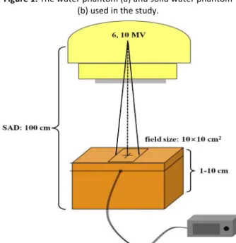
(&igure 1).
First, the distance between the ray source and the center of the ion chamber was &ixed at a SAD of 100 cm. The &ield size was 10×10 cm2 and the radiation energies of the photon rays were 6MV and 15MV. The depth interval was 1 cm (range, 1 10 cm) for each energy. The thickness of the phantom bottom was set to 5 cm to reduce the effect of the backward scattering when using the
solid phantom (&igure 2). In addition, the
Figure 2. The distance between the ray source and the center of the ion chamber was fixed at a SAD of 100 cm. The
field size was 10x10 cm2 and the radia%on energies of the photon rays were 6 MV and 15 MV.
Figure 1. The water phantom (a) and solid water phantom (b) used in the study.
(1)
where Mw(z) and Ms(z) are the ionization measurements from the water and solid phantoms for each irradiation surface size, respectively. The relative deviation at a depth of
z of material m is de&ined as equation 2.
(2)
where M is the ionization value at a depth of z and zref . Deriving the percentage deviation ∆(z)
from the relative deviation between the water and solid phantoms is achieved using equation (3) with the calibration factor obtained from equation 1 (5, 9).
(3)
RESULTS
The average values of the charge at depths in the range from 1 cm to 10 cm with intervals of 1
cm measured 30 times at each depth with a radiation energy of 6 MV for the water and soild
phantoms are shown table 1. The highest dose
when using the water phantom was
111.03±0.0047 cGy at a depth of 2 cm and the lowest dose was 87.03±0.0073 cGy at a depth of 10 cm (p<0.05). The highest dose when using the solid phantom was 111.64±0.0047 cGy at a depth of 2 cm and the lowest dose was 86.59±0.0046 cGy at a depth of 10 cm (p<0.05).
The average values of the charge at depths in the range from 1 cm to 10 cm with intervals of 1
cm measured 30 times at each depth with a radiation energy of 15 MV for the water and
soild phantoms are shown in table 2. The highest dose when using the water phantom was 107.08 cGy at a depth of 4 cm and the lowest dose was
81.94 cGy at a depth of 1 cm (p<0.05). The highest dose when using the solid phantom was
107.29 cGy at a depth of 3 cm and the lowest dose was 82.50 cGy at a depth of 1 cm (p<0.05).
The relative deviations in the measurements phantom was placed in the treatment room for
more than 1 week to minimize the impact of temperature changes on the measurements and
keep the temperature changes during the measurement as small as possible when using
the solid phantom.
The water temperature was kept within 0.1
of that of the treatment room to maintain the thermal equilibrium between the treatment
room and the water when using the water phantom. The depth in each case was considered
to be from the center of the ionization chamber to the source of the rays in each phantom during the measurement.
All the experiments were performed at a dose rate of 600 MU/min and irradiation doses of 100 MU and 80 MU to measure the charge against
the benchmark irradiation surface and the output factor on the irradiation surface, respectively. The experiment at 100 MU was
performed 30 times to calculate the average charge and the results were analyzed based on
the water phantom. Then, the charge was measured and the measured charge (nC) In the
&irst highlighted sentence, the “respectively” seems to indicate that irradiation dose of 100 MU refers to the charge against the benchmark irradiation surface and that of 80 MU refers to the output factor on the irradiation surface. Is that correct? was converted to the absorbed dose (μGy) by multiplying it by the SAD factor. This study only considers the SAD factor, because it does not use the tray, block and MLC. The SAD factors at 6 MV and 15 MV were 1.03 and 1.055, respectively. The difference in the mean charge and absorbed dose at each depth as a function of the energy was tested by ANOVA (SPSS win 18.0, USA) and, to obtain a more accurate difference, a post-hoc analysis was conducted. (p<0.05) Also, the relative deviation ratio of the water phantom to the solid phantom was calculated.
The ionization values at the depth of the solid phantom were proportional to the water
equivalent depth and were obtained by multiplying the calibration factor by the ionization measurement at an arbitrary depth
(z). The calibration factor (hw,s) was calculated
from equation 1 (5, 9).
between the two phantoms for each energy based on the water phantom were measured every 1 cm from 1 cm to 10 cm and the results were 0.034%, -0.457%, -0.167%, 0.011%, 0.117%, 0.271%, 0.349%, 0.709%, 0.376% and
0.611% at 6 MV (&igure 3) and 1.199%, 0.033%, 0.166%, 0.496%, 0.556%, 0.705%, 0.656%, 1.071%, 0.7% and 1.057% at 15 MV (&igure 4), respectively.
Table 1. The measured charge and absorbed dose at a depth of 6 MV for the water phantom and solid phantom.
Phantom Division Depth (cm) P
water phantom
charge (nC)
1 2 3 4 5 6 7 8 9 10
0.400 17.49± 0.0055 18.36± 0.0045 18.00± 0.0045 17.54± 0.0084 17.04± 0.0045 16.55± 0.0045 16.02± 0.0055 15.51± 0.0045 14.91± 0.0045 14.4± 0.0071 absorbed dose (cGy) 105.77± 0.0057 111.03± 0.0047 108.85± 0.0047 106.01± 0.0087 102.9± 0.0047 100.0± 0.0057 96.88± 0.0057 93.74± 0.0047 90.11± 0.0047 87.03± 0.0073 soild phantom
charge (nC) 17.49± 0.000 18.45± 0.0045 18.03± 0.0084 17.54± 0.0055 17.02± 0.011 16.51± 0.0089 15.97± 0.000 15.40± 0.0045 14.85± 0.0089 14.31± 0.0045 0.035 absorbed dose (cGy) 105.83± 0.000 111.64± 0.0047 109.16± 0.0087 106.19± 0.0057 103.05 ±0.011 99.96± 0.009 96.63± 0.000 93.19± 0.0047 89.92± 0.009 86.59± 0.0046
Table 2. The measured charge and absorbed dose at a depth of 15 MV for the water phantom and solid phantom.
Phantom Division Depth (cm) P
water phantom
1 2 3 4 5 6 7 8 9 10
charge (nC) 15.01± 0.0044 18.43± 0.0044 19.28± 0.044 19.26± 0.0044 18.96± 0.0054 18.72± 0.008 18.28± 0.0044 17.92± 0.0070 17.42± 0.0054 17.02± 0.0083 0.045 absorbed dose (cGy) 83.016± 0.024 101.96± 0.024 10666±0 .024 106.51± 0.024 104.88± 0.030 103.53± 0.046 101.09± 0.0024 99.09± 0.039 96.36± 0.0030 94.16± 0.046 soild phantom charge (nC) 14.83±0 .010 18.43± 0.0045 18.03± 0.0084 17.54± 0.0055 17.02±0 .011 18.59±0 .000 18.16±0 .004 17.72± 0.008 17.30± 0.0054 16.84± 0.008 0.040 absorbed dose (cGy) 82.02±0 .060 101.929 ±0.024 109.16± 0.0087 106.19± 0.0057 103.05± 0.011 102.80± 0.000 100.43± 0.024 98.0.±0 .046 95.69± 0.030 93.16± 0.0046
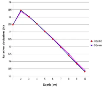
Figure 3. Comparison of the charge depending on the depth at a radia%on energy of 6 MV in the case of the water
phantom / solid water phantom.
Figure 4. Comparison of the charge depending on the depth at a radia%on energy of 15 MV in the case of the water
phantom / solid water phantom.
DISCUSSION
It is very important to precisely measure the
treatment dose and assess the error in high energy radiotherapy. Water is normally
recommended for the absolute measurement of the absorbed dose, but its disadvantages include the waterproof nature of the ionization chamber and the time required to place the ionization chamber at the correct position in the water (7, 8,
12, 13). Therefore, except for special purposes, the
solid phantom is ef&icient in terms of the time required and ease of use, including for achieving the beam data for the treatment plan system or calibrating the output of the linear accelerator. The purpose of this study was to assess the clinical usefulness of the solid phantom, which may compensate for the disadvantages of the water phantom, by comparing the radiological doses between the two types of phantom depending on their depths. The results obtained at depths from 1 to 10 cm were 0.034%, -0.457%, -0.167%, 0.011%, 0.117%, 0.271%, 0.349%, 0.709%, 0.376% and 0.611% at 6 MV and 1.199%, 0.033%, 0.166%, 0.496%, 0.556%, 0.705%, 0.656%, 1.071%, 0.7% and 1.057% at 15 MV, respectively. Kim et al. (5) compared the relative deviation depending on the depth with a white-colored polystyrene phantom and water phantom using a 10 MV X-ray beam and reported that the percentage deviation was < 0.53%. The relative deviations in (their?) study were < 0.611% and (<?) 1.057% for the 6 MV and 15 MV X-rays, respectively, where the different range of the deviation was due to the different energy intensities and depths.
Thomadsen et al. (14) compared the relative deviation for each energy level using the solid and water phantoms for electron beams ranging
from 6 to 18 MV and reported that the percentage deviation ranged from 0.46 - 0.68%.
This variation of the deviation comes from the measurement with the electron beam and not that with the X-rays. Huang et al. (15) compared the relative deviations for each energy level using the solid phantoms with the common slab and acryl and reported a result of 0.48%. As shown above, it is found that the relative
deviation depends on the phantom material.
The measurements con&irm that the solid and
water phantoms show different relative deviations depending on the energy level and
the depth. Also, another study showed that the
relative deviation depends on the phantom element and the type of radiation.
CONCLUSION
The recommended value of the relative error
in the absorbed dose based on the water phantom is ±2%. The present study shows that
the minimum and maximum errors depending
on the depths of the measurement for the various mediums and energy levels are -0.457%
and 1.199%, respectively, which conform to the recommended values. It is considered that the solid phantom is useful and may overcome the disadvantages of the water phantom, including the time required for its installation and errors in the measurement depth, while allowing the radiological dose to be precisely measured. The
data from the present study may be directly employed in the assessment of the dose by
measuring the output of the linear accelerator using the solid phantom, as well as the data analysis.
ACKNOWLEDGEMENTS
Joo-Wan Hong and Hae-Kag Lee contributed equally to this work. They are co-7irst authors.
This work was supported in part by the Soonchunhyang University Research Fund.
Con licts of interest: none to declare.
REFERENCES
1. Segawa Y, Takigawa N, Kataoka M, Takata I, Fujimoto N, Ueoka H (1997) Risk factors for development of radia%on pneumoni%s following radia%on therapy with or without chemotherapy for lung cancer. Int J Radiat Onco Biol phys, 39:91-98.
2. Armstrong J, Raben A, Zelefsky M, Burt M, Leibel S, Burman C, Kutcher G, Harrison L, Hahn, C, Ginsberg R, Rusch V, Kris M, Fuks Z (1997) Promising survival with three-dimensional
conformal radia%on therapy for non-small cell lung cancer.
Radiother Oncol, 44:17- 22.
3. Lee CG (2004) High precision radiotherapy. J Korean Med Assoc, 47:663 - 671.
4. Khan FM (1984) The Physics of Radia%on Therapy. Williams &Wilkins, Bal%more.
5. Kim JE, Cha BY, Kang SS, Park JK, Sin JW, Kim SY, Jo SH, Son DW, Choi CW, Park CH, Yoon CH, Lee JD, Park BD, Nam SH (2008) 10 MV X-ray Beam Dosimetry by Water and White Polystyrene Phantom. JRST, 31:83-87.
6. Venencia CD, Besa P (2004) Commissioning and quality assurance for intensity modulated radiotherapy with dynamic mul%leaf collimator : Experience of the Pon%ficia Universidad Catolica de Chile. Am Coll Med Phys, 5:37-54. 7. IAEA (2000) Absorbed Dose Determina%on in External Beam
Radiotherapy : An Interna%onal Code of Prac%ce for Dosimetry based on Standards of Absorbed dose to Water. Technical Report Series. 398.
8. Lee JO, Jeong DH, Kim BG (2009) Study on the Characteris%cs of Response Correc%on Factor of Ioniza%on Chamber in RW3 Solid Phantom for High Energy X-rays. JRST,
32:205-212.
9. Christ G (1995) White polystyrene as a subs%tute for water
in high energy photon dosimetry. Med Phys, 22:2097-100.
10. Ezzell GA, Galvin JM, Low D, Palta JR, Rosen I, Sharpe MB, Xia P, Xiao Y, Xing L, Yu CX (2003) IMRT subcommiKee.; AAPM Radia%on Therapy commiKee. Med Phys, 30 :2089-115.
11. Low DA, Mu%c S, Dempsey JF, Gerber RL, Bosch WR, Perez CA, Purdy JA (1998) Quan%tave dosimetryc verifica%on of an IMRT planning and delivery system. Radiother Oncol, 49: 305-16.
12. Furhang EE, Chui CS, Sgouros GA (1996) A Monte Carlo approach to pa%ent-specific dosimetry. Med Phys, 23 :1523-9.
13. Siebers JV, Keall PJ, Nahum AE, Mohan R (2000) Conver%ng absorbed dose to medium to absorbed dose to water for Monte Carlo based photon beam dose calcula%ons. Phys Med Biol, 45:983-95.
14. Thomadsen B, Constan%nou C, Ho A (1995) Evalua%on of water-equivalent plas%cs as phantom material for electron-beam dosimetry. Med Phys, 22:291-6.
15. Huang JC and Reinstein LE (2000) Evalua%on of an innova%ve plas%c cube phantom designed to improve the efficiency of accelerator QA. Autumn, 1:153-7.