INTERLEUKIN-2 BASED THERAPY FOR THE TREATMENT OF TYPE I DIABETES
MARK CHRISTOPHER JOHNSON
A dissertation submitted to the faculty of the University of North Carolina at Chapel Hill in partial fulfillment of the requirements for the degree of Doctor of Philosophy in the
Department of Microbiology and Immunology.
Chapel Hill 2012
Approved by:
Barbara Vilen, Ph.D.
Robert Maile, Ph.D.
Roland M. Tisch, Ph.D.
Scott Plevy, M.D.
ii ©2012
iii ABSTRACT
MARK CHRISTOPHER JOHNSON: INTERLEUKIN-2 BASED THERAPY FOR THE TREATMENT OF TYPE I DIABETES
(Under the direction of Dr. Roland M. Tisch)
Type I diabetes (T1D) is an autoimmune disease characterized by the destruction of the insulin producing β cells. Although multiple cell types contribute, the main
mediators of β cell destruction are pathogenic Th1 effectors (Teff). Preferential
differentiation and expansion of pathogenic Teff is partly attributed to dysregulation of FoxP3+ regulatory T cells (FoxP3+ Treg). Consequently, current strategies for treating T1D have focused on re-establishing the balance between Teff and FoxP3+ Treg. The aims of
the studies described are to: i) analyze the temporal effect of IL-2 on FoxP3+ Treg and disease incidence, ii) to test whether β cell-specific IL-2 secretion prevents T1D by expanding islet resident FoxP3+ Treg, and iii) to investigate the synergistic ability of T cell immunotherapies to induce remission in non obese diabetic (NOD) mice.
iv
relationship between systemic IL-2 expression, FoxP3+ Treg function in vivo and disease incidence.
Our second study investigated the ability of islet-localized IL-2 to prevent
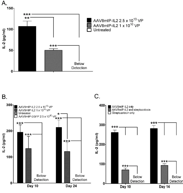
diabetes in NOD mice. We found that vaccination with a recombinant adeno-associated viral vector (rAAV) expressing IL-2 under control of the mouse insulin promoter
(AAV8mIP-IL2) prevented diabetes. Protection correlated with increased number and function of islet FoxP3+ Treg. Importantly, the effects of AAV8mIP-IL2 vaccination were islet specific. This shows that IL-2 expression driven by AAV8mIP-IL2 protected NOD mice, highlighting the potential of targeted immunotherapeutic treatment.
v
ACKNOWLEDGEMENTS
First and foremost, I would like to thank my mentor, Dr. Roland Tisch, for granting me his patience and assistance. I am indebted to him for overseeing my maturation as a scientist. I would also like to give my sincere thanks to my committee members, Dr. Stephen Clarke, Dr. Robert Maile, Dr. Scott Plevy, and Dr. Barb Vilen for granting me their valuable time and for guiding me through this arduous journey. Additionally, I would like to extend my gratitude to all members of the Tisch lab, both past and present, for their input and assistance. In particular, I would like to thank Dr. Kevin Goudy, for being a patient and irreplaceable teacher, Dr. Alaina Garland, without whom this work would not have been possible, and Dr. Charlie Kroger, for our great discussions and for being a wonderful friend. Furthermore, I would also like to thank Jude Samulski and the Samulski lab for their constant kindness and help.
vi
vii
TABLE OF CONTENTS
LIST OF TABLES ... x
LIST OF FIGURES ... xi
LIST OF ABBREVIATIONS ... xiii
CHAPTER 1. INTRODUCTION ... 1
1.1 Diabetes Mellitus ... 1
1.2 Islet Transplantation for the Treatment of T1D ... 2
1.3 Protective Immunity ... 3
1.4 Central and Peripheral Self-Tolerance ... 4
1.5 Treg Play a Key Role in Peripheral Self-Tolerance ... 7
1.6 Modes of FoxP3+ Treg-mediated Suppression ... 8
1.7 Genetic and Environmental Factors Influencing T1D Susceptibility ... 11
1.8 The Non-obese Diabetic (NOD) Mouse Model of T1D ... 13
1.9 CD4+ and CD8+ T cells are the Primary Mediators of T1D ... 14
1.10 The Role of IL-2 in Autoimmunity ... 17
1.11 Clinical Scenarios for the Treatment of T1D ... 20
viii
1.13 Gene Delivery via Recombinant Adeno-Associated (rAAV) Vectors... 22
1.14 Applications of rAAV Vector-Based Immunotherapy for T1D ... 24
1.15 Aims of the Dissertation ... 26
1.16 References ... 29
2. REDUCED IL-2 EXPRESSION IN NOD MICE LEADS TO A TEMPORAL INCREASE IN CD62LLOFOXP3+CD4+ T CELLS WITH LIMITED SUPPRESSOR ACTIVITY ... 52
2.1 Summary ... 52
2.2 Introduction ... 54
2.3 Materials and Methods ... 57
2.4 Results ... 62
2.5 Discussion... 68
2.6 References ... 85
3. ADENO-ASSOCIATED VIRUS VECTOR MEDIATED β-CELL SPECIFIC IL-2 EXPRESSION SUPPRESSES TYPE I DIABETES IN NOD MICE ... 90
3.1 Summary ... 90
3.2 Introduction ... 92
3.3 Materials and Methods ... 96
3.4 Results ... 101
3.5 Discussion... 109
ix
4. COMBINATORIAL IMMUNOTHERAPY FOR THE PREVENTION AND
TREATMENT OF TYPE I DIABETES ... 136
4.1 Summary ... 136
4.2 Introduction ... 138
4.3 Materials and Methods ... 140
4.4 Results ... 144
4.5 Discussion... 149
4.6 References ... 158
5. FUTURE PERSPECTIVES ... 162
5.1 Increased Efficacy of AAV8mIP-IL2 Immunotherapy for the Treatment of T1D ... 162
5.2 Alternative Transgene Expression for rAAV Vector Based Immunotherapy ... 165
5.3 References ... 168
x
LIST OF TABLES
xi
LIST OF FIGURES
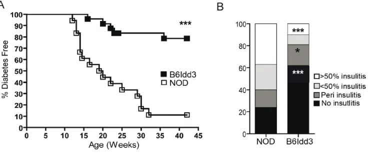
Figure 2.1: NOD.B6Idd3 female mice exhibit a reduced frequency of diabetes and
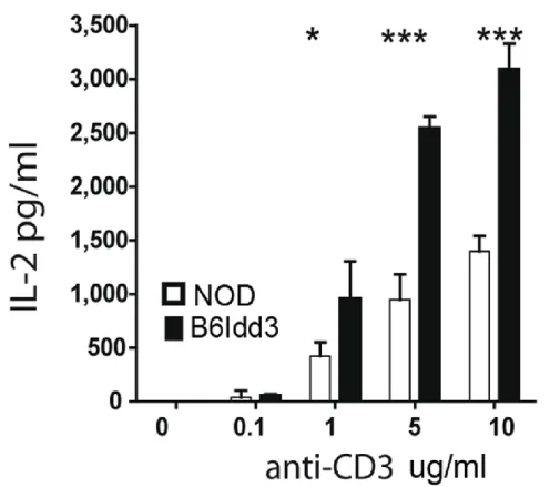
insulitis compared to NOD female mice ... 74 Figure 2.2: NOD.B6Idd3 versus NOD naïve CD4+ T cells secrete more IL-2 upon
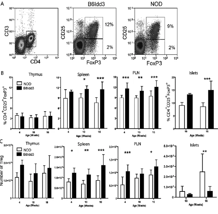
stimulation ... 75 Figure 2.3: NOD.B6Idd3 mice have an increased frequency of peripheral FoxP3+ Treg
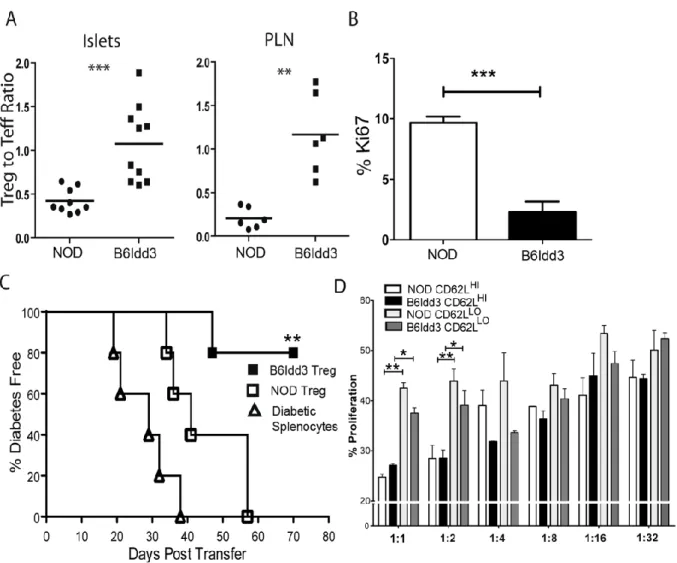
compared to age-matched NOD mice ... 76 Figure 2.4: A temporal shift in CD62Lhi- versus CD62Llo-expressing FoxP3+ Treg is
detected in NOD but not NOD.B6Idd3 female mice... 78 Figure 2.5: Proliferation of CD62LhiFoxP3+ Treg is increased in the islets of NOD.B6Idd3
versus NOD female mice ... 79 Figure 2.6: The FoxP3+Treg pool in 16-wk-old NOD.B6Idd3 versus NOD female mice
exhibits increased suppressor activity ... 80 Figure 2.7: Increased IL-2 induces an elevated frequency of CD62LhiFoxP3+ Treg in vivo
and in vitro ... 82 Figure 2.S1: The frequency of proliferating CD62LHIFoxP3+ Treg is increased in AAV-Tet-
IL-2 treated NOD mice fed doxycycline ... 83 Figure 3.1: β cell specificity of IL-2 transgene expression after AAV8mIP-IL2 vaccination ... 114 Figure 3.2: The frequency and number of islet FoxP3+ Treg are increased after short
term AAV8mIP-IL2 treatment ... 116 Figure 3.3: Treatment with AAV8mIP-IL2 increases the frequency of pSTAT5+ islet FoxP3+ Treg ... 118 Figure 3.4: Increased expansion and survival of islet FoxP3+ Treg after AAV8mIP-IL2
xii
Figure 3.5: AAV8mIP-IL2 vaccination prevents diabetes in NOD mice at a late pre-clinical
stage of T1D by increased islet FoxP3+ Treg ... 121 Figure 3.6: Qualitative changes in islet FoxP3+ Treg after AAV8mIP-IL2 treatment ... 123 Figure 3.7: FoxP3+ Treg from AAV8mIP-IL2 vaccinated mice have enhanced suppressive
capabilities ... 125 Figure 3.S1: AAV8mIP-IL2 vaccination does not prevent diabetes in CD8+ T cell transgenic
NOD.8.3 female mice ... 126 Figure 3.S2: AAV8mIP-IL2 vaccination has no effect on the frequency or phenotype of
FoxP3+ Treg in the lymphatics ... 127 Figure 4.1: The effect of exogenous IL-2 is not altered by the presence of YTS177 in vitro ... 152 Figure 4.2: Significantly increased FoxP3+ Treg frequency and pSTAT5 signaling in
combination treated normoglycemic recipients ... 153 Figure 4.3: AAV8mIP-IL2, YTS177 and YTS105 short course combination treatment
induces long term remission in recent onset diabetic NOD mice ... 155 Figure 4.4: Long term remission NOD mice have altered FoxP3+ Treg and CD4+IFNγ+ Teff
frequencies ... 156 Figure 4.5: A model of AAV8mIP-IL2 and YTS induced remission in recent onset diabetic
xiii
LIST OF ABBREVIATIONS AAV Adeno-associated virus
AICD Activated induced cell death AIRE Autoimmune regulatory APC Antigen-presenting cell Bcl-2 B cell lymphoma 2 BCR B cell receptor
CD Cluster of differentiation cDC Conventional dendritic cell cTEC Cortical thymic epithelial cell
CTLA-4 Cytotoxic T lymphocyte-associated protein 4 DC Dendritic cell
DP Double-positive DS Double-stranded
EAE Experimental autoimmune encephalomyelitis EGFP Enhanced green fluorescent protein
FoxP3 Forkhead box protein 3 GAD Glutamic acid decarboxylase
GITR Glucocorticoid-induced TNFR-related protein HLA Human leukocyte antigen
IA2 Tyrosine phosphatase-related islet antigen 2 IBD Inflammatory bowel disease
xiv IFNγ Interferon gamma
IGRP Islet-specific glucose-6-phosphatase
IL Interleukin
I.M. Intramuscular I.P. Intraperitoneal
IPEX Immunodysregulation polyendocrinopathy enteropathy X-linked LFA-1 Lymphocyte function-associated antigen 1
MHC Major histocompatibility complex MIP Mouse insulin promoter
MS Multiple sclerosis
mTEC Medullary thymic epithelial cell NK Natural Killer
NOD Non-obese diabetic mouse
PAMP Patter-associated molecular pattern PLN Pancreatic lymph node
PTPN22 Protein tyrosine phosphatase non-receptor type 22 rAAV Recombinant Adeno-associated virus
SS Single-stranded SP Single positive
STAT Signal transducer and activator of transcription T1D Type I diabetes
xv TET-ON Tetracycline on
TGFβ Transforming growth factor beta TLR Toll-like receptors
TNFα Tumor necrosis factor alpha TRA Tissue restricted antigen Treg Regulatory T cell
α Alpha
β Beta
γ Gamma
α/β Alpha/Beta
CHAPTER 1 INTRODUCTION 1.1 Diabetes Mellitus
Diabetes mellitus is a collection of metabolic diseases characterized by the body’s inability to properly produce and/or respond to insulin, resulting in high systemic blood glucose levels (1). The short peptide insulin is made by a specific cell type in the
pancreatic islets of Langerhans, known as β cells, in response to rising systemic glucose levels (2). After its release, insulin induces glucose uptake by myocytes and adipocytes, thus reducing circulating glucose levels. If left unchecked at high levels, glucose can result in a multitude of complications, thereby underscoring the importance of insulin.
2
On the other hand, type I diabetes (T1D), formerly known as juvenile diabetes, afflicts 5-10% of the total diabetic population and is found more commonly in
industrialized nations (www.diabetes.org). Unlike T2D in which insulin is still secreted, T1D is classified as an autoimmune disease characterized by the destruction of the insulin producing β cells, resulting in a deficiency in insulin (3). As a result, patients are maintained on life-long insulin therapy, given as daily injections, in order to regulate blood glucose levels. Despite insulin therapy, type I diabetics are susceptible to several complications, including increased risk for heart disease, high blood pressure, blindness, kidney disease, nerve damage, limb amputation and significantly shortened life
expectancy (4).
1.2 Islet Transplantation for the Treatment of T1D
3
as well as increased susceptibility to opportunistic infection and cancer (5-7). In spite of the widespread immune suppression, recent studies have shown that only 10% of islet transplant recipients remain insulin independent at a 5 year follow-up, even though the majority of patients experienced a decreased need for insulin and/or greater glucose stability (8). Current research is investigating the potential of encapsulating transplanted islets, preventing exposure to, and destruction by, autoimmune and allogeneic
mediated events, which may reduce the need for immunosuppressive drugs (9, 10). Although the clinical results have been promising, the long-term burden on recipients and lack of available material for transplantation has hindered the progress of islet transplantation as a viable option in the short term, necessitating the investigation of alternative approaches.
1.3 Protective Immunity
4
adaptive immune response, comprised primarily of B lymphocytes (B cells) and T lymphocytes (T cells), takes longer to develop and does so in an antigen-specific manner. B cells differentiate into antibody secreting plasma cells after recognition of antigen by the B cell receptor (BCR), in addition to “co-stimulatory” signaling (13). T cells are comprised of various subsets, including cytotoxic T cells, T helper cells and
regulatory T cells (Treg). During a normal immune response, all three subsets function in concert to control the infection, with cytotoxic and T helper cells contributing to
elimination of the pathogen, while Treg dampen the immune response after clearance to minimize potential damage to surrounding healthy tissue (14-16).
1.4 Central and Peripheral Self-Tolerance
A key property of the immune system is the capacity to distinguish between foreign and self antigens by both central and peripheral mechanisms. The latter is partly achieved by establishing “self-tolerance”. Very early in life, T cell progenitors migrate from the liver, and later the bone marrow, into the thymus. These progenitors enter the thymus near the cortico-medullary junction and begin the process of forming a hetero-dimeric α/β or γ/δ T-cell receptor (TCR) (17). This occurs through the rearrangement of the variable (V), diversity (D), and joining (J) gene segments to form functional TCRs with varying
specificities (18). After TCR expression, double-positive (DP) thymocytes randomly traffic through the cortex, interacting with cortical thymic epithelial cells (cTECs), which
5
molecules. TCR binding of these complexes with intermediate affinity and/or avidity (19, 20), delivers signals for DP thymocyte survival and subsequent lineage commitment into either a single-positive (SP) CD4+ or CD8+ T cell. Following the latter positive selection events, SP thymocytes then traffic into the medulla (17).
In the thymic medulla, SP thymocytes dwell on average 4-5 days undergoing a process known as negative selection (21, 22). Also known as clonal deletion, negative selection is the process by which SP thymocytes with a high affinity and/or avidity for self-peptide are eliminated, thereby purging T cells with an autoimmune potential (17) . This process is mediated by presentation of self-peptides by medullary thymic epithelial cells (mTECs) and thymic dendritic cells (DCs) (23). In turn, the key thymic DCs driving negative selection consist of two conventional DC (cDC) subsets, namely CD11b+CD8α -/low
migratory and CD11b-CD8α+ intrathymic cDCs (24, 25).
6
Of interest, Treg develop during the process of negative selection from late DP to SP CD4+ high avidity and/or affinity precursors that do not undergo apoptosis (14, 15, 36). It is noteworthy that mTECs and each thymic DC subset are effective in promoting immature thymocyte differentiation into Treg in vitro, suggesting that Treg development is T cell intrinsic, rather than one dependent on a specific antigen presenting cell (APC) interaction (37). Despite this, the interaction of other T cell expressed molecules
including CD28, CD40L, and LFA-1 with appropriate binding partners on mTECs and APCs are known to contribute to Treg differentiation (38). The development of “natural” Treg from thymocytes coincides with the expression of the Forkhead box protein 3 (FoxP3), which is generally thought to be a master regulator for FoxP3+ Treg function (39, 40).
After maturation, both CD4+ and CD8+ T cells egress from the thymus into the periphery. While the majority of self-reactive high affinity/avidity thymocytes are deleted during negative selection, high affinity/avidity thymocytes specific for TRA not presented by mTEC and low affinity and/or avidity self-reactive thymocytes do persist (31, 41). Therefore, a number of mechanisms exist to maintain self-tolerance in the periphery. Peripheral tolerance is in part established by the action of tolergenic APC. During an active infection, APC maturation is triggered through binding of microbial products to different innate immune sensors, most notably Toll-like receptors (TLR) (12). TLR ligation signaling results in the up-regulation of the co-stimulatory molecules
CD80/CD86 and CD40 on APC, as well as increased MHC class II and MHC class I
7
without co-stimulation, leading to tolerance induction in naïve T cells (43, 44).
Depending on the strength of signal, T cells may become nonresponsive to subsequent antigen stimulation, a state referred to as anergy, or with stronger signals T cells may be driven to undergo apoptosis. In addition, self-reactive T cells can be eliminated by apoptosis through continued recognition of self MHC complexes (45) involving a process mediated via Bim-dependent and/or Fas receptor engagement by FasL (46, 47).
1.5 Treg Play a Key Role in Peripheral Self-Tolerance
In addition to FoxP3+ Treg, which develop in the thymus, other Treg subsets
differentiate in the periphery from CD4+CD25- naïve T cells, such as Tr1 and Th3 cells (48, 49). Tr1 cells, which require TGFβ, IL-10 and IL-27 for differentiation, are classically defined by the secretion of IL-10 (50, 51). Importantly, studies have shown that antibody mediated blockade of IL-10 negates the suppressive phenotype of Tr1 cells (49). On the other hand, TGFβ1 is required for differentiation and the suppressor function of Th3 cells (48, 52). Collectively, Tr1 and Th3 function with FoxP3+ Treg to maintain peripheral tolerance and may benefit from the presence of FoxP3+ Treg for development, in a process known as “bystander suppression”. Furthermore, the combination of TGFβ1 and IL-2 in the periphery can induce the differentiation of antigen-specific adaptive FoxP3+ Treg (53). Adaptive FoxP3+ Treg have been shown to play a role in maintaining
8
Early experiments highlighted the importance of FoxP3+ Treg in maintaining peripheral tolerance. Depletion of CD4+CD25+ T cells, which compromise the majority of FoxP3+ Treg in non-autoimmune prone mice, resulted in systemic autoimmunity and inflammatory bowel disease (IBD) (54, 55). Furthermore, mutations in the human FOXP3 gene results in immunodysregulation polyendocrinopathy enteropathy X-linked
syndrome, with up to 90% of patients developing T1D, among other diseases, within a few years of birth (56, 57). This human phenotype was later recapitulated in the scurfy mouse model, which express a naturally occurring loss-of-function mutation within foxp3 (58). FoxP3+Treg suppression is mediated by both contact dependent and
independent pathways. While no one mechanism is thought to be universally required for FoxP3+ Treg mediated suppression, the combination of different mechanisms appears to contribute to the overall effectiveness of these Treg.
1.6 Modes of FoxP3+ Treg-mediated Suppression
In the steady state, FoxP3+ Treg express high levels of surface CD25, GITR, OX40, CD62L, CTLA-4 and LFA-1, but the relative contribution of each molecule to FoxP3+ Treg function has been debated. Administration of OX40 agonists in studies investigating
9
activity, suggesting that OX40 is a negative regulator of FoxP3+ Treg (60). In addition, studies have shown that stimulation of GITR through the use of an agonist antibody abrogates the protective effect of CD4+CD25+ Treg in various settings, including allograft rejection, diabetes, and gastritis, implying that GITR engagement blocks the suppressor function of FoxP3+ Treg (61-63) .
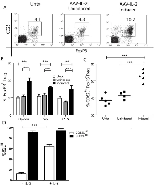
CD62L plays a role in the migration of both FoxP3+ Treg and conventional T cells in vivo to secondary lymphoid organs (64). Interestingly, FoxP3+ Treg which express high levels of CD62L have been shown to be more suppressive in T1D mouse models (65, 66) and other diseases (67). Additionally, adoptive transfer of CD62LHI FoxP3+ Treg from the spleen or draining pancreatic lymph nodes (PLN) resulted in enhanced suppression of β cell autoimmunity compared to CD62LLO FoxP3+ Treg (66). Interestingly, augmented IL-2 expression was also shown to directly correlate with increased expression of CD62L on organ resident FoxP3+ Treg (65), including the islets (M.C.J. and R.T., manuscript in preparation).
Reports also suggest that both CTLA-4 and LFA-1 expression enhance the suppressor function of FoxP3+ Treg. Deletion or blockade of CTLA-4, which is known to interact with both CD80 and CD86 on APC (68), reduces FoxP3+ Treg-mediated
suppression both in vitro and in vivo (69, 70). Importantly, elimination of CTLA-4
10
CTLA-4 up-regulation (72). Of note, one recent study has shown that CTLA-4 and LFA-1 on FoxP3+ Treg function collectively to both sequester APC away from responding T cells and cause down-regulation of the co-stimulatory molecules CD80 and CD86 (73). Lastly, CTLA-4 is thought to further alter APC function by down-regulating pro-inflammatory and/or increasing immune suppressive cytokine secretion (74, 75).
Recent studies have also shown that FoxP3+ Treg express high endogenous levels of CD39 and CD73 (76), both members of ectonucleoside triphosphate
diphosphohydrolase family (77, 78). CD39 hydrolyzes ATP, a classic immune “danger signal” into ADP/AMP, while CD73 further breaks down AMP into adenosine (77, 78). Combined, these two receptors serve to protect FoxP3+ Treg from ATP-induced death, as well as dampen ATP driven maturation of APCs (76). Furthermore, FoxP3+ Treg have been shown to lyse APCs or responding T cells through granzyme and
11
1.7 Genetic and Environmental Factors Influencing T1D Susceptibility
T1D is a multi-factorial disease, with both genetic and environmental factors
contributing to disease susceptibility (88-92). Although ~85% of T1D occurs in the absence of family history, there remains a strong genetic component (93). Among immediate relatives, the highest risk exists among siblings of diabetic patients, which show a 15-fold increase compared to siblings of non-diabetic individuals (93). In addition, children of diabetic fathers and mothers have approximately a 12% and 6% chance of developing disease, respectively, before the age of 20 years (94).
Interestingly, the incidence for the second twin developing diabetes decreases significantly with age of diagnosis in the first twin (95). Furthermore, studies have
shown that the incidence of both monozygotic twins developing diabetes is >50%, which is reduced 5-fold in dizygotic twins (93).
Currently, over 50 insulin dependent diabetes (Idd) loci have been identified in humans (90). Of interest, several genes located within these identified regions are directly related to Treg development and/or function, including CD25, CD122, IL-2, IL-10 and CTLA-4, indicating a direct link between Treg and genetic risk for T1D (90,
91)(t1dbase.org). Other known susceptibility genes include insulin, protein tyrosine phosphatase non-receptor type 22 (PTPN22), and AIRE (90, 91) (t1dbase.org).
12
The strongest genetic association with disease susceptibility is linked to the human leukocyte antigen (HLA) region, which contains genes encoding the HLA class I and II molecules. Notably, up to 90% of young type 1 diabetic patients express one or both of the HLA class II susceptibility alleles DR3 and/or DR4 (98). This has prompted increased screening of HLA, in addition to autoantibody analysis, in at risk patients (99, 100). In NOD mice, the expression of the MHC class I H2Kd/Db and MHC class II IAg7 have also been shown to directly influence disease outcome. Specifically, the role of IAg7 in diabetes is thought to be due to its unique structural properties, allowing increased binding of a unique repertoire of low affinity peptides (101).
The variable development of T1D in monozygotic twins suggests an environmental component for disease outcome. Noteworthy is that enteroviral infections have been strongly correlated to the emergence of autoantibodies in young at risk individuals (102). Enteroviruses, specifically coxsackie B virus, have been shown to infect β cells in vitro, resulting in cell death (103). Furthermore, enterovirus DNA has been detected in the islets of diabetic patients, suggesting that infection may act as an initiating trigger (104). In addition, rotaviruses have been associated with the initiation of disease, as viral proteins are known to mimic the T1D autoantigen glutamic acid decarboxylase (GAD) (105).
13
of, and total exposure to, bovine derived milk proteins may influence the development of autoantibodies against bovine proteins (89). Moreover, gluten and other similar proteins have been implicated as driving antigens in T1D as well (107). Lastly, one study has shown that helminth infection of the gastrointestinal track may inhibit diabetes development, possibly through skewing of the immune response away from a destructive type I response (see below), towards a protective type 2 response characterized by IL-4 secreting Treg (108). As a whole, these studies suggest that predicting the emergence of T1D is complicated and involves the collective influence of genetic and environmental factors.
1.8 The Non-obese Diabetic (NOD) Mouse Model of T1D
In human patients, the progressive destruction of β cells typically occurs over a number of years, resulting in the destruction of 80-90% of β cell mass at the time of clinical onset (109). Although a variety of risk factors have been identified, one of the earliest and most reliable markers is the presence of serum autoantibodies, specifically against GAD, protein tyrosine phosphatase (IA2) and/or insulin (92). Importantly, studies have shown that the presence of autoantibodies against all three correlates with the
14
The NOD mouse model has been extremely useful in the study T1D, and closely mimics the disease process in humans (101). Diabetes develops spontaneously in 80-90% of females and 20-30% of males by 35 weeks of age, suggesting a sex related component in disease progression (111). Based on studies in NOD mice, T1D is
understood to progress through a number of stages of islet inflammation or “insulitis”. The first stage, known as peri-insulitis occurs between 3 and 4 weeks of age, and is marked by the surrounding of islets by mononuclear infiltrates (101). As NOD mice age, disease progresses to the next stage known as intra-insulitis. Occurring on average at 6-10 weeks of age, intra-insulitis is noted by infiltration of the islets by mononuclear cells, resulting in β cell destruction. This process continues until a sufficient number of β cells have been destroyed leading to the onset of clinical diabetes, typically seen between 12-35 weeks in female NOD mice. The composition of the islet infiltrating population is composed of CD4+ and CD8+ T cells, NK cells, B cells, and APCs, including DC and
macrophages (111). While all cells are thought to contribute, the major mediators of β cell destruction are T cells (112).
1.9 CD4+ and CD8+ T cells are the Primary Mediators of T1D
IL-15
17 secreting Th17 cells may also be involved in this process. NOD.scid mice injected with diabetogenic BDC CD4+ T cells skewed in vitro towards a Th17 phenotype
developed T1D, although the majority of transferred cells converted to IFNγ secreting type I effectors after transfer (117). Furthermore, studies show that the frequency of Th17 cells in the infiltrating islet population is low, suggesting that Th17 cells are not the main mediators of β cell destruction (118). Interestingly, antibody mediated
neutralization of IL-17 may result in an enhanced Treg frequency through down regulation of the Th17 population (119). This result is not surprising given the mutual requirement for TGFβ in both FoxP3+ Treg and Th17 cell development (120) and warrants further investigation.
Given the importance of MHC class II in disease, early studies focused on CD4+ T cells. Adoptive transfer of diabetogenic BDC2.5 CD4+ T cell clones was sufficient to induce diabetes in appropriate recipients (121). In addition, blockade using a CD4-specific monoclonal antibody prevented diabetes in NOD mice (122). CD8+ T cells also play a key role in disease, as NOD mice either lacking MHC class I expression (123-125) or treated with anti-CD8 monoclonal antibodies (126) fail to develop diabetes.
16
suggested by some as the initiating autoantigen in mouse models, as thymic deletion of the second proinsulin isoform resulted in accelerated diabetes in NOD mice (130) , while thymus specific transgene expression of proinsulin was found to be protective (131). However, NOD mice tolerized with proinsulin had reduced incidence (132), but were not completely protected from diabetes, and the role of proinsulin in human patients is not as clear (133, 134). Despite this, the β cell specificity of diabetogenic T cells is known to increase with time, in a process known as epitope spreading (135, 136). Therefore, the potential role of other autoantigens in disease progression has been investigated.
IA-2 is known to be required for normal insulin secretion, but deletion of the IA-2 gene in NOD mice did not alter diabetes incidence, suggesting a dispensable role in driving β cell autoimmunity (137). Additionally, NOD mice deficient in GAD65 expression (138, 139) have similar diabetes incidence compared to controls. This particular
outcome was surprising, given that GAD65 is associated with early immune responses in the islets, and that GAD65 administration induces Treg and prevents diabetes in NOD mice (140, 141). Studies have indicated that administration of insulin B chains to pre-diabetic NOD mice also efficiently protects against diabetes (129). As a result, clinical trials have investigated the usefulness of insulin-based immunotherapy to suppress β cell autoimmunity in at risk and diabetic patients, with at best modest results (133, 142, 143). To further complicate matters, as of yet unidentified β cell autoantigen are
17
The large number of β cell autoantigens targeted in T1D at late preclinical and clinical stages provide a major challenge in developing immunotherapies to directly tolerize pathogenic effector T cells (Teff) (135, 136). Accordingly, an emphasis has been placed on strategies that efficiently manipulate the Treg pool in a β cell-specific manner (136, 145-147).
1.10 The Role of IL-2 in Autoimmunity
IL-2 is one member of a family of cytokines bound by receptors containing the common γ-chain, which includes IL-4, IL-7, IL-9, IL-15 and IL-21 (148). Like most members of this cytokine family, IL-2 has pleiotropic effects on the immune system. While the majority of studies have investigated the effect of IL-2 on T cells, it is also known to induce signaling in B cells, NK cells and eosinophils, among others (149-151). IL-2 is primarily secreted by activated Teff and is required for the proliferation and survival of
conventional T cells (87, 152), influencing for instance the expression of several anti-apoptotic markers, including Bcl-2 (153). Conversely, high levels of IL-2 are known to induce apoptosis in Teff through a process known as activated induced cell death (AICD) (154) . Evidence also suggests DCs are a minor source of IL-2 in vivo (155). Earlier studies investigating IL-2 showed that IL-2 deficiency surprisingly resulted in autoimmunity (156, 157). Furthermore, in mice lacking expression of either CD25 or CD122, which are
self-18
tolerance (158, 159). The emergence of autoimmunity in the absence of IL-2 was later shown to be linked to the lack of FoxP3+ Treg development and/or maintenance (87, 160, 161). In addition, antibody blockade of IL-2 was also shown to reduce FoxP3+ Treg numbers, resulting in systemic autoimmunity (162). Moreover, ectopic expression of FoxP3 (163) or adoptive transfer of wild-type FoxP3+ Treg (160) into CD122-deficient mice restored FoxP3+ Treg function and immune homeostasis.
As previously stated, FoxP3+ Treg constitutively express high levels of CD25 (54). CD25 is also up-regulated on recently activated Teff and B cells, but only transiently, making high levels of CD25 expression a reliable marker for Treg identification and/or isolation (87, 149). Additionally, FoxP3+ Treg suppressor function has been directly linked to an enhanced ability to “soak-up” exogenous IL-2 from Teff cells in vivo, due to constitutive CD25 expression (86). After binding to the IL-2R, IL-2 induces the
phosphorylation of STAT5 (164), which regulates the expression of various FoxP3+ Treg related genes, including FoxP3 itself (165, 166). Furthermore, ablation of STAT5 in mice results in a reduced FoxP3+ Treg pool (166, 167).
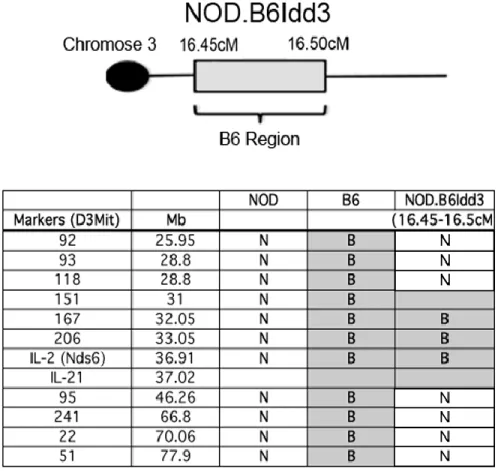
Studies in human T1D patients have shown a reduction in FoxP3+ Treg function and/or number (168, 169). In addition, polymorphisms in IL-2, CD25, and CD122 have been linked to T1D and other autoimmune diseases (170, 171). NOD mice are known to express low levels of IL-2 (153, 172, 173), which is associated with the IL-2 gene
19
shown that replacement of the NOD idd3 locus with one derived from C57BL/6 mice resulted in a significant reduction in diabetes incidence (65). This protection was
mediated by enhanced FoxP3+ Treg function and will be discussed at length in Chapter 2.
Recombinant (r) IL-2 has been successfully used to treat various human diseases. Separate studies showed that graft-versus-host-disease or Hepatitis C vasculitis were suppressed by low dose rIL-2 therapy, and a subsequent increase FoxP3+ Treg (179, 180). Of note, the majority of patients in these studies were refractory to other treatment regimes. In NOD mice, treatment with low-dose rIL-2 or with rIL-2-anti-IL-2 antibody complexes increased islet FoxP3+ Treg survival and frequency in pre-diabetic animals (153). However, increased doses of rIL-2 had significant off-target effects, resulting in systemic activation of NK cells, CD8+, and CD4+ T cells and accelerated onset of overt diabetes. Importantly, these findings underscore how critical the dose of IL-2 is for mediating a protective effect. Diabetes remission was also achieved in recent onset NOD mice treated with low dose rIL-2 alone, or in combinatorial therapy (181, 182). Despite the successes achieved in NOD mice, a recent study in human T1D patients showed that combinatorial treatment with rIL-2 and rapamycin did not significantly alter disease progression or remission (183). Early after treatment, a slight, but transient, increase in FoxP3+ Treg was seen. Nevertheless, NK cells and eosinophils were increased, and β cell autoimmunity exacerbated. Regardless, IL-2 based therapies for the treatment of T1D are still generally considered to be promising approach to manipulate β cell
20 1.11 Clinical Scenarios for the Treatment of T1D
Clinically, there are three main scenarios in which immunotherapy can be applied for
the prevention and/or treatment of T1D (8, 184-188). The first, and arguably most important, would be the prevention of T1D onset in at-risk patients. At risk individuals are typically identified by the detection of β cell autoantibodies in the serum, in addition to altered responses upon glucose challenge (189).
Secondly, immunotherapy can be applied to rescue residual β cell mass in recent onset diabetics. At the onset of clinical diabetes there is still a sufficient number of β cells remaining so that hyperglycemia can be reversed providing inflammation is effectively and rapidly suppressed (184). Furthermore, protecting residual β cell mass may provide a “starting pool” for strategies that promote β cell expansion (190). Alternatively, other cells within the pancreas, or tissues such as the liver, can be
genetically manipulated to become insulin secreting cells, as well (190, 191). In order to promote β cell differentiation or replication, however, it is essential that β cell
autoimmunity is suppressed.
21 1.12 Strategies of Immunotherapy
In order to prevent and suppress β cell autoimmunity, immunotherapies to date have
focused on re-establishing the functional balance between pathogenic Teff and Treg. This has been accomplished through several strategies, including manipulation of APC populations, dampening of Teff and/or induction or expansion of various Treg subsets. Clinically, administration of non-mitogenic anti-CD3 antibodies to recent onset diabetic patients has been shown to maintain β cell mass (192-194). The overall effectiveness of this strategy, however, is short lived and systemic depletion of T cells leaves patients susceptible to viral infection (192-194).
Administration of β cell antigens and peptides to selectively manipulate
22
and/or expanding a Treg pool sufficient to suppress the ongoing β cell autoimmunity. To combat this, the focus of various immunotherapies has been the systemic
induction/expansion of Treg, employing for instance, cytokine-based strategies (136, 145-147). Since Treg are able to suppress in a non-antigen specific manner,
enhancement in Treg number or function would be expected to be beneficial (136, 145, 146, 187, 199). In T1D mouse models, treatment with rIL-4 and rIL-10 enhances Treg populations, and prevents diabetes (200, 201). As stated previously, low dose systemic administration of rIL-2 to NOD mice results in diabetes protection through selective enhancement of islet FoxP3+ Treg (153). While effective, cytokine-based therapy typically requires continuous dosing of animals, due to the short in vivo half-life of cytokines, and may not prove feasible for long-term treatments. Furthermore, since treatment is systemic the pleiotropic effects of most cytokines are likely to result in unwanted off-target effects.
1.13 Gene Delivery via Recombinant Adeno-Associated (rAAV) Vectors
Adeno-associated virus (AAV) is a member of the Parvoviridae family in the genus
23
gene delivery in the clinic and have been effectively used for the treatment of a broad range of diseases, including macular degeneration, rheumatoid arthritis, hemophilia and Parkinson’s disease (206-209). rAAV vectors are capable of infecting both dividing and non-dividing cells (210). In addition, AAV has not been directly associated with any human disease, and typically displays low immunogenicity (202). The safety of rAAV vectors has been further increased by vector modifications that prevent self-replication. In conjunction, AAV typically exist as non-integrating circular monomers or concatemers in the nucleus, with minimal risk of genomic insertion (210).
Several capsid protein serotypes, which display a wide tissue tropism, can be used to package the AAV vectors (211, 212). The use of different serotype capsid proteins, in combination with cell- or tissue-specific promoters, permit targeted in vivo transgene expression. Recent advances in our understanding of viral receptors and capsid protein structures have resulted in the development of tissue-specific capsids. This has been accomplished through the utilization of both 1) random peptide libraries to generate capsids for previously resistant tissues (213-215), and 2) pseudotyped capsids developed through the swapping of established amino acids sequences for different serotypes (216-218). As a whole, these characteristics may lead to the
development of capsids that promote efficient and cell-specific transduction, a primary target of which could be β cells.
24
expression, thereby reducing the overall dose needed in comparison to single-stranded (ss) rAAV vectors; the latter also minimizing the likelihood of eliciting AAV vector-specific immunity (219-222). Lastly, recent changes in both the manufacturing and purification of rAAV vectors have increased the feasibility for clinical use (223).
1.14 Applications ofrAAV Vector-Based Immunotherapy for T1D
Initial studies utilized rAAV vectors to block β cell autoimmunity via systemic expression of the transgene-encoded protein. Intramuscular (I.M.) injection of rAAV vectors
encoding the β cell autoantigen GAD65 (224, 225) or IL-10 (226-228) prevented diabetes in NOD mice at both early and late pre-clinical stages of T1D. Protection was mediated primarily through induction and/or expansion of a protective Treg, in addition to
suppression of proinflammatory APC. In addition, I.M. delivery of rAAV vectors encoding human α1-anti-trypsin (229), a serine protease inhibitor, and heme-oxygenase-1 (230), a stress-response enzyme, have also been shown to suppress established β cell
autoimmunity through suppression of innate effector cells. Of note, transgene
expression in treated NOD mice persisted for a number of weeks post-injection and was both dose-dependent and stable. This suggests that immune recognition of transduced cells was minimal and highlights one of the distinct advantages rAAV vectors have over other viral-based gene delivery approaches.
25
strategies. The first involves the use of an inducible promoter, which allows for
controlled “on/off” expression of the transgene. Recent work by our group has shown that a rAAV1 vector expressing IL-2 under control of a tetracycline inducible promoter suppressed β cell autoimmunity in NOD mice at a late pre-clinical stage (231). Long-term protection was induced following only a 3 week period of IL-2 expression, which correlated with an increased frequency of islet FoxP3+ Treg. Importantly, the level and duration of IL-2 expression had no systemic effects on conventional T cells or innate effectors, such as NK cells. Collectively, these findings show that rAAV vector mediated inducible transgene expression can significantly alter β cell autoimmunity, while having minimal effects on the rest of the immune system.
A second strategy to limit the systemic effects of transgene expression in vivo is to engineer rAAV vectors with a tissue specific promoter. For the treatment of T1D, β cell-specific promoters, such as the insulin promoter, have been used (232, 233). In a streptozotocin-induced model of T1D, treatment of BALB/c mice with dsAAV8
expressing glucagon-like peptide-1 under control of the mouse insulin promoter (mIP) reversed diabetes (234). Additionally, a recent study showed that administration of a dsAAV vector expressing IL-4 driven by mIP resulted in a reduced diabetes incidence in young NOD mice (235). This protection was also shown to be at least partially due to increased Treg. In addition to limiting complications associated with systemic
26
rAAV2 vector expressing IL-4 systemically failed to prevent diabetes in NOD mice, in contrast to IL-4 expression targeted to β cells by dsAAV8 mIP-IL4 (226, 235).
While linking transgene expression specifically to the function of β cells is
desirable, the ability of rAAV vectors to specifically transduce these cells is of paramount concern. As such, both serotype and route of administration have proven to be critical for influencing the efficiency of β cell transduction. Direct intrapancreatic injection of rAAV8 expressing EGFP resulted in transduction of both acinar cells and β cells (236). Furthermore, rAAV8 was found to be superior to other serotypes, including rAAV1, rAAV2 and rAAV5, through the same route of administration.
1.15 Aims of the Dissertation
IL-2 directly affects a variety of cell types, including B cells, Teff, and FoxP3+ Treg.
27
function. We hypothesized that elevated levels of IL-2 in the pancreas of NOD.idd3 mice increases islet-resident FoxP3+ Treg and therefore prevents the onset of overt diabetes.
Treatment with rIL-2 significantly increased the survival and frequency of systemic FoxP3+ Treg in both pre-diabetic and recent onset NOD mice (153, 181, 182). Due to effects on non-FoxP3+ Treg, however, disease was exacerbated in NOD mice receiving a high dose of rIL-2. The effects of islet derived IL-2 in comparison to non-targeted systemic IL-2 has not been investigated. Accordingly, we engineered a rAAV8 vector encoding a mIP-driven IL-2 transgene (IL2). Administration of AAV8mIP-IL2 permits a direct assessment of the therapeutic effects of localized versus systemic IL-2 in NOD mice on FoxP3+ Treg and non-FoxP3+ Treg, and the progression of β cell
autoimmunity. We hypothesized that treatment with AAV8mIP-IL2 results in significant increases in islet resident FoxP3+ Treg function and fitness, resulting in protection from diabetes, while minimizing potential complications associated with systemic IL-2 delivery.
28
29
1.16 References
1. Salsali A, Nathan M. 2006. A review of types 1 and 2 diabetes mellitus and their treatment with insulin. Am J Ther 13: 349-61
2. Bell PM, Firth RG, Rizza RA. 1986. Assessment of insulin action in insulin-dependent diabetes mellitus using [6(14)C]glucose, [3(3)H]glucose, and [2(3)H]glucose. Differences in the apparent pattern of insulin resistance depending on the isotope used. J Clin Invest 78: 1479-86
3. Tisch R, McDevitt H. 1996. Insulin-dependent diabetes mellitus. Cell 85: 291-7 4. van Belle TL, Coppieters KT, von Herrath MG. 2011. Type 1 diabetes: etiology,
immunology, and therapeutic strategies. Physiol Rev 91: 79-118
5. Posselt AM, Bellin MD, Tavakol M, Szot GL, Frassetto LA, Masharani U, Kerlan RK, Fong L, Vincenti FG, Hering BJ, Bluestone JA, Stock PG. 2010. Islet transplantation in type 1 diabetics using an immunosuppressive protocol based on the anti-LFA-1 antibody efalizumab. Am J Transplant 10: 1870-80
6. Senior PA, Zeman M, Paty BW, Ryan EA, Shapiro AMJ. 2007. Changes in renal function after clinical islet transplantation: Four-year observational study. American Journal of Transplantation 7: 91-8
7. Shapiro AMJ, Lakey JRT, Ryan EA, Korbutt GS, Toth E, Warnock GL, Kneteman NM, Rajotte RV. 2000. Islet transplantation in seven patients with type 1
diabetes mellitus using a glucocorticoid-free immunosuppressive regimen. New England Journal of Medicine 343: 230-8
8. Ryan EA, Paty BW, Senior PA, Bigam D, Alfadhli E, Kneteman NM, Lakey JR, Shapiro AM. 2005. Five-year follow-up after clinical islet transplantation. Diabetes 54: 2060-9
9. Elliott RB, Escobar L, Tan PLJ, Muzina M, Zwain S, Buchanan C. 2007. Live encapsulated porcine islets from a type 1 diabetic patient 9.5 yr after xenotransplantation. Xenotransplantation 14: 157-61
30
11. Janeway CA, Medzhitov R. 2002. Innate immune recognition. Annual Review of Immunology 20: 197-216
12. Takeda K, Kaisho T, Akira S. 2003. Toll-like receptors. Annual Review of Immunology 21: 335-76
13. Coutinho A, Pobor G, Pettersson S, Leandersson T, Forsgren S, Pereira P,
Bandeira A, Martinez C. 1984. T-Cell-Dependent B-Cell Activation. Immunological Reviews 78: 211-24
14. Apostolou I, Sarukhan A, Klein L, von Boehmer H. 2002. Origin of regulatory T cells with known specificity for antigen. Nature Immunology 3: 756-63
15. Jordan MS, Boesteanu A, Reed AJ, Petrone AL, Holenbeck AE, Lerman MA, Naji A, Caton AJ. 2001. Thymic selection of CD4(+)CD25(+) regulatory T cells induced by an agonist self-peptide. Nature Immunology 2: 301-6
16. Swain SL. 1983. T-Cell Subsets and the Recognition of Mhc Class. Immunological Reviews 74: 129-42
17. Klein L, Hinterberger M, Wirnsberger G, Kyewski B. 2009. Antigen presentation in the thymus for positive selection and central tolerance induction. Nature
Reviews Immunology 9: 833-44
18. Schatz DG, Ji YH. 2011. Recombination centres and the orchestration of V(D)J recombination. Nature Reviews Immunology 11: 251-63
19. Bousso P, Bhakta NR, Lewis RS, Robey E. 2002. Dynamics of thymocyte-stromal cell interactions visualized by two-photon microscopy. Science 296: 1876-80 20. Ebert PJR, Ehrlich LIR, Davis MM. 2008. Low Ligand Requirement for Deletion and
Lack of Synapses in Positive Selection Enforce the Gauntlet of Thymic T Cell Maturation. Immunity 29: 734-45
21. Le Borgne M, Ladi E, Dzhagalov I, Herzmark P, Liao YF, Chakraborty AK, Robey EA. 2009. The impact of negative selection on thymocyte migration in the medulla. Nature Immunology 10: 823-U41
22. McCaughtry TM, Wilken MS, Hogquist KA. 2007. Thymic emigration revisited. Journal of Experimental Medicine 204: 2513-20
31
24. Li JC, Park J, Foss D, Goldschneider I. 2009. Thymus-homing peripheral dendritic cells constitute two of the three major subsets of dendritic cells in the steady-state thymus. Journal of Experimental Medicine 206: 607-22
25. Wu L, Shortman K. 2005. Heterogeneity of thymic dendritic cells. Seminars in Immunology 17: 304-12
26. Kyewski B, Klein L. 2006. A central role for central tolerance. Annual Review of Immunology 24: 571-606
27. Mathis D, Benoist C. 2009. Aire. Annual Review of Immunology 27: 287-312 28. Gabler J, Arnold J, Kyewski B. 2007. Promiscuous gene expression and the
developmental dynamics of medullary thymic epithelial cells. European Journal of Immunology 37: 3363-72
29. Gray D, Abramson J, Benoist C, Mathis D. 2007. Proliferative arrest and rapid turnover of thymic epithelial cells expressing Aire. Journal of Experimental Medicine 204: 2521-8
30. Koble C, Kyewski B. 2009. The thymic medulla: a unique microenvironment for intercellular self-antigen transfer. Journal of Experimental Medicine 206: 1505-13 31. Zehn D, Bevan MJ. 2006. T cells with low avidity for a tissue-restricted antigen
routinely evade central and peripheral tolerance and cause autoimmunity. Immunity 25: 261-70
32. Aaltonen J, Bjorses P, Perheentupa J, HorelliKuitunen N, Palotie A, Peltonen L, Lee YS, Francis F, Hennig S, Thiel C, Lehrach H, Yaspo ML. 1997. An autoimmune disease, APECED, caused by mutations in a novel gene featuring two PHD-type zinc-finger domains. Nature Genetics 17: 399-403
33. Anderson MS, Venanzi ES, Klein L, Chen ZB, Berzins SP, Turley SJ, von Boehmer H, Bronson R, Dierich A, Benoist C, Mathis D. 2002. Projection of an immunological self shadow within the thymus by the aire protein. Science 298: 1395-401 34. Bluestone JA, Herold K, Eisenbarth G. 2010. Genetics, pathogenesis and clinical
interventions in type 1 diabetes. Nature 464: 1293-300
32
36. Aschenbrenner K, D'Cruz LM, Vollmann EH, Hinterberger M, Emmerich J, Swee LK, Rolink A, Klein L. 2007. Selection of Foxp3(+) regulatory T cells specific for self antigen expressed and presented by Aire(+) medullary thymic epithelial cells. Nature Immunology 8: 351-8
37. Wirnsberger G, Mair F, Klein L. 2009. Regulatory T cell differentiation of
thymocytes does not require a dedicated antigen-presenting cell but is under T cell-intrinsic developmental control. Proceedings of the National Academy of Sciences of the United States of America 106: 10278-83
38. Sakaguchi S. 2005. Naturally arising Foxp3-expressing CD25(+) CD4(+) regulatory T cells in immunological tolerance to self and non-self. Nature Immunology 6: 345-52
39. Fontenot JD, Dooley JL, Farr AG, Rudensky AY. 2005. Developmental regulation of Foxp3 expression during ontogeny. Journal of Experimental Medicine 202: 901-6
40. Gavin MA, Rasmussen JP, Fontenot JD, Vasta V, Manganiello VC, Beavo JA, Rudensky AY. 2007. Foxp3-dependent programme of regulatory T-cell differentiation. Nature 445: 771-5
41. Liu GY, Fairchild PJ, Smith RM, Prowle JR, Kioussis D, Wraith DC. 1995. Low Avidity Recognition of Self-Antigen by T-Cells Permits Escape from Central Tolerance. Immunity 3: 407-15
42. Banchereau J, Steinman RM. 1998. Dendritic cells and the control of immunity. Nature 392: 245-52
43. Hawiger D, Inaba K, Dorsett Y, Guo M, Mahnke K, Rivera M, Ravetch JV, Steinman RM, Nussenzweig MC. 2001. Dendritic cells induce peripheral T cell
unresponsiveness under steady state conditions in vivo. Journal of Experimental Medicine 194: 769-79
44. Liu K, Iyoda T, Saternus M, Kimura Y, Inaba K, Steinman RM. 2002. Immune tolerance after delivery of dying cells to dendritic cells in situ. Journal of Experimental Medicine 196: 1091-7
45. Marrack P, Kappler J. 2004. Control of T cell viability. Annual Review of Immunology 22: 765-87
33
presentation of self-antigen occurs by a Bcl-2-inhibitable pathway mediated by bim. Journal of Experimental Medicine 196: 947-55
47. Strasser A, Harris AW, Huang DCS, Krammer PH, Cory S. 1995. Bcl-2 and Fas/APO-1 regulate distinct pathways to lymphocyte apoptosis. Embo Journal Fas/APO-14: 6Fas/APO-136-47 48. Chen YH, Kuchroo VK, Inobe J, Hafler DA, Weiner HL. 1994. Regulatory T-Cell
Clones Induced by Oral Tolerance - Suppression of Autoimmune Encephalomyelitis. Science 265: 1237-40
49. Groux H, OGarra A, Bigler M, Rouleau M, Antonenko S, deVries JE, Roncarolo MG. 1997. A CD4(+) T-cell subset inhibits antigen-specific T-cell responses and prevents colitis. Nature 389: 737-42
50. Awasthi A, Carrier Y, Peron JPS, Bettelli E, Kamanaka M, Flavell RA, Kuchroo VK, Oukka M, Weiner HL. 2007. A dominant function for interleukin 27 in generating interleukin 10-producing anti-inflammatory T cells. Nature Immunology 8: 1380-9
51. Fitzgerald DC, Zhang GX, El-Behi M, Fonseca-Kelly Z, Li H, Yu S, Saris CJM, Gran B, Ciric B, Rostami A. 2007. Suppression of autoimmune inflammation of the central nervous system by interleukin 10 secreted by interleukin 27-stimulated T cells. Nature Immunology 8: 1372-U6
52. Fukaura H, Kent SC, Pietrusewicz MJ, Khoury SJ, Weiner HL, Hafler DA. 1996. Induction of circulating myelin basic protein and proteolipid protein-specific transforming growth factor-beta 1-secreting Th3 T cells by oral administration of myelin in multiple sclerosis patients. Journal of Clinical Investigation 98: 70-7 53. Curotto de Lafaille MA, Lafaille JJ. 2009. Natural and adaptive foxp3+ regulatory
T cells: more of the same or a division of labor? Immunity 30: 626-35
54. Sakaguchi S, Sakaguchi N, Asano M, Itoh M, Toda M. 1995. Immunological Self-Tolerance Maintained by Activated T-Cells Expressing Il-2 Receptor Alpha-Chains (Cd25) - Breakdown of a Single Mechanism of Self-Tolerance Causes Various Autoimmune-Diseases. Journal of Immunology 155: 1151-64
55. Singh B, Read S, Asseman C, Malmstrom V, Mottet C, Stephens LA, Stepankova R, Tlaskalova H, Powrie F. 2001. Control of intestinal inflammation by regulatory T cells. Immunological Reviews 182: 190-200
56. Gambineri E, Torgerson TR, Ochs HD. 2003. Immune dysregulation,
34
systemic autoimmunity caused by mutations of FOXP3, a critical regulator of T-cell homeostasis. Current Opinion in Rheumatology 15: 430-5
57. Wildin RS, Smyk-Pearson S, Filipovich AH. 2002. Clinical and molecular features of the immunodysregulation, polyendocrinopathy, enteropathy, X linked (IPEX) syndrome. Journal of Medical Genetics 39: 537-45
58. Brunkow ME, Jeffery EW, Hjerrild KA, Paeper B, Clark LB, Yasayko SA, Wilkinson JE, Galas D, Ziegler SF, Ramsdell F. 2001. Disruption of a new forkhead/winged-helix protein, scurfin, results in the fatal lymphoproliferative disorder of the scurfy mouse. Nature Genetics 27: 68-73
59. Ruby CE, Yates MA, Hirschhorn-Cymerman D, Chlebeck P, Wolchok JD, Houghton AN, Offner H, Weinberg AD. 2009. Cutting Edge: OX40 Agonists Can Drive
Regulatory T Cell Expansion if the Cytokine Milieu Is Right. Journal of Immunology 183: 4853-7
60. Vu MD, Xiao X, Gao WD, Degauque N, Chen M, Kroemer A, Killeen N, Ishii N, Li XC. 2007. OX40 costimulation turns off Foxp3(+) tregs. Blood 110: 2501-10 61. Bushell A, Wood K. 2007. GITR ligation blocks allograft protection by induced
CD25(+)CD4(+) regulatory T cells without enhancing effector T-cell function. American Journal of Transplantation 7: 759-68
62. Shimizu J, Yamazaki S, Takahashi T, Ishida Y, Sakaguchi S. 2002. Stimulation of CD25+CD4+regulatory T cells through GITR breaks immunological self-tolerance. Nature Immunology 3: 135-42
63. Suri A, Shimizu J, Katz JD, Sakaguchi S, Unanue ER, Kanagawa O. 2004. Regulation of autoimmune diabetes by non-isletspecific T cells - a role for the
glucocorticoidinduced TNF receptor. European Journal of Immunology 34: 447-54 64. Venturi GM, Conway RM, Steeber DA, Tedder TF. 2007. CD25(+)CD4(+)
regulatory T cell migration requires L-selectin expression: L-selectin
transcriptional regulation balances constitutive receptor turnover. Journal of Immunology 178: 291-300
65. Goudy KS, Johnson MC, Garland A, Li CW, Samulski RJ, Wang B, Tisch R. 2011. Reduced IL-2 expression in NOD mice leads to a temporal increase in
35
66. Szanya V, Ermann J, Taylor C, Holness C, Fathman CG. 2002. The Subpopulation of CD4(+) CD25(+) splenocytes that delays adoptive transfer of diabetes
expresses L-selectin and high levels of CCR7. Journal of Immunology 169: 2461-5 67. Taylor PA, Panoskaltsis-Mortari A, Swedin JM, Lucas PJ, Gress RE, Levine BL, June
CH, Serody JS, Blazar BR. 2004. L-Selectin(hi) but not the L-selectin(lo) CD4(+)25(+) T-regulatory cells are potent inhibitors of GVHD and BM graft rejection. Blood 104: 3804-12
68. Salomon B, Lenschow DJ, Rhee L, Ashourian N, Singh B, Sharpe A, Bluestone JA. 2000. B7/CD28 costimulation is essential for the homeostasis of the
CD4(+)CD25(+) immunoregulatory T cells that control autoimmune diabetes. Immunity 12: 431-40
69. Read S, Malmstrom V, Powrie F. 2000. Cytotoxic T lymphocyte-associated
antigen 4 plays an essential role in the function of CD25(+)CD4(+) regulatory cells that control intestinal inflammation. J Exp Med 192: 295-302
70. Takahashi T, Tagami T, Yamazaki S, Uede T, Shimizu J, Sakaguchi N, Mak TW, Sakaguchi S. 2000. Immunologic self-tolerance maintained by CD25(+)CD4(+) regulatory T cells constitutively expressing cytotoxic T lymphocyte-associated antigen 4. J Exp Med 192: 303-10
71. Wing K, Onishi Y, Prieto-Martin P, Yamaguchi T, Miyara M, Fehervari Z, Nomura T, Sakaguchi S. 2008. CTLA-4 control over Foxp3(+) regulatory T cell function. Science 322: 271-5
72. Schneider H, Valk E, da Rocha Dias S, Wei B, Rudd CE. 2005. CTLA-4 up-regulation of lymphocyte function-associated antigen 1 adhesion and clustering as an alternate basis for coreceptor function. Proc Natl Acad Sci U S A 102: 12861-6 73. Onishi Y, Fehervari Z, Yamaguchi T, Sakaguchi S. 2008. Foxp3+ natural regulatory
T cells preferentially form aggregates on dendritic cells in vitro and actively inhibit their maturation. Proc Natl Acad Sci U S A 105: 10113-8
74. Dejean AS, Beisner DR, Ch'en IL, Kerdiles YM, Babour A, Arden KC, Castrillon DH, DePinho RA, Hedrick SM. 2009. Transcription factor Foxo3 controls the
magnitude of T cell immune responses by modulating the function of dendritic cells. Nature Immunology 10: 504-13
36
76. Borsellino G, Kleinewietfeld M, Di Mitri D, Sternjak A, Diamantini A, Giometto R, Hopner S, Centonze D, Bernardi G, Dell'Acqua ML, Rossini PM, Battistini L,
Rotzschke O, Falk K. 2007. Expression of ectonucleotidase CD39 by Foxp3(+) Treg cells: hydrolysis of extracellular ATP and immune suppression. Blood 110: 1225-32
77. Airas L, Hellman J, Salmi M, Bono P, Puurunen T, Smith DJ, Jalkanen S. 1995. Cd73 Is Involved in Lymphocyte Binding to the Endothelium - Characterization of Lymphocyte Vascular Adhesion Protein-2 Identifies It as Cd73. Journal of
Experimental Medicine 182: 1603-8
78. Mizumoto N, Kumamoto T, Robson SC, Sevigny J, Matsue H, Enjyoji K, Takashima A. 2002. CD39 is the dominant Langerhans cell associated ecto-NTPDase:
Modulatory roles in inflammation and immune responsiveness. Nature Medicine 8: 358-65
79. Cao XF, Cai SF, Fehniger TA, Song JL, Collins LI, Piwnica-Worms DR, Ley TJ. 2007. Granzyme B and perforin are important for regulatory T cell-mediated
suppression of tumor clearance. Immunity 27: 635-46
80. Gondek DC, Lu LF, Quezada SA, Sakaguchi S, Noelle RJ. 2005. Cutting edge: Contact-mediated suppression by CD4(+)-CD25(+) regulatory cells involves a granzyme B-dependent, perforin-independent mechanism. Journal of Immunology 174: 1783-6
81. Collison LW, Workman CJ, Kuo TT, Boyd K, Wang Y, Vignali KM, Cross R, Sehy D, Blumberg RS, Vignali DAA. 2007. The inhibitory cytokine IL-35 contributes to regulatory T-cell function. Nature 450: 566-U19
82. Garin MI, Chu CC, Golshayan D, Cernuda-Morollon E, Wait R, Lechler RI. 2007. Galectin-1: a key effector of regulation mediated by CD4(+)CD25(+) T cells. Blood 109: 2058-65
83. Levings MK, Bacchetta R, Schulz U, Roncarolo MG. 2002. The role of IL-10 and TGF-beta in the differentiation and effector function of T regulatory cells. International Archives of Allergy and Immunology 129: 263-76
84. Nakamura K, Kitani A, Strober W. 2001. Cell contact-dependent
37
85. Pop SM, Wong CP, Culton DA, Clarke SH, Tisch R. 2005. Single cell analysis shows decreasing FoxP3 and TGF beta 1 coexpressing CD4(+)CD25(+) regulatory T cells during autoimmune diabetes. Journal of Experimental Medicine 201: 1333-46 86. Pandiyan P, Zheng L, Ishihara S, Reed J, Lenardo MJ. 2007. CD4+CD25+Foxp3+ regulatory T cells induce cytokine deprivation-mediated apoptosis of effector CD4+ T cells. Nat Immunol 8: 1353-62
87. Malek TR, Castro I. 2010. Interleukin-2 receptor signaling: at the interface between tolerance and immunity. Immunity 33: 153-65
88. Hirschhorn JN. 2003. Genetic epidemiology of type 1 diabetes. Pediatric Diabetes 4: 87-100
89. Knip M, Veijola R, Virtanen SM, Hyoty H, Vaarala O, Akerblom HK. 2005.
Environmental triggers and determinants of type 1 diabetes. Diabetes 54 Suppl 2: S125-36
90. Maier LM, Wicker LS. 2005. Genetic susceptibility to type 1 diabetes. Curr Opin Immunol 17: 601-8
91. Mehers KL, Gillespie KM. 2008. The genetic basis for type 1 diabetes. Br Med Bull 88: 115-29
92. Rose NR. 2008. Predictors of autoimmune disease: autoantibodies and beyond. Autoimmunity 41: 419-28
93. Steck AK, Rewers MJ. 2011. Genetics of type 1 diabetes. Clin Chem 57: 176-85 94. Steck AK, Barriga KJ, Emery LM, Fiallo-Scharer RV, Gottlieb PA, Rewers MJ. 2005.
Secondary attack rate of type 1 diabetes in Colorado families. Diabetes Care 28: 296-300
95. Redondo MJ, Yu L, Hawa M, Mackenzie T, Pyke DA, Eisenbarth GS, Leslie RD. 2001. Heterogeneity of type I diabetes: analysis of monozygotic twins in Great Britain and the United States. Diabetologia 44: 354-62
96. Anderson MS, Su MA. 2011. Aire and T cell development. Curr Opin Immunol 23: 198-206
38
98. Redondo MJ, Fain PR, Eisenbarth GS. 2001. Genetics of type 1A diabetes. Recent Prog Horm Res 56: 69-89
99. Achenbach P, Warncke K, Reiter J, Naserke HE, Williams AJ, Bingley PJ, Bonifacio E, Ziegler AG. 2004. Stratification of type 1 diabetes risk on the basis of islet autoantibody characteristics. Diabetes 53: 384-92
100. Bingley PJ, Bonifacio E, Williams AJ, Genovese S, Bottazzo GF, Gale EA. 1997. Prediction of IDDM in the general population: strategies based on combinations of autoantibody markers. Diabetes 46: 1701-10
101. Anderson MS, Bluestone JA. 2005. The NOD mouse: a model of immune dysregulation. Annu Rev Immunol 23: 447-85
102. Hiltunen M, Hyoty H, Knip M, Ilonen J, Reijonen H, Vahasalo P, Roivainen M, Lonnrot M, Leinikki P, Hovi T, Akerblom HK, Tuomilehto J, Lounamaa R, Toivanen L, Virtala E, Pitkaniemi J, Fagerlund A, Flittner M, Gustafsson B, Haggqvist C, Hakulinen A, Herva L, Hiltunen P, Huhtamaki T, Huttunen NP, Huupponen T, Hyttinen M, Joki T, Jokisalo R, Kaar ML, Kallio S, Kaprio EA, Kaski U, Knip M, Laine L, Lappalainen J, Maenpaa J, Makela AL, Niemi K, Niiranen A, Nuuja A, Ojajarvi P, Otonkoski T, Pihlajamaki K, Pontynen S, Rajantie J, Sankala J, Schumacher J, Sillanpaa M, Stahlberg MR, Strahlmann CH, Uotila T, Vare M, Varimo P,
Wetterstrand G. 1997. Islet cell antibody seroconversion in children is temporally associated with enterovirus infections. Journal of Infectious Diseases 175: 554-60 103. Roivainen M, Ylipaasto P, Savolainen C, Galama J, Hovi T, Otonkoski T. 2002.
Functional impairment and killing of human beta cells by enteroviruses: the capacity is shared by a wide range of serotypes, but the extent is a characteristic of individual virus strains. Diabetologia 45: 693-702
104. Ylipaasto P, Klingel K, Lindberg AM, Otonkoski T, Kandolf R, Hovi T, Roivainen M. 2004. Enterovirus infection in human pancreatic islet cells, islet tropism in vivo and receptor involvement in cultured islet beta cells. Diabetologia 47: 225-39 105. Honeyman MC, Stone NL, Harrison LC. 1998. T-cell epitopes in type 1 diabetes
autoantigen tyrosine phosphatase IA-2: Potential for mimicry with rotavirus and other environmental agents. Molecular Medicine 4: 231-9
106. Hypponen E, Laara E, Reunanen A, Jarvelin MR, Virtanen SM. 2001. Intake of vitamin D and risk of type 1 diabetes: a birth-cohort study. Lancet 358: 1500-3 107. Hummel M, Naserke HE, Bonifacio E, Ziegler AG. 2002. Elimination of dietary
39
108. Saunders KA, Raine T, Cooke A, Lawrence CE. 2007. Inhibition of autoimmune type 1 diabetes by gastrointestinal helminth infection. Infection and Immunity 75: 397-407
109. Foulis AK, Liddle CN, Farquharson MA, Richmond JA, Weir RS. 1986. The
histopathology of the pancreas in type 1 (insulin-dependent) diabetes mellitus: a 25-year review of deaths in patients under 20 years of age in the United
Kingdom. Diabetologia 29: 267-74
110. Verge CF, Gianani R, Kawasaki E, Yu L, Pietropaolo M, Chase HP, Eisenbarth GS. 1996. Number of autoantibodies (against insulin, GAD or ICA512/IA2) rather than particular autoantibody specificities determines risk of type I diabetes. Journal of Autoimmunity 9: 379-83
111. Kikutani H, Makino S. 1992. The murine autoimmune diabetes model: NOD and related strains. Adv Immunol 51: 285-322
112. Bach JF. 1994. Insulin-dependent diabetes mellitus as an autoimmune disease. Endocr Rev 15: 516-42
113. Serreze DV, Chapman HD, Varnum DS, Gerling I, Leiter EH, Shultz LD. 1997. Initiation of autoimmune diabetes in NOD/Lt mice is MHC class I dependent. Journal of Immunology 158: 3978-86
114. Nagata M, Santamaria P, Kawamura T, Utsugi T, Yoon JW. 1994. Evidence for the Role of Cd8+ Cytotoxic T-Cells in the Destruction of Pancreatic Beta-Cells in Nonobese Diabetic Mice. Journal of Immunology 152: 2042-50
115. Bendelac A, Carnaud C, Boitard C, Bach JF. 1987. Syngeneic Transfer of Autoimmune Diabetes from Diabetic Nod Mice to Healthy Neonates -
Requirement for Both L3t4+ and Lyt-2+ T-Cells. Journal of Experimental Medicine 166: 823-32
116. Tisch R, Wang B. 2008. Dysrulation of T Cell Peripheral Tolerance in Type 1 Diabetes. Advances in Immunology: Immunopathogenesis of Type 1 Diabetes Mellitus, Vol 100 100: 125-49
117. Bending D, De la Pena H, Veldhoen M, Phillips JM, Uyttenhove C, Stockinger B, Cooke A. 2009. Highly purified Th17 cells from BDC2.5NOD mice convert into Th1-like cells in NOD/SCID recipient mice. J Clin Invest 119: 565-72
40
119. Emamaullee JA, Davis J, Merani S, Toso C, Elliott JF, Thiesen A, Shapiro AM. 2009. Inhibition of Th17 cells regulates autoimmune diabetes in NOD mice. Diabetes 58: 1302-11
120. Lee YK, Mukasa R, Hatton RD, Weaver CT. 2009. Developmental plasticity of Th17 and Treg cells. Curr Opin Immunol 21: 274-80
121. Haskins K. 2005. Pathogenic T-cell clones in autoimmune diabetes: More lessons from the NOD mouse. Advances in Immunology, Vol 87 87: 123-62
122. Shizuru JA, Taylor-Edwards C, Banks BA, Gregory AK, Fathman CG. 1988.
Immunotherapy of the nonobese diabetic mouse: treatment with an antibody to T-helper lymphocytes. Science 240: 659-62
123. Katz J, Benoist C, Mathis D. 1993. Major Histocompatibility Complex Class-I Molecules Are Required for the Development of Insulitis in Nonobese Diabetic Mice. European Journal of Immunology 23: 3358-60
124. Serreze DV, Leiter EH, Christianson GJ, Greiner D, Roopenian DC. 1994. Major Histocompatibility Complex Class-I Deficient Nod-B2m(Null) Mice Are Diabetes and Insulitis Resistant. Diabetes 43: 505-9
125. Wicker LS, Leiter EH, Todd JA, Renjilian RJ, Peterson E, Fischer PA, Podolin PL, Zijlstra M, Jaenisch R, Peterson LB. 1994. Beta-2-Microglobulin-Deficient Nod Mice Do Not Develop Insulitis or Diabetes. Diabetes 43: 500-4
126. Wang B, Gonzalez A, Benoist C, Mathis D. 1996. The role of CD8(+) T cells in the initiation of insulin-dependent diabetes mellitus. European Journal of
Immunology 26: 1762-9
127. Lennon GP, Bettini M, Burton AR, Vincent E, Arnold PY, Santamaria P, Vignali DAA. 2009. T Cell Islet Accumulation in Type 1 Diabetes Is a Tightly Regulated, Cell-Autonomous Event. Immunity 31: 643-53
128. Lieberman SM, DiLorenzo TP. 2003. A comprehensive guide to antibody and T-cell responses in type 1 diabetes. Tissue Antigens 62: 359-77
129. Panagiotopoulos C, Trudeau JD, Tan R. 2004. T-cell epitopes in type 1 diabetes. Curr Diab Rep 4: 87-94