Functional Transcriptomics in Diverse Intestinal Epithelial
Cell Types Reveals Robust MicroRNA Sensitivity in Intestinal
Stem Cells to Microbial Status
*
□SReceived for publication, November 29, 2016, and in revised form, December 23, 2016 Published, JBC Papers in Press, January 4, 2017, DOI 10.1074/jbc.M116.770099
Bailey C. E. Peck‡§1, Amanda T. Mah¶, Wendy A. Pitman§, Shengli Ding储, P. Kay Lund‡¶储, andX Praveen Sethupathy‡§2
From the‡Curriculum in Genetics and Molecular Biology,§Department of Genetics,¶Department of Nutrition, and储Department of Cell Biology and Physiology, University of North Carolina at Chapel Hill, Chapel Hill, North Carolina 27599
Edited by Xiao-Fan Wang
Gut microbiota play an important role in regulating the devel-opment of the host immune system, metabolic rate, and at times, disease pathogenesis. The factors and mechanisms that mediate interactions between microbiota and the intestinal epithelium are not fully understood. We provide novel evidence that micro-biota may control intestinal epithelial stem cell (IESC) prolifer-ation in part through microRNAs (miRNAs). We demonstrate that miRNA profiles differ dramatically across functionally dis-tinct cell types of the mouse jejunal intestinal epithelium and that miRNAs respond to microbiota in a highly cell type-specific manner. Importantly, we also show that miRNAs in IESCs are more prominently regulated by microbiota compared with miRNAs in any other intestinal epithelial cell subtype. We iden-tify miR-375 as one miRNA that is significantly suppressed by the presence of microbiota in IESCs. Using a novel method to knockdown gene and miRNA expressionex vivoenteroids, we demonstrate that we can knock down gene expression in Lgr5ⴙ IESCs. Furthermore, when we knock down miR-375 in IESCs, we observe significantly increased proliferative capacity. Under-standing the mechanisms by which microbiota regulate miRNA expression in IESCs and other intestinal epithelial cell subtypes will elucidate a critical molecular network that controls intestinal homeostasis and, given the heightened interest in miRNA-based therapies, may offer novel therapeutic strategies in the treatment of gastrointestinal diseases associated with altered IESC function.
The intestinal epithelium is a single layer of cells exposed to the intestinal lumen and is composed of multiple cell types including the proliferative intestinal epithelial stem cells (IESCs)3and progenitor cells (also known as transit amplifying cells) as well as differentiated absorptive enterocytes and secre-tory goblet, Paneth, and enteroendocrine cells (EECs). IESCs divide to yield more rapidly proliferating progenitors that give rise to all of the other intestinal epithelial cell (IEC) types and drive continuous renewal of the intestinal epithelium every
⬃3–5 days (1). Proper renewal facilitates important intestinal epithelial functions including barrier integrity to protect against invasion of harmful toxins present in the intestinal lumen, nutrient digestion and absorption, and production of hormones that regulate systemic energy homeostasis. These physiological processes are mediated in part by interactions with resident microbiota (2). Studies using germ-free animals have demonstrated that gut microbiota influence intestinal barrier function, nutrient absorption, proliferation, differenti-ation, cellular signaling, and migration (3, 4). However, the molecular factors and mechanisms underlying microbiota-me-diated control of IEC functions, particularly IESC proliferation, are unknown.
microRNAs (miRNAs) have emerged as critical regulatory factors of many biological processes in numerous tissues and are known to confer phenotypic robustness in response to envi-ronmental stimuli (5). However, less is known about miRNA expression and function in the intestinal epithelium compared with most other tissues. Recently, miRNAs were implicated in the regulation of IEC physiology (6, 7). McKennaet al.(6) dem-onstrated in mice that the IEC-specific knock-out ofDicer1, an essential enzyme for canonical miRNA biogenesis, results in altered IEC proliferation, differentiation, nutrient absorption, and impaired barrier function, indicating that miRNAs are likely important modulators of intestinal homeostasis (6). Fur-thermore, the presence of microbiota in the gut has been shown to alter miRNA expression profiles in intestinal macrophages
*This work was supported, in whole or in part, by National Institutes of Health Grants UNC GMB T32GM007092 (NIGMS), 5R25GM55336 (a UNC IMSD Diversity Fellowship; NIGMS to B. C. P.), F31DK105747 (NIDDK to B. C. P.), P30DK034987 (NIDDK; to P. S.), and R01DK105965 (NIDDK; to P. S.). This work was also supported by 14CSA20660001 (American Heart Association; to P. S.) and 1-16-ACE-47 (American Diabetes Association; to P. S.). The authors declare that they have no conflicts of interest with the contents of this article. The content is solely the responsibility of the authors and does not necessarily represent the official views of the National Institutes of Health.
□S
This article containssupplemental File 1.
Raw sequencing data and counts are available through GEO accession number GSE81126.
1To whom correspondence may be addressed: Curriculum in Genetics and
Molecular Biology, School of Medicine, 5100H Genetic Medicine Bldg. CB#7264, 120 Mason Farm Rd., UNC Chapel Hill, Chapel Hill, NC 27599. E-mail: [email protected].
2To whom correspondence may be addressed: Dept. of Genetics, School of
Medicine, 5091 Genetic Medicine Bldg. CB#7264, 120 Mason Farm Rd., UNC Chapel Hill, Chapel Hill, NC 27599. Tel.: 919-966-6387; E-mail: [email protected].
3The abbreviations used are: IESC, intestinal epithelial stem cell; miRNA,
microRNA; IEC, intestinal epithelial cell; EEC, enteroendocrine cell; GF, germ-free; CV, conventionalized; EGFP, enhanced GFP; CR, conventionally raised; NS, non-sorted; RPMMM, reads per million mapped to miRNAs; PCA, principal component analysis; LNA, locked nucleic acid; FC, -fold change; FDR, false discovery rate; UNC, University of North Carolina at Chapel Hill; qRT, quantitative real-time; RQV, relative quantitative value.
crossmark
(8) as well as in whole intestine (9, 10). Understanding the mechanisms by which microbiota regulate miRNA and gene expression in IESCs and other IEC subtypes will elucidate a critical molecular network that controls intestinal homeostasis and, given the heightened interest in miRNA-based therapies, may offer novel therapeutic strategies in the treatment of gas-trointestinal diseases associated with altered IESC function. However, no study to date has investigated miRNA expression and activity across the functionally distinct IEC subtypes, and cell type-specific effects of microbiota on miRNAs is com-pletely unknown. We hypothesized that each IEC population has a distinct miRNA profile and that miRNAs respond to gut microbiota in a cell type-specific manner in order to control function and overall homeostasis of the intestinal epithelium.
Results
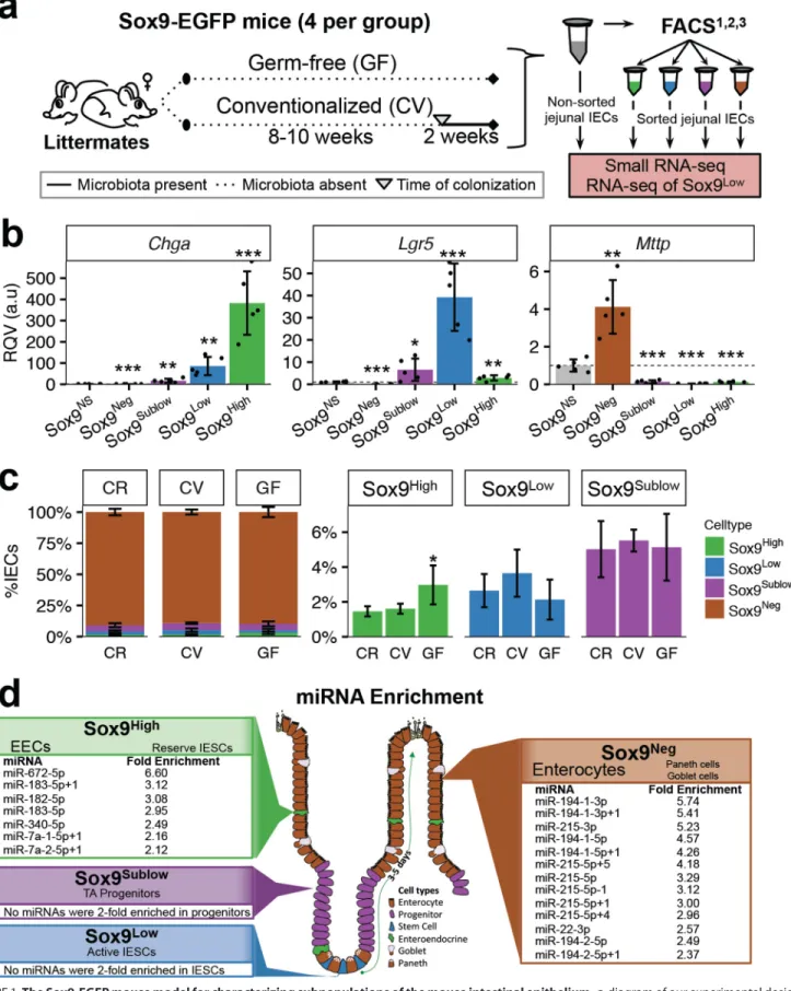
Germ-free Mice Have an Altered Jejunal IEC Composition Compared with Their Conventionalized and Conventionally Raised Counterparts—We selected the well characterized Sox9-EGFP transgenic mouse model to evaluate miRNA expression and effect of microbiota in functionally distinct IECs. This model was originally created by GENSAT (11), who developed the model by randomly inserting into the mouse genome a BAC (bacterial artificial chromosome) containing the EGFP gene driven by the cloned genomic regions upstream and downstream ofSox9(12). A beneficial feature of this model is that EGFP expression is fully penetrant within the mouse intes-tine, which permits the isolation and analysis of four dis-tinct IEC populations (12). Applying the same fluorescence-activated cell sorting (FACS)-based approach used to isolate IESCs from the commonly used Lgr5-EGFP model, which dem-onstrates mosaic expression among crypts in the intestine, both actively cycling IESCs (Sox9Low) and transit-amplifying
pro-genitor cells (Sox9Sublow) can be isolated from the Sox9-EGFP
mouse intestine (12, 13). Moreover, two additional differenti-ated cell populations can also be isoldifferenti-ated on the basis of vari-able EGFP intensity, including Sox9Neg(mostly differentiated
enterocytes as well as goblet cells and Paneth cells) and Sox9High(primarily EECs as well as reserve/quiescent⫹4 stem
cells) (12–17).
To evaluate the effect of microbiota on miRNA expression in IECs, we first took a conventionalization approach (Fig. 1a). A 2-week conventionalization was selected because previous studies have shown this to be a time point at which the gene expression profile begins stabilizing in the small intestine of young mice after conventionalization (18 –20). After generat-ing germ-free (GF) Sox9-EGFP animals at the University of North Carolina at Chapel Hill (UNC) Gnotobiotic core facility, we selected four pairs of female GF Sox9-EGFP littermates from four different litters born between February and July 2015. One littermate from each pair was randomly selected at 8 –10 weeks of age for conventionalization. The 2-week convention-alization resulted in a slightly decreased body weight relative to the remaining germ-free sibling along with a commensurate increase in liver weight (Fig. 2). However, no significant differ-ences were observed in the length of the small intestine or colon between GF and conventionalized (CV) animals (Fig. 2). IECs were collected from the mid-region of the small intestine
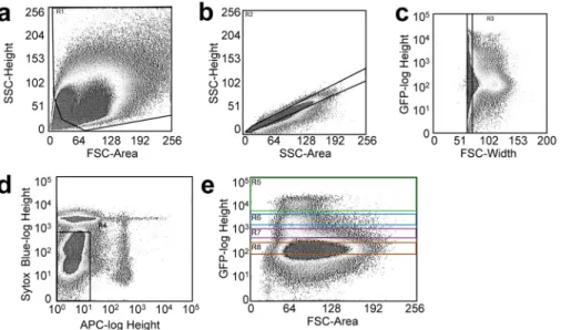
(“Experimental Procedures”), hereafter referred to as jejunum, of the GF and CV animals, and FACS was performed based on Sox9-EGFP intensity (Fig. 1a). Special care was taken to gate out cellular debris, dead and dying cells, immune cells, and mul-tiplets during FACS (see “Experimental Procedures”; Fig. 3). Additionally, a strict gating scheme was used to avoid contam-ination between cell populations. To evaluate the accuracy of the sorting in our hands, we performed qRT-PCR on sorted cells from 10-week-old female conventionally raised Sox9-EGFP mice and demonstrated clear enrichment ofChgain the Sox9Highpopulation,Lgr5in the Sox9Lowpopulation, andMttp
in the non-sorted cells (which consists primarily of enterocytes) and the Sox9Negpopulation (Fig. 1b). As these genes are known
to be enriched in EECs, IESCs, and enterocytes, respectively, these data confirm the utility and accuracy of the sorting method for isolating functionally distinct subpopulations of the intestinal epithelium.
Next we compared the abundances of each major IEC sub-population in GF and CV animals to conventionally raised (CR) chow-fed animals. We found very similar abundances of Sox9Sublowcells (transit amplifying) and Sox9Negcells
(entero-cytes, Paneth, and goblet) in CV and GF populations relative to CR mice (Fig. 1c). Notably, GF mice had significantly more Sox9Highcells (EECs) than CR mice (⫺fold change⫽2.04,p⫽
0.04; Fig. 1c), which is consistent with previous studies compar-ing EECs in the jejunum of GF and CR rodents (21, 22). Also, there were on average fewer Sox9Low cells (actively cycling IESCs) in GF mice relative to CR and CV mice, which has not been shown before but could help explain previous reports sug-gesting reduced proliferation in the small intestine of GF ani-mals (20, 23–25).
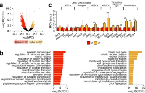
IESCs Demonstrate Robust Transcriptional Differences bet-ween Distinct Microbial States—To evaluate the transcriptional changes that occur in response to conventionalization in the IESCs of GF and CV animals, we performed RNA sequencing analysis on the Sox9Lowpopulation (which we will refer to as
IESCs for simplicity). We identified 823 genes and long, non-coding RNAs (lncRNAs) significantly elevated in GF IESCs and 334 genes and lncRNAs significantly elevated in CV IESCs (Fig. 4a). Gene Ontology Biological Process (26, 27) enrichment analysis using Enrichr (28) revealed that genes elevated in CV IESCs are most significantly overrepresented in pathways related to proliferation such as “mitotic cell cycle” and “nuclear division” (Fig. 4b). The genes elevated in GF IESCs genes were associated with processes related to hormone secretion and transport (Fig. 4b). Consistent with these findings, in CV IESCs we observed that established markers of proliferation (Ccnb1,
Cdk1, and Mki67) were significantly up-regulated, positive transcriptional regulators of IESC proliferation and self-re-newal (Gata4andGata6) were up-regulated, and negative reg-ulators of IESC proliferation and self-renewal (Bmp4) were down-regulated (Fig. 4c). Also, some, but not all, classic mark-ers of EECs were up-regulated in GF IESCs (Fig. 4c), which could indicate some priming for cells to enter the EEC lineage, consistent with our observation that GF mice have more Sox9Highcells. Known markers of reserve (quiescent) stem cells
FIGURE 1.The Sox9-EGFP mouse model for characterizing subpopulations of the mouse intestinal epithelium.a, diagram of our experimental design.b, data from qRT-PCR analysis showing the normalized expression ofChga,Lgr5, andMttpin sorted Sox9-EGFP populations relative to normalized expression in non-sorted cells (Sox9NS) from 10-week-old CR female Sox9-EGFP mice (n⫽5). Data are displayed as RQVs, which are in arbitrary units (a.u.). Significance was
determined for each cell type in comparison to Sox9NSby Student’s two-tailed unpairedttest and is denoted as: *,p⬍0.05; **,p⬍0.01; ***,p⬍0.001.c, mean
percentage of each IEC subtype sorted from jejunum of CR, GF, and CV mice (n⫽4 each). Significance was determined for each category in comparison to CR by Student’s two-tailed unpairedttest and is denoted as: *,p⬍0.05.d, schematic showing location and types of IECs in the Sox9-EGFP mouse. Listed are miRNAs with a mean normalized expression (across CR, CV, and GF) that is at least 2-fold greater in one particular IEC subtype relative to all others. Only miRNAs with RPMMM⬎400 in at least one sample were included in the analysis.Error barsdepict S.D.
and enterocytes (Elf3). These data confirm that the Sox9Low
cells are indeed enriched for IESCs and that CV IESCs harbor a gene signature consistent with increased proliferative capacity. As miRNAs are known regulators of proliferation and differen-tiation, we performed small RNA sequencing of each of the functionally distinct IEC subpopulations from four CR, GF, and CV animals.
miRNAs Show Cell Type-specific Expression across Function-ally Distinct Populations of IECs—Total RNA was isolated from the four sorted populations from each animal as well as from non-sorted IECs (NS IECs; NS IECs were purified by FACS but not sorted based on Sox9-EGFP intensity). Small RNA-se-quencing was performed in three batches, two of which con-tained small RNA libraries from sorted and unsorted IECs from two GF animals and two CV animals. The third batch contained libraries of the four CR animals. miRNAs and their isoforms (called isomiRs) were quantified using miRquant, our previ-ously described method (see “Experimental Procedures” for details) (29). To test our hypothesis that miRNAs are differen-tially expressed among functionally distinct IEC subtypes, we evaluated miRNAs with an expression level of at least 400 reads per million mapped to miRNAs (RPMMM) in one or more samples, identifying 149 robustly expressed miRNAs across all IEC populations.
Many miRNAs were uniquely enriched in one IEC subtype relative to all others (⬎2-fold more highly expressed than any other cell type across all samples; Fig. 1d). For example, we found that miR-215 and miR-194 were enriched in Sox9Neg cells, which consist primarily of enterocytes. Both of these miRNAs are processed from a single primary miRNA transcript on Chr1 and were previously shown to be induced by HNF4␣ during differentiation of Caco-2 colon carcinoma cells (30). Five miRNAs were enriched in Sox9Highcells (EECs and reserve
stem cells) including miR-182–5p and miR-183–5p (Fig. 1d), which are also generated from a single primary miRNA tran-script. Consistent with enrichment in a subpopulation of cells composed largely of EECs, miR-182 has been shown to have important functions in other endocrine cells, specifically, pan-creatic beta cells (31). Unexpectedly, we did not find any miRNAs enriched in the Sox9LowIESCs or Sox9Sublow
progen-itors, which are the only actively proliferating cell populations (Fig. 1d).
FIGURE 2.Physiological parameters observed at the time of sacrifice of female GF and 2-week CV mice at 10 –12 weeks of age (nⴝ4 each).a, weight of spleen in grams.b, length of small intestine (SI) from pyloric sphinc-ter to caecum in centimesphinc-ters (cm).c, length of the colon from caecum to anus in cm.d, ratio of spleen weight to body weight (BW).e, body weight in grams.
f, liver weight in grams.g, ratio of liver weight to body weight. Significance was determined by a Student’s two-tailed pairedttest is denoted as: *,p⬍0.05.
To further assess variability within and across samples, we performed principal component analysis (PCA) to reduce dimensionality and assess the effect of microbial presence on each sample. The first three principal components (PC1-PC3) captured 67% of the variability across samples. Using biplots, we evaluated segregation of samples by cell type and mouse condition (GF, CV, or CR). We showed that miRNA expression profiles were sufficient to cluster most samples by their respec-tive cell types regardless of microbial status (Fig. 5,aandb). For example, Sox9Neg cells and NS IECs were tightly clustered, which is expected given that NS IECs are composed of 85–90% Sox9Negcells. When we compared the samples using PC1 and
PC2, GF Sox9LowIESCs did not cluster together with CV and
CR Sox9LowIESCs (Fig. 5a), indicating that IESCs are
particu-larly sensitive to the presence or absence of microbiota. How-ever, when PC1 and PC3 were projected, clear cell type-specific clustering was observed regardless of mouse condition (Fig. 5b), suggesting that the subset of miRNAs loaded into PC2 (⬃40 miRNAs) contributed to the grouping observed in Fig. 5a. Taken together, these data indicate strong cell type-specific expression of miRNAs across IEC populations and a robust effect of microbial presence on the IESC population specifi-cally. Both observations are supported by visualization of the most highly expressed miRNAs across GF and CV samples (Fig. 5c).
miRNAs of IESCs Show More Distinct Expression Differences between GF and CV States Than Other IEC Types—To evaluate the cell type-specific sensitivity to microbial status and to account for batch and littermate effects, we used a linear mod-eling approach (see “Experimental Procedures”). We found the expression levels of 11 miRNAs (miR-34a-5p, miR-200c-3p, miR-200c-3p-1, miR-143-3p, miR-130b-3p, miR-140 –3p⫹1, miR-378 –3p⫹1, miR-20a-5p, miR-17-5p, miR-93-5p, and
miR-29a-3p) to be significantly influenced by microbial status across all cell populations. These miRNAs in general were ele-vated in CV mice across all cell populations, although the mag-nitude of the effect was always most pronounced in the Sox9Low (IESC) population. When we assessed changes specific to each cell type, we were surprised to find that although many miRNAs were altered significantly in the IESC population in the CV state relative to GF, no miRNAs were identified as significantly changed specifically in the Sox9High, Sox9Sublow, or Sox9Neg populations (Fig. 6a,supplemental File 1). A total of 19 miRNAs in IESCs were significantly different in expression depending on microbial status (Fig. 6b). This finding underscores the highly cell type-specific effect of microbial presence on miRNAs.
Of these 19 microbiota-sensitive miRNAs in IESCs, miR-375-3p was ⬃2.5-fold (false discovery rate (FDR) ⫽ 0.003) reduced in CV IESCs compared with GF IESCs and was the most highly expressed (Fig. 6,aandb). Notably, miR-375-3p was 2.4- and 7.2-fold more highly expressed than the next-most significant microbiota-sensitive miRNA in the CV and GF IESC populations, respectively (Fig. 6b). We also found that its isomiRs, miR-375-3p-1 and miR-375-3p⫹1, were also both sig-nificantly down-regulated in IESCs upon conventionalization ((⫺fold change (FC) ⫽ ⫺2.45, FDR⫽ 0.0006; FC⫽ ⫺2.47, FDR⫽0.02, respectively; Fig. 6b). qRT-PCR on Sox9Lowcells
confirmed that the miR-375-3p family was significantly down-regulated by conventionalization (FC⫽ ⫺3.85,p⫽0.03; Fig. 6c). Of note, miR-375-3p exhibited rather low expression in Sox9Sublowprogenitors and Sox9Negcells (Fig. 6d). Although
miR-375-3p is highly expressed in Sox9HighEECs, it was not altered in EECs by conventionalization and was only signifi-cantly down-regulated by microbiota in the IESCs.
FIGURE 4.RNA-seq analysis of the Sox9Lowpopulation from GF and CV mice.a, volcano plot showing differentially expressed genes in Sox9LowIESCs
between GF and CV mice (n⫽4 animals per condition). Thehorizontal dashed gray lineindicates a FDR of 0.05.Vertical dashed gray linesindicate FC⫽1.5 and
⫺1.5. Significantly differentially expressed genes are colored inorangeandred, representing genes that at enriched in CV or GF IESCs, respectively.b, Top Enrichr Gene Ontology Biological Process enrichment terms for genes significantly up-regulated in GF (left) or CV (right) IESCs.c, RQVs, in arbitrary units (a.u.) of normalized counts for selected genes in CV relative to GF IESCs (n⫽4 animals per condition), including markers of reserve or quiescent IESCs (rIESCs), and actively cycling IESCs (aIESCs). Significance determined using DEseq2 differential expression analyses is denoted as: *, FDR⬍0.05; **, FDR⬍0.01; ***, FDR⬍ 0.001.Error barsdepict S.D.
Knockdown of Gene Expression in IESCs of ex Vivo Enteroids using Gymnosis—To functionally evaluate the effect of the observed miRNA and gene expression changes, we sought out methods to suppress miRNA/gene expression in IESCs ofex vivoenteroid culture systems, which have been shown to main-tainin vivocellular composition and molecular gene expression profiles over time (32). We evaluated the use of gymnosis, a term coined by the Troels Koch laboratory in 2009 (33), to describe a process of introducing modified or locked nucleic acids (LNA) complementary to a specific gene or miRNA into cells without the use of traditional transfection reagents. Gym-nosis has been used previously to knockdown gene expression in enteroids (34); however, knockdown capacity specifically in IESCs has not been evaluated. To determine whether IESCs of
ex vivo cultured enteroids can take up LNAs introduced through the media and/or Matrigel via gymnosis and down-regulate target gene/miRNA expression (Fig. 7a), we tested knockdown efficacy of an LNA against EGFP (LNA-EGFP) in Lgr5-EGFP⫹ enteroids. Specifically, we identified EGFP⫹ crypts immediately after seeding into Matrigel and followed the growth of those enteroids over the course of 8 days and mea-sured the EGFP signal after 1MLNA-EGFP treatment (Fig. 7, b– e). As Lgr5-EGFP crypts exhibit mosaic expression of EGFP (in our colony,⬃1 in 30 crypts are EGFP⫹), qRT-PCR analysis to assess changes inEGFPwas inconclusive (data not shown). However, based on fluorescence imaging, we observed an appreciable depletion of EGFP by day 4 (Fig. 7b). We also dem-onstrated that knockdown efficiency improved with repeated
LNA-EGFP treatment over time (Fig. 7c). Moreover, whereas 1
Mand 5Mconcentrations of LNA-EGFP were sufficient to
achieve considerable knockdown of EGFP, a 500 nM concentra-tion was not, indicating a clear dose-dependent effect (Fig. 7d). Finally, as there are multiple enteroids per well, we sought to assess the level of within-well variability of the knockdown efficiency by surveying all EGFP⫹crypts from a single well treated with LNA-EGFP. Overall, EGFP knockdown effi-ciency appeared to be high across all of the EGFP⫹enteroids that were visualized (Fig. 7e). Taken together, these data indi-cate successful LNA-mediated knockdown of EGFP gene expression in IESCs ofex vivoLgr5-EGFP enteroids using gym-nosis and support the general use of gymgym-nosis for knockdown of gene expression in IESCs of enteroids.
Knockdown of miR-375-3p in Enteroids Results in Increased Proliferation—To test the functional effect of miR-375-3p sup-pression, we knocked-down miR-375-3p by gymnosis in enteroids from GF Sox9-EGFP crypts. We achieved a robust
⬃700-fold knockdown of miR-375-3p at day 8 after treatment with an LNA inhibitor of miR-375-3p (LNA-375; Fig. 8a). At both days 4 and 8, LNA-375-treated enteroids exhibited signif-icantly increased budding (Fig. 8,band c), a marker of IESC proliferative capacity (35, 36), relative to Mock or enteroids treated with a scrambled LNA (LNA-SCR). This finding was confirmed when the experiments were repeated in enteroids established from crypts isolated from conventionally raised Lgr5-EGFP (Fig. 8d) and wild-type C57BL/6J mice (Fig. 8e). Consistent with this finding, whole mount staining of the enteroids also showed increased signal for Ki67 upon knock-down of miR-375-3p (Fig. 8f), although no difference in en-teroid size (Fig. 8,gandh) or passage efficiency was observed (data not shown). These data indicate that miR-375-3p is likely a potent regulator of IESC proliferation and that microbiota may regulate IESC renewal in part via modulation of miR-375-3p (Fig. 9).
Discussion
In this study we provided novel evidence that miRNAs are sensitive to the presence of gut microbiota in a cell type-specific manner. Microbiota exert the strongest effect on host miRNA expression in the Sox9Lowpopulation, which is highly enriched in IESCs (12–14, 16, 17). Subpopulation analysis was necessary to identify this effect, as IESCs make up only 1–3% of all IEC types. miR-375-3p was identified as significantly down-regu-lated in the IESC population in response to microbial presence, and follow-up experimentsex vivodemonstrated miR-375-me-diated control of IESC expansion and proliferation, thereby providing a mechanism by which microbiota may regulate these processes during conventionalizationin vivo. miR-375-3p has been associated previously with the regulation of
prolifera-tion and differentiaprolifera-tion in several tissues (34, 38, 39). It is pre-dicted to target many members of the Wnt/-catenin and Hippo signaling pathways but so far has only been experimen-tally shown to directly inhibit Frizzled-8 (39) and Yap1 (40). miR-375-3p has been knocked down systemically in mice, and although the authors did not study intestinal proliferation, they observed an increased rate of intestinal transit (41). miR-375-3p is best studied in the context of pancreatic endocrine cell differentiation and function (42– 44), and more recently, Knudsenet al.(34) identified a role for miR-375-3p in regulat-ing EEC differentiation as well. We found that although miR-375-3p is robustly expressed in both IESCs and EECs, it is sen-sitive to microbiota only in IESCs (Fig. 6d). This observation might suggest cell type-specific microbial signaling pathways and cell type-specific roles for miR-375-3p.
Our RNA sequencing analysis, the first to our knowledge comparing GF and CV IESCs, demonstrates substantial gene expression differences in IESCs between the GF and CV states. CV IESCs showed up-regulation of genes associated with pro-liferation. Of note, our data indicate a⬃4-fold increase inLgr5
mRNA expression (Fig. 4c). Microbiota may regulate Wnt sig-naling upstream of Lgr5, an R-spondin ligand receptor, as known upstream regulators of Lgr5 are also altered by the pres-ence of microbiota, includingGata4,Gata6, andBmp4(Fig. 4c) (45, 46). This is a finding that deserves further investigation.
An unexpected finding was that GF IESCs (Sox9Low) have a
gene and miRNA expression profile demonstrating some simi-larity to Sox9Highcells. Given the careful sorting protocol and
the observation that Sox9High-enriched genes and miRNAs
change in both the upward and downward directions within the Sox9Lowpopulation, our finding is unlikely to be driven solely
by contamination between populations. One possible explana-tion is that Sox9Lowcells are primed for the EEC lineage in the
absence of microbial influence. Alternatively, one of the caveats of the Sox9-EGFP model is that although the Sox9High
popula-tions consist primarily of EECs, they also include a small popu-lation of reserve stem cells (17). It is, therefore, possible that microbiota influence the maintenance of reserve stem cells in addition to their role in regulating actively cycling IESCs through miR-375-3p, although the gene expression data do not fully support this hypothesis. Although outside the scope of this study, more research including single cell analyses will need to be conducted to delineate more precisely the differences between GF and CV IESCs as well as to determine which miRNAs are involved in the maintenance of active and quiescent IESC states.
An important added value of our study is the first ever map of miRNA expression across different IEC subtypes and the cell type-specific influence of microbial conventionalization on
FIGURE 6.Cell population-specific differences in miRNA expression between GF and CV revealed through linear modeling analysis.a, for each miRNA that met an expression threshold of RPMMM⬎400 in 1⫹samples, the FDR of the cell population condition covariate interactionpvalue of our linear model analysis is plotted. miRNAs are ordered by average expression across all IEC subtypes across all categories (GF, CV, and CR), and thevertical red dashed line
indicates FDR⫽0.05. Cell population is signified by color and shape.b, the mean RPMMM of the 19 miRNAs identified as significant (FDR⬍0.05) in the Sox9Low
population (inpanel a) are plotted for both GF and CV mice (n⫽4 each). Thexaxis is shown on a square root scale.c, qRT-PCR data confirming sequencing results for miR-375 expression in the Sox9Lowpopulation from CV and GF mice (n⫽4 for each condition). Data are shown in a standardbox-and-whisker plot
miRNA expression. We also provide evidence that IESC micro-biota-sensitive miR-375-3p influences IEC proliferation, most likely through physiological maintenance of actively cycling IESC. Of course many questions still remain, including how microbiota influence miRNA expression in IESCs. This phe-nomenon may be explained by direct and/or indirect
mecha-nisms. Regarding the former, although thus far bacteria have only been found to reside within the crypts of the caecum and colon, where microbial density is highest (47), it nevertheless remains a possibility that bacteria residing within the jejunal crypt may directly influence miRNAs in the stem cell subpop-ulation. Indirect mechanisms are also possible, such as changes
FIGURE 7.Gymnosis was effective for knocking down gene expression in Lgr5-EGFPⴙstem cells.a, schematic of miRNA knockdown in enteroids using gymnosis.b, representative images of EGFP⫹enteroids identified at day 0 and tracked over the course of 8 days using fluorescence microscopy are shown after gymnosis using no LNA (Mock), 1MLNA-SCR, or 1MLNA-EGFP. FITC alone (top), stacked Bright Field (BF), FITC, and TxRed image (bottom).c, time course of EGFP⫹enteroids treated with 1MLNA-SCR or 1MLNA-EGFP.d, level of knockdown of EGFP in EGFP⫹enteroids 8 days after treatment with three different concentrations of LNA-EGFP or LNA-SCR (5M, 1M, and 500 nM). An example of an EGFP⫺enteroid is included from a Mock-treated well.Top corner insetsshow enteroids at day 0 confirming positive EGFP expression.e, all EGFP⫹enteroids from one well of LNA-EGFP treated enteroids are shown at day 8. EGFP⫺ enteroids within the frame are marked with ared x.b– e, unless otherwise indicated, images shown in each panel for each treatment condition are from:i, FITC/TxRed stacked fluorescence image;ii, stacked Bright Field (BF), FITC, and TxRed (merged) image. TxRed is used to estimate autofluorescence, commonly observed in the enteroid center. In all images,dashedandsolid white linesindicate the region within the image that has minimal projection into the z-frame (focused). FITC signal in betweendashedandsolid lineswas used to determine EGFP knockdown efficacy.
in the microenvironment (metabolites and bacterial endotox-ins) or through indirect signaling by immune or mesenchymal cells, which were not profiled in this study. Although outside the scope of this analysis, further research is certainly war-ranted to investigate the interesting relationship between host miRNAs and resident microbiota.
It is also reasonable to consider in future studies how changes in miRNA expression in IESCs or other cell populations in response to environmental stimuli may in turn directly or indi-rectly influence gut microbial load and/or composition. As IESCs give rise to all IEC types, small changes in miRNA expres-sion in these cells may result in altered intestinal epithelial
com-position. We have already shown in a previous publication that manipulation of a single miRNA promotes enterocyte differen-tiation (48), and it is likely that other miRNAs influence the differentiation of other IEC types. A change in relative number or secretions of secretory cells, including goblet and Paneth cells, could affect the composition of the gut microbiome (49 – 51). Of note, miRNAs can be loaded into the extracellular ves-icles that are secreted both basolaterally and apically into the lumen (52). A recent study suggests that these luminally located miRNAs may regulate the composition of the gut micro-biome (53). Investigating the mutual regulatory relationship between resident microbiota and host intestinal epithelial
FIGURE 8.Ex vivoknockdown of miR-375 using gymnosis in enteroids resulted in increased proliferation.a, RQVs are shown for miR-375-3p in Mock-, LNA-375-, and LNA-SCR-treated enteroids from female GF Sox9-EGFP mice at day 8 as measured by qRT-PCR.Error barsreflect S.D.a.u., arbitrary units.b, representative images at day 1, day 4, and day 8, after treatment with no LNAs (Mock), LNA-375, or LNA-SCR (enteroids shown are from the same experiments for which quantitative data are shown inpanels aandc). Sox9-EGFP expression (FITC/green) is overlaid on the bright field images.c– e, mean percent of enteroids with 0, 1, 2, or 3⫹buds at day 4 and day 8 after treatment with no LNAs (Mock), 500 nMLNA-375, or 500 nMLNA-SCR are shown for enteroids generated from female GF Sox9-EGFP mice (mock,n⫽12; LNA-375,n⫽11; LNA-SCR,n⫽9) (c), male Lgr5-EGFP mice (mock,n⫽7; LNA-375n⫽6; LNA-SCR,
n⫽6) (d), and female wild-type C57BL/6J mice (mock,n⫽16; LNA-375,n⫽16; LNA-SCR,n⫽12) (e).f, confocal images of whole mount enteroids stained for Ki67 and DNA (DAPI).g, representative 4⫻images of wells from the same experiments represented inpanel care shown.h, distribution of enteroid sizes (m2)
miRNAs is of substantial interest and potential therapeutic relevance.
It is important to note that each segment of the intestinal epithelium has distinct physiological roles and differing magni-tudes of microbial load. Our study only examined changes in response to microbial presence in IECs from the mid-section of the small intestine (which we referred to as jejunum throughout for simplicity). In the future it would be important to assess differences in cell type-specific responses to microbiota along the entire length of the intestine. Additionally, it would be interesting to investigate cell type-specific responses to micro-biota in other populations not sorted herein, including goblet and Paneth cell populations. These cell types do not express Sox9-EGFP and are rare cell populations in the Sox9Neg
frac-tion, which is composed primarily of enterocytes. Nevertheless, Paneth and goblet cells may experience robust changes in response to microbial presence based on their known func-tions. Although our current study focuses on the Sox9-EGFP model, which precluded examining these populations, they deserve attention in future work.
From an experimental standpoint, our study also provided validation of an important new tool to knock down gene expres-sion in IESCs of intestinal enteroids using gymnosis, a tech-nique that does not rely on cytotoxic transfection reagents (33, 34). Although not fully investigated in this study, it is possible that gymnosis allows for the uptake of LNAs into other IEC types in addition to IESCs. In one of our previous studies of an intestinal cell line we demonstrated stable knockdown of target miRNAs for 21 days after a single LNA transfection (48), which highlights the remarkable stability of LNAs in culture. Further investigation of the stability of LNAs in enteroids as well as the knockdown efficacy in other IEC types is warranted to evaluate the full utility of this assay. Nevertheless, because knockdown of gene expression in IESCs has proven quite difficult, requiring
electroporation and adenoviral mediate knockdown, this study presents a quick and affordable alternative to knockdown gene and miRNA expression in IESCs of enteroids.
Conclusions
In summary, we provide novel data on the miRNA landscape in four distinct cell populations from the intestinal epithelium and demonstrate that miRNA profiles are very different across the IEC subtypes and also that miRNA sensitivity to microbial status is highly cell type-specific. IESCs demonstrate robust gene and miRNA expression changes at 14 days post-conven-tionalization. We investigate one IESC microbiota-sensitive miRNA, miR-375-3p, and show that its suppression inex vivo
enteroids results in significantly increased proliferative capac-ity, providing one possible mechanism by which microbiota regulate proliferation of IESCsin vivo. We believe the data pro-vided herein progress the field and provide the scientific com-munity a valuable resource through which researchers can ini-tiate novel studies into miRNAs and microbiota-mediated regulation of intestinal physiology, homeostasis, and disease pathogenesis.
Experimental Procedures
Animals—All animal studies were approved by the Univer-sity of North Carolina at Chapel Hill’s Institutional Animal Care and Use Committee. The original source and mainte-nance ofSox9-EGFPmice have been described elsewhere (12– 14). GF Sox9-EGFP mice on a C57BL/6J background were gen-erated at the UNC Gnotobiotic Core Facility. Four pairs of female GF littermates were used in these experiments at 8 –10 weeks of age. Each pair came from separate litters born between April and July 2015. GF mice were housed with animals of the same sex from the same litter on Envigo 7070C Tekland Dia-mond Dry Cellulose bedding. Four age-matched convention-ally raised Sox9-EGFP animals and wild-type C56BL/6J animals were included as controls in each individual FACS experiment. Conventionally raised and conventionalized mice were bedded on Andersons irradiated1⁄4-inch Bed-O’cobs laboratory animal
bedding. The small RNA-seq data presented for conventionally raised animals was generated in a separate experiment, which isolated each IEC population from female conventionally raise animals fed a standard chow diet at 30-weeks of age. The qRT-PCR data presented in Fig. 1bwere generated using FACS-iso-lated cells (described below) from five 10-week-old female conventionally raised animals. Crypt culture studies were per-formed using female conventionally raised C56BL/6J, female GF Sox9-EGFP animals, and male conventionally raised Lgr5-EGFP-IRES-creERT2 mice. Conventionally raised animal colo-nies were maintained several generations at the University of North Carolina at Chapel Hill.
Conventionalization—For each littermate pair, 0.2– 0.7 g of fresh fecal pellets were collected on separate days from multiple animals across 6 – 8 cages in the conventionally raised Sox9-EGFP animal colony housed at UNC and were frozen at⫺80 °C until reconstitution. Less than 1 h before conventionalization, the fecal sample was thawed on ice and then reconstituted at 1 g/10 ml cold PBS under anaerobic conditions. The fecal slurry was passed through a 100-m filter to remove debris, and 1 ml
FIGURE 9.Current working model of miR-375-3p-mediated effects of microbiota on IESC proliferation.Previous research shows increased intes-tinal epithelial proliferation upon conventionalization of GF mice. We found that miR-375-3p is down-regulated in IESCs from CV mice relative to GF mice and thatex vivoknockdown of miR-375-3p in enteroids results in increased proliferative capacity.
was aliquoted into a fresh microcentrifuge tube. For each litter-mate pair, one GF animal was conventionalized (CV) using pre-pared fecal slurry and administered by oral gavage at 10l/g body weight. To ensure conventionalization, whiskers and anus were swabbed, and the remaining slurry was painted onto sev-eral pieces of food left on the bottom of the animals’ cage. CV animals were housed individually throughout the duration of conventionalization with access to food and waterad libitum.
IEC Isolation and FACS—After a 2-week conventionaliza-tion, both the CV and GF animals were anesthetized using iso-fluorane, then euthanized by cervical dislocation. The small intestine was removed and divided into 3 equal sections. The proximal and distal 10 cm were considered duodenum and ileum, respectively. The middle section was considered jeju-num and used for all studies. Jejujeju-num was flushed with ice-cold PBS to remove the contents, and total IECs were prepared for FACS as previously described (14). IECs were sorted using a Mo-Flo XDP cell sorter (Beckman-Coulter, Fullerton, CA) at the University of North Carolina Flow Cytometry Core Facility using previously described gating parameters (13–15). Conven-tionally raised age-matched Sox9-EGFP animals were included in each individual sorting experiment and used to set Sox9-EGFP gates. CD31-APC (BioLegend, San Diego, CA), CD45-APC (BioLegend), and annexin-V-CD45-APC (Life Technologies), and Sytox-Blue (Life Technologies) staining excluded immune cells, endothelial cells, and apoptotic cells, respectively. Sox9-EGFP cells were then subsequently sorted based on Sox9-Sox9-EGFP intensity directly into RNA lysis buffer (Norgen Biotek, Thor-old, ON, Canada). Additionally, NS IECs were collected for each animal, except one conventionalized mouse (CV314) which did not have enough remaining sample to isolate a NS IEC population. NS IECs were purified by FACS to exclude non-epithelial and dying cells but were not sorted based on Sox9-EGFP intensity. Due to the density of cells, Sox9Negcells
were sorted into cell culture media, then pelleted after sorting by centrifugation. Total RNA was isolated using either the Nor-gen Total RNA kit (Sox9Neg and NS) or Norgen Single-Cell
RNA Purification kit (Sox9High, Sox9Low, Sox9Sublow; Norgen)
as per the manufacturer’s instructions. Nanodrop 2000 was used to quantify RNA.
mRNA Library Preparation and Sequencing—mRNA se quencing libraries were prepared from 10 ng of total RNA using the Clontech SMARTer Ultra Low Input library preparation kit combined with Nextera XT DNA sample preparation kit (Illu-mina) by the UNC High Throughput Sequencing Core Facility (as per the Clontech sample preparation guide). Four libraries were randomly pooled per lane and sequenced 100 bp single-end on a HiSeq2000 platform at the UNC High Throughput Sequencing Core Facility. Seven bases were trimmed from the beginning of each read using Trimmomatic (v0.36) (54) to elim-inate the remaining SMART adapter sequences, then reads were mapped and aligned to the GENCODE (55–57) mouse transcriptome (vM10) using STAR (v2.4.2a) (58) and SALMON (v0.6.0) (59). Transcript counts were then imported into R (v3.3.1) and were normalized and differential expression of genes quantified using DESeq2 (v1.12.4) (37, 60). Raw sequenc-ing data as well as counts are available through GEO accession GSE81126.
Small RNA Library Preparation and Sequencing—The small RNA sequencing was done at Genome Sequencing Facility of Greehey Children’s Cancer Research Institute at University of Texas Health Science Center at San Antonio. Libraries were prepared using an average of 50 ng of total RNA using the Tri-Link CleanTag Small RNA Ligation kit (TriTri-Link Biotechnolo-gies, San Diego, CA) and the suggested library preparation method. Six to seven libraries were pooled per lane and were sequenced single-end 50⫻on the HiSeq2000 platform. One GF
Sox9Sublowsample failed during sequencing. However, for the
remaining samples we received an average of 26.5 million reads per sample. Raw sequencing data and miRNA quantification tables for all samples can be accessed through GEO record GSE81126.
Bioinformatics—Sequencing quality was extremely high as assessed using FASTQC. Reads were trimmed and aligned to the mouse genome (mm9) as previously described (29) with the following modification; only contigs with⬎1 read alignment were passed into the to Shrimp alignment pipeline. An average of 58.9% of reads mapped to the mouse genome across samples (mapping statistics can be found insupplemental File 1). Due to the large number of reads mapping throughout the genome in GF315 NS IECs, Shrimp failed to align this sample, and it was eliminated from further analysis. Annotated miRNAs with a RPMMM expression threshold of⬎400 in at least 1 sample were used in further analyses. One aberrant CV Sox9Sublow
sample was identified on the basis of poor clustering by PCA and hierarchical clustering analyses and was removed from subsequent analyses.
Enteroid Culture—Jejunum was isolated and flushed with cold PBS (Gibco catalog #14190-144, ThermoFisher Scientific, Waltham, MA), opened, and divided into 6-cm sections. Sec-tions were placed in cold high glucose DMEM and rocked to remove excess fecal matter. Each section was then placed in 3 mMEDTA (cat 46-034-Cl, Corning, Corning, NY) diluted in PBS and rocked at 4 °C for 15 min. The luminal side of the tissue was gently scraped to remove villi and placed into fresh 3 mM
EDTA/PBS and rocked an additional 30 min at 4 °C. Sections were shaken for 2 min in ice-cold PBS to remove crypts then filtered through a 70-m cell strainer and counted. Crypts were resuspended into Reduced Growth Factor Matrigel (catalog #356230, BD Biosciences). Advanced DMEM/F-12 (Gibco, ThermoFisher) supplemented with GlutaMAX (Gibco catalog #35050-061, ThermoFisher), Pen/Strep (Gibco catalog #15140, ThermoFisher), HEPES (Gibco catalog #15630-080, Thermo-Fisher), 1⫻N2 supplement (Gibco catalog #17502-048, Thermo-Fisher), 1 ng/l EGF (catalog #2028-EG, R&D Systems, Min-neapolis, MN), 2 ng/l Noggin (catalog #250-38, PeproTech, Rocky Hill, NJ), 10 ng/l murine R-spondin (catalog #3474-RS-050, R&D Systems), and Y27632 (catalog #ALX-270-333-M025, Enzo Life Sciences, Farmingdale, NY) was added. For 48-well plates, 400 crypts were added to 10l of Matrigel, and 250l of media were added to each well. For 96-well plates, 250 crypts were added to 5l of Matrigel, and 125l of media were added to each well. Either the media or Matrigel was supple-mented with PBS or an equal volume of LNA. For miRNA stud-ies, LNAs were added at 500 nMand include miRCURY LNA
Exiqon, Woburn, MA) or Negative Control A (catalog #199006, Exiqon). For studies knocking down EGFP, a custom LNA long RNA Standard GapmeR was designed to target EGFP (Exiqon, Design ID: 590367-1), and Negative Control A Gapmer (Exiqon, catalog #300610) was used in concentrations ranging from 500 nM to 5 M. For EGFP knockdown studies,
Lgr5-EGFP⫹crypts were identified at day 0 for followup analyses. Enteroids were counted at day 1, and bud formation was assessed at day 4 and day 8 using an Olympus IX83 Inverted Microscope fixed with a live imaging incubator. Images of EGFP⫹enteroids were take every 2 days. Media were supple-mented with half the original starting concentration of LNA or PBS when changed at day 4, and growth factors were supple-mented every other day. Enteroids were harvested at day 8, and RNA was isolated using the Norgen Total RNA isolation kits as per manufacturer’s instructions.
Whole Mount Staining of Enteroids—For whole mount stain-ing, enteroids were fixed in 2% paraformaldehyde, then per-meabilized using 0.5% Triton X-100 (Sigma) diluted in PBS. Enteroids were blocked using 10% normal goat serum diluted in our immunofluorescence buffer, which consisted of 0.1% bovine serum albumin (Sigma), 0.2% Triton X-100, and 0.05% Tween 20 (Sigma) in PBS. Then, enteroids were stained using antibodies against and Ki67 (ab15580, 1:250, Abcam, Cam-bridge, MA) diluted in immunofluorescence buffer. Nuclear staining was done using Hoechst 33342 (1:5000, catalog #H3570, ThermoFisher) diluted in immunofluorescence buff-er. Confocal imaging was performed at the UNC Microscopy Core Facility on a Zeiss CLSM 710 Spectral Confocal Laser Scanning Microscope.
Analysis of Enteroid Area—At day 8, a z-stack of 4⫻bright field images capturing the entire well was taken every 10m throughout the Matrigel patty. Each enteroid was measured for area at its maximal projection within the z-stack as an estimation of enteroid size using ImageJ. All enteroids that reached at least 1000m2within each well were included.
Three wells from each condition were analyzed from a single experiment.
Validation of miRNA Expression Levels—miRNA expression in the CV and GF animals was validated by qRT-PCR using Taqman assays (Applied Biosystems, Foster City, CA). Relative quantitative value (RQV) for miRNAs was determined relative to control geneU6. Gene expression in the CR-sorted cell pop-ulations was determined by qRT-PCR using mouse Taqman assays (Applied Biosystems), and RQV was determined relative to control genesGapdhandRps9.
Linear Model—The model covariates include cell type (T), condition (C), littermate pair (P), and sequencing group (G) as well as an interaction term between cell type and condition (Equation 1).
YmiR⫽0⫹TT⫹CC⫹InteractionT⫻C⫹PP⫹GG
⫹⑀miR (Eq. 1)
To determine significance, a multiple testing correction (FDR) was performed onpvalues for each covariate across all miRNAs.
Author Contributions—B. C. E. P. conceptualized the experiments, conducted the experiments, analyzed and interpreted the data, pre-pared the figures, and drafted and revised the manuscript. A. T. M. worked with the UNC Gnotobiotic core facility to develop the GF Sox9-EGF model. W. A. P. conducted the experiments and analyzed and interpreted the data. S. D. assisted with experimental design and provided technical advice and assistance with data interpretation. P. K. L. provided assistance with conceptualization and experimen-tal design and contributed to the revision of the manuscript. P. S. obtained funding, provided assistance with conceptualization and experimental design, interpreted the data, supervised the study, and revised the manuscript.
Acknowledgments—We thank R. Eric Blue for help in developing the GF Sox9-EGFP resource, Elaine Glenny for critical assistance with the anaerobic chamber and the protocol for conventionalization, Dr. C. Lisa Kurtz, Dr. Emily Moorefield, Dr. Jeanette Baran-Gale, and Dr. Kasia Kedziora for technical assistance and training, Dr. John Rawls, Dr. Scott Magness, Dr. Susan Henning, Dr. Michael Shanahan, and Dr. Ajay Gulati for helpful discussions as well as Felicia Heyward and UNC Flow Cytometry Core Facility, the UNC Gnotobiotic Core Facil-ity, Dr. Zhao Lai and the UTHSCSA Genome Sequencing FacilFacil-ity, and Dr. Bob Bagnell and the UNC Microscopy Services Laboratory for critical services. The UNC Gnotobiotic Core Facility is supported through National Institutes of Health Grants P39DK034987 and P40OD010995.
References
1. Creamer, B., Shorter, R. G., and Bamforth, J. (1961) The turnover and shedding of epithelial cells.Gut2,110 –118
2. Velasquez-Manoff, M. (2015) Gut microbiome: the peacekeepers.Nature
518,S3–S11
3. Everard, A., and Cani, P. D. (2014) Gut microbiota and GLP-1.Rev. En-docr. Metab. Disord.15,189 –196
4. Jaladanki, R. N., and Wang, J.-Y. (2011)Regulation of gastrointestinal mu-cosal growth(Granger, D. N., and Granger, J. P., eds) Morgan & Claypool Life Sciences, San Rafael, CA
5. Siciliano, V., Garzilli, I., Fracassi, C., Criscuolo, S., Ventre, S., and di Ber-nardo, D. (2013) miRNAs confer phenotypic robustness to gene networks by suppressing biological noise.Nat. Commun.4,2364
6. McKenna, L. B., Schug, J., Vourekas, A., McKenna, J. B., Bramswig, N. C., Friedman, J. R., and Kaestner, K. H. (2010) MicroRNAs control intestinal epithelial differentiation, architecture, and barrier function. Gastroenter-ology139,1654 –1664
7. Biton, M., Levin, A., Slyper, M., Alkalay, I., Horwitz, E., Mor, H., Kredo-Russo, S., Avnit-Sagi, T., Cojocaru, G., Zreik, F., Bentwich, Z., Poy, M. N., Artis, D., Walker, M. D., Hornstein, E., Pikarsky, E., and Ben-Neriah, Y. (2011) Epithelial microRNAs regulate gut mucosal immunity via epithe-lium-T cell crosstalk.Nat. Immunol.12,239 –246
8. Wang, D., Xia, M., Yan, X., Li, D., Wang, L., Xu, Y., Jin, T., and Ling, W. (2012) Gut microbiota metabolism of anthocyanin promotes reverse cho-lesterol transport in mice via repressing miRNA-10b.Circ. Res.111,
967–981
9. Dalmasso, G., Nguyen, H. T., Yan, Y., Laroui, H., Charania, M. A., Ayya-durai, S., Sitaraman, S. V., and Merlin, D. (2011) Microbiota modulate host gene expression via microRNAs.PLoS ONE6,e19293
10. Archambaud, C., Sismeiro, O., Toedling, J., Soubigou, G., Be´cavin, C., Lechat, P., Lebreton, A., Ciaudo, C., and Cossart, P. (2013) The intestinal microbiota interferes with the microRNA response upon oralListeria in-fection.mBio.4,e00707-13
11. Gong, S., Zheng, C., Doughty, M. L., Losos, K., Didkovsky, N., Schambra, U. B., Nowak, N. J., Joyner, A., Leblanc, G., and Hatten, M. E., and Heintz, N. (2003) A gene expression atlas of the central nervous system based on bacterial artificial chromosomes.Nature425,917–925
12. Formeister, E. J., Sionas, A. L., Lorance, D. K., Barkley, C. L., Lee, G. H., and Magness, S. T. (2009) Distinct SOX9 levels differentially mark stem/progenitor populations and enteroendocrine cells of the small intestine epithelium.Am. J. Physiol. Gastrointest. Liver Physiol.296,
G1108 –G1118
13. Gracz, A. D., Ramalingam, S., and Magness, S. T. (2010) Sox9 expression marks a subset of CD24-expressing small intestine epithelial stem cells that form organoidsin vitro.Am. J. Physiol. Gastrointest. Liver Physiol.
298,G590 –G600
14. Van Landeghem, L., Santoro, M. A., Krebs, A. E., Mah, A. T., Dehmer, J. J., Gracz, A. D., Scull, B. P., McNaughton, K., Magness, S. T., and Lund, P. K. (2012) Activation of two distinct Sox9-EGFP-expressing intestinal stem cell populations during crypt regeneration after irradiation.Am. J. Physiol. Gastrointest. Liver Physiol.302,G1111–G1132
15. Mah, A. T., Van Landeghem, L., Gavin, H. E., Magness, S. T., and Lund, P. K. (2014) Impact of diet-induced obesity on intestinal stem cells: hyper-proliferation but impaired intrinsic function that requires insulin/IGF1.
Endocrinology155,3302–3314
16. Andres, S. F., Simmons, J. G., Mah, A. T., Santoro, M. A., Van Landeghem, L., and Lund, P. K. (2013) Insulin receptor isoform switching in intestinal stem cells, progenitors, differentiated lineages and tumors: evidence that IR-B limits proliferation.J. Cell Sci.126,5645–5656
17. Roche, K. C., Gracz, A. D., Liu, X. F., Newton, V., Akiyama, H., and Magness, S. T. (2015) SOX9 maintains reserve stem cells and pre-serves radioresistance in mouse small intestine.Gastroenterology149,
1553–1563.e10
18. El Aidy, S., van Baarlen, P., Derrien, M., Lindenbergh-Kortleve, D. J., Hoo-iveld, G., Levenez, F., Dore´, J., Dekker, J., Samsom, J. N., Nieuwenhuis, E. E., and Kleerebezem, M. (2012) Temporal and spatial interplay of mi-crobiota and intestinal mucosa drive establishment of immune homeosta-sis in conventionalized mice.Mucosal Immunol.5,567–579
19. Camp, J. G., Frank, C. L., Lickwar, C. R., Guturu, H., Rube, T., Wenger, A. M., Chen, J., Bejerano, G., Crawford, G. E., and Rawls, J. F. (2014) Microbiota modulate transcription in the intestinal epithelium without remodeling the accessible chromatin landscape. Genome Res. 24,
1504 –1516
20. El Aidy, S., Merrifield, C. A., Derrien, M., van Baarlen, P., Hooiveld, G., Levenez, F., Dore´, J., Dekker, J., Holmes, E., Claus, S. P., Reijngoud, D.-J., and Kleerebezem, M. (2013) The gut microbiota elicits a profound meta-bolic reorientation in the mouse jejunal mucosa during conventionalisa-tion.Gut62,1306 –1314
21. Uribe, A., Alam, M., Johansson, O., Midtvedt, T., and Theodorsson, E. (1994) Microflora modulates endocrine cells in the gastrointestinal mu-cosa of the rat.Gastroenterology107,1259 –1269
22. Sharma, R., and Schumacher, U. (1996) The diet and gut microflora influ-ence the distribution of enteroendocrine cells in the rat intestine. Experi-entia52,664 – 670
23. Sommer, F., and Ba¨ckhed, F. (2013) The gut microbiota: masters of host development and physiology.Nat. Rev. Microbiol.11,227–238 24. Ishikawa, K., Satoh, Y., Tanaka, H., and Ono, K. (1986) Influence of
con-ventionalization on small-intestinal mucosa of germ-free Wistar rats: quantitative light microscopic observations.Acta Anat.127,296 –302 25. Guenet, J. L., Sacquet, E., Gueneau, G., and Meslin, J. C. (1970) Action
de la microflore totale du rat sur i’activite´ mitotique des cryptes de lieberku¨hn.C. R. Acad. Sci. (Paris)270,3087–3090
26. Gene Ontology Consortium. (2015) Gene Ontology Consortium: going forward.Nucleic Acids Res.43,D1049 –D1056
27. Ashburner, M., Ball, C. A., Blake, J. A., Botstein, D., Butler, H., Cherry, J. M., Davis, A. P., Dolinski, K., Dwight, S. S., Eppig, J. T., Harris, M. A., Hill, D. P., Issel-Tarver, L., Kasarskis, A., Lewis, S., Matese, J. C., Richardson, J. E., Ringwald, M., Rubin, G. M., and Sherlock, G. (2000) Gene ontology: tool for the unification of biology. The Gene Ontology Consortium.Nat. Genet.25,25–29
28. Chen, E. Y., Tan, C. M., Kou, Y., Duan, Q., Wang, Z., Meirelles, G. V., Clark, N. R., and Ma’ayan, A. (2013) Enrichr: interactive and collabor-ative HTML5 gene list enrichment analysis tool.BMC Bioinformatics
14,128
29. Baran-Gale, J., Fannin, E. E., Kurtz, C. L., and Sethupathy, P. (2013) Beta cell 5⬘-shifted isomiRs are candidate regulatory hubs in type 2 diabetes.
PLoS ONE8,e73240
30. Hino, K., Tsuchiya, K., Fukao, T., Kiga, K., Okamoto, R., Kanai, T., and Watanabe, M. (2008) Inducible expression of microRNA-194 is regulated by HNF-1 during intestinal epithelial cell differentiation. RNA 14,
1433–1442
31. Melkman-Zehavi, T., Oren, R., Kredo-Russo, S., Shapira, T., Mandelbaum, A. D., Rivkin, N., Nir, T., Lennox, K. A., Behlke, M. A., Dor, Y., and Horn-stein, E. (2011) miRNAs control insulin content in pancreatic-cells via downregulation of transcriptional repressors.EMBO J.30,835– 845 32. Gru¨n, D., Lyubimova, A., Kester, L., Wiebrands, K., Basak, O., Sasaki, N.,
Clevers, H., and van Oudenaarden, A. (2015) Single-cell messenger RNA sequencing reveals rare intestinal cell types.Nature525,251–255 33. Stein, C. A., Hansen, J. B., Lai, J., Wu, S., Voskresenskiy, A., Høg, A., Worm,
J., Hedtja¨rn, M., Souleimanian, N., Miller, P., Soifer, H. S., Castanotto, D., Benimetskaya, L., Ørum, H., and Koch, T. (2010) Efficient gene silencing by delivery of locked nucleic acid antisense oligonucleotides, unassisted by transfection reagents.Nucleic Acids Res.38,e3– e3
34. Knudsen, L. A., Petersen, N., Schwartz, T. W., and Egerod, K. L. (2015) The microRNA repertoire in enteroendocrine cells: Identification of miR-375 as a potential regulator of the enteroendocrine lineage.Endocrinology156,
3971–3983
35. Fuller, M. K., Faulk, D. M., Sundaram, N., Shroyer, N. F., Henning, S. J., and Helmrath, M. A. (2012) Intestinal crypts reproducibly expand in culture.
J. Surg. Res.178,48 –54
36. Seiler, K. M., Schenhals, E. L., von Furstenberg, R. J., Allena, B. K., Smith, B. J., Scaria, D., Bresler, M. N., Dekaney, C. M., and Henning, S. J. (2015) Tissue underlying the intestinal epithelium elicits proliferation of intesti-nal stem cells following cytotoxic damage.Cell Tissue Res.361,427– 438 37. Love, M. I., Huber, W., and Anders, S. (2014) Moderated estimation of-fold change and dispersion for RNA-seq data with DESeq2.Genome Biol.
15,550
38. Yan, J.-W., Lin, J.-S., and He, X.-X. (2014) The emerging role of miR-375 in cancer.Int. J. Cancer135,1011–1018
39. Wang, Y., Huang, C., Reddy Chintagari, N., Bhaskaran, M., Weng, T., Guo, Y., Xiao, X., and Liu, L. (2013) miR-375 regulates rat alveolar epithelial cell trans-differentiation by inhibiting Wnt/⫺catenin pathway.Nucleic Acids Res.41,3833–3844
40. Zhang, Z.-W., Men, T., Feng, R.-C., Li, Y.-C., Zhou, D., and Teng, C.-B. (2013) miR-375 inhibits proliferation of mouse pancreatic progenitor cells by targeting YAP1.Cell. Physiol. Biochem.32,1808 –1817
41. Nezami, B. G., Mwangi, S. M., Lee, J. E., Jeppsson, S., Anitha, M., Yarandi, S. S., Farris, A. B., 3rd, and Srinivasan, S. (2014) MicroRNA 375 mediates palmitate-induced enteric neuronal damage and high-fat diet-induced de-layed intestinal transit in mice.Gastroenterology146,473– 483.e3 42. Poy, M. N., Hausser, J., Trajkovski, M., Braun, M., Collins, S., Rorsman, P.,
Zavolan, M., and Stoffel, M. (2009) miR-375 maintains normal pancreatic alpha- and beta-cell mass.Proc. Natl. Acad. Sci. U.S.A.106,5813–5818 43. Nathan, G., Kredo-Russo, S., Geiger, T., Lenz, A., Kaspi, H., Hornstein, E.,
and Efrat, S. (2015) MiR-375 promotes redifferentiation of adult human cells expandedin vitro.PLoS ONE10,e0122108
44. Poy, M. N., Eliasson, L., Krutzfeldt, J., Kuwajima, S., Ma, X., Macdonald, P. E., Pfeffer, S., Tuschl, T., Rajewsky, N., Rorsman, P., and Stoffel, M. (2004) A pancreatic islet-specific microRNA regulates insulin secretion.
Nature432,226 –230
45. Walker, E. M., Thompson, C. A., and Battle, M. A. (2014) GATA4 and GATA6 regulate intestinal epithelial cytodifferentiation during develop-ment.Dev. Biol.392,283–294
46. Whissell, G., Montagni, E., Martinelli, P., Hernando-Momblona, X., Sev-illano, M., Jung, P., Cortina, C., Calon, A., Abuli, A., Castells, A., Castellvi-Bel, S., Nacht, A. S., Sancho, E., Stephan-Otto Attolini, C., Vicent, G. P., Real, F. X., and Batlle, E. (2014) The transcription factor GATA6 enables self-renewal of colon adenoma stem cells by repressing BMP gene expres-sion.Nat. Cell Biol.16,695–707
48. Peck, B. C., Sincavage, J., Feinstein, S., Mah, A. T., Simmons, J. G., Lund, P. K., and Sethupathy, P. (2016) miR-30 family controls proliferation and differentiation of intestinal epithelial cell models by directing a broad gene expression program that includes SOX9 and the ubiquitin ligase pathway.
J. Biol. Chem.291,15975–15984
49. Sasabe, J., Miyoshi, Y., Rakoff-Nahoum, S., Zhang, T., Mita, M., Davis, B. M., Hamase, K., and Waldor, M. K. (2016) Interplay between microbial d-amino acids and host d-amino acid oxidase modifies murine mucosal defence and gut microbiota.Nat. Microbiol.1,16125
50. Cobo, E. R., Kissoon-Singh, V., Moreau, F., and Chadee, K. (2015) Colonic MUC2 mucin regulates the expression and antimicrobial activity of -de-fensin 2.Mucosal Immunol.8,1360 –1372
51. Salzman, N. H., Hung, K., Haribhai, D., Chu, H., Karlsson-Sjo¨berg, J., Amir, E., Teggatz, P., Barman, M., Hayward, M., Eastwood, D., Stoel, M., Zhou, Y., Sodergren, E., Weinstock, G. M., Bevins, C. L., Williams, C. B., and Bos, N. A. (2010) Enteric defensins are essential regulators of intesti-nal microbial ecology.Nat. Immunol.11,76 – 83
52. van Niel, G., Raposo, G., Candalh, C., Boussac, M., Hershberg, R., Cerf-Bensussan, N., and Heyman, M. (2001) Intestinal epithelial cells secrete exosome-like vesicles.Gastroenterology121,337–349
53. Liu, S., da Cunha, A. P., Rezende, R. M., Cialic, R., Wei, Z., Bry, L., Com-stock, L. E., Gandhi, R., and Weiner, H. L. (2016) The host shapes the gut microbiota via fecal microRNA.Cell Host Microbe19,32– 43
54. Bolger, A. M., Lohse, M., and Usadel, B. (2014) Trimmomatic: a flexible trimmer for Illumina sequence data.Bioinformatics30,2114 –2120 55. Mudge, J. M., and Harrow, J. (2015) Creating reference gene
annota-tion for the mouse C57BL6/J genome assembly.Mamm. Genome26,
366 –378
56. Harrow, J., Denoeud, F., Frankish, A., Reymond, A., Chen, C.-K., Chrast, J., Lagarde, J., Gilbert, J. G., Storey, R., Swarbreck, D., Rossier, C., Ucla, C., Hubbard, T., Antonarakis, S. E., and Guigo, R. (2006) GENCODE: produc-ing a reference annotation for ENCODE.Genome Biol.7,S4.1–9 57. Harrow, J., Frankish, A., Gonzalez, J. M., Tapanari, E., Diekhans, M.,
Koko-cinski, F., Aken, B. L., Barrell, D., Zadissa, A., Searle, S., Barnes, I., Bignell, A., Boychenko, V., Hunt, T., Kay, M.,et al.(2012) GENCODE: the refer-ence human genome annotation for The ENCODE Project.Genome Res.
22,1760 –1774
58. Dobin, A., Davis, C. A., Schlesinger, F., Drenkow, J., Zaleski, C., Jha, S., Batut, P., Chaisson, M., and Gingeras, T. R. (2013) STAR: ultrafast univer-sal RNA-seq aligner.Bioinformatics29,15–21
59. Patro, R., Duggal, G., and Kingsford, C. (2015) Salmon: Accurate, versatile and ultrafast quantification from rna-seq data using lightweight align-ment.bioRxiv10.1101/021592
60. Anders, S., and Huber, W. (2010) Differential expression analysis for se-quence count data.Genome Biol.11,R106 .