Review
The Rockefeller University Press
J. Gen. Physiol. 2017 Vol. 149 No. 12 1065–1089
T
he J
ou
rn
al o
f G
e
ne
ra
l P
hy
si
o
lo
g
y
IntroductionThe release of Ca2+ ions from intracellular mem-brane-bound stores is a key step in a wide variety of bio-logical functions. The release of Ca2+ is predominantly mediated by two related Ca2+ release channel fami-lies: the ryanodine receptors (RyRs; Meissner, 1994; Franzini-Armstrong and Protasi, 1997; Hamilton and Serysheva, 2009; Clarke and Hendrickson, 2016) and inositol 1,4,5-trisphosphate receptors (Patterson et al., 2004; Seo et al., 2015; Baker et al., 2017). Both types of Ca2+ release channels are localized in the ER and in muscle in a specialized subcompartment, the SR. Other intracellular organelles that store and release Ca2+ in-clude mitochondria and acidic endosomal structures. One distinct feature of the RyRs is their modification by the plant alkaloid ryanodine, hence their name, which distinguishes them from other Ca2+ channels. The RyRs are comprised of four polypeptides each of ∼5,000 amino acids and four FK506-binding proteins (FKBPs) each of ∼110 amino acids. RyRs are high-conductance, monovalent- and divalent-conducting channels regu-lated by multiple factors that include Ca2+, Mg2+, ATP, calmodulin (CaM), protein kinases and phosphatases, and redox active species. Recent determination of the high-resolution cryo-electron microscopy (cryo-EM) structure of the skeletal muscle RyR1 and cardiac mus-cle RyR2 isoforms has provided insight into the complex interacting regulatory mechanisms of SR Ca2+ release
(Efremov et al., 2015; Yan et al., 2015; Zalk et al., 2015; Bai et al., 2016; des Georges et al., 2016; Peng et al., 2016; Wei et al., 2016). RyR-like structures were iden-tified in colony-forming choanoflagellates that evolved more than 600 million years ago (Mackrill, 2012). Alter-ations in the activity of the RyRs have been implicated in a number of muscle diseases. More than 100 RyR1 mu-tations are potentially associated in skeletal muscle with malignant hyperthermia (MH), central core disease (CCD), and multi-minicore disease (MmD; Robinson et al., 2006; Treves et al., 2008), and more than 150 RyR2 mutations are potentially linked to catecholaminergic polymorphic ventricular tachycardia (CPVT), arrhyth-mogenic right ventricular dysplasia type 2 (ARVD2), and idiopathic ventricular fibrillation in cardiac muscle (George et al., 2007; Leenhardt et al., 2012). Here, the experimental and recent structural studies that led to our current understanding of RyR regulatory mecha-nisms and their structural basis are reviewed.
Mechanisms of SR Ca2+ release in mammalian skeletal and cardiac muscle
Rapid release of Ca2+ in striated muscle is triggered by an action potential that spreads rapidly over the surface membrane into tubular infoldings (T-tubule) of the sur-face membrane. A distinguishing feature is that cardiac muscle excitation–contraction (E-C) coupling depends on extracellular Ca2+, whereas skeletal muscle E-C cou-pling does not. This finding led to the formulation of two principal mechanisms for E-C coupling: the Ca2+- induced Ca2+ release (CICR) hypothesis in heart (Fa-biato, 1983) and the mechanical coupling hypothesis
Large-conductance Ca2+ release channels known as ryanodine receptors (RyRs) mediate the release of Ca2+ from
an intracellular membrane compartment, the endo/sarcoplasmic reticulum. There are three mammalian RyR iso-forms: RyR1 is present in skeletal muscle; RyR2 is in heart muscle; and RyR3 is expressed at low levels in many tissues including brain, smooth muscle, and slow-twitch skeletal muscle. RyRs form large protein complexes
comprising four 560-kD RyR subunits, four ∼12-kD FK506-binding proteins, and various accessory proteins
in-cluding calmodulin, protein kinases, and protein phosphatases. RyRs share ∼70% sequence identity, with the
greatest sequence similarity in the C-terminal region that forms the transmembrane, ion-conducting domain
comprising ∼500 amino acids. The remaining ∼4,500 amino acids form the large regulatory cytoplasmic “foot”
structure. Experimental evidence for Ca2+, ATP, phosphorylation, and redox-sensitive sites in the cytoplasmic
structure have been described. Exogenous effectors include the two Ca2+ releasing agents caffeine and
ryano-dine. Recent work describing the near atomic structures of mammalian skeletal and cardiac muscle RyRs provides a structural basis for the regulation of the RyRs by their multiple effectors.
The structural basis of ryanodine receptor ion channel function
Gerhard Meissner
Department of Biochemistry and Biophysics, School of Medicine, University of North Carolina, Chapel Hill, NC
© 2017 Meissner This article is distributed under the terms of an Attribution– Noncommercial–Share Alike–No Mirror Sites license for the first six months after the publication date (see http ://www .rupress .org /terms /). After six months it is available under a Creative Commons License (Attribution–Noncommercial–Share Alike 4.0 International license, as described at https ://creativecommons .org /licenses /by -nc -sa /4 .0 /).
Correspondence to Gerhard Meissner: [email protected]
Abbreviations used: ARVD, arrhythmogenic right ventricular dysplasia; Bsol, bridging solenoid; CaM, calmodulin; CaMK, Ca2+/CaM-dependent protein kinase; CCD, central core disease; CHA PS, 3-[(3-cholamidopropyl)dimethylammonio]-1-pro-panesulfonate; CPVT, catecholaminergic polymorphic ventricular tachycardia; cryo-EM, cryo-electron microscopy; Csol, central solenoid; CSQ, calsequestrin; DHPR, dihydropyridine receptor; E-C, excitation–contraction; FKBP, FK506-binding protein; FRET, fluorescence resonance energy transfer; GSH, glutathione; GSSG, GSH disulfide; Jsol, junctional solenoid; MH, malignant hyperthermia; MmD, multi-minicore disease; RyR, ryanodine receptor.
on August 16, 2019 jgp.rupress.org
Downloaded from
in vertebrate skeletal muscle (Schneider and Chandler, 1973; Ríos and Pizarro, 1991). Voltage- and dihydropyr-idine-sensitive (L-type) Ca2+ channels (dihydropyridine receptors [DHPRs], Cav1.2s) located in the surface membrane and T-tubule in cardiac muscle open closely apposed SR Ca2+ release channels (RyR2s) that result in the release of massive amounts of Ca2+. In contrast, in mammalian skeletal muscle, voltage-sensing L-type Ca2+ channels (DHPRs, Cav1.1s) in the T-tubule open closely apposed SR Ca2+ release channels (RyR1s) through di-rect protein–protein interactions.
Structural arrangement of RyRs in skeletal and cardiac muscle
Skeletal and cardiac muscle SRs have longitudinal (free) and junctional SRs that are structurally and functionally distinct membrane regions. Longitudinal SR is a sarcotubular network that surrounds the myo-fibrils and contains a Ca2+ pump responsible for Ca2+ uptake by SR. The junctional SR is mainly involved in Ca2+ release and houses RyR ion channels near L-type Ca2+ channels in the plasmalemma (RyR2) and T- tubule (RyR1 and RyR2; Franzini-Armstrong and Pro-tasi, 1997). Groupings of L-type Ca2+ channels and RyRs in so-called Ca2+ release units or couplons comprise 10– 100 RyR1 in fast- and slow-twitch rat, guinea pig, frog, and toadfish skeletal muscle (Franzini-Armstrong et al., 1999). Large protein structures termed “feet” represent the large cytoplasmic domains of the RyR1s that span the narrow gap separating the two membranes. Mor-phological evidence suggests a well-defined interaction between T-tubular Cav1.1 and SR RyR1 channels in skel-etal muscle. Clusters of four particles, tetrads represent-ing four Cav1.1s, are located opposite four subunits of every other RyR1 (Block et al., 1988). The formation of Cav1.1 tetrads depends on the presence of apposing RyR1s (Takekura et al., 1995; Protasi et al., 1998). How-ever, surface membrane–SR junctions were observed in mouse skeletal muscle primary cultures lacking RyR1. This suggests that other proteins are involved in the close linkage of the two membrane systems. SR junc-tional proteins that assist in the formation of the triad junction include triadin (Caswell et al., 1991; Guo and Campbell, 1995), junctin (Jones et al., 1995), mitsugu-min 29 (Takeshima et al., 1998), and STAC3 (Campiglio and Flucher, 2017).
Two populations of RyR1s in skeletal muscle, one cou-pled to tetrads and one not, raised the question of how unlinked RyRs are activated. One suggestion was that Ca2+ released by Cav1.1-linked RyR1s activates Cav1.1-un-linked RyR1s by a Ca2+-induced Ca2+ release mechanism seen in cardiac muscle (Ríos and Pizarro, 1991). How-ever, CICR is slow in the presence of ATP and Mg2+ compared with physiological Ca2+ release (review by Endo, 2009). Ca2+ did not trigger the release of Ca2+ in mouse permeabilized skeletal muscle in the presence
of physiological concentrations of ATP and Mg2+, which also suggested that Ca2+ is not the principal activator of RyR1 (Figueroa et al., 2012). On the other hand, it was proposed that Ca2+ may terminate the release of Ca2+ by binding to Ca2+ inactivation sites. Posterino and Lamb (2003) observed an increase in SR Ca2+ release from skinned rat muscle fibers in the presence of ∼400 µM 1,2-bis(2-aminophenoxy)ethane-N,N,N′,N′-tetraacetic acid tetrakis(acetoxymethyl ester) (BAP TA), a fast Ca2+-chelating agent. Sztretye et al. (2011) noted that BAP TA changed the amplitude and kinetics of Ca2+ re-lease in mammalian fibers. An alternative mechanism is that neighboring RyR1s are physically and functionally linked, which leads in lipid bilayers to the simultaneous opening and closing of two or more channels, termed coupled gating (Marx et al., 1998).
Clusters of tetrads of Cav1.1s in skeletal muscle lo-cated opposite the four subunits of every other RyR1 resulted in a Cav1.1/RyR1 ratio of 1.4:1.9 (Bers and Stif-fel, 1993). A ratio of less than two suggested that not all tetrads were complete, as observed in freeze-fracture studies (Protasi et al., 1997).
The location and arrangement of DHPRs and RyRs in cardiac muscle is more diverse. The Cav1.2/RyR2 ratio is lower, ranging from 0.27 in rabbit to 0.1 in fer-ret myocytes (Bers and Stiffel, 1993). SR junctional do-mains containing closely packed groupings of RyR2s that range in size from a few to several 100 RyR2s form discrete junctional domains with the surface membrane (peripheral couplings) and T-tubule (diads). In cardiac cells lacking T-tubules (e.g., conduction cells, avian hearts), junctional SR is near the surface membrane and in extended junctional SR away from the surface membrane (Sommer and Johnson, 1979). Frog ventri-cle contains few if any RyRs, which suggests a reliance on calcium entry through surface membrane Ca2+ chan-nels for muscle contraction (Tijskens et al., 2003).
rapid contractions are required in muscle (O’Brien et al., 1993). Spontaneous or evoked activation of a few RyRs results in spatially limited Ca2+ release events, known as Ca2+ sparks (Cheng et al., 1993). Ca2+ sparks are observed in skeletal muscle that express RyR3, and in cardiac muscle.
Preparations for studying RyR function
RyR function has been studied using intact and skinned fibers, fragmented SR, and purified RyR preparations. There are two ways of accessing the intracellular com-partment in muscle cells without disrupting the SR membrane (review by Stephenson, 1981). One is to me-chanically remove the surface membrane from intact skeletal muscle cells. This procedure consists of physi-cally rolling back a portion of the sarcolemma of a single fiber (Natori, 1954). T-tubules will reseal in mechan-ically skinned fiber preparations. SR Ca2+ release and contraction can therefore be elicited by depolarizing T-tubule by ion substitution (Donaldson, 1985; review by Lamb, 2002). Fabiato and Fabiato (1975) studied the mechanism of CICR in cardiac muscle by removing the surface membrane from fragments of single car-diac cells by microdissection. To study SR Ca2+ release in the absence of T-tubule depolarization, muscle cells were treated with low concentrations of saponin, a de-tergent that disrupts cholesterol-containing membranes (surface membrane and T-tubule) but not membranes that lack cholesterol (SR). Skinned fiber studies were performed using mammalian skeletal muscle and am-phibian skeletal muscle preparations that contain the RyR3 isoform, in addition to RyR1. These studies iden-tified three principal properties of the RyRs: SR Ca2+ release is stimulated by Ca2+ and adenine nucleotides and inhibited by Mg2+.
Fragmentation of SR during homogenization of rab-bit skeletal muscle and subsequent fractionation by differential and density centrifugation yielded “light” and “heavy” SR vesicles according to sedimentation properties (Meissner, 1975). Light vesicles correspond to the longitudinal SR and contain the SR Ca2+ pump as the major protein. Heavy SR vesicles derived from the junctional region of the SR contain “Ca2+ release channels” that are activated by Ca2+ and adenine nu-cleotides and inhibited by Mg2+ (Meissner, 1984; Meis-sner et al., 1986). Another important advance was the isolation of membrane fractions enriched in T-tubule segments sandwiched between two junctional SR com-partments (Mitchell et al., 1983). These junctional complexes are known as triads. Microsomal mem-brane fractions enriched in ryanodine-sensitive Ca2+ release channels were also isolated from other excit-able tissues, such as cardiac muscle, smooth muscle, and brain. In these cases, however, the membrane fractions were typically of a lower purity than those from rabbit skeletal muscle.
Purification and identification of RyRs
Isolation of the SR Ca2+ release channel is facilitated by the identification of ryanodine as a channel-specific li-gand (Fleischer et al., 1985; Pessah et al., 1985; Sutko et al., 1985; Meissner, 1986). Ryanodine is toxic and has different pharmacological effects depending on muscle type and activity. Ryanodine can increase or decrease the contractile force (Jenden and Fairhurst, 1969). Be-cause the drug binds with high specificity and dissoci-ates slowly from the high-affinity site in the receptor (either membrane-bound or detergent-solubilized), [3H]ryanodine is an ideal probe to monitor the isola-tion of RyRs from a variety of tissues and species.
RyRs were initially isolated from rabbit skeletal muscle SR membrane preparations as a spanning protein com-plex comprised of 300-kD subunits (Kawamoto et al., 1986) and multiprotein ryanodine-binding complexes that displayed a pharmacology (Pessah et al., 1986, 1987; Inui et al., 1987b; Lai et al., 1987) and morphology char-acteristic of the feet structures in junctional SR (Inui et al., 1987b; Lai et al., 1988a). Insertion of rabbit skeletal muscle (Smith et al., 1985, 1986) and canine cardiac muscle (Rousseau et al., 1986) SR vesicles and purified [3H]ryanodine-binding protein complexes (Imagawa et al., 1987; Lai et al., 1988b; Smith et al., 1988; Anderson et al., 1989) in lipid bilayers showed that RyR1 and RyR2 are Ca2+- and ATP-activated, monovalent and divalent ion–conducting channels composed of four ∼565-kD polypeptides. The high molecular weight RyR polypep-tide was associated with isoform-specific immunophilins (FKBPs) that migrated on SDS acrylamide gels as 12-kD proteins (Collins, 1991; Timerman et al., 1996).
Cloning and sequencing rabbit, human, and pig RyR1 (Takeshima et al., 1989; Zorzato et al., 1990; Fujii et al., 1991), rabbit and human cardiac RyR2 (Nakai et al., 1990; Otsu et al., 1990; Tunwell et al., 1996), and rabbit RyR3 (Hakamata et al., 1992) revealed that the mam-malian RyRs are a family of closely related Ca2+ -conduct-ing channels composed of four polypeptides of ∼5,000 amino acids each.
Structure of mammalian RyRs
transmem-brane domain (Wagenknecht et al., 1989; Radermacher et al., 1994; Serysheva et al., 1995). Frozen hydrated specimens at ∼10-Å resolution indicated that the closed (nonconducting; Ludtke et al., 2005; Samsó et al., 2005) and open (conducting; Samsó et al., 2009) RyR1 has a pore structure characteristic of the voltage-gated ion channel family. Along with revealing the global struc-ture, cryo-EM in combination with fluorescence reso-nance energy transfer (FRET) measurements identified regulatory sites on RyR1 and RyR2 such as the FKBP site (Wagenknecht et al., 1996; Sharma et al., 2006; Gir-genrath et al., 2013) and sites of the Ca2+-free and Ca2+ -bound forms of CaM (Wagenknecht et al., 1994; Samsó and Wagenknecht, 2002; Huang et al., 2012).
Recent cryo-EM studies provide more detailed insights into the structure of intact RyRs. The closed and open pore structures of RyR1 and RyR2 were determined at 3.6- to ∼6-Å resolution (Efremov et al., 2015; Yan et al.,
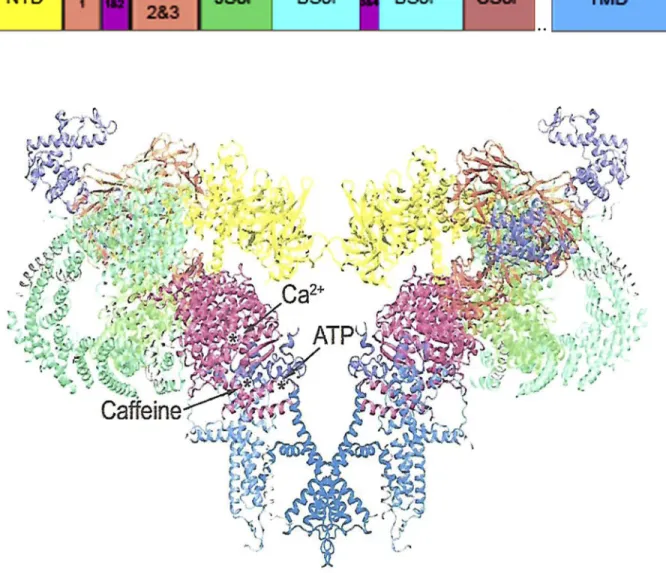
2015; Zalk et al., 2015; Bai et al., 2016; des Georges et al., 2016; Peng et al., 2016; Wei et al., 2016). More than two thirds of the molecular mass of RyRs was resolved. SR membranes from rabbit skeletal muscle (Efremov et al., 2015; Yan et al., 2015; Zalk et al., 2015; Bai et al., 2016; des Georges et al., 2016; Wei et al., 2016) and porcine cardiac muscle (Peng et al., 2016) were solubilized using the zwitterionic detergent 3-[(3-cholamidopropyl)dime-thylammonio]-1-propanesulfonate (CHA PS). RyRs were purified on sucrose gradients or by affinity chromatog-raphy using glutathione-transferase–tagged FKBP12 and FKBP12.6. Closed RyR2 channels were applied to grids in buffers containing 5 mM EDTA and the nonionic de-tergent digitonin (Peng et al., 2016). To obtain open channels, RyR2 was transferred to a buffer containing 20 µM Ca2+ and 20 µM PCB95, a RyR activator. Closed RyR1 structures were determined in buffers containing EGTA and Tween 20 (Yan et al., 2015) or CHA PS and a Figure 1. Open RyR1 channel structure. The structure (PDB code 5TAL) reveals the major domains and the location of Ca2+-, ATP-,
low concentration of lipid (Zalk et al., 2015; des Georges et al., 2016). To obtain closed and open RyR1s, Bai et al. (2016) applied the purified RyR1 channel complex to grids in a buffer that contained Tween 20 or CHA PS, respectively. Wei et al. (2016) determined the struc-ture of the closed RyR1 channel in a buffer containing amphipol A8-35, an amphipathic surfactant that stabi-lizes and maintains solubilized membrane proteins in detergent-free solutions. The open RyR1 channel was maintained in the presence of 100 µM Ca2+ and 10 µM ruthenium red, an RyR channel blocker. With some ex-ceptions, retention of function of the purified RyRs was not determined. Four different channel states were ob-tained according to single-channel measurements that kept RyR1 in a closed state (nominal 0 µM Ca2+), par-tially open state (30 µM Ca2+; 0 µM Ca2+, 2 mM ATP, and 5 mM caffeine), or fully open state (30 µM Ca2+, 2 mM ATP, and 5 mM caffeine; Fig. 1). Addition of 10 µM ry-anodine converted channels to an open subconducting state (des Georges et al., 2016).
Determination of the high-resolution closed- and open-channel structures provides a better understand-ing of the mechanisms of channel gatunderstand-ing and ion per-meation and how they are altered in RyR-associated diseases. Each RyR subunit has been subdivided into
∼10 domains (Fig. 1 and Table 1). These include (1) the N-terminal domain that is a hotspot of RyR1 and RyR2 disease mutations, (2) the SPRY1 domain that is part of the binding site for FKPB12, (3) the junctional solenoid (Jsol) and bridging solenoid (Bsol), where the latter contains PKA and Ca2+/CaM-dependent protein kinase II (CaMKII) phosphorylation sites, (4) the cen-tral solenoid (Csol) domain that contains two EF hand Ca2+ binding motifs, (5) the transmembrane domain that forms the conductance pathway for Ca2+, and (6) the C-terminal domain (CTD) that forms part of the Ca2+, ATP, and caffeine activation sites. Crystal struc-tures of smaller RyR fragments at resolutions of <2 Å and covering N-terminal, SPRY1, SPRY2, RY1 and 2, and RY3 and 4 domains have clarified the functional
role of ligands and disease mutations (Yuchi and Van Petegem, 2016). A comprehensive description of the RyR1 domain structure is provided by des Georges et al. (2016) and Samsó (2017).
Cav1.1–RyR1 interactions
A unique property of the mammalian skeletal muscle RyR1 is its direct linkage to the skeletal muscle Cav1.1 channel isoform, which acts in E-C coupling as a volt-age-sensing molecule rather than as a Ca2+ channel. Cav1.1 is comprised of a pore-forming α1 subunit and
α2δ, β, and γ subunits. Early death of dysgenic mice lacking the α1 subunit (Beam et al., 1986) and β-null mutant mice (Gregg et al., 1996) suggested that both subunits are required for skeletal muscle E-C coupling. Several regions of Cav1.1 α1 and β subunits interacted with RyR1 and vice versa (review by Bannister, 2016). Detailed structural information on the interaction be-tween the intact Cav1.1 and RyR1 is lacking, in part because a Cav1.1–RyR1 complex has not been isolated. However, obtaining this structural information should be facilitated by the availability of the recently deter-mined near-atomic resolution structure of the intact RyR1 (Yan et al., 2015; Zalk et al., 2015; Bai et al., 2016; des Georges et al., 2016; Wei et al., 2016) and Cav1.1 (Wu et al., 2016) complexes.
RyRs are high-conductance, cation-selective channels
Fusion of heavy rabbit skeletal muscle vesicles with pla-nar lipid bilayers provided the initial evidence that the SR membrane contains a calcium-conducting chan-nel of 125 pS (Smith et al., 1985). Chanchan-nel activities recorded in the presence of Ca2+, ATP, and ruthenium red were compared with results obtained in vesicle flux measurements according to a rapid quench protocol. Results indicated that the 125-pS channel mediates rapid Ca2+- and ATP-gated release of Ca2+ from skeletal muscle SR vesicles. Subsequent lipid bilayer studies with canine cardiac muscle SR vesicles (Rousseau et al., 1986) and purified [3H]ryanodine-binding protein complexes Table 1. Cryo-EM domain nomenclature of RyR1
Domain name Alternative names Colora Sequenceb
N-terminal domain Yellow 1–627
SPRY1 Light orange 628–849
RY1 and 2 P1 Light magenta 850–1,054
SPRY2 and 3 Light orange 1,055–1,656
Jsol Handle Green 1,657–2,144
Bsol HD1&2 Cyan 2,145–3,613
Includes RY3 and 4 P2 Orange 2,735–2,938
Csol Central domain Tomato 3,667–4,174
Includes EF hand motifs 4,060–4,134
TaF 4,175–4,253
Transmembrane domain Blue 4,541–4,956
C-terminal domain Brown 4,957–5,037
aDomain color in Fig. 1.
(Imagawa et al., 1987; Lai et al., 1988b; Smith et al., 1988; Anderson et al., 1989) established that RyR1 and RyR2 are Ca2+- and ATP-activated, Mg2+-inhibited, mon-ovalent and divalent ion–conducting channels with a PCa/PK ratio of ∼7–8.
Sequence comparisons and mutagenesis studies pro-vided evidence that the RyRs have a pore structure sim-ilar to K+ channels (Balshaw et al., 1999; Zhao et al., 1999; Gao et al., 2000; Du et al., 2001). RyR amino acid residues between the two C-terminal S5 and S6 mem-brane-spanning segments were suggested to be lumi-nally located, with a pore helix and an amino acid motif (GGG IG) similar to the selectivity filter motif (TV/ IGYG) of K+ channels. However, there are important differences among the RyRs relative to K+ channels. One is that RyRs conduct both monovalent and divalent cations with the following relative specificity: K+ > Rb+ > Na+≈ Cs+ > Li+, and Ba2+ > Sr2+ > Ca2+ > Mg2+ (Lindsay et al., 1991; Tinker and Williams, 1992). A second dif-ference is that single-channel measurements with per-meant and imperper-meant organic cations suggest a wider minimum radius of 3.3–3.5 Å for RyR2 (Tinker and Williams, 1993) than the experimentally determined minimum radius of 1.5 Å for K+ channels (Hille, 1973). Mutagenesis studies showed that two negatively charged amino acid residues (RyR1-D4899 and E4900) immedi-ately after the selectivity filter motif in the luminal vesti-bule and two negatively charged residues (RyR1-D4938 and D4945) in the cytosolic vestibule have a critical role in RyR1 ion permeation and selectivity (Wang et al., 2005; Xu et al., 2006).
Construction of a RyR1-Δ183-4006 deletion mutant and C-terminal construct of 1,377 amino acid resi-dues showed that the RyR1 C-terminal portion forms a monovalent conducting channel activated by Ca2+ and modified by ryanodine (Bhat et al., 1997). The two C-terminal transmembrane segments that included the pore helix and connecting loops formed a homote-trameric assembly that conducted K+ and Ca2+ and also Cl, which suggests the loss of a cation-specific pathway (Euden et al., 2013b).
Cryo-EM data support the view that RyR1 has a pore structure characteristic of the voltage-gated channel family. Samsó et al. (2005) and Ludtke et al. (2005) de-termined the pore structure of the closed (nonconduct-ing) RyR1 at a resolution of ∼10 Å. The pore-forming region as visualized by Ludtke et al. (2005) consisted of a long inner helix comprised of 31 amino acid residues and a pore helix of 15 residues. An increased resolution of ∼4 Å showed that Ile4937 in the S6 pore lining helix forms the hydrophobic constriction site in the closed RyR1 with a pore radius of less than 1 Å, rendering the channel impermeable to Ca2+ (Yan et al., 2015; Zalk et al., 2015). In the open RyR2 state, the pore constriction site was shifted to Gln4864 (corresponding to Gln4933 in RyR1), widening the RyR2 minimal pore radius to
∼2 Å (Peng et al., 2016). Ion-pulling simulations gener-ated an open-channel conformation of RyR1 (Mowrey et al., 2017) from the 3.8-Å closed state of RyR1 (Yan et al., 2015). The pore constriction site at RyR1-Gln4933 had a minimum open-channel pore radius of ∼2.8 Å (Mowrey et al., 2017), in reasonable agreement with the experimentally derived minimum radius of 3.3–3.5 Å (Tinker and Williams, 1993). The cryo-EM structure and model data suggest that channel opening involves a rotation of the upper portion of the pore-lining S6 helix away from the fourfold channel axis. Wei et al. (2016) used the RyR channel blocker ruthenium red to obtain an open channel. At variance with the aforemen-tioned studies, Ile4937 in the closed RyR1 conferred a pore radius of 2.5 Å, which increased to 4.9 Å in the “open ruthenium-red–stabilized” channel, allowing the passage of hydrated Ca2+ ions.
The RyR1 S6 pore lining helix has two conserved hinge glycines (Gly4934 and Gly4941) associated with channel opening. Determination of the pore structure of the closed (nonconducting) RyR1 at a resolution of
∼10 Å suggested that the inner S6 helix has a bend at RyR1-Gly4934 (Ludtke et al., 2005). Samsó et al. (2009) determined the cryo-EM structure of both the open and closed RyR1. As observed for the high-resolution struc-tures of K+ channels (Jiang et al., 2002), the inner helix had an outward bend on a glycine (Gly4934 or Gly4941) in the open but not closed channel. This change along with other cytoplasmic structural changes widened the radius of a cytoplasmic ion gate from ∼4 to ∼6 Å, al-lowing the flow of ions through the channel. Because cryo-EM had a resolution of only ∼10 Å, the positions of pore residue side chains and the structure of loops connecting the helices remained unknown (Ludtke et al., 2005; Samsó et al., 2009).
The two conserved glycines were replaced with un-charged amino acid residues of an increased side chain volume. Substitution of RyR2-Gly4864 (corresponding to RyR1-Gly4934) with alanine resulted in no significant change of RyR2 function, whereas replacement with va-line and prova-line profoundly altered channel gating and ion permeation (Euden et al., 2013a). Replacement of RyR1-Gly4934 and Gly4941 with alanine altered RyR1 channel function in single-channel measurements (Mei et al., 2015). Mutations further increasing the side chain volume at these positions (RyR1-G4934V and G4941I) re-sulted in loss of function. Molecular modeling suggested that the two glycines facilitate RyR channel function by providing flexibility and minimizing amino acid clashes.
the selectivity filter and open-channel RyR2-Gln4864 residue. A Poisson–Nernst–Planck density functional theory model accurately modeled the role of nega-tively charged residues in the luminal (RyR1-Asp4899 and Glu4900; Wang et al., 2005) and cytosolic (RyR1-Asp4938 and Asp4945; Xu et al., 2006) vestibules of RyR1 in generating the high ion conductances of RyRs (Gillespie et al., 2005). Selectivity was attributed to charge-space competition, as Ca2+ could accommodate the most charge in the least space compared with K+ (Gillespie et al., 2005; Gillespie, 2008). Although the large dehydration energy but high conductance of Mg2+ suggest that ion dehydration does not play a major role in RyR ion permeation (Gillespie et al., 2014), the exact hydration states of Ca2+ ions remain to be determined as they pass through the pore. Gillespie and Fill (2008) predicted that RyR ion channels must mediate their own K+ countercurrents to minimize the formation of a
membrane potential during SR Ca2+ release that would otherwise rapidly impede further release of Ca2+.
Regulation of RyRs by Ca2+
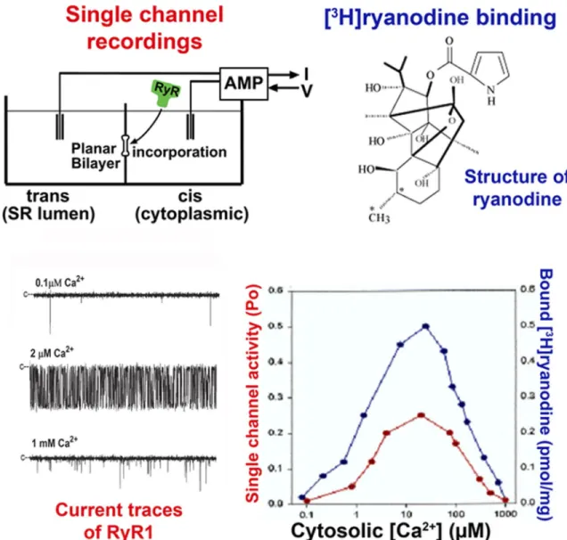
The mechanism of skeletal muscle and cardiac SR Ca2+ release has been extensively studied using isolated SR membrane and purified RyR preparations by applying three complementary methods. Macroscopic ion fluxes were measured from actively or passively loaded SR ves-icles using rapid flow, quench, and filtration protocols. Microscopic monovalent ion and Ca2+ currents were re-corded through single Ca2+ release channels incorpo-rated into lipid bilayers (Fig. 2). In a widely used but less direct method, RyR channel activity was probed using the specific plant alkaloid ryanodine (Sutko et al., 1997; Fig. 2). All three methods showed that RyRs are acti-vated by micromolar Ca2+ and millimolar ATP and are inhibited by millimolar Mg2+ and Ca2+.
Figure 2. Ca2+ dependence of RyR1. Data obtained using the planar lipid bilayer and [3H]ryanodine binding methods. With
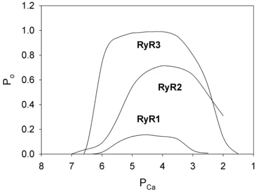
Evidence for cytosolic RyR sites involved in Ca2+ activa-tion. RyRs are activated by micromolar cytosolic Ca2+ and inhibited by millimolar cytosolic Ca2+ in the ab-sence of other channel effectors. A biphasic behavior implies there are at least two classes of Ca2+-binding sites: a high-affinity activation site and a low-affinity in-activation site. However, differences in Ca2+ regulation of the mammalian RyR isoforms have been observed. In single-channel measurements, RyR2 and RyR3 iso-forms are activated to a greater extent by cytosolic Ca2+ than RyR1 and require higher cytosolic Ca2+ concen-trations to be inhibited when Ca2+ is the sole activating ligand (Fig. 3). Similarly, in avian and amphibian skel-etal muscle that express both mammalian RyR1 and RyR3 homologues, RyR3 is activated by Ca2+ to a greater extent than RyR1 (Percival et al., 1994; Mu-rayama and Ogawa, 2004).
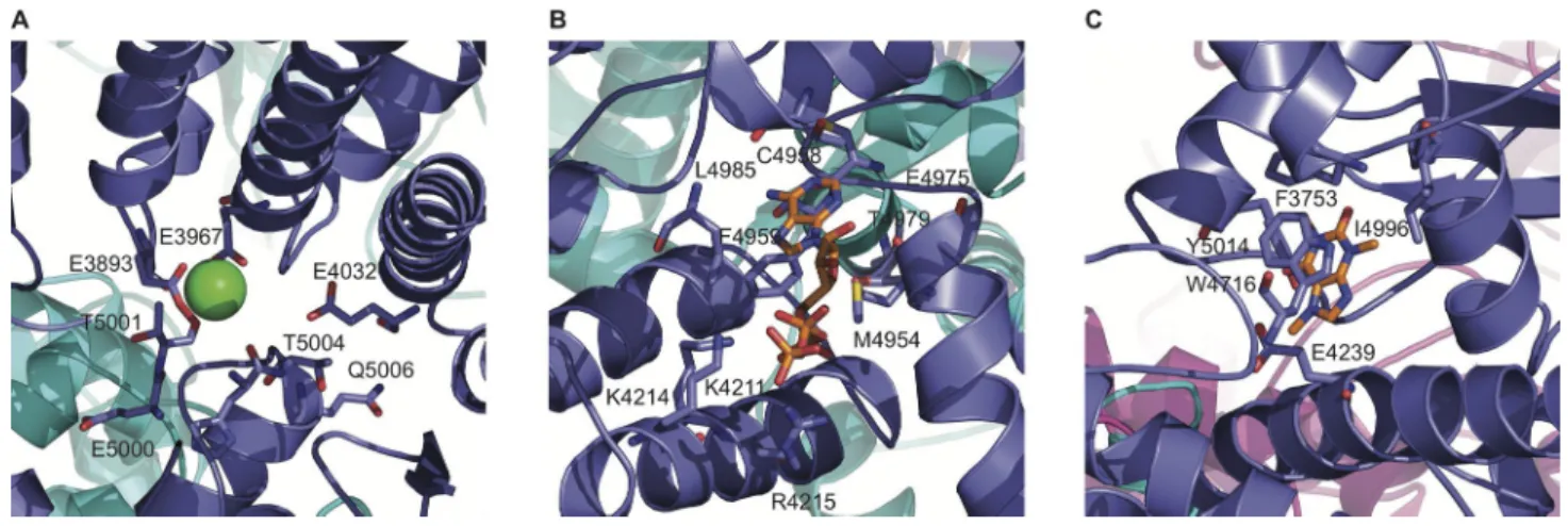
Comparison of cryo-EM maps prepared in the ab-sence or preab-sence of 30 µM Ca2+ revealed a Ca2+ -bind-ing site at the interdomain interfaces of the RyR1 Csol and C-terminal domains (des Georges et al., 2016). The site encompasses three amino acids conserved among the RyR and IP3R families directly interacting with Ca2+, RyR1-Glu3893, and Glu3967 of the Csol and Thr5001 of the C-terminal domain (Fig. 4 A). Two amino acids, Gln3970 and His3895, indirectly interact with Ca2+. The channel apparently remains closed with 30 µM Ca2+ as the sole activating ligand, even though single-channel measurements showed a channel open probability (Po) of 0.22. Similarly, opposite results were obtained using ATP and caffeine alone, whereas the three ligands Ca2+, ATP, and caffeine opened the channel in the structural and single-channel studies. des Georges et al. (2016)
proposed that Ca2+ or ATP/caffeine alone primed the channel for opening, but all three ligands were re-quired to overcome a barrier for opening. An alterna-tive explanation is that the number of open channels under the conditions of Ca2+ or ATP/caffeine was too low to be scored as a separate class.
Mutagenesis has indicated that additional regions are involved in the regulation of RyRs by Ca2+. In one early study, RyR1-Δ183-4006 deletion mutant was activated by micromolar Ca2+, which suggests a Ca2+-binding site dif-ferent from the one identified by cryo-EM (Bhat et al., 1997). Substitution of RyR1-Glu4032 by alanine and the corresponding amino acid residue in the RyR3 isoform reduced Ca2+-gated channel activity (Chen et al., 1998; Du and MacLennan, 1998; Fessenden et al., 2001). However, RyR1-Glu4032 was not a part of a Ca2+-binding site, but rather stabilized the interaction between two RyR1 regions (des Georges et al., 2016). Other exam-ples include CCD mutations located in the pore-form-ing region of RyR1 in skeletal muscle (McCarthy et al., 2000; Dirksen and Avila, 2002; Xu et al., 2008), sites in S2 (Gomez and Yamaguchi, 2014), S4–S5 linker (Murayama et al., 2011; Ramachandran et al., 2013), N-terminal region of RyR2 (Liu et al., 2015), and S6 of RyR2 (Sun et al., 2016). Taken together, both struc-tural and functional studies indicate that the transition of the closed to Ca2+-activated open channel involves the coordinated motion of multiple regions outside the Ca2+-binding site.
Evidence for cytosolic RyR sites involved in Ca2+ inactiva-tion. Efremov et al. (2015) compared two RyR1 cryo-EM structures in the absence of Ca2+ and at 10 mM Ca2+ at a Figure 3. Ca2+ dependence of single
purified RyR1, RyR2, and RyR3. Shown are channel Po values as a function of
cytosolic free Ca2+ in 250 mM K+, pH
resolution of 6.1 Å. Differences in the Ca2+-free and Ca2+-bound maps were interpreted to indicate that binding of Ca2+ to a two–EF hand motif in the Csol do-main (Fig. 1 and Table 1) increased the mean gate di-ameter, which indicated formation of an open channel. However, functional studies were not presented. More-over, the addition of 10 mM Ca2+ results in a nearly fully closed and not open RyR1 channel (Fig. 3). Therefore, it seems that Ca2+ ions bound to sites involved in Ca2+ inactivation and not activation of RyR1.
Earlier [3H]ryanodine binding studies with RyR1/ RyR2 chimeras suggested that Ca2+ inactivation sites are located in the C-terminal quarter of RyR1 (Du and MacLennan, 1999). In this region, two EF1 and EF2 hand motifs are located in a CaM-like domain of RyR1 (amino acids 4,064–4,210; Xiong et al., 2006). A pep-tide corresponding to the CaM-like domain bound Ca2+ with an apparent affinity of 60 µM (Xiong et al., 2006). Two smaller recombinant proteins from rabbit skeletal muscle RyR1 (amino acids 4,069–4,139), cardiac mus-cle RyR2 (amino acids 4,025–4,095), and lobster skel-etal muscle containing the two EF hand motifs bound two Ca2+ with millimolar affinities, implying that sites in these regions have an inhibitory role (Xiong et al., 1998). Deletion and mutations of the two–EF hand motif in the full-length RyR2 suggested that the EF-hand domain has a role in RyR2 regulation by luminal Ca2+ but is not required for activation by cytosolic Ca2+ (Guo et al., 2016). Fessenden et al. (2004) assessed the functional significance of five potential Ca2+ regulatory sites in RyR1, taking into account that, in addition to the two–EF hand motifs, there are three negatively charged EF-hand-like sequences encompassing amino acid resi-dues 4,253–4,264, 4,407–4,416, and 4,489–4,499. Muta-genesis of the five motifs in RyR1 did not reveal major functional differences in response to depolarization or caffeine compared with wild-type RyR1-expressing myotubes (Fessenden et al., 2004). However, in a bind-ing assay usbind-ing the RyR specific ligand [3H]ryanodine,
a mutant with a scrambled EF1 hand motif exhibited altered Ca2+ activation and inactivation. In a scrambled EF2 hand mutant, [3H]ryanodine binding was lost, but Ca2+-dependent activity was maintained in single-chan-nel recordings. Studying a large number of RyR1/RyR2 chimeras, Gomez and Yamaguchi (2014) found that two RyR regions are involved in Ca2+-dependent inac-tivation, one region containing the two–EF hand mo-tifs and a second that included the S2 transmembrane segment. Among the two–EF hand motifs, EF1 more strongly controlled the isoform-specific inactivation of the RyRs by Ca2+.
Evidence for luminal RyR sites involved in regulation by luminal Ca2+. Both cytosolic and luminal Ca2+ regulate RyRs. Accordingly, dependence of channel activity on Ca2+ has suggested the presence of at least three types of Ca2+-binding sites, high-affinity (activating) and low-af-finity (inactivating) sites accessible from the cytosol in large cytosolic foot region of the RyRs, and luminal Ca2+-binding sites, whose occupancy in skeletal and car-diac muscle depends on SR Ca2+ load (Ford and Po-dolsky, 1972; Fabiato and Fabiato, 1979; Shannon et al., 2000; Györke et al., 2002).
channel (Sitsapesan and Williams, 1994, 1995). Differ-ences in the number of channel events and the duration of open and closed events suggested that binding of ATP or sulmazole to cytosolic channel sites uncovered luminal Ca2+-binding sites. Diaz-Sylvester et al. (2011) compared the regulation of RyR2 by luminal Mg2+, Ca2+, Sr2+, and Ba2+. RyR2 activation depended on cytosolic Ca2+ and caffeine concentrations. Although a greater activation by divalent cations compared with Cs+ was observed in the absence of caffeine at 100 μM cytosolic Ca2+, caffeine was required at 100 nM cytosolic Ca2+ for luminal Ca2+ and Sr2+ (but not Mg2+ and Ba2+) to acti-vate RyR2 (Diaz-Sylvester et al., 2011). This suggested the presence of two luminal activating cation-binding sites, one specific for Ca2+ and another that bound Ca2+ and Mg2+. Gaburjakova and Gaburjakova (2016) reported that at 100 nM cytosolic Ca2+ and millimolar caffeine, RyR2 was fully activated by luminal Ca2+ and Sr2+. Higher caffeine concentrations were required for full activation by luminal Mg2+ and Ba2+. A luminal S1-S2 EF hand–related loop region was suggested to bind with an affinity of Ca2+ > Sr2+ >Mg2+≈ Ba2+.
A “store overload-induced Ca2+ release” (SOI CR) in cardiac muscle can generate Ca2+ waves and Ca2+ -trig-gered arrhythmias. Loss of luminal Ca2+ activation sug-gested the presence of a luminal regulatory site (Jones et al., 2017). A point mutation in RyR2 (RyR2-E4872A corresponding to RyR1-E4942A) eliminated Ca2+ regula-tion by luminal Ca2+ but not by cytosolic Ca2+. In mouse hearts, the RyR2-E4872 mutation suppressed SOI CR and Ca2+ triggered ventricular tachycardia (Chen et al., 2014).
Evidence for cytosolic RyR sites involved in regulation by luminal Ca2+. An alternative suggestion is that luminal
Ca2+ regulates RyRs by binding to cytosolic regulatory sites after their passage through the open channel (also referred to as feed-through; Herrmann-Frank and Leh-mann-Horn, 1996; Tripathy and Meissner, 1996; Xu and Meissner, 1998; Uehara et al., 2017). To distinguish lu-minal and cytosolic Ca2+ regulatory sites, voltage- and Ca2+-dependent regulation of single RyR1 channels was reported (Tripathy and Meissner, 1996). Single chan-nels were partially opened by cytosolic ATP in the pres-ence of low cytosolic Ca2+. As luminal Ca2+ was increased to micromolar concentrations, increasing channel ac-tivity was initially only seen at negative holding poten-tials that supported a luminal-to-cytosolic flow through the channels. A further increase in luminal Ca2+ to mil-limolar levels resulted in a decrease in channel activity. The results suggested that luminal Ca2+ flowing through the skeletal muscle Ca2+ release channel regulates chan-nel activity by accessing cytosolic Ca2+ activation and in-activation sites.
A possible resolution of the different mechanisms proposed for the luminal regulation of RyRs is that it involves Ca2+ sensing sites on both the luminal and
cyto-solic channel sites (Laver and Honen, 2008). Modeling of single ATP-activated RyR2 channel activities indi-cated four Ca2+ regulatory sites: luminal (L) activation site, cytosolic activation (A) site, and cytosolic high-af-finity (I2) and low-afhigh-af-finity (I1) inactivation sites. Lumi-nal Mg2+ inhibited RyR2 by competing for Ca2+ at the L and A sites and binding to the I1 site. As discussed later, luminal Ca2+ also regulates RyRs by binding calse-questrin (CSQ), a low-affinity, high-capacity luminal Ca2+-binding protein.
Regulation by Mg2+
Mg2+ is thought to inhibit RyRs by (a) competing with Ca2+ for a high-affinity cytosolic Ca2+ activation site, (b) binding with similar affinity as Ca2+ to a low-affinity cyto-solic Ca2+ inhibitory site, (c) binding to luminal regula-tory sites, and (d) reducing the flow of Ca2+ through the channel (Meissner et al., 1986, 1997; Smith et al., 1986; Gusev and Niggli, 2008; Laver and Honen, 2008). Mg2+ also has an “activating” effect. Suppression of rat RyR2 and recombinant rabbit RyR2 activities at 10–100 µM Ca2+ was relieved by Mg2+ in [3H]ryanodine binding assays (Chugun et al., 2007; Gomez and Yamaguchi, 2014). As discussed later, Mg2+ forms a MgATP complex that modulates the regulation of the RyRs by Ca2+.
Regulation by other divalent and trivalent cations
Among nonphysiological ions tested, Sr2+ was optimal in activating RyR2 at 1 mM, whereas Ba2+ did not acti-vate RyR2 (Liu et al., 1998; Diaz-Sylvester et al., 2011). Both inhibited RyR1 and RyR2 at high millimolar con-centrations (Meissner et al., 1997; Diaz-Sylvester et al., 2011). Fe2+ but not Fe3+ inhibited [3H]ryanodine bind-ing by competbind-ing with Ca2+ for the RyR2 Ca2+ activation site (Kim et al., 1995). Lanthanides (Tb3+, Eu3+, Sm3+) are nonconducting ions that activated or inactivated RyR1 at submicromolar to micromolar levels by binding to cytosolic high-affinity Ca2+ activating and low-affinity Ca2+ inactivating sites (Hadad et al., 1994; Sárközi et al., 2017). Voltage-dependent regulation by cytosolic Eu3+ suggested that lanthanides bind to cytosolic activating Ca2+-binding sites in the electrical field of the channel, whereas the inactivating sites are located outside the electrical field (Sárközi et al., 2017).
Regulation by monovalent cations and anions
Nonphysiological assay media have been used to study RyR function. These include 50 mM CaHEP ES/TrisHEP ES or 0.25 M cesium methanesulfonate in single-chan-nel measurements with SR membrane preparations to suppress SR K+ and Cl− channel activities, or 0.25 M
KCl in [3H]ryanodine binding studies or single-chan-nel measurements with purified RyR preparations. However, RyR activities are greatly affected by the ionic composition and strength of the assay medium. An in-crease in KCl or NaCl stimulated Ca2+ release from SR vesicles and increased [3H]ryanodine binding and sin-gle-channel Po (Michalak et al., 1988; Chu et al., 1990; Ogawa and Harafuji, 1990; Meissner et al., 1997; Liu et al., 1998). Using the [3H]ryanodine ligand binding assay, RyR1 and RyR2 Ca2+-dependent channel activities were oppositely affected by (a) the competitive binding of monovalent cations to high-affinity Ca2+ activating binding sites, and (b) binding of Cl− to unknown anion
activating sites (Meissner et al., 1997; Liu et al., 1998).
Regulation by adenine nucleotides
Comparison of cryo-EM maps prepared in the absence or presence ATP and caffeine revealed an ATP-binding site at the cytoplasmic extension of S6 and elements of the C-terminal domain (des Georges et al., 2016). The site encompasses eight RyR1 amino acids. The adenine base of ATP contacts RyR1-Met4954, Phe4959, Thr4979, and Leu4985; the ribose ring of ATP likely interacts with Glu4955; and the triphosphate tail of ATP with positively charged Lys4211, Lys4214, and Arg4215 (Fig. 4 B). Chan-nels apparently remained closed in the presence of ATP and caffeine as the sole activating ligands, even though single-channel measurements showed a channel Po of 0.13. des Georges et al. (2016) proposed that, similar to Ca2+ alone, ATP/caffeine alone primed the channel for opening, and that in addition, Ca2+ was required to open RyR1. Cross-linking studies using 2-azidoadenosine
5′-trisphosphate 2′3′-biotin-long-chain-hydrazone identi-fied four glycine-rich consensus motifs in the N-terminal 95-kD fragment of RyR1, which suggests there may be additional nucleotide-binding sites (Popova et al., 2012).
Ca2+ and Mg2+ greatly affect the activation of RyRs by ATP. In single-channel measurements in the absence of cytosolic Ca2+ and Mg2+, millimolar ATP weakly ac-tivated the essentially closed RyR1 (Tripathy and Meis-sner, 1996) and RyR2 (Kermode et al., 1998). On the other hand, RyR1 and RyR2 were nearly fully activated by ATP in the presence of micromolar Ca2+. Other nine nucleotides such as ADP, AMP, adenosine, or ade-nine enhanced the release of Ca2+ from skeletal muscle SR vesicles (Morii and Tonomura, 1983; Meissner, 1984) with a decreased potency compared with AMP-PCP (Meissner, 1984). In single channel measurements, ADP was less effective in activating RyR2 than ATP (Ker-mode et al., 1998). Other nucleotides such as CTP, GTP, ITP, or UTP had no substantial effect on the release of Ca2+ (Morii and Tonomura, 1983; Meissner, 1984).
Most cellular ATP is present as MgATP complex that represents the predominant biologically active form of ATP in cells. An increase in Mg2+ that increases MgATP and decreases uncomplexed ATP decreased RyR activ-ity (Meissner et al., 1986; Xu et al., 1996; Walweel et al., 2014). However, whether MgATP or ATP is a physiolog-ical regulator of RyRs is unclear. In support of an inter-action with “free” ATP is that ATP activates RyRs in the absence of Mg2+, and three positively charged amino acid residues in the ATP-binding site complement the negatively charged triphosphate tail of ATP. Experimen-tal evidence whether ATP or MgATP is the preferred physiological regulator of RyR was not obtained by des Georges et al. (2016) because the cryo-EM studies were done in the absence of Mg2+
.
Regulation of RyRs by posttranslational modifications
In addition to the regulation by Ca2+ and other small molecules, RyRs are regulated by posttranslational mod-ifications involving phosphorylation and redox modifi-cation of sites in the large cytosolic domain of the RyRs.
RyR cysteine redox modifications. RyRs contain a large number of amino acid residues that are potential tar-gets for reactive oxygen and nitrogen molecules gener-ated in working muscle under physiological or pathological conditions. These include free thiols, amines, and tyrosines. Most attention has been directed toward the role of cysteines and their modification by redox active molecules. RyRs contain a large number of cysteines: 100 in RyR1 and 89 in RyR2 subunit, and 1 in FKBP12 and 2 in FKBP12.6. It is therefore not surpris-ing that cysteines are involved in the regulation of RyR activity.
re-ducing agent. However, muscle pO2 is ∼10 mm Hg, and muscle cells have a high ratio of reduced to oxidized glutathione (GSH). Use of the lipophilic thiol–specific probe monobromobimane indicated that nearly half of the 100 cysteines in the RyR1 subunit were free in the presence of 5 mM reduced GSH at pO2 ∼10 mm Hg (Eu et al., 2000). An increase in oxygen tension from ∼10 mm Hg to ambient air (pO2 ∼150 mm Hg) in the pres-ence of 5 mM GSH resulted in loss of 6–8 free thiols/ RyR1 subunit. Substitution of GSH with oxidized GSH (GSH disulfide [GSSG]) at pO2 ∼10 mm Hg or ambient air reduced the number of free thiols/RyR1 subunit to
∼26 and increased RyR1 activity (Sun et al., 2001b). The number of free thiols in RyR2-enriched fractions in the presence of 5 mM GSH was 58 cysteines /mg protein at pO2 ∼10 mm Hg and 50 cysteines/mg protein at ∼150 mm Hg (Sun et al., 2008). In the presence of 5 mM GSSG, a lower number of free cysteines was measured. However, because RyR2 was only partially purified, the exact number of GSH- and pO2-sensitive thiols in native RyR2 remains to be determined. The results suggest that RyR1 and RyR2 have functional thiols that respond to cellular pO2 and the GSH/GSSG redox state.
Treatment of SR vesicles with reduced GSH at pO2
∼150 mm Hg and mass spectrometric analysis of tryp-tic fragments showed that monobromobimane reacted with 40 cysteines in the majority of experiments, with 20 additional monobromobimane reactive cysteines less often detected (Petrotchenko et al., 2011). Seven RyR1 cysteines (Cys1040, 1303, 2436, 2565, 2606, 2611, and 3635) were selectively labeled by 7-diethylamino- 3-(4′-maleimidylphenyl)-4-methylcoumarin under con-ditions that led to loss of RyR1 redox sensitivity (Voss et al., 2004). Nine RyR1 cysteines (Cys36, 315, 811, 906, 1591, 2326, 2363, 3193, and 3635) were endogenously modified, and another three cysteines (Cys253, 1040, and 1303) were modified by exogenous reactive oxygen and nitrogen molecules (Aracena-Parks et al., 2006). Sun et al. (2013) identified 13 oxygen tension-sensitive cysteines (Cys36, 566, 762, 845/854, 1674, 2305/2310, 2555, 2606, 2611, 2704, and 4238). Another eight cys-teines (Cys120, 253, 305, 490, 1686, 2021, 3892, and 4663) were oxidized by an NADH oxidase at high pO2.
Mutagenesis studies have shown that cysteines located in the Jsol and Bsol domains and a poorly resolved re-gion of RyR1 (des Georges et al., 2016) may serve a redox regulatory function. Substitution of cysteines with serine or alanine identified three cysteines (Cys1781, 2436, and 2606) that responded to a change in GSH redox potential (Petrotchenko et al., 2011). At physio-logical O2 concentrations, nitric oxide (NO) specifically S-nitrosylated Cys3635 (Sun et al., 2001a). Cys3635 is part of the high-affinity CaM-binding domain of RyR1, which provides an explanation for the observation that NO was effective only in the presence of CaM. In addi-tion, mutagenesis of Cys4958 and Cys4961 in C2H2-type
Zn2+ finger motif of RyR1 resulted in an inactive chan-nel, which supported a depolarization-dependent Ca2+ entry mechanism (Hurne et al., 2005). Mutagenesis of RyR2-Cys4888 and Cys4891 eliminated caffeine activa-tion of RyR2 (Peng et al., 2016).
RyR phosphorylation. The large cytoplasmic foot region of RyRs has many potential phosphorylation sites (Takeshima et al., 1989; Zorzato et al., 1990) that may be targeted by protein kinases and phosphatases includ-ing PKA, CaMKII, and phosphatases 2A (PP2A), PP1, and PP2B (Hohenegger and Suko, 1993; Marx et al., 2000, 2001; Dulhunty et al., 2001; Shin et al., 2002). RyR1 has one and RyR2 has three well-established phos-phorylation sites per RyR subunit. Early studies indi-cated that canine RyR2-Ser2809 is phosphorylated by CaMKII and by PKA to a lesser extent (Witcher et al., 1991). Subsequent studies showed that cAMP-activated kinase (PKA) phosphorylates Ser2843 in rabbit RyR1 (Suko et al., 1993), mouse RyR2 at Ser2030 (Xiao et al., 2005), and human RyR2 at Ser2808 (Marx et al., 2000). CaMKII uniquely phosphorylated recombinant RyR2 at Ser2815 (Wehrens et al., 2004). RyR1 was phosphory-lated at Ser2843 (corresponding to Ser2809 in RyR2) by cAMP-, cGMP-, and CaMKs (Suko et al., 1993). Phos-phorylation of threonine (Suko et al., 1993) and differ-ent functional effects of CaMKII, PKA, and cGMP-dependent protein kinase (Takasago et al., 1991; Hain et al., 1995) suggested the presence of additional phosphorylation sites.
Phosphorylation of rabbit RyR2-Ser2809 in the P2 domain induced a more flexible conformation that fa-vored the transition from the closed to open channel states (Dhindwal et al., 2017). Crystal structures of the P2 domain of RyR1 (amino acids 2,734–2,940; Sharma et al., 2012; Yuchi et al., 2012) and the correspond-ing P2 domains of RyR2 and RyR3 (Yuchi et al., 2012) have been reported. In vitro phosphorylation showed that PKA phosphorylated four residues corresponding to human residues Ser2808, Thr2810, Ser2811, and Ser2814 in the RyR2 P2 domain peptide (Yuchi et al., 2012). CaMKII also phosphorylated four residues (but Thr2876 instead of Thr2810).
complexes, which resulted in defective RyR2 regulation. However, other studies have challenged these findings (Stange et al., 2003; Xiao et al., 2005; Alvarado et al., 2017). A recent study indicates complex PKA phosphor-ylation-mediated regulation of RyR2-Ser2809. Both the complete loss of phosphorylation or maximal phosphor-ylation of Ser2809 increased the SR leak in rabbit ven-tricular myocytes (Bovo et al., 2017).
Modulation of RyR activity by accessory proteins
RyR activity is affected by a large number of proteins that interact with the RyRs, such as the FKBPs, CaM, S1001, triadin, junctin, and anchoring proteins for ki-nases and phosphatases.
FKBPs. The small 12- and 12.6-kD FKBPs are
predomi-nantly associated with RyR1 and RyR2, respectively (Lam et al., 1995) and are generally considered part of the massive RyR complexes. FKBPs belong to the family of immunophilins and exhibit cis/trans isomerase activ-ity, and their pharmacological removal using rapamycin or FK506 functionally uncouples groups of channels and increases channel activity (Brillantes et al., 1994). Dissociation of FKBPs induced the formation of sub-states in single-channel measurements (Brillantes et al., 1994; Ma et al., 1995; Ahern et al., 1997), whereas in other studies, full conductance was maintained (Barg et al., 1997; Mei et al., 2013). Cryo-EM studies located FKBP12 (fused to GSH S-transferase) to a relatively large area of RyR1 (Wagenknecht et al., 1996), which in subsequent studies was narrowed to a site at the inter-face of three peripheral domains corresponding to the SPRY1, SPRY3, and Jsol domains (Samsó et al., 2006; Sharma et al., 2006). Crystal structure analysis of the RyR2-SPRY1 domain along with FRET and mutagenesis studies and docking to cryo-EM maps suggested FKBP binding to a hydrophobic cluster within SPRY1 (Yuchi et al., 2015).
CaM. CaM is a 16.7-kD cytosolic protein that regulates SR Ca2+ release by direct binding to RyRs and through other proteins that interact with RyRs and bind CaM. Other major contributors are the voltage-regulated sur-face membrane Ca2+ channels (Cav1s), CaM-dependent protein kinase (CaMKII), and CaM-stimulated protein phosphatase (calcineurin). CaM inhibits all three mam-malian RyR isoforms at free [Ca2+] >1 µM. At low free Ca2+ concentrations (<1 µM), CaM activates RyR1 and RyR3 channel activity, whereas RyR2 is inhibited by CaM (Ikemoto et al., 1998; Fruen et al., 2000; Balshaw et al., 2001; Yamaguchi et al., 2005). RyR1 and RyR2 bind with nanomolar affinity 1 apoCaM (Ca2+-free form of CaM) or 1 CaCaM (Ca2+-bound form of CaM) per RyR subunit (Balshaw et al., 2001; Yamaguchi et al., 2005).
Trypsin digestion and site-directed mutagenesis demonstrated that RyRs have a single conserved
high-af-finity CaM-binding domain per RyR subunit (RyR1 amino acids 3,614–3,643, RyR2 amino acids 3,581– 3,610, and RyR3 amino acids 3,467–3,498) that inter-acts with Ca2+-free or Ca2+-bound forms of CaM (Moore et al., 1999; Rodney et al., 2001; Yamaguchi et al., 2001, 2003, 2005). Crystal structure, nuclear magnetic reso-nance, and FRET data showed that the CaM-binding domain binds both CaM lobes in a complex formed by CaCaM and peptide corresponding to the CaM-bind-ing domain of RyR1 (Maximciuc et al., 2006). Alterna-tively, CaCaM bound only the C-lobe, with the N-lobe potentially binding to another RyR1 region. This may explain why several RyR1 CaCaM- and apoCaM-binding sites were observed, using synthetic peptides and fusion proteins (Chen and MacLennan, 1994; Guerrini et al., 1995; Lau et al., 2014). Studies with RyR1/RyR2 chime-ras and mutants indicated that N-terminal sites and two predicted Ca2+ binding motifs (EF1 RyR1-4081-4092 and EF2 RyR1-4116-4127) are involved in the isoform-spe-cific regulation by CaM at less than 1 µM Ca2+ (Xu et al., 2017). Determination of temperature dependence of CaM binding showed major differences in the energet-ics of CaM binding to and CaM dissociation from RyR1 and RyR2 (Meissner et al., 2009).
Cryo-EM studies with RyR1 suggest that the CaM-bind-ing site is at least 10 nm away from the transmembrane channel of the receptor and that Ca2+ binding to CaM (and RyR1) causes an ∼33-Å shift of the binding site (Wagenknecht et al., 1994; Samsó and Wagenknecht, 2002). CaCaM is located in clefts formed by structural domains corresponding to the Jsol (amino acids 1,657– 2,144) and Bsol (amino acids 2,145–3,613) domains, with the latter domain located close to the high-affinity CaM-binding site (Wagenknecht et al., 1997). The se-quence corresponding to the high-affinity CaM-binding domain was not sufficiently resolved in high-resolution cryo-EM studies to reveal its structure in intact RyRs (des Georges et al., 2016; Peng et al., 2016).
commonly implicated in cardiomyopathies. In support of a role of CaM in terminating SR Ca2+ release, CaM prolonged the closed durations of RyR2 in the presence of ATP and Mg2+, allowing Ca2+ to diffuse away from the release sites and eliminating reopening of these chan-nels by Ca2+ (Xu and Meissner, 2004).
S100A1. S100A1 is a small Ca2+-binding protein that modulates the activity of multiple Ca2+-handling pro-teins in skeletal and cardiac muscle. S100A1 modulated striated muscle function by binding to the CaM-binding domain of RyR1 (Prosser et al., 2008; Wright et al., 2008). S100A1 inhibited RyR2 but not the single-site mutant RyR2-L3591D of the RyR2 CaM-binding domain in single-channel measurements (Yamaguchi et al., 2013). A more recent FRET study suggested that CaM and S100A1 can concurrently modulate RyR1 and RyR2 function without S100A1 competing for CaM at the RyR CaM-binding site (Rebbeck et al., 2016).
Other RyR accessory proteins. RyRs interact with a large number of additional proteins such as triadin, junctin, CSQ, anchoring proteins for kinases and phosphatases, and homer proteins. RyR interacting sites for these pro-teins have been described. The RyRs form a junctional quaternary complex with triadin, junctin, and CSQ. Tri-adin and junctin have a single transmembrane-span-ning domain and interact directly with the RyRs. Redox-sensitive cysteines on RyR1 and triadin have a role in regulating RyR1 function (Liu and Pessah, 1994). Three positively charged lysines in full-length 95-kD triadin bind to three negatively charged RyR1 residues located in S5 pore helix (RyR1-Asp4878) and S6 pore helix (Asp4907, Glu4908) linkers (Lee et al., 2004; Goonasekera et al., 2007). Junctin-binding sites are less well defined. Different junctin domains may bind to cytosolic and luminal sites (Altschafl et al., 2011; Li et al., 2015).
CSQ is a low-affinity, high-capacity Ca2+ storage pro-tein (MacLennan and Wong, 1971) concentrated in the heavy vesicle fraction derived from the SR terminal cisternae of skeletal muscle (Meissner, 1975). Cloning studies indicated the presence of two CSQ isoforms (Zorzato et al., 1994). CSQ1 is present in fast-twitch skeletal muscle and CSQ2 in cardiac muscle. Both CSQ isoforms are expressed in slow-twitch skeletal muscle (Arai et al., 1991; Biral et al., 1992; Murphy et al., 2009). The two isoforms contain a large number of negatively charged aspartic and glutamic acid residues, with CSQ2 having an extended negatively charged C terminus. As-sociation with the RyR complex involved the binding of triadin and junctin KEKE motifs (Guo and Campbell, 1995; Zhang et al., 1997; Kobayashi et al., 2000) to as-paragine-rich regions in the C terminus of CSQ (Shin et al., 2000; Beard and Dulhunty, 2015). CSQs formed lin-ear polymers (Park et al., 2004) and dissociated (Zhang
et al., 1997) from their binding proteins as the Ca2+ con-centration increased.
In addition to increasing Ca2+ store size, CSQ mod-ulates RyR channel activity. A deficiency in SR luminal cardiac CSQ in humans (Postma et al., 2002; Lahat et al., 2004), mice (Knollmann et al., 2006), and cardio-myocytes (Terentyev et al., 2003) resulted in an imbal-ance of Ca2+ handling and CPVT. Overexpression of CSQ2 suppressed SR Ca2+ transients and led to severe cardiac hypertrophy in mice (Jones et al., 1998; Sato et al., 1998; Terentyev et al., 2003). Abnormal Ca2+ han-dling in association with cardiac myopathies was also observed in mice and cardiomyocytes that lacked or overexpressed the RyR2-accessory proteins triadin and junctin (Gergs et al., 2007; Kirchhof et al., 2007; Yuan et al., 2007; Chopra et al., 2009).
Marx et al. (2001) identified several anchoring pro-teins that mediate an interaction between RyR2 and accessory kinases and phosphatases through conserved leucine/isoleucine motifs. Spinophilin targeted PP1 to the N-terminal domain, PR130 directed PP2A to the SPRY 3 domain, PR130 directed PP2A to a region over-lapping with SPRY3 domain, and A-kinase anchoring protein targeted PKA to Bsol domain. Homer proteins modulate RyR activity by binding putative proline-con-taining motifs in the RyR SPRY1 and Jsol domains (Feng et al., 2002; Pouliquin and Dulhunty, 2009).
Effects of ryanodine and caffeine
(Lai et al., 1989; Pessah and Zimanyi, 1991; reviewed by Sutko et al., 1997).
Caffeine binds to a site encompassing the S2-S3 linker and CTD, contacting residues Trp4716 and RyR1-Ile4996, respectively (des Georges et al., 2016; Fig. 4 C). In contrast to ryanodine, caffeine (1,3,7-trimethyl-xanthine) activated purified RyR1 by increasing chan-nel open probability without significantly affecting single-channel conductance (Rousseau et al., 1988). Another difference is that caffeine activates the Ca2+ release channel without loss of sensitivity to regulation by Ca2+, Mg2+, and ATP. Among 30 xanthines tested, 1,7-dimethylxanthine and 1-hexyl-3,7-dimetylxanthine (pentifylline) were most effective in activating RyR1 in a [3H]ryanodine binding assay (Liu and Meissner, 1997).
RyRs and muscle disorders
Naturally occurring mutations in RyR1 give rise to a variety of muscle diseases that include MH, CCD, and MmD (Treves et al., 2008) A list of mutations potentially associated with RyR1 is available at http ://www .dmd .nl / nmdb2 /variants .php ?select _db =RYR1 &action =search _ all &order. MH is an inherited disease that causes a rapid rise in body temperature and inappropriate muscle contraction when the affected persons receive general anesthesia and MH-linked RyR1 mutations release Ca2+ ions from the SR. MH-linked RyR1 mutations were initially mapped to the N-terminal and central domains of RyR1; however, more recently identified MH mutations are distributed throughout the RyR1 coding sequence (Robinson et al., 2006; Stowell, 2008; Treves et al., 2008). MH is treated with dantrolene, a muscle relaxant that reduces the rate of SR Ca2+ release (Zucchi and Ronca-Testoni, 1997). Recent studies have shown that two RyR1 ligands, CaM (Oo et al., 2015) and an increase in metabolite Mg2+ from MgATP hydrolysis
during increased muscle activity (Choi et al., 2017), facilitate dantrolene inhibition. CCD and MmD are congenital diseases associated with a dysfunctional RyR1 (Treves et al., 2008). In many cases, dominant RyR1 mutations linked to CCD localize to the C-terminal domain of the channel. RyR1 with homozygous mutations associated with core myopathies often do not conduct Ca2+, whereas heterozygous RyR1 channels composed of wild-type and mutant subunits conduct variable amounts of Ca2+ (Xu et al., 2008). More than 150 RyR2 mutations have been potentially linked to cardiac diseases such as CPVT and ARVD2 (George et al., 2007; Leenhardt et al., 2012). CPVT is an inherited cardiac disorder characterized by life-threatening arrhythmias elicited by stress and emotional disturbances. ARVD is an inherited RyR2-linked disorder that results in arrhythmias in the right ventricle.
Conclusion
Determination of RyR structure at near atomic resolu-tion has opened the way for systematic studies on the molecular properties of a key component of skeletal and cardiac muscle function, as well as in other tissues that express RyRs such as brain and smooth muscle. Understanding the structure–function relationships of RyRs will help to explain the molecular mechanisms associated with myocardial myopathies. The combined results of structural, functional, and computational ap-proaches have contributed basic biochemical, pharma-cological, and electrophysiological information on RyR function that has set the stage for future studies of the complex regulatory mechanisms of RyRs by Ca2+ and other small molecules, posttranslational modifications, and accessory proteins. However, the recent identifica-tion of RyR-binding sites may not sufficiently explain their regulation by Ca2+ and ATP, with additional
activa-Figure 5. Effect of ryanodine on sin-gle purified RyR1. Single-channel re-cordings of K+ current of purified RyR1
incorporated in a planar lipid bilayer. The top trace shows the appearance of an subconductance state with Po