JCB:
Report
The Rockefeller University Press $30.00
Introduction
Throughout development, establishment of functional synaptic
contacts is pivotal for the correct wiring of neurons and
ulti-mately proper brain function in adulthood. Therefore, it is of
utmost importance to fully comprehend the cascade of events
comprising synapse formation. One such event,
presynap-tic differentiation, corresponds to the organized clustering of
presynaptic material in specific locations along the axon (Jin
and Garner, 2008), which is induced by a cohort of presynaptic
differentiating proteins, including adhesive or secreted factors
(Chia et al., 2013). However, the intra-axonal on-site
down-stream events triggering assembly of presynaptic material at
spots of axodendritic contact are still poorly understood.
Con-trol of protein turnover by the ubiquitin–proteasome system
(UPS) has been shown to act locally at synapses (Segref and
Hoppe, 2009), but its involvement in vertebrate presynapse
for-mation is still unknown.
Steady-state levels of ubiquitin (Ub) are required for
proper synapse formation. The ataxia mice Ups14
axJ, with a
loss-of-function mutation in the deubiquitinase Usp14, display
severe structural and functional alterations at the
neuromus-cular junction (NMJ; Chen et al., 2009, 2011). These defects
are completely rescued by restoration of neuronal Ub levels
(Chen et al., 2009, 2011). A pioneering study in the
Drosophila
melanogaster
NMJ concluded that a tight
ubiquitination/deu-biquitination balance is crucial for synapse development, thus
revealing a role for synaptic ubiquitinated proteins (DiAntonio
et al., 2001). In fact, ubiquitinated proteins are highly enriched
at the vicinity of the active zone of
Drosophila
NMJs (Tian and
Wu, 2013). Moreover, the presynaptic ubiquitinated proteome
includes both structural and signaling proteins as well as
pro-teins with known roles in synaptogenesis (Franco et al., 2011;
Na et al., 2012). Despite the wealth of knowledge on UPS
deg-radation at the synapse, the physiological significance of such a
complex presynaptic ubiquitinated proteome is far from being
understood. In the present study, we demonstrate that the UPS
Differentiation of the presynaptic terminal is a complex and rapid event that normally occurs in spatially specific axonal
regions distant from the soma; thus, it is believed to be dependent on intra-axonal mechanisms. However, the full nature
of the local events governing presynaptic assembly remains unknown. Herein, we investigated the involvement of the
ubiquitin–proteasome system (UPS), the major degradative pathway, in the local modulation of presynaptic
differentia-tion. We found that proteasome inhibition has a synaptogenic effect on isolated axons. In addition, formation of a stable
cluster of synaptic vesicles onto a postsynaptic partner occurs in parallel to an on-site decrease in proteasome
degrada-tion. Accumulation of ubiquitinated proteins at nascent sites is a local trigger for presynaptic clustering. Finally,
protea-some-related ubiquitin chains (K11 and K48) function as signals for the assembly of presynaptic terminals. Collectively,
we propose a new axon-intrinsic mechanism for presynaptic assembly through local UPS inhibition. Subsequent on-site
accumulation of proteins in their polyubiquitinated state triggers formation of presynapses.
The proteasome controls presynaptic differentiation
through modulation of an on-site pool of
polyubiquitinated conjugates
Maria J. Pinto,
1,2Pedro L. Alves,
4Luís Martins,
1Joana R. Pedro,
1Hyun R. Ryu,
5Noo Li Jeon,
5,6Anne M. Taylor,
7and
Ramiro D. Almeida
1,3,81CNC - Center for Neuroscience and Cell Biology, 2PhD Program in Experimental Biology and Biomedicine, Center for Neuroscience and Cell Biology, and 3Institute for
Interdisciplinary Research, University of Coimbra, 3004-517 Coimbra, Portugal
4Instituto de Educação e Cidadania, 3770-033 Mamarrosa, Portugal
5Institute of Advanced Machinery and Design and 6Department of Mechanical and Aerospace Engineering, Seoul National University, Seoul 151-744, Korea 7Joint Department of Biomedical Engineering, University of North Carolina at Chapel Hill and North Carolina State University, Chapel Hill, NC 27599 8School of Allied Health Technologies, Polytechnic Institute of Porto, 4400-330 Vila Nova de Gaia, Portugal
© 2016 Pinto et al. This article is distributed under the terms of an Attribution–Noncommercial– Share Alike–No Mirror Sites license for the first six months after the publication date (see http ://www .rupress .org /terms). After six months it is available under a Creative Commons License (Attribution–Noncommercial–Share Alike 3.0 Unported license, as described at http ://creativecommons .org /licenses /by -nc -sa /3 .0 /).
Correspondence to Ramiro D. Almeida: [email protected]
Abbreviations used in this paper: ANO VA, analysis of variance; CCD, charged-coupled device; CNQX, 6-cyano-7-nitroquinoxaline-2,3-dione; DIC, differential interference contrast; DIV, day in vitro; FLIP, fluorescence loss in pho-tobleaching; FOV, field of view; ICC, immunocytochemistry; NMJ, neuromus-cular junction; PDL, poly-d-lysine; PDMS, polydimethylsiloxane; ROI, region of interest; SV, synaptic vesicle; Ub, ubiquitin; UPS, ubiquitin–proteasome system; WB, Western blot; wt, wild type.
THE
JOURNAL
OF
CELL
acts locally to control the assembly of new presynapses by
reg-ulating accumulation of an on-site pool of polyubiquitinated
proteins that acts as a hub for presynaptic assembly.
Results and discussion
Inhibition of the proteasome in isolated axons has a synaptogenic effect
To understand the axonal intrinsic processes underlying
forma-tion of presynaptic clusters, we relied on microfluidic devices
for the isolation of axons (Fig. S1 A and Fig. 1 A; Taylor et al.,
2005, 2009; Cristovão et al., 2014; Neto et al., 2014). We used
this platform to specifically inhibit the proteasome in axons
with clasto-lactacystin
β
-lactone or MG132 (Fig. S1, B and C),
here referred to as local or axonal proteasome inhibition. To
evaluate the involvement of UPS in presynaptic differentiation,
we first characterized the time course of presynaptic clustering
upon local proteasome inhibition. Both MG132 and
β
-lactone
caused a robust increase in the density of presynaptic clusters
that peaked at 1 h with a decrease afterward (Fig. 1, B and C;
and Fig. S1 D), which is likely caused by disassembly of the
newly generated orphan presynapses (Yamada et al., 2013). The
rapid assembly of presynaptic clusters (1 h) is in agreement with
the proposed time line for synapse formation (Friedman et al.,
2000). We therefore used this time point in subsequent
experi-ments. We validated the clustering specificity of our presynaptic
phenotype by excluding the possibility of a random increase in
markers’ total levels caused by less degradation (Fig. S1, E–G).
of releasing the dye upon stimulation (Fig. 1 G), thus showing
that local proteasome inhibition leads to the formation of new
functional presynaptic sites.
To evaluate the relevance of localized proteasome
inhibi-tion for presynaptic differentiainhibi-tion in an axodendritic synapse,
we used microfluidic chambers specialized for the
compart-mentalization of synapses (Fig. 1 H; Taylor et al., 2010). The
excitatory synaptic vesicle (SV) marker VGluT1mCherry was
presynaptically expressed with axons of transduced neurons
crossing to the synaptic compartment (Fig. S1 J) and forming
synaptic contacts with dendrites (Fig. S1, J and K). Proteasome
inhibition in the synaptic compartment increased the number of
presynaptic clusters formed on dendrites (Fig. 1, I–K). Analysis
of VGluT1mCherry-containing presynaptic clusters, whose
so-matodendritic elements were not exposed to treatment,
demon-strates that inhibition of the proteasome in distal axons enhances
their capability of establishing synapses with a postsynaptic
partner. Collectively, these results demonstrate a role for
pro-teasome inhibition at the initial stages of presynaptic assembly.
Presynaptic assembly is accompanied by an on-site decrease in proteasome activity
We next asked whether contact with a postsynaptic partner
in-duces changes in the rate of proteasome-mediated degradation
along the axon. To address this question, formation of stable
presynaptic clusters on beads (Fig. 2) and dendrites (Fig. 3) was
monitored using live imaging. Neurons in microfluidic devices
coexpressing VGluT1mCherry (presynaptic reporter) and
Ub-G76VGFP (degradation reporter in which GFP bears a signal for
rapid ubiquitination and proteasome clearance; Fig. S1 B;
Dan-tuma et al., 2000) extended their axons into the axonal
compart-ment, to which beads were subsequently added (Fig. S2, A and
B). Upon contact with a bead, axons responded rapidly (20 min)
with an on-bead increased intensity of the degradation reporter
that remained elevated, with no detectable changes in adjacent
(off-bead) segments (Fig. 2, A and B). This bead-specific
en-hancement of Ub
G76VGFP intensity was not due to increases in
axonal volume, at least during the first 1 h of contact (Fig. S2, C
and D). Moreover, FRAP and fluorescence loss in
photobleach-ing (FLIP) on bead-contactphotobleach-ing axons indicated that it was not
solely dependent on diffusion from adjacent regions (Fig. 2,
D–F) or due to increased retention of the reporter at bead
con-tact sites (Fig. 2, G–I), respectively. Combined, these results
indicate that local changes in Ub
G76VGFP are due to decreased
UPS activity at axonal domains contacting beads. Clustering of
VGluT1mCherry was later observed at 150 min of bead contact
(Fig. 2, A and C). Moreover, poststaining for Bassoon revealed
that its clustering was enhanced on beads that were capable of
rapidly increasing reporter intensity (Fig. S2, E and F).
We then addressed this issue in axon–dendrite contacts by
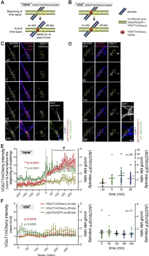
monitoring presynaptic clustering on axons contacting MAP2
+structures (Fig. S2, G–I). We quantified changes in the axonal
degradation rate occurring at the site of a newly formed
VGluT1-mCherry cluster (“new”) in comparison to a preexisting cluster
(“old”) on dendrites (Fig. 3, A and B). During formation of a
new stable VGluT1mCherry cluster, there was a significant
in-crease in the ratio of Ub
G76VGFP intensity between the site of
clustering and a nonsynaptic adjacent site (Ub
G76VGFP intensity
on/off-site ratio; Fig. 3, C and E). Occasional Ub
G76VGFP
ac-cumulation events accompanied by VGluT1mCherry clustering
occurred fleetingly but did not form stable clusters. On the
con-trary, no local changes in the degradation reporter signal were
observed throughout the lifetime of an old puncta (Fig. 3, D and
F). In conclusion, assembly of an excitatory presynaptic
termi-nal onto a postsynaptic partner is preceded and accompanied by
a localized reduction in the activity of the proteasome.
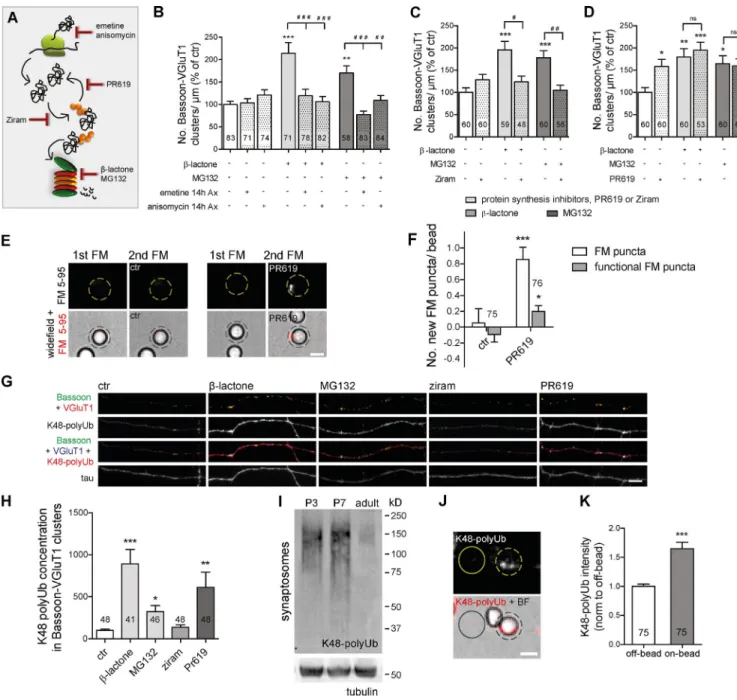
Proteasome inhibition-induced presynaptic accumulation of ubiquitinated conjugates as the trigger for presynaptic differentiation
To unmask the local mechanism generating new presynaptic
clusters, we first assessed its dependence on protein synthesis
(Fig. 4 A). The synaptogenic effect of axonal proteasome
in-hibition relied on axonal protein synthesis (Fig. 4 B and Fig.
S3 A) but not on soma synthesized proteins (Fig. S3, D and E).
The fact that presynaptic assembly was only reverted when
pre-ceded by long-term (14 h) axonal protein synthesis inhibition
(Fig. 4 B and Fig. S3 A), in opposition to an acute (1 h)
treat-ment (Fig. S3, B and C), indicated that rapid accumulation of no
longer degraded proteins from a pool of previously synthesized
ones underlies the presynaptogenic effect. Interestingly,
forma-tion of sensory-motor synapses in
Aplysia californica
has also
been suggested to require local protein synthesis (Lyles et al.,
2006). Nonetheless, the difference in terms of synapse type and
organisms’ complexity between this study and ours points to
the possibility of different mechanisms controlling presynaptic
assembly through local translation.
Because proteasome blocking triggers an extensive
ac-cumulation of ubiquitinated proteins (Fig. S3, J and K) while
reducing free Ub levels (Fig. S3, H and I), we next wondered
whether the effect would be dependent on de novo
ubiquiti-nation. We used an inhibitor of the E1 Ub-activating enzyme,
ziram (Fig. 4 A), which prevents formation of E1–Ub
conju-gates (Rinetti and Schweizer, 2010), and so no degradation of
nonubiquitinated proteins occurs. Treatment of isolated axons
with ziram alone had no effect on the number of presynaptic
clusters, but when in combination with proteasome inhibitors,
it led to a complete reversion of their clustering effect (Fig. 4 C
and Fig. S3 F). This result demonstrated that accumulation of
proteins in their ubiquitinated state is required for local
pro-teasome inhibition-induced presynaptic assembly. To further
validate this hypothesis, we used a broad-range inhibitor of
deubiquitinases, PR619 (Fig. 4 A; Altun et al., 2011), which
expectedly led to an accumulation of ubiquitinated conjugates
(Fig. S3, J and K). Similar to proteasome inhibitors, PR619 also
increased the number of presynaptic clusters along axons, yet
no cumulative effect was observed (Fig. 4 D and Fig. S3 G),
suggesting that both inhibitors act through the same
mecha-nism. Furthermore, local inhibition of deubiquitination resulted
in a significant increase in the number of new FM puncta on
beads (Fig. 4, E and F).
synaptic expression of K48 Ub chains is higher at
develop-mental stages coincident with the peak of synaptogenesis in
the hippocampus (Fig. 4 I; Mody et al., 2001), which
recapitu-lates the pattern observed in whole brain extracts (Chen et al.,
2011). Moreover, contact with beads induces accumulation of
K48 polyubiquitinated proteins in the contacting axon (Fig. 4,
J and K). Altogether, these results demonstrate that
polyubiq-uitinated conjugates bearing the K48 polyUb tag accumulate at
nascent presynaptic sites.
Collectively, this set of data suggests that accumulation of
polyubiquitinated conjugates at the site of a nascent presynapse,
in response to a local halt in proteasome degradation, functions
as the trigger for presynaptic clustering.
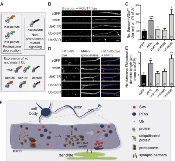
A role for proteasome-related polyUb chains in presynaptic assembly
strongly increased the number of presynaptic clusters (Fig. 5,
B and C; and Fig. S3, T–V) and the number of total and
func-tional FM puncta along dendrites (Fig. 5, D and E; and Fig.
S3 W), which were suppressed by prevention of K11 and K48
polyubiquitination, as opposed to K63. We thus concluded that
up-regulation of polyubiquitinated conjugates upon expression
of Ub triggers presynaptic assembly. Interestingly, Ub chains
with expected roles in proteasome degradation (K11 and K48)
were required. Both proteasome inhibition and prevention of
protein polyubiquitination reduce global protein degradation.
The difference resides in the state of the substrates being
ac-cumulated: polyubiquitinated versus mono- or
nonubiquiti-nated. Accordingly, the lack of synaptogenic effect of UbK48R
(or UbK11R) further supports the hypothesis that the effect
of proteasome inhibitors is attributed to the remaining pool
of polyubiquitinated substrates. We hereby demonstrate that
K11- and K48-polyubiquitinated proteins are the triggers for
presynaptic formation.
Figure 3. On-site decreased proteasome ac-tivity during formation of stable VGluT1 clusters
on dendrites. (A–D) VGluT1mCherry puncta on
A proteasome inhibition-derived polyUb nest for presynaptic assembly
In this study, we unmask a new on-site UPS-related mechanism
controlling formation of presynaptic sites. Our results suggest
a model in which transient and local reduction of proteasome
activity after contact with a postsynaptic partner leads to an
on-site accumulation of proteins in their polyubiquitinated state
(K48 and K11), which in turn functions as a nesting platform
for the clustering of presynaptic material and subsequently,
pre-synaptic differentiation (Fig. 5 F).
the
Drosophila
embryo, K48 and K11 Ub chains represent 57%
and 11%, respectively, of the neuronal ubiquitome (Franco et al.,
2011). These studies show that at the peak of synaptogenesis,
the UPS is highly active. Somewhat similar to the effect on
sen-sory synaptic inputs in
A. californica
motoneurons (Zhao et al.,
2003), we show enhanced presynaptic assembly upon local
pro-teasome inhibition. Hence, one can speculate that in the young
brain, high proteasome activity acts as a brake on the formation
of presynaptic sites, which will be relieved upon
spatiotempo-ral reduction. The idea that local decreased proteasome activity
dictates the onset of synaptogenesis implies that the developing
axon has ways of promoting endogenous proteasome inhibition.
Indeed, dynamic modifications can alter proteasome activity in
neurons (Upadhya et al., 2006; Djakovic et al., 2009; Tai et al.,
2010; Caldeira et al., 2013; Huang et al., 2013); however, this
has yet to be linked to presynaptogenic cues. Contradictorily, in
Caenorhabditis elegans
axons, the extent of presynaptic
differ-entiation is controlled by localized function of E3 ligases, either
by removing kinases that suppress synaptogenesis (Liao et al.,
2004; Nakata et al., 2005) or by eliminating unwanted terminals
(Ding et al., 2007). These studies reveal a role for enhanced
pro-teasome clearance in the control of nascent presynapses, which
together with our results indicate that UPS may govern synapse
formation in diverse ways.
In this study, we gathered a substantial set of data
demon-strating that formation of presynaptic clusters is triggered by an
on-site accumulation of polyubiquitinated proteins. Interestingly,
the two types of chains known to target proteins for degradation
(K11 and K48; Thrower et al., 2000; Kravtsova-Ivantsiv and
Ciechanover, 2012) were identified as the prime signals. So far,
few studies have revealed roles for K48 and K11 Ub chains other
than this classical labeling (Ye et al., 2003; Flick et al., 2004;
Dynek et al., 2010; Goto et al., 2010; Hay-Koren et al., 2011).
Herein, we propose a novel role for these two types of Ub
link-ages in signaling presynaptic assembly. In agreement, various
presynaptic proteins are known to be ubiquitinated (Segref and
Hoppe, 2009; Bingol and Sheng, 2011; Franco et al., 2011; Na
et al., 2012). Future efforts should be directed at identifying the
polyubiquitinated local players instructing presynaptic
forma-tion. We predict these might be presynaptic scaffolds or elements
of the axonal cytoskeleton machinery, which are currently
be-lieved to scaffold nascent presynapses (Chia et al., 2013). We
fur-ther hypothesize that transient enrichment of proteasome-related
polyubiquitinated proteins may act as a hub for the recruitment
Figure 5. K48 and K11 polyubiquitination triggers formation of presynaptic sites. (A) Altering levels of endogenous polyUb chains by overexpressing wt and mutant Ub. (B and D) wtUb led to increased presynaptic clusters in isolated axons (B) and FM puncta along dendrites (MAP2, blue; D), which did not occur for UbK11R and UbK48R mutants. eGFP, control. Bars, 5 µm. (C) Number of presynaptic clusters per axonal length (percentage of eGFP).of SV protein transport vesicles and piccolo-Bassoon transport
vesicles and formation of en passant boutons (Fig. 5 F).
Analo-gously, some strategies for DNA repair involve recruitment of
repair machinery to the site of DNA lesion by the recognition of
loci of extensively ubiquitinated histones (Jackson and Durocher,
2013). Also, activation of the NF-
κ
B pathway requires
recruit-ment of kinases to a highly polyubiquitinated signaling complex
formed by activation of transmembrane receptors (Kulathu and
Komander, 2012). This study opens the exciting question of how
a pool of polyubiquitinated conjugates functions as an
intracel-lular signal to presynaptic assembly. Furthermore, we anticipate
the need to identify the axonal machinery responsible for the
recognition and decoding of these polyUb signals.
Materials and methods
Constructs
F(syn)WRBN-VGluT1mCherry, a vector for lentiviral expression of a fusion version of VGluT1 to mCherry, was provided by E. Herzog (In-terdisciplinary Institute for Neuroscience, Bordeaux, France; Herzog et al., 2011). The VGluT1mCherry coding sequence was amplified by PCR (primers 5′-CCA CCT CTA GAC GCG TGC CGC CGC CAT GGA GTT CCG GCA GGA GGA GTT-3′ and 5′-CTG GTC GGA TCA TTG GGC CCT TAC TTG TAC AGC TCG TCC ATG CCG-3′) and cloned into pSinRep5 vector (Invitrogen) through ApaI and MluI sites by a cloning kit (639648; In-Fusion HD; Takara Bio Inc.), so that a Sindbis viral expression ver-sion of this fuver-sion protein was generated. The degradation reporter Ub-G76VGFP (plasmid 11941; Addgene; Dantuma et al., 2000) and GFP-Ub
(plasmid 11928; Addgene) were provided by C. Duarte (University of Coimbra, Coimbra, Portugal). To generate a plasmid for Sindbis viral– mediated expression of the degradation reporter, the UbG76V-GFP coding sequence was amplified by PCR (primers 5′-CCA CCT CTA GAC GCG TGC CGC CGC CAT GCA GAT CTT CGT GAA GAC TCTG-3′ and 5′ -CTG GTC GGA TCA TTG GGC CCT TAC TTG TAC AGC TCG TCC ATG CCG AGA GT-3′) and cloned into pSinRep5 vector through ApaI and MluI sites by the cloning kit. To generate a Sindbis viral construct for the expression of Ub, the coding sequence of the wtUb was amplified from GFP-Ub by PCR (primers 5′-CAC CAC CAC CTC TAG ACG GGC CGC ATG CAG ATC TTC GTG AA-3′ and 5′-AGG GGC GGA ATC TAG ATC ACC CAC CTC TGA GAC GGA GTAC-3′) and cloned into pSinRep-IRES-eGFP vector (provided by U. Hengst, Columbia University, New York, NY; Wu et al., 2005) through the XbaI site by the cloning kit. In the resulting plasmid, pSinRep-wtUb-IRES-eGFP, the expression of Ub is under control of the subgenomic promoter, whereas eGFP expression is controlled by a ribosome entry site (IRES). The empty vector pSin-Rep-IRES-eGFP was used as the control. Site-directed mutagenesis to the coding sequence of Ub inserted into the pSinRep-wtUb-IRES-eGFP plasmid was performed by a site-directed mutagenesis kit (200521; Qui-kChange II XL; Agilent Technologies). Lysines in positions 11, 48, and 63 of Ub sequence were mutated to arginine to generate the Ub mutant forms UbK11R (primers 5′-GAA GAC TCT GAC TGG TAG GAC CAT CAC CCT CGA-3′ and 5′-TCG AGG GTG ATG GTC CTA CCA GTC AGA GTC TTC-3′), UbK48R (primers 5′-AGA GGC TGA TCT TTG CTG GAA GAC AGC TGG AAGA-3′ and 5′-TCT TCC AGC TGT CTT CCA GCA AAG ATC AGC CTCT-3′), and UbK63R (primers 5′-CCT GTC TGA CTA CAA CAT CCA GAG AGA GTC CAC CCT-3′ and 5′-AGG GTG GAC TCT CTC TGG ATG TTG TAG TCA GAC AGG-3′), respectively.
Antibodies
We used the following primary antibodies: mouse monoclonal Bassoon (1:400, immunocytochemistry [ICC]; 1:1,000, Western blot [WB];
ADI-VAM-PS003; Enzo Life Sciences), rabbit monoclonal K48 polyUb (Apu2; 1:500, ICC; 1:1,000, WB; 05-1307; EMD Millipore), chicken polyclonal MAP2 (1:5,000, ICC; AB5543; EMD Millipore), chicken polyclonal tau (1:1,000, ICC; ab75714; Abcam), mouse monoclonal tu-bulin (1:300,000, WB; T7816; Sigma-Aldrich), mouse monoclonal tuj1 (1:1,000, ICC; MMS-435P; Covance), rabbit polyclonal Ub (1:200, ICC; 1:1,000, WB; Z0458; Dako), guinea pig polyclonal VGluT1 (1:1,500, ICC; AB5905; EMD Millipore), and rabbit polyclonal VGluT1 (1:5,000, WB; 135503; Synaptic Systems). As for secondary antibodies, alkaline phosphatase–conjugated antibodies (Jackson Im-munoResearch Laboratories, Inc.) were used for blotting, and Alexa Fluor 350–, 488–, 568–, and 647–conjugated antibodies (1:1,000; Thermo Fisher Scientific) were used for immunocytochemistry.
Microfluidic devices for neuron culture
Microfluidic devices consist of a molded polydimethylsiloxane (PDMS) chamber assembled in a glass coverslip (Taylor et al., 2003). The molds for the PDMS devices used in this study were fabricated by N.L. Jeon. Microfluidic devices were prepared and assembled similarly to those previously described (Taylor et al., 2005). In brief, PDMS was prepared from a silicon elastomer kit (Sylgard 184; Dow Corning) and cured in molds, and each device was properly cleaned, sterilized with 75% ethanol, and air dried in the culture hood. Glass coverslips (Assistent) were cleaned in 65% nitric acid and extensively washed with mQH2O (five washes for 30 min each), rinsed twice in 100% ethanol, dried, and sterilized under UV radiation for at least 15 min. The PDMS mold was properly assembled on the glass coverslip under sterile conditions. Coverslips were double coated with PDL (0.1 mg/ml overnight at 37°C) and laminin (2 µg/ml for 2 h at 37°C). Before plating cells, devices were washed once with plating medium (MEM supplemented with 0.026 M NaHCO3, 0.025 M glucose, 1 mM sodium pyruvate, and 10% FBS).
Primary culture of hippocampal neurons
Primary cultures of rat hippocampal neurons were prepared as previ-ously described (Baptista et al., 2010; Baeza et al., 2012), with minor changes. After dissection, hippocampi from E18 Wistar rat embryos were dissociated in 0.045% trypsin/0.01% vol/vol deoxyribonuclease in HBSS for 15 min at 37°C. Hippocampi were then washed once in plating medium containing 10% FBS, thus stopping trypsin activity, and mechanically dissociated in fresh plating medium, and cell density was determined. Cells were plated in plating medium: in 450-µm mi-crofluidic devices, 7 × 10⁴ cells were plated in the somal compartment; in synapse formation chambers, 7 × 10⁴ and 105 cells were plated in the presynaptic and postsynaptic compartment, respectively. Neurons were allowed to attach for 2–4 h, and then plating medium was replaced with culture medium (neurobasal medium supplemented with 2% B27, 25 µM glutamate, 0.5 mM glutamine, and 1:400 penicillin/streptomy-cin). In microfluidic devices, as a way of reducing glutamate excitotox-icity in growing axons, glutamate-free culture medium was added to the axonal compartment of 450-µm microfluidic devices and to the syn-aptic compartment of synapse formation chambers. Cells were main-tained in a humidified incubator with 5% CO2/95% air at 37°C. At day in vitro (DIV) 3/4, the mitotic inhibitor 5-fluorodeoxyuridine (10-µM final concentration) was added to reduce contamination with glia cells. Cells were allowed to grow, and, unless otherwise indicated, experi-ments were performed at DIV 7/8, which corresponds to the peak of synaptogenesis in primary hippocampal cultures (Fletcher et al., 1994).
Synaptosome preparation
were dissected and homogenized in a motor-driven glass Teflon homog-enizer (30 stokes and 900 rpm at 4°C) in Hepes-buffered sucrose buffer (0.32 M sucrose and 4 mM Hepes, pH 7.4) supplemented with prote-ase and phosphatprote-ase inhibitors (0.2 mM PMSF, 1 µg/ml chymostatin/ leupeptin/antipain/pepstatin, 0.1 mM sodium ortovanadate [Na3VO4], and 50 mM NaF). The homogenate was then centrifuged at 900 g for 15 min at 4°C, and the supernatant was collected and centrifuged at 18,000 g for 15 min at 4°C to yield the synaptosomal fraction. It was further washed by resuspension in Hepes-buffered sucrose buffer and centrifugation at 18,000 g for 15 min at 4°C. Quantification of pro-tein was performed by the bicinchoninic acid assay, and 40-µg samples were denatured with denaturating buffer (62.5 mM Tris-HCl, pH 6.8, 10% vol/vol glycerol, 2% vol/vol SDS, 0.01% weight/vol bromophenol blue, and 5% vol/vol β-mercaptoethanol, added fresh) and boiled at 95°C for 5 min before running the WB.
Generation of Sindbis and lentivirus
For the generation of Sindbis virus, the pSinRep construct expressing the desired gene of interest and the helper plasmid DH26S were lin-earized with either XhoI, PacI, or NotI and properly treated for the removal of RNase contamination. Synthesis of RNA from linearized DNAs was performed by in vitro transcription using the mMES SAGE mMAC HINE SP6 kit (1340; Ambion). BHK-1 cells were electropo-rated with 12 µg DH26S RNA and 12 µg of the desired pSinRep RNA, and production of virus was allowed to occur for 24–36 h. Supernatant was then collected, and virus particles were purified by centrifugation at 60,000 g for 2 h and 20 min at 15°C. The viral pellet was then resus-pended in PBS (137 mM NaCl, 2.7 mM KCl, 10 mM Na2HPO4, and 1.8 mM KH2PO4, pH 7.4) with 0.1% BSA and stored at −80°C. The virus titer was determined in BHK-1 cells, and the volume of virus for infection was adjusted so that >85% of neurons were transduced. For expression of Ub and its mutant forms in microfluidic devices, expres-sion was allowed to occur for 18–20 h. For live-imaging experiments, cells were incubated with virus for 6–8 h before imaging.
For the generation of lentivirus, HEK293T cells were transfected using calcium phosphate transfection with the lentiviral expression vector F(syn)WRBN-VGluT1mCherry and three lentiviral packaging vectors, pLP1, pLP2, and pLP-VSVG, for the expression of gag/pol
genes, rev gene, and VSVG envelope glycoprotein gene, respectively. The supernatant containing virus particles was collected 48–60 h after transfection and concentrated by centrifugation at 60,000 g
for 2 h at 22°C. The viral pellet was then resuspended in PBS with 0.1% BSA and stored at −80°C. After infection, expression of pro-tein occurred for 48–60 h.
PDL-coated beads
Aliphatic amine latex beads were prepared as previously described (Taylor et al., 2013). They were incubated with PDL for 30 min at 37°C, centrifuged to remove supernatant, washed twice in sterile mQH2O, and diluted in culture medium. 30 µl of bead suspension was added to the axonal compartment of microfluidic devices and incu-bated at 37°C for the indicated period of time. For live-imaging experi-ments, beads were diluted in Hepes-buffered solution imaging medium (119 mM NaCl, 5 mM KCl, 2 mM CaCl2, 2 mM MgCl2, 30 mM glu-cose, and 10 mM Hepes, pH 7.4).
Drug treatment
Treatment of cells with UPS or protein synthesis inhibitors was per-formed in conditioned medium. For proteasome inhibitors, a preincu-bation of 30 min and 15 min was performed with β-lactone and MG132, respectively. When cells were cotreated with proteasome inhibitors and protein synthesis inhibitors PR619 or ziram, both inhibitors were
added simultaneously. All inhibitors were diluted in conditioned me-dium from a 1,000× stock in DMSO and added to cells, except for the protein synthesis inhibitors emetine and anisomycin, which were di-luted in mQH2O. Equal amounts of DMSO or water were added to the control. β-Lactone, emetine, and anisomycin were applied at 10 µM. As for MG132, PR619, and ziram, a concentration of 1 µM was used.
Biochemistry
Biochemical analysis of protein levels was performed by WB as pre-viously described (Shin et al., 2010; Baptista et al., 2014), with minor changes. Cells were first washed twice in cold PBS. Protein extracts were then prepared by scrapping cells in radioimmunoprecipitation assay lysis buffer (150 mM NaCl, 50 mM Tris-HCl, pH 7.4, 5 mM EGTA, 1% Triton X-100, 0.5% deoxycholate, and 0.1% SDS, pH 7.5, freshly supplemented with 50 mM NaF, 1.5 mM Na3VO4, 0.1 mM PMSF, and 1 µg/ml chymostatin/leupeptin/antipain/pepstatin). Lysates were sonicated and centrifuged at 16,100 g for 10 min at 4°C, and the supernatant was collected. Quantification of protein was performed by the bicinchoninic acid assay, and samples (40 µg in 40 µl) were dena-tured with denaturating buffer and boiled at 95°C for 5 min. Protein ex-tracts were electrophoresed in a Tris-glycine-SDS buffer (25 mM Tris, 192 mM glycine, and 0.1% weight/vol SDS, pH 8.3) in 7.5% (Bas-soon and VGluT1), 15% (free Ub), or 4–15% gradient (ubiquitinated conjugates with Ub and Apu2 antibodies) polyacrylamide gel 1.5 mm thick. Electrotransfer onto a polyvinylidene difluoride membrane was performed either overnight at 40 V at 4°C or by using an equivalent protocol for rapid transfer (250 mA for 4 h or 250 mA for 6 h at 4°C depending on the protein of interest weight). Membranes were washed once with TBS (20 mM Tris and 137 mM NaCl) with 0.1% vol/vol Tween 20 (TBS-T) and blocked for 1 h at RT in TBS-T with 5% nonfat dry milk or 3% BSA. Membranes were again washed for three times with TBS-T and incubated with the primary antibody diluted in TBS-T containing 5% or 0.5% weight/vol nonfat dry milk or 3% BSA. Incu-bation was performed either overnight at 4°C or at RT for 1 h. After three washes, membranes were incubated for 1 h with alkaline phos-phatase–conjugated secondary antibodies (anti-mouse or anti-rabbit, depending on the primary antibody host species) at RT, washed again for three times, and resolved with enhanced chemifluorescence sub-strate for a maximum of 5 min. Membranes were scanned with a gel and blot imaging system (Storm 860; GE Healthcare), and quantifica-tion was performed using ImageQuant software under linear exposure conditions. Whenever necessary, membranes were stripped with 0.2 M NaOH for 20 min and reprobed.
Live-cell imaging
Live-imaging experiments for fluorescently tagged proteins (eGFP and mCherry; excitation wavelength of 488 and 561 nm) and for the FM 5-95 lypophilic styryl dye (T23360; Thermo Fisher Scientific; excitation wavelength of 560 nm) were performed at RT (∼20°C) in imaging me-dium (119 mM NaCl, 5 mM KCl, 2 mM CaCl2, 2 mM MgCl2, 30 mM glucose, and 10 mM Hepes, pH 7.4). Acquisition was performed using a spinning disk confocal imaging system (CSU-X1-M1N-E; Yokogawa Electric Corporation) configured for a motorized inverted microscope (IX81; Olympus) driven by iQ 3.1 software (Andor Technology). Im-ages were collected with a 60× water objective (1.2 NA; UPL SAPO; Olympus) and an electron-multiplying charged-coupled device (CCD) camera (IXON-X3; Andor Technology).
microgrooves into the axonal compartment) were selected. Data were collected sequentially from several defined positions (a maximum of 15 per device). Focal drift during the experiment was corrected auto-matically using the autofocus feature of the Olympus system. Laser intensities were kept as low as possible, and acquisition at 2 × 2 bin-ning was performed. In experiments involving beads, three frames were captured of the selected positions on the axonal compartment before addition of beads, and then beads were added to both compartments (the same volume was added to both sides to prevent focus drift caused by unbalanced preparation). Time-lapse was resumed 10 min after addition of beads. Frames were acquired as z stacks (35–40 slides, 8–10-µm range) every 10 min.
For the FM 5-95 dye experiments, each cycle of FM dye loading/ unloading comprised the following steps: FM 5-95 dye loading solution containing a high KCl concentration (imaging medium with 90 mM KCl supplemented with 10 µM FM 5-95 dye, 20 µM 6-cyano-7-nitro-quinoxaline-2,3-dione [CNQX], and 50 µM D-AP5) to promote depo-larization was added to microfluidic devices for 1 min and washed once with 10 µM FM 5-95 dye in imaging medium for 1 min. An additional three washes with 1 mM Advasep-7 in imaging medium, for 1 min each, were performed for optimal removal of FM dye excess. This me-dium was then replaced by imaging meme-dium containing 20 µM CNQX and 50 µM D-AP5, and the device was placed on the microscope stage. CNQX and D-AP5, which are α -amino-3-hydroxy-5-methyl-4-isoxaz-ole propionic acid (AMPA)/kainate and N-methyl-d-aspartate (NMDA) receptor antagonists, respectively, were added to the medium to block recurrent excitation. Positive and negative electrodes were placed on each well of either the somal or axonal compartment, and unloading of the FM dye was induced by electrical stimulation, which was per-formed by a two-channel stimulus generator (model STG4002; Multi Channel Systems) in current mode with an asymmetric waveform (−480 µA for 1 ms and 1,600 µA for 300 µs) at 20 Hz for 1,200 pulses for 1 min. At least four frames were acquired before stimulation. The frames were acquired as z stacks (35–40 slides, 8–10-µm range) every 15 s for 5 min in a 2 × 2 binning mode. FM puncta that could unload >5% of their content after 1 min of electrical stimulation were consid-ered functional presynaptic clusters (Taylor et al., 2013). For detection of functional FM puncta along dendrites (Fig. 5, D and E), one cycle of FM dye loading/unloading protocol was performed on the somal side of microfluidic devices after 18–20-h viral infection with the Ub con-structs. Under the brightfield light, positions to image were carefully chosen to include nearly the same number of cell bodies. Dendrites were later detected by retrospective immunocytochemistry for MAP2.
For the experiments in which we were interested in monitoring the appearance of new FM puncta on beads (Fig. 1, D–G; and Fig. 4, E and F), beads were first added to the axonal compartment of micro-fluidic devices and incubated for 3 h. Then, a first cycle of FM 5-95 dye was performed, followed by complete destaining with high KCl imaging medium for 1 min and two washes with imaging medium containing 20 µM CNQX and 50 µM D-AP5. An image after FM dye destaining was acquired for later subtraction from the initial image of the second FM dye cycle. Cultures were then treated with proteasome inhibitors, PR619 or DMSO, for 1 h. A second cycle of FM dye with live-monitored unloading at exactly the same position was then per-formed to look for the formation of new functional FM puncta. The position of the beads and the microgrooves were instrumental for local-izing the imaging position between the first and second FM dye cycles. FRAP/FLIP experiments were performed at 37°C with a con-focal microscope (LSM 710; ZEI SS) driven by Zen Black 2012 soft-ware (ZEI SS) with a 63× oil differential interference contrast (DIC) objective (M17n; NA 1.40; Plan-Apochromat) at a magnification of 2×. Neurons in microfluidic devices expressing UbG76V-GFP for 12 h were
used, and imaging was performed on the axonal compartment. Medium was replaced for imaging medium, and beads were added to the axo-nal compartment for 20 min before FRAP/FLIP imaging. Imaging was confined to the time interval between 20 min and 1 h after bead contact, and acquisition was done in a single z-slice manner. Bleach was accom-plished by performing 10 bleach iterations with the 488-nm laser power set to 100% (100× greater than for acquisition) in a total bleach time of 0.45 s at 5 × 2–µm regions of interest (ROIs) carefully placed along axons. Before bleaching, three baseline images were taken. For FRAP, regions to bleach were chosen at off-bead and on-bead axonal sites. For the latter, a distinction was made between beads causing an increased intensity of UbG76V-GFP (on-bead with UbG76V-GFP increase) and the ones that did not alter its fluorescence along axons (on-bead without UbG76V-GFP increase). A minimal distance of 45 µm was maintained between bleached and control ROIs. After a single bleaching event, changes in fluorescence were analyzed at bleached and control ROIs every 1 s for a total of 50 s. For FLIP, repeated bleaching was per-formed at a 5-µm distance from the measurement site (off-bead, on-bead with UbG76V-GFP, or on-bead without UbG76V-GFP). An image was taken after each bleaching episode in a total experiment time of 130 s. Changes in fluorescence were analyzed at the bleaching region (FLIP ROI), at the measurement region (analysis ROI), and at regions of nonbleached axons (control ROI) to control for fluorescence decay as a result of bleaching throughout the experiment.
Immunocytochemistry
Immunocytochemistry was performed similarly to that previously described (Baptista et al., 2013). Cells were fixed in prewarmed 4% paraformaldehyde (in PBS with 4% sucrose) for 10 min at RT. A pre-fixation of 5 min in 1% paraformaldehyde was performed to prevent damaging the population of isolated axons. Cultures were washed three times in PBS and then permeabilized in PBS with 0.25% Triton X-100 for 5 min at RT and washed once in PBS before blocking for 30 min in PBS with 3% BSA. Preparations were incubated with the mix of primary antibodies in 3% BSA either overnight at 4°C or for 2 h at 37°C, washed three times in PBS, and incubated with the mix of secondary antibodies for 1 h at RT in 3% BSA. Cultures were again washed, this time twice in PBS with 0.1% Triton X-100 and once in PBS, and the coverslip was rinsed in mQH2O and mounted in prolong mounting media with or without DAPI. For microfluidic chambers, the PDMS device was disassembled from the coverslip only before mount-ing on the microscope glass.
Microscopy of antibody-labeled cultures
collected by using the microgrooves as coordinates. In brief, micro-grooves were numbered according to the orientation of the device on the microscope stage, and at the end of acquisition, each position of the microgroove nearer to its center was marked down. The position was then corrected manually by comparing the DIC image acquired at the end of the live experiment with that of the fixed preparation. Moreover, the plug-in Align images by line ROI in ImageJ was further used to guarantee perfect alignment.
Quantitative imaging analysis
Quantification of fluorescence images was performed using ImageJ software (National Institutes of Health). For fixed cells, samples within an experiment were simultaneously stained and imaged with identical settings (exposure time and fluorescence light intensity were kept con-stant throughout acquisition). Images of random fields of view (FOVs) of either isolated axons or cell bodies were taken. Selection of ROIs to acquire fluorescent images was performed either on the axonal or somal marker to avoid bias acquisition. All images were converted to 8 bit for quantification purposes.
To quantify the number of presynaptic puncta along axons, the axonal marker image was used to select populations of axons to quan-tify. Axonal length was determined by performing analysis (ImageJ plug-in Analyze skeleton) of a “skeletonized” version of the axonal marker. The sum of the length of all the axonal branches identified in an image was used as the axonal length. Correspondent images of synaptic markers were thresholded (threshold values conserved in individual experiments), and particle analysis was performed to cal-culate number and area of puncta. To quantify the number of pre-synaptic clusters (Bassoon-VGluT1 clusters), the presence or absence of VGluT1 puncta within Bassoon puncta ROIs was determined, and the total number of Bassoon ROIs containing VGluT1 puncta was di-vided by the axonal length. Analysis was limited to Bassoon puncta bigger than the smaller quantifiable object (0.05 µm2) in accordance with our imaging settings (Waters, 2009) in order to exclude the possible contribution of mobile piccolo-Bassoon transport vesicles (0.02 µm2; Zhai et al., 2001; Shapira et al., 2003), thus narrowing down analysis to nascent presynaptic sites. A minimum of 12 micro-scope FOVs of the axonal side were analyzed per individual exper-iment in each condition.
For quantification of live experiments, confocal slices were sum projected in ImageJ, and alignment of frames within each video was performed by TurboReg or StackReg plug-ins and converted to 8 bit. In experiments involving accumulation of synaptic material on beads, analysis was performed similarly to a study by Lucido et al. (2009). The brightfield image was used to locate beads in contact with axons, and ROIs for individual beads and adjacent sites along the axon were created (on-bead and off-bead, respectively). Change in signal was quantified by measuring the fluorescence intensity at each bead and correspondent off-bead site in each individual frame of the time-lapse video. Individual values were then normalized to the fluorescence in-tensity in that site at the frame preceding addition of beads (time 0, 0 min) both for on-bead and off-bead sites.
For the experiment in which formation of presynaptic clusters on dendrites was monitored (Fig. 3, A–F), digital videos of the time-lapse sequence for each position were prepared and carefully analyzed to detect formation of stable VGluT1mCherry clusters in sites not de-tected at prior time points (“new”) or clusters stable at approximately the same location throughout the entire time-lapse (“old”). Puncta were only considered new if stable until the end of the time-lapse for at least 30 min. After identification of new and old puncta, their locations were overlaid on the corresponding posthoc MAP2 immunostained images (see Microscopy of antibody-labeled cultures for further
de-tails on acquisition of images after retrospective labeling). Alignment of VGluT1mCherry and UbG76V-GFP videos with MAP2 retrospective ones was done according to the brightfield images of the same region taken at the end of the time-lapse and the one after immunostaining by the ImageJ plug-in Align images by line ROI. The alignment correction used for DIC images was applied to VGluT1mCherry and UbG76V-GFP, and new and old VGluT1mCherry clusters formed onto dendrites were considered. ROIs encompassing the whole VGluT1mCherry cluster were created at the site of clustering (on-site) and equal-sized ROIs at adjacent axonal sites (site; Fig. 3, A and B, on-site and off-site correspond to dashed and solid boxes, respectively). Change in sig-nal was quantified by measuring the fluorescence intensity at each ROI in each individual frame of both VGluT1-mCherry and UbG76V-GFP time-lapse videos and used to generate the dataset presented in Fig. 3 (E and F). For VGluT1mCherry, individual values were normalized to the fluorescence intensity in that site at time 0. For new and old puncta, time 0 is considered to be the frame before clustering was initiated or the first frame of the time-lapse, respectively. For new puncta, we considered the beginning of clustering as the frame at which the stable puncta first appeared, recognized as a high increase in fluorescent sig-nal (at least a 30% increase in sigsig-nal intensity in relation to the previous frame). For UbG76V-GFP, the ratio of its intensities between the on- and off-site was calculated.
For the FM dye experiments, the brightfield image was used to create ROIs encompassing beads or dendrites that contact with axons. Then, the number of FM puncta on each bead or along each dendritic segment was quantified by performing particle analysis in ImageJ. To evaluate the unloading capacity of FM puncta, quantification was adapted from previous work (Taylor et al., 2013). Each puncta inten-sity is measured throughout the registered sequence of images (frames every 15 s for 5 min), normalized to the frame before stimulation, and corrected for the baseline slope (calculated from the change in intensity in the three frames preceding stimulation). Puncta that unloaded >5% of their FM dye content after 1 min of stimulation were considered as functional (Taylor et al., 2013). For the experiment with a double FM dye cycle (Fig. 1, D–G; and Fig. 4, E and F), the net gain in func-tional and total FM puncta was calculated by subtracting the number of puncta after and before treatment.
For quantification of axonal volume, 0.23-µm–spaced z stacks were acquired from mApple-expressing axons in the axonal com-partment. Volume at designated ROIs was quantified with the ImageJ plug-in 3D object counter (Bolte and Cordelières, 2006), which de-termines the number of voxels in an object and calibrates it into xyz dimensions, with the threshold kept constant between on-bead and the adjacent ROI. 3D surface reconstructions were obtained in ImageJ with the plug-in ImageJ 3D viewer.
For FRAP and FLIP, change in fluorescence at designated ROIs was obtained from the sequence of images, and values were normalized to the frame preceding bleaching. For quantification of FRAP experi-ments, data for each FRAP event were fitted to a one-phase exponential association function (y = y0 + [plateau − y0] × [1 − exp(−x/τ)], with y0 constrained to between 0 and 1) in Prism 5 (GraphPad Software). Individual values of the plateau and time constant (τ) were obtained for each curve fitting and compared. The recovery fraction was calculated as equal to the plateau × 100 (Fig. 2 F). For FLIP experiments, data for each FLIP event were fitted to a one-phase exponential decay function (y = [y0 − plateau] × exp[−x/τ] + plateau, with y0 constrained to be-tween 0 and 1) in Prism 5 software. Individual values of the plateau and time constant (τ) were obtained for each curve fitting. The retained fraction was calculated as equal to (1 − plateau) × 100 (Fig. 2 I, left).
Statistical analysis
Results are presented as mean values ± SEM. Graphs and statistical analysis were performed in Prism 5 software. Statistical significance was assessed by nonparametric tests. Mann-Whitney test or Wilcoxon paired t test was performed for comparisons of changes between two groups. For comparisons between multiple groups, we used the Kruskal- Wallis test followed by the Dunn’s multiple comparison test. For the live-imaging data, we performed two-way analysis of variance (ANO VA) with time as the repeated measure to assess changes between two groups throughout time.
Online supplemental material
Fig. S1 provides control experiments validating the specific inhibition of the proteasome in isolated axons and its synaptogenic effect. Fig. S2 gathers additional information that supports a decreased proteasome activity at sites of newly formed presynaptic clusters. Fig. S3 provides supporting information for the triggering of presynaptic assembly by proteasome-related ubiquitinated conjugates. Online supplemental material is available at http ://www .jcb .org /cgi /content / full /jcb .201509039 /DC1.
Acknowledgments
We thank Dr. Etienne Herzog for plasmids and Drs. Carlos Duarte and Ulrich Hengst for plasmids and comment on the manuscript. We thank Dr. Luísa Cortes for technical microscope support and Drs. Ana Luísa Carvalho, Carlos Duarte, and João Peça for helpful discussions.
This work was supported by Fundação para a Ciência e Tecnologia (FCT) with grant SFRH/BD/51196/2010; by Federación Española de Enfermedades Raras through Programa Operacional Factores de Competitividade; by national funds through FCT (PTDC/SAU-NEU/104100/2008, EXPL/NEU-NMC/0541/2012, and UID/ NEU/04539/2013); and by a Marie Curie Actions International re-integration grant (249288), Seventh Framework program.
A.M. Taylor and N.L. Jeon are inventors of the microfluidic chambers to compartmentalize neurons (US 7419822 B2), and both have finan-cial interest in Xona Microfluidics, LLC. The authors declare no further competing financial interests.
Submitted: 8 September 2015 Accepted: 24 February 2016
References
Altun, M., H.B. Kramer, L.I. Willems, J.L. McDermott, C.A. Leach, S.J. Goldenberg, K.G.S. Kumar, R. Konietzny, R. Fischer, E. Kogan, et al. 2011. Activity-based chemical proteomics accelerates inhibitor development for deubiquitylating enzymes. Chem. Biol. 18:1401–1412. http ://dx .doi .org /10 .1016 /j .chembiol .2011 .08 .018
Baeza, J.L., B.G. de la Torre, C.M. Santiveri, R.D. Almeida, M.T. García-López, G. Gerona-Navarro, S.R. Jaffrey, M.Á. Jiménez, D. Andreu, R. González-Muñiz, and M. Martín-Martínez. 2012. Cyclic amino acid linkers stabilizing key loops of brain derived neurotrophic factor. Bioorg. Med. Chem. Lett. 22:444–448. http ://dx .doi .org /10 .1016 /j .bmcl .2011 .10 .107 Baptista, F.I., M.J. Pinto, F. Elvas, R.D. Almeida, and A.F. Ambrósio. 2013.
Diabetes alters KIF1A and KIF5B motor proteins in the hippocampus.
PLoS One. 8:e65515. http ://dx .doi .org /10 .1371 /journal .pone .0065515 Baptista, F.I., M.J. Pinto, F. Elvas, T. Martins, R.D. Almeida, and A.F. Ambrósio.
2014. Diabetes induces changes in KIF1A, KIF5B and dynein distribution in the rat retina: implications for axonal transport. Exp. Eye Res. 127:91– 103. http ://dx .doi .org /10 .1016 /j .exer .2014 .07 .011
Baptista, M.S., C.V. Melo, M. Armelão, D. Herrmann, D.O. Pimentel, G. Leal, M.V. Caldeira, B.A. Bahr, M. Bengtson, R.D. Almeida, and C.B. Duarte. 2010. Role of the proteasome in excitotoxicity-induced cleavage of
glutamic acid decarboxylase in cultured hippocampal neurons. PLoS One. 5:e10139. http ://dx .doi .org /10 .1371 /journal .pone .0010139 Bingol, B., and M. Sheng. 2011. Deconstruction for reconstruction: the role of
proteolysis in neural plasticity and disease. Neuron. 69:22–32. http ://dx .doi .org /10 .1016 /j .neuron .2010 .11 .006
Bolte, S., and F.P. Cordelières. 2006. A guided tour into subcellular colocalization analysis in light microscopy. J. Microsc. 224:213–232. http ://dx .doi .org /10 .1111 /j .1365 -2818 .2006 .01706 .x
Caldeira, M.V., M. Curcio, G. Leal, I.L. Salazar, M. Mele, A.R.A. Santos, C.V. Melo, P. Pereira, L.M.T. Canzoniero, and C.B. Duarte. 2013. Excitotoxic stimulation downregulates the ubiquitin-proteasome system through activation of NMDA receptors in cultured hippocampal neurons.
Biochim. Biophys. Acta. 1832:263–274. http ://dx .doi .org /10 .1016 /j .bbadis .2012 .10 .009
Chen, P.C., L.N. Qin, X.M. Li, B.J. Walters, J.A. Wilson, L. Mei, and S.M. Wilson. 2009. The proteasome-associated deubiquitinating enzyme Usp14 is essential for the maintenance of synaptic ubiquitin levels and the development of neuromuscular junctions. J. Neurosci. 29:10909–10919. http ://dx .doi .org /10 .1523 /JNE URO SCI .2635 -09 .2009
Chen, P.C., B.J. Bhattacharyya, J. Hanna, H. Minkel, J.A. Wilson, D. Finley, R.J. Miller, and S.M. Wilson. 2011. Ubiquitin homeostasis is critical for synaptic development and function. J. Neurosci. 31:17505–17513. http :// dx .doi .org /10 .1523 /JNE URO SCI .2922 -11 .2011
Chia, P.H., P. Li, and K. Shen. 2013. Cellular and molecular mechanisms underlying presynapse formation. J. Cell Biol. 203:11–22. http ://dx .doi .org /10 .1083 /jcb .201307020
Cristovão, G., M.J. Pinto, R.A. Cunha, R.D. Almeida, and C.A. Gomes. 2014. Activation of microglia bolsters synapse formation. Front. Cell. Neurosci.
8:153. http ://dx .doi .org /10 .3389 /fncel .2014 .00153
Dantuma, N.P., K. Lindsten, R. Glas, M. Jellne, and M.G. Masucci. 2000. Short-lived green fluorescent proteins for quantifying ubiquitin/proteasome-dependent proteolysis in living cells. Nat. Biotechnol. 18:538–543. http :// dx .doi .org /10 .1038 /75406
DiAntonio, A., A.P. Haghighi, S.L. Portman, J.D. Lee, A.M. Amaranto, and C.S. Goodman. 2001. Ubiquitination-dependent mechanisms regulate synaptic growth and function. Nature. 412:449–452. http ://dx .doi .org /10 .1038 /35086595
Ding, M., D. Chao, G. Wang, and K. Shen. 2007. Spatial regulation of an E3 ubiquitin ligase directs selective synapse elimination. Science. 317:947– 951. http ://dx .doi .org /10 .1126 /science .1145727
Djakovic, S.N., L.A. Schwarz, B. Barylko, G.N. DeMartino, and G.N. Patrick. 2009. Regulation of the proteasome by neuronal activity and calcium/ calmodulin-dependent protein kinase II. J. Biol. Chem. 284:26655– 26665. http ://dx .doi .org /10 .1074 /jbc .M109 .021956
Dynek, J.N., T. Goncharov, E.C. Dueber, A.V. Fedorova, A. Izrael-Tomasevic, L. Phu, E. Helgason, W.J. Fairbrother, K. Deshayes, D.S. Kirkpatrick, and D. Vucic. 2010. c-IAP1 and UbcH5 promote K11-linked polyubiquitination of RIP1 in TNF signalling. EMBO J. 29:4198–4209. http ://dx .doi .org /10 .1038 /emboj .2010 .300
Fletcher, T.L., P. De Camilli, and G. Banker. 1994. Synaptogenesis in hippo-campal cultures: evidence indicating that axons and dendrites become competent to form synapses at different stages of neuronal development.
J. Neurosci. 14:6695–6706.
Flick, K., I. Ouni, J.A. Wohlschlegel, C. Capati, W.H. McDonald, J.R. Yates, and P. Kaiser. 2004. Proteolysis-independent regulation of the transcription factor Met4 by a single Lys 48-linked ubiquitin chain. Nat. Cell Biol.
6:634–641. http ://dx .doi .org /10 .1038 /ncb1143
Franco, M., N.T. Seyfried, A.H. Brand, J. Peng, and U. Mayor. 2011. A novel strategy to isolate ubiquitin conjugates reveals wide role for ubiquitination during neural development. Mol. Cell. Proteomics. 10. http ://dx .doi .org /10 .1074 /mcp .M110 .002188
Friedman, H.V., T. Bresler, C.C. Garner, and N.E. Ziv. 2000. Assembly of new individual excitatory synapses: time course and temporal order of synaptic molecule recruitment. Neuron. 27:57–69. http ://dx .doi .org /10 .1016 /S0896 -6273(00)00009 -X
Goto, E., Y. Yamanaka, A. Ishikawa, M. Aoki-Kawasumi, M. Mito-Yoshida, M. Ohmura-Hoshino, Y. Matsuki, M. Kajikawa, H. Hirano, and S. Ishido. 2010. Contribution of lysine 11-linked ubiquitination to MIR2-mediated major histocompatibility complex class I internalization. J. Biol. Chem.
285:35311–35319. http ://dx .doi .org /10 .1074 /jbc .M110 .112763 Hay-Koren, A., M. Caspi, A. Zilberberg, and R. Rosin-Arbesfeld. 2011. The
EDD E3 ubiquitin ligase ubiquitinates and up-regulates β-catenin. Mol. Biol. Cell. 22:399–411. http ://dx .doi .org /10 .1091 /mbc .E10 -05 -0440 Herzog, E., F. Nadrigny, K. Silm, C. Biesemann, I. Helling, T. Bersot, H. Steffens,
R. Schwartzmann, U.V. Nägerl, S. El Mestikawy, et al. 2011. In vivo imaging of intersynaptic vesicle exchange using VGL UT1Venus knock-in
Huang, Q., H. Wang, S.W. Perry, and M.E. Figueiredo-Pereira. 2013. Negative regulation of 26S proteasome stability via calpain-mediated cleavage of Rpn10 subunit upon mitochondrial dysfunction in neurons. J. Biol. Chem.
288:12161–12174. http ://dx .doi .org /10 .1074 /jbc .M113 .464552 Jackson, S.P., and D. Durocher. 2013. Regulation of DNA damage responses by
ubiquitin and SUMO. Mol. Cell. 49:795–807. http ://dx .doi .org /10 .1016 /j .molcel .2013 .01 .017
Jin, Y., and C.C. Garner. 2008. Molecular mechanisms of presynaptic differentiation. Annu. Rev. Cell Dev. Biol. 24:237–262. http ://dx .doi .org /10 .1146 /annurev .cellbio .23 .090506 .123417
Kravtsova-Ivantsiv, Y., and A. Ciechanover. 2012. Non-canonical ubiquitin-based signals for proteasomal degradation. J. Cell Sci. 125:539–548. http ://dx .doi .org /10 .1242 /jcs .093567
Kulathu, Y., and D. Komander. 2012. Atypical ubiquitylation - the unexplored world of polyubiquitin beyond Lys48 and Lys63 linkages. Nat. Rev. Mol. Cell Biol. 13:508–523. http ://dx .doi .org /10 .1038 /nrm3394
Liao, E.H., W. Hung, B. Abrams, and M. Zhen. 2004. An SCF-like ubiquitin ligase complex that controls presynaptic differentiation. Nature. 430:345– 350. http ://dx .doi .org /10 .1038 /nature02647
Lucido, A.L., F. Suarez Sanchez, P. Thostrup, A.V. Kwiatkowski, S. Leal-Ortiz, G. Gopalakrishnan, D. Liazoghli, W. Belkaid, R.B. Lennox, P. Grutter, et al. 2009. Rapid assembly of functional presynaptic boutons triggered by adhesive contacts. J. Neurosci. 29:12449–12466. http ://dx .doi .org /10 .1523 /JNE URO SCI .1381 -09 .2009
Lyles, V., Y. Zhao, and K.C. Martin. 2006. Synapse formation and mRNA localization in cultured Aplysia neurons. Neuron. 49:349–356. http ://dx .doi .org /10 .1016 /j .neuron .2005 .12 .029
Mody, M., Y. Cao, Z. Cui, K.Y. Tay, A. Shyong, E. Shimizu, K. Pham, P. Schultz, D. Welsh, and J.Z. Tsien. 2001. Genome-wide gene expression profiles of the developing mouse hippocampus. Proc. Natl. Acad. Sci. USA.
98:8862–8867. http ://dx .doi .org /10 .1073 /pnas .141244998
Na, C.H., D.R. Jones, Y. Yang, X. Wang, Y. Xu, and J. Peng. 2012. Synaptic protein ubiquitination in rat brain revealed by antibody-based ubiquitome analysis.
J. Proteome Res. 11:4722–4732. http ://dx .doi .org /10 .1021 /pr300536k Nakata, K., B. Abrams, B. Grill, A. Goncharov, X. Huang, A.D. Chisholm, and
Y. Jin. 2005. Regulation of a DLK-1 and p38 MAP kinase pathway by the ubiquitin ligase RPM-1 is required for presynaptic development. Cell.
120:407–420. http ://dx .doi .org /10 .1016 /j .cell .2004 .12 .017
Neto, E., C.J. Alves, D.M. Sousa, I.S. Alencastre, A.H. Lourenço, L. Leitão, H.R. Ryu, N.L. Jeon, R. Fernandes, P. Aguiar, et al. 2014. Sensory neurons and osteoblasts: close partners in a microfluidic platform. Integr Biol (Camb). 6:586–595. http ://dx .doi .org /10 .1039 /c4ib00035h Newton, K., M.L. Matsumoto, I.E. Wertz, D.S. Kirkpatrick, J.R. Lill, J. Tan,
D. Dugger, N. Gordon, S.S. Sidhu, F.A. Fellouse, et al. 2008. Ubiquitin chain editing revealed by polyubiquitin linkage-specific antibodies. Cell.
134:668–678. http ://dx .doi .org /10 .1016 /j .cell .2008 .07 .039
Peng, J., D. Schwartz, J.E. Elias, C.C. Thoreen, D. Cheng, G. Marsischky, J. Roelofs, D. Finley, and S.P. Gygi. 2003. A proteomics approach to understanding protein ubiquitination. Nat. Biotechnol. 21:921–926. http ://dx .doi .org /10 .1038 /nbt849
Petersen, A., A. Honarvar, and M. Zetterberg. 2010. Changes in activity and kinetic properties of the proteasome in different rat organs during development and maturation. Curr. Gerontol. Geriatr. Res.2010. http ://dx .doi .org /10 .1155 /2010 /230697
Ribeiro, L.F., T. Catarino, S.D. Santos, M. Benoist, J.F. van Leeuwen, J.A. Esteban, and A.L. Carvalho. 2014. Ghrelin triggers the synaptic incorporation of AMPA receptors in the hippocampus. Proc. Natl. Acad. Sci. USA. 111:E149–E158. http ://dx .doi .org /10 .1073 /pnas .1313798111 Rinetti, G.V., and F.E. Schweizer. 2010. Ubiquitination acutely regulates
presynaptic neurotransmitter release in mammalian neurons. J. Neurosci.
30:3157–3166. http ://dx .doi .org /10 .1523 /JNE URO SCI .3712 -09 .2010 Sadowski, M., R. Suryadinata, A.R. Tan, S.N.A. Roesley, and B. Sarcevic. 2012.
Protein monoubiquitination and polyubiquitination generate structural diversity to control distinct biological processes. IUB MB Life. 64:136– 142. http ://dx .doi .org /10 .1002 /iub .589
Segref, A., and T. Hoppe. 2009. Think locally: control of ubiquitin-dependent protein degradation in neurons. EMBO Rep. 10:44–50. http ://dx .doi .org /10 .1038 /embor .2008 .229
Shapira, M., R.G. Zhai, T. Dresbach, T. Bresler, V.I. Torres, E.D. Gundelfinger, N.E. Ziv, and C.C. Garner. 2003. Unitary assembly of presynaptic active zones from Piccolo-Bassoon transport vesicles. Neuron. 38:237–252. http ://dx .doi .org /10 .1016 /S0896 -6273(03)00207 -1
Shen, W., B. Wu, Z. Zhang, Y. Dou, Z.R. Rao, Y.R. Chen, and S. Duan. 2006. Activity-induced rapid synaptic maturation mediated by presynaptic cdc42 signaling. Neuron. 50:401–414. http ://dx .doi .org /10 .1016 /j .neuron .2006 .03 .017
Shin, S.B.Y., R.D. Almeida, G. Gerona-Navarro, C. Bracken, and S.R. Jaffrey. 2010. Assembling ligands in situ using bioorthogonal boronate ester synthesis. Chem. Biol. 17:1171–1176. http ://dx .doi .org /10 .1016 /j .chembiol .2010 .09 .008
Tai, H.C., H. Besche, A.L. Goldberg, and E.M. Schuman. 2010. Characterization of the brain 26S proteasome and its interacting proteins. Front. Mol. Neurosci. 3:1–19.
Taylor, A.M., S.W. Rhee, C.H. Tu, D.H. Cribbs, C.W. Cotman, and N.L. Jeon. 2003. Microfluidic multicompartment device for neuroscience research.
Langmuir. 19:1551–1556. http ://dx .doi .org /10 .1021 /la026417v Taylor, A.M., M. Blurton-Jones, S.W. Rhee, D.H. Cribbs, C.W. Cotman, and
N.L. Jeon. 2005. A microfluidic culture platform for CNS axonal injury, regeneration and transport. Nat. Methods. 2:599–605. http ://dx .doi .org /10 .1038 /nmeth777
Taylor, A.M., N.C. Berchtold, V.M. Perreau, C.H. Tu, N. Li Jeon, and C.W. Cotman. 2009. Axonal mRNA in uninjured and regenerating cortical mammalian axons. J. Neurosci. 29:4697–4707. http ://dx .doi .org /10 .1523 /JNE URO SCI .6130 -08 .2009
Taylor, A.M., D.C. Dieterich, H.T. Ito, S.A. Kim, and E.M. Schuman. 2010. Microfluidic local perfusion chambers for the visualization and manipulation of synapses. Neuron. 66:57–68. http ://dx .doi .org /10 .1016 /j .neuron .2010 .03 .022
Taylor, A.M., J. Wu, H.C. Tai, and E.M. Schuman. 2013. Axonal translation of
β-catenin regulates synaptic vesicle dynamics. J. Neurosci. 33:5584– 5589. http ://dx .doi .org /10 .1523 /JNE URO SCI .2944 -12 .2013
Thrower, J.S., L. Hoffman, M. Rechsteiner, and C.M. Pickart. 2000. Recognition of the polyubiquitin proteolytic signal. EMBO J. 19:94–102. http ://dx .doi .org /10 .1093 /emboj /19 .1 .94
Tian, X., and C. Wu. 2013. The role of ubiquitin-mediated pathways in regulating synaptic development, axonal degeneration and regeneration: insights from fly and worm. J. Physiol. 591:3133–3143. http ://dx .doi .org /10 .1113 /jphysiol .2012 .247940
Upadhya, S.C., L. Ding, T.K. Smith, and A.N. Hegde. 2006. Differential regulation of proteasome activity in the nucleus and the synaptic terminals. Neurochem. Int. 48:296–305. http ://dx .doi .org /10 .1016 /j .neuint .2005 .11 .003
Waters, J.C. 2009. Accuracy and precision in quantitative fluorescence microscopy. J. Cell Biol. 185:1135–1148. http ://dx .doi .org /10 .1083 /jcb .200903097
Wu, K.Y., U. Hengst, L.J. Cox, E.Z. Macosko, A. Jeromin, E.R. Urquhart, and S.R. Jaffrey. 2005. Local translation of RhoA regulates growth cone collapse. Nature. 436:1020–1024. http ://dx .doi .org /10 .1038 /nature03885 Xu, P., D.M. Duong, N.T. Seyfried, D. Cheng, Y. Xie, J. Robert, J. Rush,
M. Hochstrasser, D. Finley, and J. Peng. 2009. Quantitative proteomics reveals the function of unconventional ubiquitin chains in proteasomal degradation. Cell. 137:133–145. http ://dx .doi .org /10 .1016 /j .cell .2009 .01 .041
Yamada, T., Y. Yang, J. Huang, G. Coppola, D.H. Geschwind, and A. Bonni. 2013. Sumoylated MEF2A coordinately eliminates orphan presynaptic sites and promotes maturation of presynaptic boutons. J. Neurosci.
33:4726–4740. http ://dx .doi .org /10 .1523 /JNE URO SCI .4191 -12 .2013 Ye, Y., H.H. Meyer, and T.A. Rapoport. 2003. Function of the
p97-Ufd1-Npl4 complex in retrotranslocation from the ER to the cytosol: dual recognition of nonubiquitinated polypeptide segments and polyubiquitin chains. J. Cell Biol. 162:71–84. http ://dx .doi .org /10 .1083 /jcb .200302169 Zhai, R.G., H. Vardinon-Friedman, C. Cases-Langhoff, B. Becker,
E.D. Gundelfinger, N.E. Ziv, and C.C. Garner. 2001. Assembling the presynaptic active zone: a characterization of an active one precursor vesicle.
Neuron. 29:131–143. http ://dx .doi .org /10 .1016 /S0896 -6273(01)00185 -4 Zhao, Y., A.N. Hegde, and K.C. Martin. 2003. The ubiquitin proteasome system